- •Burn Care and Treatment
- •Contents
- •1.1 Initial Assessment and Emergency Treatment
- •Box 1.1. Primary and Secondary Survey
- •1.2 Fluid Resuscitation and Early Management
- •1.2.1 Fluid Resuscitation
- •1.2.2 Endpoint of Burn Resuscitation
- •1.2.4 Role of Colloids, Hypertonic Saline, and Antioxidants in Resuscitation
- •1.2.4.1 Colloids
- •1.2.4.2 Hypertonic Saline
- •1.2.4.3 Antioxidants: High-Dose Vitamin C
- •1.3 Evaluation and Early Management of Burn Wound
- •1.3.1 Evaluation of Burn Depth
- •1.3.2 Choice of Topical Dressings
- •1.3.3 Escharotomy
- •1.3.4 Operative Management
- •References
- •2: Pathophysiology of Burn Injury
- •2.1 Introduction
- •2.2 Local Changes
- •2.2.1 Temperature and Time Effect
- •2.2.2 Etiology
- •2.2.3 Pathophysiologic Changes
- •2.2.4 Burn Size
- •2.3 Systemic Changes
- •2.3.1 Edema Formation
- •2.3.3.1 Resting Energy Expenditure
- •2.3.3.2 Muscle Catabolism
- •2.3.3.3 Glucose and Lipid Metabolism
- •2.3.4 Renal System
- •2.3.5 Gastrointestinal System
- •2.3.6 Immune System
- •2.4 Summary and Conclusion
- •References
- •3: Wound Healing and Wound Care
- •3.1 Introduction
- •3.2 Physiological Versus Pathophysiologic Wound Healing
- •3.2.1 Transforming Growth Factor Beta
- •3.2.2 Interactions Between Keratinocytes and Fibroblasts
- •3.2.3 Matrix Metalloproteinases (MMP)
- •3.3.1 Burn Wound Excision
- •3.3.2 Burn Wound Coverage
- •3.3.3 Autografts
- •3.3.4 Epidermal Substitutes
- •3.3.5 Dermal Substitutes
- •3.3.6 Epidermal/Dermal Substitutes
- •3.4 Summary
- •References
- •4: Infections in Burns
- •4.1 Burn Wound Infections
- •4.1.1 Diagnosis and Treatment of Burn Wound Infections
- •4.1.1.1 Introduction
- •4.1.2 Common Pathogens and Diagnosis
- •4.1.3 Clinical Management
- •4.1.3.1 Local
- •4.1.3.2 Systemic
- •4.1.4 Conclusion
- •4.4 Guidelines for Sepsis Resuscitation
- •References
- •5: Acute Burn Surgery
- •5.1 Introduction
- •5.2 Burn Wound Evaluation
- •5.3 Escharotomy/Fasciotomy
- •5.4 Surgical Burn Wound Management
- •5.5.1 Face
- •5.5.2 Hands
- •5.6 Treatment Standards in Burns Larger Than Sixty Percent TBSA
- •5.7 Temporary Coverage
- •5.9.1 Early Mobilisation
- •5.9.2 Nutrition and Anabolic Agents
- •Bibliography
- •6.1 Introduction
- •6.2 Initial and Early Hospital Phase
- •6.2.1 Blood Pressure
- •6.2.1.1 Resuscitation
- •6.2.1.2 Albumin
- •6.2.1.3 Transfusion
- •6.2.1.4 Vasopressors
- •6.2.2 Urine Output
- •6.2.4 Respiration
- •6.2.4.1 Ventilation Settings
- •6.2.5 Inhalation Injury
- •6.2.6 Invasive and Noninvasive Thermodilution Catheter (PiCCO Catheter)
- •6.2.7 Serum Organ Markers
- •6.3 Later Hospital Phase
- •6.3.1 Central Nervous System
- •6.3.1.1 Intensive Care Unit-Acquired Weakness
- •6.3.1.2 Thermal Regulation
- •6.3.2 Heart
- •6.3.3 Lung
- •6.3.3.1 Ventilator-Associated Pneumonia
- •6.3.4 Liver/GI
- •6.3.4.1 GI Complications/GI Prophylaxis/Enteral Nutrition
- •6.3.4.2 Micronutrients and Antioxidants
- •6.3.5 Renal
- •6.3.6 Hormonal (Thyroid, Adrenal, Gonadal)
- •6.3.7 Electrolyte Disorders
- •6.3.7.1 Sodium
- •6.3.7.2 Chloride
- •6.3.7.3 Phosphate and Magnesium
- •6.3.7.4 Calcium
- •6.3.8 Bone Demineralization and Osteoporosis
- •6.3.9 Coagulation and Thrombosis Prophylaxis
- •Conclusion
- •References
- •7.1 Introduction
- •7.2.1 Glucose Metabolism
- •7.2.2 Fat Metabolism
- •7.2.3 Protein Metabolism
- •7.3 Attenuation of the Hypermetabolic Response
- •7.3.1.1 Nutrition
- •Nutritional Route
- •Initiation of Nutrition
- •Amount of Nutrition
- •Composition of Nutrition (Table 7.1)
- •7.3.1.2 Early Excision
- •7.3.1.3 Environmental Support
- •7.3.1.4 Exercise and Adjunctive Measures
- •7.3.2 Pharmacologic Modalities
- •7.3.2.1 Recombinant Human Growth Hormone
- •7.3.2.2 Insulin-Like Growth Factor
- •7.3.2.3 Oxandrolone
- •7.3.2.4 Propranolol
- •7.3.2.5 Insulin
- •7.3.2.6 Metformin
- •7.3.2.7 Other Options
- •7.4 Summary and Conclusion
- •References
- •8.1 Introduction
- •8.2 Knowledge Base
- •8.2.1.1 Incidence
- •8.3 Aetiology and Risk Factors
- •8.3.1 Pathophysiology
- •8.3.1.1 Severity Factors
- •Box 8.1. Burn Severity Factors
- •8.3.2 Local Damage
- •8.3.3 Fluid and Electrolyte Shifts
- •8.4 Cardiovascular, Gastrointestinal and Renal System Manifestations
- •8.4.1 Types of Burn Injuries
- •8.4.1.1 Clinical Manifestations
- •Box 8.2. Primary Survey Assessment
- •Box 8.3. Signs and Symptoms of Hypovolemic Shock
- •Box 8.4. Physical Findings of Inhalation Injury
- •Box 8.5. Signs and Symptoms of Vascular Compromise
- •Box 8.6. Secondary Survey Assessment
- •8.5 Clinical Management
- •8.5.1 Nonsurgical Care
- •Box 8.7. Secondary Survey Highlights
- •Box 8.8. First Aid Management at the Scene
- •Box 8.9. Treatment of the Severely Burned Patient on Admission
- •Box 8.10. Fluid Resuscitation Using the Parkland (Baxter) Formula
- •Box 8.11. Properties of Topical Antimicrobial Agents
- •Box 8.12. Criteria for Burn Wound Coverings
- •8.5.2 Surgical Care
- •8.5.3 Pharmacological Support
- •8.5.4 Psychosocial Support
- •References
- •9.1 Electrical Injuries
- •9.1.1 Introduction
- •9.1.2 Diagnosis and Management
- •9.2 Chemical Burns
- •9.3 Cold Injury (Frostbite)
- •References
- •10.1 Introduction
- •10.2 Pathophysiology
- •10.3 Scarring
- •10.4 Therapy
- •10.5 Psychological Aspects
- •10.6 Return to Work
- •10.8 Exercise
- •10.9 Summary
- •References
- •11: Burn Reconstruction Techniques
- •11.1 From the Reconstructive Ladder to the Reconstructive Elevator
- •11.2 The Reconstructive Clockwork
- •11.2.1 General Principles
- •11.3 Indication and Timing of Surgical Intervention
- •11.4 The Techniques of Reconstruction
- •11.4.1 Excision Techniques
- •11.4.1.1 W-Plasty and Geometric Broken Line Closure
- •11.4.2 Serial Excision and Tissue Expansion
- •11.4.3 Skin Grafting Techniques
- •11.4.4 Local Skin Flaps
- •11.4.4.1 Z-Plasty
- •11.4.4.2 Double Opposing Z-Plasty
- •11.4.4.3 ¾ Z-plasty or half-Z
- •11.4.4.4 Musculocutaneous (MC) or Fasciocutaneous (FC) Flap Technique
- •11.4.5 Distant Flaps
- •11.4.5.1 Free Tissue Transfer
- •11.4.5.2 Perforator Flaps
- •11.4.6 Composite Tissue Allotransplantation
- •11.4.7 Regeneration: Tissue Engineering
- •11.4.8 Robotics/Prosthesis
- •11.5 Summary
- •References
- •Appendix
- •Sedatives and Pain Medications
- •Index
168 |
L.-P. Kamolz and S. Spendel |
|
|
|
Tissue |
|
Robotic, |
Engineering |
|
Prothetic |
Skin |
|
substitutes |
||
|
Composite |
Skin-Grafts |
Flaps |
Tissue Allo- |
|
|
Tx |
|
|
Direct Tissue closure
expansion
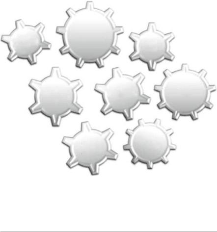
Fig. 11.1 The reconstructive clockwork: the interlocking wheels of a clockwork illustrate the integration of different reconstructive methods
tissues that is missing and which allows defect coverage with tissue of similar contour, texture, and color [4, 5].
11.2The Reconstructive Clockwork
In clinical daily routine, combinations of different techniques are often applied in order to permit new reconstructive possibilities for the patient, but neither the reconstructive ladders of Mathes and Nahai in 1982 nor the reconstructive elevator permits a real combination of the different reconstructive procedures and techniques.
The image of interlocking wheels of a clockwork [6] (Fig. 11.1) illustrates the integration of different reconstructive methods even more impressive than the conventional reconstructive ladder and elevator.
11.2.1 General Principles
Hypertrophic scars and scar contraction with concomitant functional impairment are the most common problems that require correction or reconstruction. Choosing