

NUCLEAR POWER PLANTS
.pdfRadiobiological Characterization Environment Around Object "Shelter" |
235 |
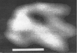
The particles of the correct spherical or oval-oblong, which are easily identified (spores, cocci, etc.) [Gusev M.V. & Mineeva L.A. 1992] and particles of irregular forms seen in presented figures. It is shown by electron microprobe elemental analysis that the major part of these particles consists of organic matter and it is of biological origined [V.B. Rybalka, G.F. Smirnova & G.I.Petelign, 2005].
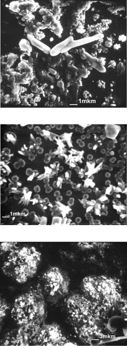
Fig. 5. The appearance of the surface of 4th level impactor. Cocci from 10 to 2 micron and submicron particles of indefinite form are visible
Fig. 6. The appearance of the surface of 4th level impactor. It is seen a large number of particles that are aggregates of loosely coupled small particles
Thus, there are a large number of particles consisting of organic matter in the investigated samples of aerosols originated from “Shelter” premises. A significant number of these particles are identified as the cells of microorganisms and spores.
Sub-micron hot particles (smHP) There is high-dispersive hot particles, which needs a special attention. Such kind of particles is a product of spontaneous dust productivity phenomenon which means dust generation from surfaces of irradiated nuclear fuel and LFCM surfaces. Such a phenomenon was discovered experimentally [Baryakhtar V.G., Gonchar V.V. & Zhidkov A.V. 1997] and for irradiated fuel was confirmed in [Walker C. 2000] later. The smHP grade distribution and possible physical mechanisms responsible for it was later
236 |
Nuclear Power Plants |
identified in [Baryakhtar V., V.Gonchar & Zhidkov A. 2002]. Typical particle grade is 150 nm for fuel HP and near 50 nm for LFCM particles; all the particles have a complicated internal structure [Baryakhtar V., Gonchar V. & Zhidkov A. 2002]. Their radionuclide composition and specific radioactivity does correspond to those for FCM [Baryakhtar V.G., Gonchar V.V. & Zhidkov A.V. 1997]. Such kind of HP practically cannot be trapped by standard respirators, which usually at personnel’s disposal. Annual estimated activity generated in a form of such kind aerosol does equal to a few tens kilograms of irradiated fuel [Baryakhtar V.G., Gonchar V.V. & Zhidkov A.V. 1997].
Such sub-micron HP (Figures 7, 8) to be considered as the most dangerous radiationhazardous agent regarding to a few reasons: their behaviour in biological liquids is similar to those for the particles in true liquid solutions, smHP does not need solubility of fuel matrix for penetrating all natural biological barriers originated from cell membranes. Submicron HP aerosol provides near 80% of total inhalation dose and to be an agent determining effective dose formation for “Shelter” object personnel [Bondarenko O.A., Aryasov P.B. & Melnichuk D.V. 2001]. Existing national regulatory documents on radiation safety does not establish a tolerable concentration of such kind aerosols in the “Shelter” object atmosphere because any attempt to classify them (in accordance to accepted classification) turned out to be doubtful. There are, however, explanations in regulatory document СПРБ-ОУ (in Ukrainian) (Appendix 1), which prescripts what should be done when real aerosols characteristics differs from the typical ones. According to that document, when planned activity stipulates thermal or chemical impact on FCM congestions or heavily contaminated “Shelter” object elements and when revealing in aerosols of non-oxide uranium or TUE chemical compositions one should establish the tolerable concentration for α- and β-emitters in air basing on results of additional special investigation.
Water of the lower marks and technological waters Shelter. It was established that concentration of -emitting radionuclides reaches 3.8 1011 Bq/l, while the concentration of uranium measured up to 0.3 g/l in several thousand cubic meters of "block" and the technological water in premises of lower marks. These water accumulations influence the state of nuclear security of the object “Shelter”. They may lead to a change in breeding properties of system "FBM + fragments of the core + water” and the emergence of emissions of short-lived radioisotopes such as iodine. This radionuclide is known to cause the spectrum of thyroid diseases, including cancer.
Fig. 7. Dust particles complicated internal structure, formed in the fuel-bearing materials. The length of the white line - 80 nm.

Radiobiological Characterization Environment Around Object "Shelter" |
237 |
Fig. 8. Spontaneous destruction of the fuel particles to the submicron particles of "hot particles" as a result of processes of radiation defect in the fuel-bearing materials. The length of the white line – 80 nm.
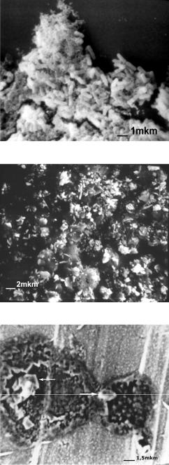
The concentration of radioactivity elements occurs in the silts as the drying pools during the summer-autumn period. Thus silts in “Shelter” represent a real risk as a source of radioactive aerosols. Different kind of microorganisms are found in water accumulations at zero marks of "Shelter" – bubbler pool (Figures 9 - 12) and on the surface of the walls (Figures 13 - 16) [Rybalka V.B., Rybalko S.I. & Zimin Yu.I. 2001, Zhdanova N.N., Zaharchenko V.A. & Tugaj T.I. 2005, Rybalka V.B., Smirnova G.F. & Petelin G.I. 2005, Petelin G.I., Zimin Yu.I., Tepikin V.E. 2003, Rybalka V., Klechkovskaja E., Serbinovich V. 2001]. The experimental investigation of water samples of the Shelter are presented in Figures 9 - 13.
As follows from figures 9 and 10, in the waters of the Shelter are present microbial community in various forms and sizes. In some samples well-reviewed spores size of about 0,1 micron. There is a large number of different units and formations. The biomass of the samples has a very high level of specific activity (137Cs to 3,9 x10¹⁰ Bq/m3; 90Sr to 7, 9 x10⁹ Bq/m3, 238+239+240 Pu to 1,1 x10⁵ Bq/m3).
Fig. 9. The appearance of content drops of water from bubbler pool.
238 |
Nuclear Power Plants |
Fig. 10. The appearance of content drops of water from bubbler pool.
Fig. 11. The appearance of content drops from water from bubbler pool.
Fig. 12. The appearance of content drops from water "Shelter
Radiobiological Characterization Environment Around Object "Shelter" |
239 |
Fig. 13. The appearance of content drops of water from bubbler pool.
Fig. 14. The appearance of content drops of water from bubbler pool.
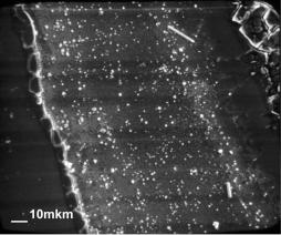
Fig. 15. The fuel particles from swabs from the walls after the burning of organic matter in a vacuum.
240 |
Nuclear Power Plants |
Fig. 16. The appearance of the microbes isolated from swabs of material from the walls after drying a drop of suspension on the glass
Microscopic siza fungi widely exist in the microbiota of the Shelter together with bacteria represented. It is shown that the defeat of microscopic fungi growing in areas with low levels of contamination (from one to 100 mR/hr) [Zhdanova N.N., Zakharchenko V.A. & Tugay T.I. 2005]. The life-cycle reduction, increased radioresistance, increasing the frequency of occurrence of positive radiotropic reactions and radiostimulation, high photosensitivity, that correlated with radiotropic response are typical for fungi-extermophyles isolated from such premises. In these premises increasing the risk of fungal biodegradation, increasing the probability of development of active agents of onychomycosis, skin lesions, lung infections, otitis, invasive fungal infections, as well as the selection of radio resistant organisms with unpredictable invasive characteristics. Lack in human natural immunity to these microbes, resistance of new cultures to the action of traditional medicines, high-speed distribution of microbes (biomass doubling from 15 minutes to 2 hours), the possibility of "transfer" the genetic information with hazardous properties to other bacteria, protozoa, algae, fungi and higher organisms can provide a very serious threat for people. It is known that in the light roof of sarcophagus has a large number of defects and inside the "Shelter" are circulating constant vertical wind currents. So it is quite realistic assumption that some of these microorganisms, especially in the form of spores, can potentially overcome the filters and act to the respiratory tract and lungs of people working inside this object at one side and at another side may carry away outside "Shelter".
2.2 Elucidation of radiobiological effects that can influence the formation of the structure of morbidity
The small doses of radiation can increase the likelihood of developing cancer [Brenner D.J., Doll R. & Goodhead D.T. 2003, NCR 2006] and is possibility appearing morbidity non-cancer origin [Hildebrandt G. 2010]. The main outcome of the 25-year study of morbidity in different categories of exposed persons in connection with the Chernobyl catastrophe is a significant increase in primary morbidity is not associated with tumor pathology. Not found increased risk of leukemia even among those engaged in reconstruction work. Only recently become apparent relationship of radiation exposure
Radiobiological Characterization Environment Around Object "Shelter" |
241 |
with increasing number of non-oncological pathology such as cardiovascular diseases [Preston D.L, Shimuzu Y. & Pierce D.A. 2003]
Statistically significant increase in the spread of non-neoplastic diseases is shown in [Bouzounov O.V., Tereshchenko V.M. 2010]. At the same time structure of morbidity leading place is occupied by diseases of digestive, circulatory and nervous system The maximum level and the largest number with the non-neoplastic diseases statistically confirmed by link determined in the dose > 0,25 Gy for subcohort study group. Long-term effects of chronic low-intensity exposure to low doses for exposed persons in this category until the end is unclear.
The rapid growth of non-tumor morbidity in exposed populations reflects certain changes in the systems that control the growth, development and aging, namely stereotypes matching sequence and intensity of reading the genetic information in different cells. It is shown that this task cannot perform any neural mechanism, or hormonal agents with their ability to alter the rate of metabolic processes. Neurotransmitters, hormones and their receptors do not possess sufficient ontogenetic variability and dispersal [Poletayev A.B. 2008].
According to modern ideas exactly a physiological autoimmunity throughout life provides a readout of genetic information in different cells of the whole organism [Churilov L.P. 2008, Maltsev V.N. 1983, Zaichik A.Sh. & Churilov L.P. 2008]. Singularity of the function of autoantibodies (AuAB) compared with other regulatory substances is considerably longer their half-lives ranged from 10 to 50 days. Therefore the system of autoimmunity has greater inertia. Autoantibodies regulate slow the physiological processes that continue some days and weeks [Ashmarin I.P., Freidlin V.P. 2005]. It is postulated that a mild autoimmune response to their own antigens is a necessary condition for the normal functioning of the immune system and is a prerequisite for the normal regulation and synchronization of cellular functions and morphogenesis [Churilov L.P. 2008]. Additional conditions for unmasking antigens of tissues and organs and represent them immune cells with subsequent increased production of specific AuAB appear as a result of exposure to ionizing radiation.
It is well known that oxygen absorbed by the mitochondria is converted into adenosine triphosphoric acid (ATP). About 5% of oxygen consumed by tissues is converted into free radicals such as superoxide, hydrogen peroxide, hydroxyl radical, singlet oxygen, peroxynitrite (reactive oxygen species, ROC) with unpaired valence electrons. Most of the ROS are produced continuously in cells as byproducts of normal cellular metabolism (mainly due to a small leakage of electrons to the mitochondrial respiratory chain, as well as other reactions in the cytoplasm), and do not cause damage to cells. An excess of ROS under intense ionizing radiation exceeds the protective capabilities cells and can cause serious cell disorders (eg, depletion of ATP). The increase of free radical molecules and their products have a place in the development of the state of homeostasis, has been called oxidative stress. Slow development of oxidative stress triggers apoptosis, and its intensive development leads to necrosis. Postradiation apoptosis is characterized by maintaining the integrity of the cell plasma membrane and the lack of exposure of intracellular contents from cells of the immune system. In the end, remnants of apoptotic cells are removed by exfoliation in intraorganic space and subsequently excreted from the body. But the shortage of ATP, in particular after irradiation, the energy dependence of the mechanisms of apoptosis are disabled, and the cell dies with loss of cell membrane integrity and release of macromolecular
242 |
Nuclear Power Plants |
components (eg, ALT, AST, etc.) into the intercellular space. Necrosis caused an immune response in the form of inflammation - leukocyte infiltration of the affected tissue, interstitial fluid accumulation and subsequent induction of specific immune responses (specifically sensibilized T-lymphocytes and autoantibodies) to the unmasked and recognized by lymphocytes of intracellular components. According to data of many researchers the AuAB are primarily the attribute of the norm. They can be identified in healthy individuals. [Churilov L.P. 2008, Zaichik A. Sh. & Churilov L.P. 2008b, Poletayev A.B. 2005, Cohen I.R. 2005, Harel M. & Shoenfeld Y. 2006, Shoenfeld Y. 2008]. The AuAB involved in the process of apoptosis, cleaning the body from catabolic products, modulation of the activity of many enzymes and hormones, as well as perform the transport function [Poletayev A.B. 2008a]. It was shown that antibodies against nuclear antigens can penetrate into the cell nucleus in vivo and stimulate the synthesis of RNA and DNA in target cells [Zaichik A. Sh. & Churilov L.P. 1988].
Notkins in 2007 hypothesized that natural AuAB can be very informative not only precursors of autoimmune diseases, but also a variety of somatic diseases and syndromes [Notkins A.L. 2007]. It is important that changes in the content of organ natural AuAT in most cases, ahead of the clinical manifestation of appropriate forms of pathology. If for example the content of “cardiotropic", hepatotropic", "neurotropic" AuAB a concrete person within the borders of the norm, this suggests that the intensity of apoptosis, respectively, cells of the heart, liver or nerve tissue does not go beyond the norm. Persistent changes, for example, from "hepatotropic" AuAB should be regarded as a sign of the possible formation of a pathological process in liver tissue, even if at the time of examination are no clear clinical symptoms or specific biochemical changes [Poletayev A.B. 2008a, Churilov L.P. 2008, Notkins A.L. 2007, Zedman, A.J.W. & Vossenaar E.R. 2004].
The important role of the abolition of immune tolerance in the occurrence of non-viral hepatitis research shows serum levels of antibodies to liver-specific lipoprotein (LSP) for various categories of people. [Kovalev V.A. & Senyuk O.F., 2008].
The LSP which was first isolated by Meyer zum Burschenfelde and Miescher in 1971 is considered to be specific poly antigen for the liver. Native LSP is mixture of antigenic determinants of the substrate from the membranes of hepatocytes and contains soluble and membrane components were isolated by gel filtration (chromatography) supernatant after ultracentrifugation of liver homogenate [Manns M, Gerken G. & Kyriatsoulis A, 1987, Ballot E., Homberg J. C. & Johanet C. 2000]. It is known that LSP is found not only in the liver, but also in some other organs [Garcia-Buey., Garcia-Monzon C. & Rodriguez S. 1995]. Therefore, the total increase in antibody levels to the PSL can be seen as a sign of abolition of immune tolerance to many organs and tissues of the human body [Kovalev V.A. & Seniuk O.F., 2008].
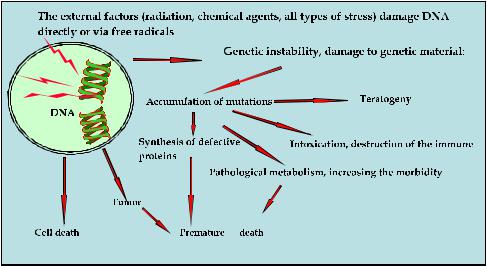
There are three outcomes for the cell, if the cellular radiation damage is not adequately repaired. The cell may die, or will delay it or keep playing with the viability of new qualities, or mutations as the basis for the development of remote descendants (See figure 17). The consequences of the first approach in the development of cells after irradiation described below.
The linear non-threshold concept is used as the primary standard for radiation protection and risk assessment for many years. It suggests that damage induced by low doses of radiation do not contribute significantly to increased risk of disease because a significant
Radiobiological Characterization Environment Around Object "Shelter" |
243 |
amount of endogenous genome damage occurs during life and they are restored in cells with high probably. In fact endogenous damages (ED) constantly appear in the cells. Some of them are due to thermodynamic processes, the hydrolysis reaction, while others arise from the effects of free radicals generated by cell during its life and still others are a necessary component of metabolism (DNA breaks accompany the process of differentiation, recombination, etc.) [Lindahl T. 1993, Bont R.D. & Van Larebeke N. 2004]. According to [Lindahl T., 1993] per one day in the DNA of one cell may have more than 50,000 endogenous damages as single-strand breaks (SSB) and 10 ones as double-strand breaks (DSB).
Fig. 17. Effects of radiation exposure on the genetic apparatus of cells.
Many studies suggest that most of the radiation damage (RD) occurring in the DNA of cells differ significantly in their chemical nature from the ED. The main difference between DNA damage induced by ionizing radiation, from the ED was its the complexity of their chemical nature, and clustering. The proportion of complex, critical for the fate of the cell damage is much higher when exposed to IR.
When treatment of mammalian cells to H2O2 ratio of DSB to SSB is 1: 10 000 [Bradley M.O. & Kohn K.W. 1979] while under the influence of IR is much higher -1:20 [Shikazono N., Noguchi M., & Fujii K. 2009]. Many of the DNA RD are not accidental, are located in close proximity and have the cluster grouping. They are formed as a result of coincidence of two or more single damages within 1-2 rotation the DNA helix [Hada M., Georgakilas A.C. 2008, Sutherland B.M., Bennet P.V. & Sidorkina O. 2000]. Especially the massive clustering of DNA damage occurs when the ionization tracks pass along chromatin fiber. In this case, they may cover DNA regions with an average size of about 2000 base pairs [Radulescu I., Elmroth K. & Stenerlow B. 2004]. At the same time, we know that the probability of occurrence of endogenous clustered DNA damage is extremely low [Bennett P.V., Cintron N.S. & Gros L. 2004]. Accumulation of ED does not occur because in the cells are constantly functioning mechanisms of reparation, specifically targeted at removing various types damages [Friedburg E.C., Walker G.C. & Siede W. 2006].
244 |
Nuclear Power Plants |
The complex nature of RD of DNA and the presence of cluster groups can be regarded as the first cause, which creates difficulties for repairing systems cells. Damages repair processes within the cluster can break down in various stages of excision repair and lead to the formation of additional or inaccurate DSB repair, important for cell survival, mutagenesis, and the risk of malignancy [Ide H., Shoulkamy M.I. & Nakano T. 2010]. The second reason for the low efficiency of repair of RD may be a relatively low amount of DNA damage. Therefore, radiation effects in low dose range and low power are certain features associated not only with destructive modifications but with deducing the cellular genome at a different level of activity. High doses of ionizing radiation via activation of cell cycle control points - checkpoints, blocking the synthetic phase of the cell cycle (S) and the transition from G1 to S phase and G2 to M (mitotic) phase and support the repair of DNA. In this case, small doses can not activate the G2/M chekpoynt-arrest and DNA repair are not activated when the number of DSB DNA damage and the MNF up to 10 - 20 per cell.
In this case, heterochromatin little relaxes, and access of repair enzymes to sites of DNA damage worsens [Fernet M, Megnin-Chanet F. & Hall J. 2010, Grudzenskia S., Rathsa A., Conrada S., 2010, Marples B., Wouters B.G. & Collis S.J. 2004, Gaziyev A.I. 2011]. There is the third of the possible causes of low efficiency of repair of critical DNA damages.
The major part of radiation induced DNA damages are represented by DSB and crosslink between strands, which slowly and inefficiently repaired and are responsible for various end effects - from the radiation death of cells to the appearance of chromosome aberration, gene mutations and neoplastic transformation. [Pfeiffer P., Gottlich B. & Reichenberger S. 1996]. Therefore radiation effects in low dose range and low rate dose have the certain features associated not only with destructive changes than with deducing the cellular genome at another level of activity. Analysis of many studies suggests that DNA damage caused by the IR, increase linearly with dose, but the reaction of cells on these lesions, the efficiency of repair of the most complicated critical damages can be nonlinear. Because irradiation is decreased expression of proteins, enzymes of different systems providing the stability of DNA as a result of the accumulation of DNA damage is not recovered or recovered with errors and fix mutations. Dysfunction of many genes, regulatory systems and cellular processes that are ultimately linked to the development of various pathologies, including carcinogenesis. A significant part of replication errors - spontaneous mutations - can be harmful to an organism. There are the basis for the occurrence of hereditary diseases, carcinogenesis, etc. were revealed.
2.3 The study of the radiological effects of low and middle doses in model experiments on linear animals
It is known that some of the surviving cells after irradiation can produce functionally modified descendants, who have for many generations with a high frequency occur de novo chromosome aberrations and predominantly point mutations, in certain cases increase cell death by apoptosis [Little J.B. 2000, Seymour C.B., Mothersill C. & Alper T. 1986, Baverstock K. 2000]. The accumulation of such mutations suggests a high probability disorders in the bases caused by oxidative stress [Little D.B. 2007]. It is known that even in non-irradiated cells residing in close proximity to irradiated cells, induction of chromosomal instability is possible.
Placing of non-irradiated cells (NirC) in the culture medium of irradiated cells (IrC), soon leads to the appearance in the descendants of (NirC) of chromatid and chromosome aberrations, micronuclei, gene mutations that increase the content of the transformed cells