


11
Head and Neck Lesions
James J. Chandler and Doreen M. Agnese
Objectives
1.To provide a survey of head and neck surgery, designed as an introduction to this field.
2.To help the physician, surgical resident, or medical student develop an understanding of the diagnosis and treatment of primary cancers of various head and neck sites.
3.To enable the reader to develop an approach to a neck mass and to be able to discuss diagnostic methods and treatment.
4.To be able to answer such questions as: What are the more common neck masses in children and their embryonic origins? What is the relationship of alcohol and tobacco products to cancer? How is the risk of cancer of the thyroid assessed?
5.To develop an understanding of thyroid malignancies and their cells of origin.
6.To be able to develop a plan for diagnosis and treatment of salivary gland tumors and of primary hyperparathyroidism.
7.To develop an understanding of thyroiditis.
Case
A 48-year-old man is seen at your office. He noted a lump in the anterior neck while shaving a week ago; the lump is not painful or tender and has not changed. He has been completely well. On examination, you find a 2-cm-diameter lump just to the left of the midline, at the anterior margin of the sternocleidomastoid muscle. The lump is moderately firm, and it moves up when he swallows. The lump seems to be in the edge of the thyroid lobe. See Algorithms 11.1 and 11.2.
177
178 J.J. Chandler and D.M. Agnese
|
|
|
History and physical exam |
|
|
|
|
||||
Intraoral, |
|
|
|
|
|
Neck |
|
|
|||
pharyngeal, |
|
|
|
|
|
|
|
|
|
||
nasal |
Upper neck |
|
Mid-neck |
Supraclavicular |
|||||||
|
|
|
|
|
|
|
|
|
|
|
|
|
|
|
See Algorithm |
Thyroid |
See Algorithms |
See Algorithm |
|||||
Biopsy ± |
11.3 |
|
see Algorithm |
11.2 and 11.3 |
11.3 |
|
|||||
Refer to surgical oncology, |
|
|
|
11.2 |
|
|
|
|
|||
|
|
|
|
|
|
|
|
|
|||
oral surgery, |
|
|
|
|
|
|
|
|
|
||
head and neck surgery, |
|
|
|
|
|
|
|
|
|
||
|
|
|
Thyroid cancer |
|
|
||||||
plastic surgery |
|
|
|
|
|
||||||
|
|
|
See Algorithm |
|
|
|
|
||||
|
|
|
|
|
|
|
|
|
|
||
|
|
|
|
|
|
11.4 |
|
|
|
|
|
|
|
Scalp |
|
|
|
|
|
|
|
||
|
|
|
|
|
|
|
|
|
|
|
|
|
|
|
|
|
|
|
|
|
|||
Longtime lump |
Brown lesion |
|
|
|
|
|
|
||||
nontender superfacial, |
|
|
|
|
|
|
|
|
|
||
just under skin |
? Melanoma |
|
|
|
|
|
|
||||
|
|
|
|
|
|
|
|
|
|
|
|
It's a wen |
Surgery consult |
|
|
|
|
|
|
||||
|
|
|
|
|
|
|
|
|
|||
|
|
|
derm oncology |
|
|
|
|
|
|
||
Excise, local |
|
|
|
|
|
|
|
|
|
||
anesthesia |
|
|
|
|
|
|
|
|
|
||
|
Embedded tick? |
|
|
|
|
|
|
||||
send to ER
Algorithm 11.1. Algorithm for approach to a patient with a mass in the head or neck.
Introduction
Problems presented that are centered in the region of the head and neck are best addressed while simultaneously considering the regional anatomy (which is reliable, with minimal anatomic variation between patients) and the patient’s medical and social history.
For example, the patient in the case presented above stated that he never smoked and that he drank only an occasional glass of wine.
There is a close relationship of high alcohol intake or the use of smokeless tobacco with cancers of the oral cavity and pharynx, and there is a close relationship of tobacco smoking and alcohol intake with cancers of the esophagus and the entire respiratory tract.
The patient in our case would not be expected to have a cancer primary in any of these areas, given his social history.
Risk Factors
Tobacco, in its various forms, is a risk factor for the development of head and neck cancer. These forms include inhaled tobacco, chewing

11. Head and Neck Lesions 179
tobacco, and snuff (often referred to as “snoose” in the western states), which is held against the cheek or gums. Betel nut chewing, common in the western Pacific basin and South Asia, also is associated with increased risk. Most cases of head and neck cancer are associated with a significant history of alcohol consumption coupled with a history of tobacco use. Marijuana use and some viruses have been implicated to play a causative role in the development of head and neck malig-
|
|
|
Physical Exam of Thyroid Nodule |
|
|||
|
Ultrasound |
|
|
|
1 Abundant colloid |
||
|
|
Fine-needle |
|||||
|
|
|
|
||||
|
|
|
aspiration (FNA) biopsy |
and many lymphocytes |
|||
|
|
|
|||||
Multiple |
Single |
few follicular cells |
|||||
Cold |
|
|
|||||
|
|
or |
Surgery or |
|
|||
Observe |
|
Scan |
Hot |
|
|||
|
observe |
|
|||||
|
|
|
|
|
|||
Symptomatic? |
FNA for largest |
|
FNA: an enlarging |
||
|
|
nodule |
|
|
|
|
|
|
Total or subtotal |
|
|
thyroidectomy |
|
|
2a) “Suspicious”
b)“Malignant”
c)Many follicular cells  Surgery little or no colloid
d)Hürthle cells
a)Female with 4-cm single nodule and “follicular neoplasm”
b)Male with 2.5-cm nodule and “follicular neoplasm”
c)“Highly suspicious” for cancer
d)“Malignant”
“Total,” 98+% thyroidectomy
Alternative approach—now less common: Total lobe on one side and
“near-total lobectomy” other side
Cancer? See Algorithm 11.4
Symptomatic
Observe
Total or subtotal
thyroidectomy
Indeterminate cells
Insufficient for diagnosis
Re-biopsy
“Follicular neoplasm” (smaller)
Total lobectomy
Final diagnosis: malignant
Return patient to O.R. in next 2–3 days and complete a total thyroidectomy, or wait 6 weeks
Algorithm 11.2. Algorithm for the evaluation and diagnosis of a thyroid nodule.
180 J.J. Chandler and D.M. Agnese
nancies. Radiation therapy for acne or skin warts can be followed by skin or thyroid cancers years later.
History
Important points to elicit in the history of the patient presenting with a mass in the head and neck region are:
•the exact location of the lesion
•the length of time the lesion has been present
•the rate of growth of the lesion: rapid enlargement implies infection or malignancy
•the presence of pain or tenderness: cancer usually is not painful unless there is a superimposed infection or nerve invasion
•the presence of an unpleasant odor: bacterial tonsillitis, a foreign body in a child’s nasal passage, and squamous cell carcinoma of the tonsil or base of tongue with superimposed bacterial infection all are noteworthy for the associated odor
•history of difficulty swallowing
•painful or tender persistent lesion in the mouth
•referred pain to the ear
•hoarseness
•weight loss
•history of radiation exposure.
A thorough family history can be helpful. Specific questions regarding family members with goiter, multiple endocrine neoplasia syndrome, or a high incidence of skin cancers should be asked.
In addition to a history of the use of tobacco and alcohol, a history of the use of other nonprescription substances should be sought. For example, cocaine use may result in intranasal lesions.
A complete sexual history should be obtained. Oropharyngeal sexually transmitted diseases have been reported. Risk factors for HIV and AIDS may be identified that may alter the differential diagnosis.
The patient in our case was asked about these points, but nothing contributory was found. This was a neck lump without symptoms, discovered suddenly during a morning shave.
Head and Neck Examination
Inspection (see Algorithm 11.3)
Many lesions in the head and neck can be identified using simple inspection. On the scalp, epidermal inclusion cysts (known as “wens”) easily can be appreciated; a puncta often is not visible, and skin color is normal. The external ear protrudes and especially is prone to damage from sun exposure. A horn-like, hard little lesion that can be torn off, producing a shallow ulcer, is referred to as actinic keratosis. This lesion is a precursor of squamous cell carcinoma. Patients with these lesions are managed appropriately by referral to a dermatologist or head and neck surgeon for treatment. Any ulcerated skin lesion demands

Superior to hyoid
Probably a lymph node
Follows
No infection
infection
history
Surgery Observe referral X months
Or FNA
AT/near midline
At hyoid
Elevates when tongue protrudes
It,s a thyroglossal duct cyst
Surgery
|
|
|
|
Supraclavicular |
||||
At or below |
|
|
|
|
||||
Hard? |
|
|||||||
thyroid cartilage |
|
|||||||
|
|
|
|
Patient >40 |
|
|||
|
|
|
|
|
||||
Moves with |
|
Cancer diagnosis? |
||||||
|
|
|
|
|||||
thyroid cartilage |
FNA |
|
||||||
|
|
|
|
CXR |
||||
|
|
|
|
|||||
|
|
|
|
Biopsy |
||||
|
|
|
|
|
||||
Likely thyroid |
|
|
|
|
CT chest |
|||
|
|
|
||||||
See A11.2 |
|
TB? |
||||||
|
|
|||||||
|
|
|
|
|
||||
|
|
|
|
|
||||
|
|
|
|
Referral |
|
|||
|
|
|
|
Infection, |
|
|||
|
|
|
|
Disease or |
|
|||
|
|
|
|
pulmonary |
|
|||
|
|
|
|
medicine |
|
|||
|
|
Lateral middle neck and |
|
|||||
|
|
|
nonpulsatile |
|
||||
|
|
|
|
|
|
|
||
|
|
|
|
|
|
|
|
|
|
|
|
FNA BX |
|
||||
"Thyroid"cells |
Brown cloudy fluid |
|||||||
|
|
|
|
|||||
|
|
|
|
|
|
|
|
|
|
|
|
|
Branchial cleft cyst |
||||
Likely cancer |
||||||||
|
|
|
|
|||||
|
|
|
|
|||||
|
|
|
|
Surgery referral |
||||
Surgery referral |
||||||||
|
|
|
|
|||||
(See A11.4) |
|
|
|
|
||||
Lateral upper neck
Rubbery |
|
|
|
|
||
Pulsatile |
||||||
patient <40 |
||||||
|
|
|
|
|||
Lymphoma |
|
|
|
|
||
|
Refer vascular |
|||||
|
|
|
||||
|
|
|
||||
CXR-open |
|
surgery |
||||
biopsy |
Just below |
|||||
consider |
ext. ear |
|||||
|
|
Nontender |
||||
|
|
|
|
|
||
|
|
|
|
|
|
|
|
|
Parotid |
||||
|
|
|
|
|
|
|
|
|
|
|
|
|
|
Lateral neck |
Refer surgery |
|||||
|
|
|
|
|||
? Met. cancer |
|
|
|
|
||
FNA BX |
|
|
|
|
||
Just under mandible
Node vs. submaxillary gland
FNA BX and check mouth
Algorithm 11.3. Algorithm for the evaluation and management of a neck mass.
Lesions Neck and Head .11
181
182 J.J. Chandler and D.M. Agnese
biopsy so that a diagnosis can be made and appropriate treatment given. Skin lesions that have changed during a period of observation, have irregular borders, display variegated pigmentation, or bleed when rubbed must be referred for excision or biopsy.
These may be melanomas, requiring complete removal with curative intent.
Gray-white plaques on the lower lip may be seen. These lesions are called leukoplakia, and a small percentage subsequently develop cancer. Squamous cell carcinomas of the tongue usually are firm, tender, and painful. They often are raised and on the side of the tongue.
Cancers of the gums, tonsillar pillars, and inner surfaces of the cheeks generally are redder than the adjacent surfaces. A patient who seems sick and shows a swollen tonsil near the midline may have a peritonsillar abscess, which requires urgent drainage. A bluish cyst in the floor of a child’s mouth is rare and is a ranula.
Brown patches on the lips signal Peutz-Jeghers syndrome, associated with intestinal polyps that can bleed or obstruct. Skin tumors of various size consisting of tiny blood vessels, hemangiomas, can be found anywhere in the head and neck region. Epidermal inclusion cysts or sebaceous cysts commonly are found behind the lower external ear, in areas of acne activity, the posterior neck, and the earlobes (especially at the site of skin or ear piercing). The pink or red skin blotches on sun-exposed skin may be malignant or premalignant and generally require diagnosis and possible treatment from a specialist in skin conditions. Darkly pigmented spots or skin blotches that leave a gray, roughened zone when the surface is lightly scraped are seborrheic keratoses, related to skin aging.
Cervical lymph nodes that are obvious on inspection or palpation mandate a complete examination of the head and neck. A firm unilateral neck mass in an adult is cancer until proven otherwise (see Algorithm 11.3). Many of these are cervical metastases from squamous cell carcinoma of the head and neck. Deviation of the tongue to the side of the lesion may be appreciated when the patient protrudes the tongue, suggesting 12th cranial nerve invasion by cancer.
A painless, hard mass in the lower neck in a patient who lives in crowded conditions or is immunocompromised, with or without another known to have tuberculosis, may be scrofula, a tuberculosis lymph nodal mass.
A lump in the upper midline of the anterior neck may be a thyroglossal duct cyst (see Algorithm 11.3). If located further up, under the chin, it will be an enlarged submental lymph node. If you stand to the side and ask that the tongue be put out, elevation of this lump with tongue protrusion is diagnostic of a thyroglossal duct cyst.
Branchial cleft cyst presents at the anterior border of the sternocleidomastoid muscle or just in front of the external ear’s tragus. Thyroid enlargement, diffuse or nodular, has been termed goiter. The swelling in the thyroid often is easily visible, as in our case.
When inspecting the thyroid, try sitting lower than the patient, with your eyes at the level of her/his midneck, using some light from the side. Ask the patient to swallow. When the patient swallows, the
11. Head and Neck Lesions 183
thyroid slides up and down and the thyroid nodule or multinodular goiter easily is seen.
Palpation
A thyroid nodule often can be appreciated moving up and down under the sternocleidomastoid muscle, as you palpate more deeply lateral to the trachea. Lipomas may present in the supraclavicular areas. These lesions have well-defined borders and are relatively soft. Masses in the neck may represent malignant or inflammatory disease. Enlarged lymph nodes tend to be found along the course of the jugular vein and are termed high-jugular lymph nodes when located in the upper neck, below the angle of the jaw. Firm, nontender masses in the neck that are not easily moved are likely cancer metastatic to cervical lymph nodes.
Infections of the tonsils or teeth also can result in enlargement of neck lymph nodes, but these nodes are tender. When cancer metastasizes to the upper jugular nodes, the most common primary sites are the base of the tongue, the nasopharynx, and the tonsillar areas. Cancer metastatic to mid-jugular nodes—lymph nodes in the central lateral neck under the muscle—most commonly originates from the thyroid lobe on that side. Supraclavicular lymph node metastases generally are from cancer sites below the clavicles. Keep in mind, however, that lung cancer can and does spread anywhere (see
Algorithm 11.3).
Palpation of the thyroid gland is best performed by facing the patient, placing the index finger on the thyroid cartilage (Adam’s apple) to stabilize it while curling the fingers of the opposite hand around the sternocleidomastoid muscle, resting the thumb on the thyroid isthmus. The neck muscles should be relaxed. When the patient is asked to swallow, the thyroid lobe slips up and down between your fingers and thumb, allowing you to appreciate a nodule in that thyroid lobe. The neck lump in our case patient was firm. It moved up with the thyroid lobe when he swallowed.
Examination of the oral cavity requires palpation for completeness.
A moistened, gloved finger gently sweeps over the gum surfaces, the floor of the mouth, and the tongue, searching for rough or tender areas. With the patient breathing through the mouth, one quickly can sweep across the base of the tongue to the epiglottis. Bimanual examination especially is useful for the floor of the mouth and can be used for cheek surfaces and for the tongue.
Special Examination Techniques
Special examination techniques are performed by surgical oncologists and head and neck surgical specialists. Fiberoptic laryngoscopes are passed through the nose for direct examination of the vocal cords and nearby areas. Pediatric (3.6 mm in diameter) and anesthesia (4.0 mm) bronchoscopes, both fiberoptic, also are useful for this examination. A complete examination, searching for a primary cancer site, requires general anesthesia. The examination relies on the use of fiberoptic instruments to look into and at all surfaces that can be reached,
184 J.J. Chandler and D.M. Agnese
including the nasopharynx and sinuses, and the performance of appropriate biopsies. Esophagoscopy and bronchoscopy are added when the primary cancer site has not been found: about 3% of patients with metastatic cancer found in a cervical lymph node will have a final unknown primary classification.
Imaging studies also play an important diagnostic role. Computed tomography (CT) scan with contrast is most useful in evaluating suspicious cervical lymph nodes. For suspected or known supraclavicular nodes involved with cancer or for lymphoma anywhere above the clavicles, CT of the chest with contrast is used. Adenocarcinoma diagnosed by cervical lymph node biopsy indicates the need for further studies, possibly including mammography and endoscopy. Magnetic resonance imaging (MRI) is superior to CT for evaluating neural elements. Positron emission tomography (PET) scanning may supplant the use of some modalities, but currently the cost factor argues against its use except in special circumstances. Ultrasound is used to assess the entire thyroid gland (see Algorithm 11.2). Ultrasound can determine whether a lesion is cystic or solid: a thyroid lesion demonstrated on ultrasound is benign if it is entirely cystic. Radioisotope scanning also may be useful; nodules that take up less isotope than the surrounding thyroid tissue are termed “cold” and have a much higher chance of being malignant than “hot” nodules (1% incidence of cancer in “hot” nodules). Sestamibi scan often is able to locate a parathyroid benign tumor (adenoma).
Biopsy Techniques
Fine-needle aspiration (FNA) is the initial biopsy technique used for the diagnosis of thyroid lesions and neck masses, with few exceptions (see Algorithm 11.2). In the case presented, FNA was the first step chosen by the thyroid surgeon to whom this 48-year-old healthy man with a suspected thyroid nodule was referred. Using local anesthetic, the lump (nonpulsatile) is fixed between fingers of the nondominant hand, and a needle attached to a small syringe (for best suction) is passed into the lesion, then quickly passed in and part way out of the mass, “chopping” firm tissue to free cells to be aspirated. An experienced cytopathologist should evaluate this specimen.
Fine-needle aspiration for diagnosis of a thyroid nodule may not be definitive. If the material is deemed “insufficient for diagnosis,” a repeat FNA should be performed. The presence of abundant colloid or lymphocytes suggests benign disease, with the indication for surgery resting on factors other than suspicion of malignancy. “Papillary cancer,” “suspicious for papillary cancer,” and “follicular neoplasm” are phrases in the pathology report that argue for surgical removal, since a significant number of these patients will have a malignancy.
Biopsy of an intraoral lesion can be taken with a scalpel or using a dermal “punch” biopsy technique. Biopsy for suspected lymphoma (see Algorithm 11.3) requires open surgical biopsy of some or all of the lymph node. The complete diagnosis requires more tissue than can
11. Head and Neck Lesions 185
be obtained with needle aspiration or needle core biopsy. An open biopsy in the neck always is done by a surgeon familiar with the planning for possible neck dissection, because a diagnosis of squamous cell cancer in a node mandates the excision of the biopsy incision site as part of a curative operation.
On the face, surgeons plan to take a little normal-appearing skin with the biopsy, while cosmetically planning the best approaches for removal of a suspected cancer. In assessing a pigmented lesion anywhere on the skin, a possible melanoma, “shave” biopsy is never appropriate because the depth of invasion determines the plan for surgical cure. Punch biopsy at the thickest part of the lesion or excisional biopsy with a tiny margin is preferred as the initial diagnostic biopsy when melanoma is suspected.
The case patient’s biopsy was sufficient for diagnosis. The pathologist described the cytology as “follicular neoplasm,” and an operation was recommended to the patient. He concurred, after learning about the options, the procedure, and the significant risks. A preoperative ultrasound study of the neck revealed no abnormality except for a left thyroid lobe solid nodule, 1.8 cm in diameter.
Benign Lesions of the Head and Neck
Congenital
Thyroglossal duct cysts are in the midline, may enlarge quickly with infection, and elevate with tongue protrusion (see Algorithm 11.3). These lesions are removed completely (including the central portion of the hyoid bone) with general anesthesia. A midline mass in a baby or young child may be lingual thyroid tissue. These masses may require excision if they cause obstruction. It is important to recognize that this might be the only functional thyroid tissue present; this means that normal thyroid must be identified by scanning technique before any surgical intervention is planned.
Dermoid cysts, consisting of elements from all three germ cell layers, are rare in the head and neck.
First branchial cleft sinus or cyst presents in the preauricular skin, lying close to the parotid gland. This lesion always is deep and difficult to remove completely. Incomplete removal is followed by recurrence. Second branchial cleft cyst presents at the anterior border of the sternocleidomastoid muscle in the middle or lower neck or as a large tender infected mass under the muscle. Diagnosis is made by aspirating brown turbid fluid. After treatment of the acute infection, the child or young adult returns for elective surgical removal. A cystic hygroma is a large, soft mass in the side of the neck above the clavicle. These complex, cystic lesions present in infancy and are difficult to remove; suspected cases should be referred to a pediatric surgeon for definitive management.
In older patients, the differential diagnosis of a mass presenting in the upper neck must be considered: metastatic cancer, carotid body
186 J.J. Chandler and D.M. Agnese
tumor, carotid artery aneurysm, branchial cleft cyst, or a primary cancer (see Algorithm 11.3).
Salivary gland tumors are most common in the parotid gland, and the majority of these are benign (75–85%). Most parotid tumors are “mixed tumors” or pleomorphic adenomas. All parotid tumors are removed by surgeons experienced in dissecting parotid tissue off the seventh cranial nerve. In the case of malignant tumors of the parotid, the nerve is no longer sacrificed (unless it is grossly involved with cancer), and the area is treated by irradiation after surgery. Tumors of other salivary glands are more likely to be malignant.
Infections
In a child or teenager, upper neck masses usually are enlarged lymph nodes draining an infected area. In the posterolateral neck, lateral to the sternocleidomastoid, and in the posterior triangle, these lumps almost always are inflamed nodes draining a zone of scalp infection. However, thyroid cancer in a node can present here. A mass in the thyroid or adjacent to the thyroid is relatively common in all ages with the exception of infancy. Malignancy always must be considered.
Consider doing an FNA.
Scrofula (tuberculous lymphadenitis in the neck) is treated medically after diagnosis has been made. One actually might avoid the usual skin test in this case because the intermediate tuberculin test could result in a huge reaction, with skin slough of the forearm. Chest x-ray, CT of the neck and chest, sputum, or, better yet, early morning gastric washings for tuberculosis (TB) smear and culture should result in a positive diagnosis in a patient with a tuberculous infection severe enough to result in scrofula.
Ludwig’s angina is a severe, spreading, acute infection that arises from mixed mouth bacterial flora. It involves the floor of the mouth and produces pain and tenderness under the jaw in the midline. Immediate referral is essential because some patients require emergency drainage in addition to antibiotics to protect the airway.
Vincent’s angina (“trench mouth”) develops from poor hygiene and ulcerations in the gums, and is noted by fetid odor, acute infection, and rapid spreading. This condition is managed with antibiotics as well. Referral usually is indicated, because differentiation from Ludwig’s angina is important.
Vascular (see Algorithm 11.3)
A carotid body tumor easily can be mistaken for a low, lateral parotid gland tumor. If a hard lump is right over the likely site of the carotid bulb, Doppler color flow study and possibly CT should precede any needle biopsy or surgical removal. Aneurysms of the carotid artery and a tortuous innominate artery present as pulsatile masses in the lateral neck. While color flow Doppler clarifies these diagnoses, consultation with a vascular surgeon should be strongly considered.
11. Head and Neck Lesions 187
Parathyroid
The two superior parathyroid glands arise from the fourth branchial pouches, along with the lateral thyroid lobes. The two inferior glands arise from the third branchial pouches and normally lie more anterior than the superior two. Primary hyperparathyroidism (pHPT) results in elevated serum calcium (Ca2+) levels and usually is picked up on a routine blood serum laboratory study. Confirmation of pHPT comes from finding elevated serum Ca2+ with elevated parathyroid hormone (PTH). This condition can result in bone demineralization, fractures, severe arthritis, renal failure, ureteral stones, acute pancreatitis, peptic ulcer, and mental changes. However, most patients are asymptomatic at the time of diagnosis.
Cure for pHPT is surgical. Since the majority of cases are caused by a single parathyoid adenoma, identification of the site of the adenoma, if possible, allows a more rapid procedure that usually requires only a short stay after surgery. Thus, with pHPT diagnosed with a radioisotope scan (sestamibi scan) demonstrating the site of the single adenoma, the surgeon can remove the enlarged gland and check the probability of cure with a rapid PTH level intraoperatively. Bloods for this test are drawn before and after removal of the adenoma. The PTH level falls within 5 minutes to a level consistent with cure after removing the single adenoma responsible for pHPT. In about 4% of cases there are two adenomas; in about 15% the cause of pHPT is hyperplasia, which usually involves all four glands. In the event that the sestamibi scan is not able to find a single adenoma or rapid PTH assay is not helpful or available, the surgeon plans a more elaborate procedure, requiring finding all parathyroids before removing any. To aid in locating these glands, some use intravenous methylene blue dye preoperatively. To aid locating a single adenoma, one can use a sestamibi scan preoperatively and then use a gamma-detecting probe to pick up the radioactive emissions in the operating room.
Thyroid
Diffuse enlargement and nodular masses of the thyroid are the most common neck masses. History and physical examination should be done first, before laboratory evaluation, imaging studies, or biopsy (see
Algorithm 11.2).
Thyroiditis
Chronic lymphocytic (Hashimoto’s) thyroiditis is found virtually only in women, can be nodular, and leads to hypothyroidism. Surgery is reserved for those with the late fibrosis that can develop, causing tracheal or esophageal compression symptoms, and for cases in which cancer is suspected. Subacute thyroiditis produces a swollen and tender thyroid. Medical endocrinologists treat these cases with antiinflammatory medication.
Hyperthyroidism
A diffuse goiter with signs and symptoms of hypermetabolic activity, elevated thyroxine, and low thyroid stimulating hormone levels is con-
188 J.J. Chandler and D.M. Agnese
sistent with primary hyperthyroidism—Graves’ disease. The treatment for this condition is medical, with an antithyroid agent used initially, sometimes with a beta-blocker added, and radioactive iodine used for recurrence. Women who are pregnant or anticipate the possibility of pregnancy should not receive antithyroid drugs or radiation therapy due to the risk of resultant fetal hypothyroidism. In these cases and some others, surgical intervention may be warranted. Medical follow-up is necessary, both to assess thyroid function and to decide on hormone replacement therapy.
Before any surgery on the thyroid, one must be certain either that the patient is euthyroid or that the hyperthyroid state is controlled to avoid the potentially lethal complication of “thyroid storm.” This dangerous condition is caused by release of thyroid hormone from the thyroid gland during surgical manipulation and results in severe tachycardia, fever, and other signs of hypermetabolism. For this reason, antithyroid drug plus a beta-blocker are given to “cool off” the thyroid and stop the symptoms and signs of Graves’ disease before surgery. If the thyroxine level in the serum has not fallen to a safe level for surgery, beta-blockers are continued for 4 or 5 days postoperatively. Iodine usually is given in an oral form for a week before operating on a hyperactive thyroid so as to block the release of thyroid hormone and to make the gland firmer and less vascular.
Thyroid Nodules (see Algorithm 11.2)
Multinodular goiters usually are benign, with nodules composed of colloid. These goiters may enlarge and compress the trachea and/or esophagus. It is important to remember that an individual nodule in a multinodular goiter may be malignant. If a nodule is notably larger than others or enlarges during a period of observation, biopsy is recommended. Finding a “follicular” lesion indicates the need for surgical referral. Finding a solitary thyroid nodule indicates the need for further evaluation if the diameter is greater than 0.8 cm on ultrasound. Fine-needle aspiration, often with guidance by ultrasound, is the best initial diagnostic modality. Radioisotope scan then may be indicated to discover a “cold” nodule. If a nodule is large enough to be seen easily or if symptoms are present, surgical intervention is considered, regardless of the results of FNA. If the biopsy yields primarily thyroid follicle cells, surgical referral is indicated.
While attempted “suppression” of a possibly malignant thyroid nodule through the administration of oral thyroid hormone formerly was a popular first step, the recommendation by a majority of medical and surgical endocrinologists today is surgical removal and evaluation by the pathologist. Some simple rules of thumb indicate the risk of malignancy in a thyroid nodule:
1.Most thyroid lesions and problems occur in women; a woman with a single nodule at age 40 is least likely, compared with women of other ages, to have a malignancy (about 10% likelihood in the surgical literature).
2.The chance of cancer increases as the age of the patient increases or decreases from age 40. A woman at age 20 or age 60 with a single
11. Head and Neck Lesions 189
nodule has about a 25% chance of having cancer in the single nodule.
3.A male, for reasons unknown, has a two to three times greater likelihood of thyroid cancer as compared with a female of the same age with the same size thyroid nodule.
4.The larger the follicular neoplasm of the thyroid, the more likely it is cancer. A 4-cm thyroid tumor composed of thyroid follicular cells has a more than 50% likelihood of being cancer.
5.Firm neck lymph node, hoarse voice, lung nodule, bone pain or lesion, and hard and fixed thyroid mass are some of the signs of aggressive cancer.
The patient in our case had a left thyroid lobe resection. This tissue was sent to the pathologist with request for rapid section diagnosis. The pathologist, via intercom into the operating room, reported that there was a 1.8-cm-diameter follicular lesion in the left thyroid lobe. She could not see any definite sign of malignancy in the sections studied. The surgeon closed the neck, and the patient went home 6 hours later; he was able to swallow and felt only some mild discomfort.
Salivary Glands
Parotid tumors are much more frequent than tumors in the other salivary glands, and most are benign. All tumors are removed under general anesthesia, dissecting the gland containing the tumor off the facial nerve. The tumor is never just lifted out of the glandular tissue because doing so leads to a high rate of recurrence, with difficulty of cure thereafter. Pleomorphic adenoma, the most common benign parotid tumor, can become very large, and removal should be undertaken early when cure and safe removal are much easier. Submandibular (also termed “submaxillary”) gland enlargement is less common. Fine-needle aspiration is useful. Tumors of submandibular, sublingual, and minor salivary glands are more likely malignant, and all are treated by complete removal of the gland.
Be warned—the submandibular gland can be enlarged because of blockage of the orifice of Stensen’s duct by a “stone” or by cancer of the floor of the mouth. Check the area behind the teeth, below the tongue.
Sites of Head and Neck Cancer
Skin
Premalignant and low-grade skin cancers are common, but melanoma is the more feared lesion, and we constantly must be on the lookout for it. Therefore, plan biopsy for any pigmented lesion that has changed, is asymmetric, has irregular borders, has variegated color pattern, or is ulcerated. Seborrheic keratoses are the “age spots” seen on the skin; some of these are difficult to differentiate from melanoma. The scalp may be hiding a malignancy, a wen, a buried tick, or the site of a Lyme disease–carrying tick bite. Check for these.

190 J.J. Chandler and D.M. Agnese
Figure 11.1. Oral Cavity includes lips, floor of mouth, anterior two thirds of tongue, buccal mucosa, hard palate, upper and lower alveolar ridge, and retromolar trigone. (Reprinted from Bradford CR. Head and neck malignancies. In: Norton JA, Bollinger RR, Chang AE, et al, eds. Surgery: Basic Science and Clinical Evidence. New York: Springer-Verlag, 2001, with permission.)
Oral Cavity
The oral cavity includes the lips, buccal mucosa, oral tongue (anterior two thirds), floor of mouth, hard palate, upper and lower alveolar ridges, and retromolar trigone (Fig. 11.1). Virtually all malignancies here are squamous cell in origin. They often are painful, generally raised or ulcerated, and firmer to the touch than surrounding tissue. The pain from superficial infection often can be relieved through antibiotic treatment. A patient with a suspected lesion is referred immediately for early biopsy, and, if necessary, a multidisciplinary treatment plan for cancer cure can be instituted. Cancers of this region affect speech and swallowing patterns. A new onset of hoarseness or painful or difficult swallowing, especially when coupled with a history of tobacco or alcohol use, should prompt a thorough evaluation to identify a primary cancer. The therapy for oral cavity cancers is dependent on the site and cancer stage at presentation.
Squamous cell carcinoma of the lip, almost always the lower lip, is the most common oral cavity malignancy. Early lesions are treated with wide excision. Neck dissection is indicated when neck metastases are present or when the primary cancer is large. Additional treatment is given in cases in which a margin is involved, if there is perineural, vascular, or lymphatic invasion, and for large primary tumors (>3 cm). Carcinoma of the buccal mucosa is rare, often arising from areas of leukoplakia. Early-stage lesions not involving bony structures are well treated with radiation therapy. More advanced lesions are treated with resection, followed by radiation.

11. Head and Neck Lesions 191
Cancers of the oral tongue often are associated with occult cervical lymph node metastases. Selective neck dissection is combined with primary resection (usually hemiglossectomy) in all but the most superficial lesions. Forty percent to 70% of patients with cancers of the floor of the mouth larger than 2 cm have occult lymph node metastases. Because of this, surgical resection includes selective neck dissection or cervical lymph node irradiation. Early cancers of the retromolar trigone or alveolar ridge are treated effectively by transoral resection. More advanced lesions may require mandibulectomy and neck dissection, followed by postoperative radiation. Lesions of the palate and all abnormal-appearing lesions need biopsy.
Pharynx
The pharynx is a muscular tube that extends from the base of the skull to the cervical esophagus. It consists of three subdivisions—the nasopharynx, the oropharynx, and the hypopharynx (Fig. 11.2). The nasopharynx extends from the nose openings to the soft palate, and about 2% of the squamous cell cancers of the head and neck begin in this part of the pharynx. These may present with nose bleed, nasal obstruction, headache, or unilateral hearing loss. The majority of these cancers are associated with enlarged cervical lymph nodes at the time of presentation. Due to the difficulties of surgery in this region, early
Figure 11.2. Sagittal view of the face and neck depicting the subdivisions of the pharynx as described in the text. (Reprinted from Bradford CR. Head and neck malignancies. In: Norton JA, Bollinger RR, Chang AE, et al, eds. Surgery: Basic Science and Clinical Evidence. New York: Springer-Verlag, 2001, with permission.)
192 J.J. Chandler and D.M. Agnese
cancers are treated most appropriately with primary radiation therapy. In more advanced lesions, chemotherapy is added.
The oropharynx includes the tonsillar fossa and anterior and posterior tonsillar pillars, tongue base, uvula, and lateral and posterior pharyngeal walls. Cancers of the oropharynx commonly present with chronic sore throat and ear pain, and later-stage patients may notice voice change, difficulty swallowing, or pain upon opening the mouth. Small cancers without cervical lymph node involvement can be treated equally well with surgical excision or primary radiation therapy. Advanced cancers require multimodality therapy. Cancers of the base of the tongue are very challenging. These often are diagnosed late, metastases are more common, and there is significant morbidity associated with treatment. Early cancers are treated best with radiation in order to preserve function.
The hypopharynx extends from the hyoid bone to the level of the cricoid cartilage. Cancers in this zone are very aggressive and generally have poor outcome irrespective of the therapy chosen.
Larynx
The larynx is composed of three parts—the supraglottis, the glottis, and the subglottis (Figs. 11.3 and 11.4). The supraglottic larynx consists of the epiglottis, the aryepiglottic folds, the arytenoids, and the false vocal cords. The glottis includes the true vocal cords and the anterior and posterior commissures. The subglottic larynx extends from the lower portion of the glottic larynx to the hyoid bone. The primary symptom associated with laryngeal cancer is hoarseness, but airway obstruction, painful swallowing, neck mass, and weight loss may occur. In general, early-stage disease can be managed with radiation therapy or conservation surgery. More advanced cancers require laryngectomy, with or without neck dissection, and postoperative radiation therapy or induction (“neoadjuvant”) chemotherapy plus radiation therapy.
Sinuses and Nasal Cavity
These cancers are rare, and most are squamous cell cancers. Multiple other cell types, including melanoma, occur. Most cancers present late and all suspected cases should be referred early. Biopsy and careful staging studies by CT, MRI, and possibly PET are essential for planning treatment.
Salivary Glands
Cancers of the salivary glands can arise in major glands (including the parotid, submandibular, and sublingual) and minor glands. Surgery is the main form of treatment. Malignant tumors of the parotid gland are treated with total parotidectomy with preservation of the facial nerve, unless the nerve is involved directly. If the cancer is “high grade,” selective or modified radical neck dissection is added, then usually followed by postoperative radiation therapy.

11. Head and Neck Lesions 193
Figure 11.3. Sagittal view of the larynx depicting the subdivisions of the larynx. The preepiglottic space is that area anterior to the epiglottis bordered by the hyoid bone superiorly and the thyrohyoid membrane and superior rim of the thyroid cartilage anteriorly. (Reprinted from Bradford CR. Head and neck malignancies. In: Norton JA, Bollinger RR, Chang AE, et al, eds. Surgery: Basic Science and Clinical Evidence. New York: Springer-Verlag, 2001, with permission.)
Figure 11.4. Laryngoscopic view of endolarynx. Relevant structures are identified. (Reprinted from Bradford CR. Head and neck malignancies. In: Norton JA, Bollinger RR, Chang AE, et al, eds. Surgery: Basic Science and Clinical Evidence. New York: Springer-Verlag, 2001, with permission.)

194 J.J. Chandler and D.M. Agnese
Thyroid (see Algorithm 11.4)
Because intraoperative frozen section analysis of thyroid tissue cannot always distinguish benign from malignant follicular lesions, the experienced thyroid surgeon must plan the surgical procedure based on the operative findings, size of the tumor, and all of the factors noted earlier indicating the risk of cancer. Whether to remove all or most of the thyroid gland has been controversial (Tables 11.1 and 11.2). An excellent discussion of the evidence for these approaches is found in R.J. Weigel’s chapter on thyroid surgery in Surgery: Basic Science and Clinical Evidence, edited by J.A. Norton et al, published by Springer-Verlag,
|
|
THYROID CANCER |
|
|
|
|
|
Lymphoma: |
|
|
|
|
|
|
Hürthle cell cancer: |
Medullary cancer: |
|||
(do not take up iodine, |
total thyroidectomy |
chemotherapy |
||
produce much thyroglobulin) |
and central neck |
± radiotherapy |
||
Total thyroidectomy |
complete node dissection |
|
||
Node dissection if node is positive |
|
|
|
|
Lifetime suppression of TSH |
|
|
|
|
Follow thyroglobulin level |
|
|
|
|
|
|
Cancer (Papillary/Follicular Types) |
|
|
In “low-risk” patients:
Total thyroidectomy
Lifetime T4 suppression Surgical follow-up Endocrine follow-up
Node(s) positive?
Modified neck dissection on affected side, along with total thyroidectomy
Consider whole body scan in 1–2 years
Surgical follow-up
Endocrine follow-up
In “high-risk” patients:
Total thyroidectomy
For papillary and non-Hürthle cell cell follicular cancer
Six weeks off thyroid hormone and iodine
Radioactive iodine Rx
Lifetime suppression of
TSH with p.o. T4
Papillary cancer, thyroglobulin
less than 5, scan probably unnecesary
Algorithm 11.4. Algorithm for the treatment of thyroid cancer.

Table 11.1. Proponents supporting less than total thyroidectomy (level II evidence).
|
|
|
Mean |
|
|
|
Total |
Risk stratification |
follow-up |
|
|
Authors |
patients |
(basis) |
(years) |
Outcome |
Conclusions |
Nguyen and |
155 |
(AMES) |
9 |
Mortality TT vs. <TT; |
For low-risk patients, conservative |
Dilawari 1995a |
|
141 low |
|
2.3% vs. 1.85% (NS) |
resection is adequate |
Shaha et al |
1038 |
(AMES) |
20 |
Local recurrence, Lob vs. |
Avoid less than lobectomy; for |
1997b |
|
465 low |
|
<Lob; 27% vs. 4% (p = .005) |
low-risk patients, no advantage |
|
|
|
|
Local recurrence, TT vs. Lob; |
in recurrence or survival for total |
|
|
|
|
1% vs. 4% (p = .1) |
thyroidectomy vs. lobectomy |
|
|
|
|
Overall failure, TT vs. Lob; |
|
|
|
|
|
8% vs. 13% (p = .06) |
|
Sanders and |
1019 |
(AMES) |
13 |
Recurrence TT vs. Lob: |
For low-risk (AMES) patients, |
Cady 1998c |
|
790 low |
|
Low risk; 5% vs. 5% |
lobectomy is adequate |
|
|
|
|
High risk; 29% vs. 34% (NS) |
|
Wanebo et al |
347 |
(Age) |
|
10-yr mortality TT vs. Lob: |
No benefit of total thyroidectomy |
1998d |
|
216 low |
|
Low; 16.5% vs. 12.4% (NS) |
in any risk group |
|
|
103 intermediate |
|
Intermediate; 75.4% vs. 33.5% (NS) |
|
|
|
28 high |
|
High; 65% vs. 20% (NS) |
|
TT, total thyroidectomy; Lob, lobectomy.
a Nguyen KV, Dilawari RA. Predictive value of AMES scoring system in selection of extent of surgery in well differentiated carcinoma of thyroid. Am Surg 1995;61:151–155.
b Shaha AR, Shah JP, Loree TR. Low-risk differentiated thyroid cancer: the need for selective treatment. Ann Surg Oncol 1997;4:328–333.
c Sanders LE, Cady B. Differentiated thyroid cancer: reexamination of risk groups and outcome of treatment. Arch Surg 1988;133:419–425.
d Wanebo H, Coburn M, Teates D, Cole B. Total thyroidectomy does not enhance disease control or survival even in high-risk patients with differentiated thyroid cancer. Ann Surg 1998;227:912–921.
Source: Reprinted from Weigel RJ. Thyroid. In: Norton JA, Bollinger RR, Chang AE, et al, eds. Surgery: Basic Science and Clinical Evidence. New York: Springer-Verlag, 2001, with permission.
Lesions Neck and Head .11
195

Table 11.2. Proponents supporting total thyroidectomy (level II evidence).
|
|
|
|
Mean |
|
|
|
|
|
Total |
Risk stratification |
follow-up |
|
|
|
Authors |
patients |
(basis) |
(years) |
Outcome |
Conclusions |
||
|
|
|
|
|
|
||
DeGroot et al |
269 |
I: 128 |
12 |
Recurrence TT/NT vs. ST/Lob; |
Decreased risk of recurrence for TT/NT |
|
|
|
1990a |
|
II: 89 |
|
(p < .016) |
vs. ST/Lob |
|
|
|
|
III: 29 |
|
20-yr mortality TT/NT vs. ST/Lob; |
Decreased mortality for tumors > 1.0 cm |
|
|
|
|
IV: 20 |
|
10% vs. 20% (p < .004) |
for TT/NT vs. ST/Lob |
|
|
|
|
(class) |
|
(tumors > 1.0 cm) |
|
|
Samaan et al |
1599 |
I: 670 |
11 |
Mortality TT vs. Lob vs. <Lob; 9% |
In patients not receiving RAI, decreased |
||
|
1992b |
|
II: 563 |
|
vs. 15% vs. 19% (p < .003) |
recurrence and mortality for TT |
|
|
|
|
III: 271 |
|
|
Trend for improved outcome with TT |
|
|
|
|
IV: 95 |
|
|
for patients receiving RAI |
|
|
|
|
(class) |
|
|
|
|
Mazafferi and |
1355 |
I: 170 |
15.7 |
30-yr recurrence (class II, III) TT |
Total thyroidectomy results in lower |
||
|
Jhiang 1994c |
|
II: 948 |
|
vs. Lob; 26% vs. 40% (p < .002) |
recurrence and mortality compared to |
|
|
|
|
III: 204 |
|
30-yr mortality TT vs. Lob; 6% vs. |
lesser resections |
|
|
|
|
IV: 33 |
|
9% (p = .02) |
|
|
Loh et al 1997d |
|
(class) |
|
|
|
|
|
700 |
I: 516 |
11.3 |
10-yr recurrence TT vs. Lob; 23% |
Patients undergoing less than total |
|||
|
|
|
II: 57 |
|
vs. 46% (p < .0001) |
thyroidectomy had higher recurrence |
|
|
|
|
III: 104 |
|
10-yr mortality TT vs. Lob; 5% vs. |
and mortality |
|
|
|
|
IV: 23 |
|
11% (p < .01) |
|
|
|
|
|
(TNM) |
|
|
|
|
|
|||||||
TT, total thyroidectomy; Lob, lobectomy; ST/Lob, subtotal thyroid lobectomy; NT, near-total thyroidectomy; RAI, radioactive iodine. |
|
||||||
a |
DeGroot LJ, Kaplan EL, McCormick M, Straus FH. Natural history, treatment and course of papillary thyroid carcinoma. J Clin Endocrinol Metab 1990;71:414–424. |
||||||
b |
Samaan NA, Schultz PN, Hickey RC, et al. The results of various modalities of treatment of well differentiated thyroid carcinoma: a retrospective review of 1599 |
||||||
patients. J Clin Endocrinol Metab 1992;75:714–720. |
|
|
|
|
|||
c |
Mazzaferri EL, Jhiang SM. Long-term impact of initial surgical and medical therapy on papillary and follicular thyroid cancer. Am J Med 1994;97:418–428. |
||||||
d |
Loh K-C, Greenspan FS, Gee L, Miller TR, Yeo PPB. Pathological tumor-node-metastasis (pTNM) staging for papillary and follicular thyroid carcinomas: a retrospec- |
||||||
tive analysis of 700 patients. J Clin Endocrinol Metab 1997;82:3553–3562.
Source: Reprinted from Weigel RJ. Thyroid. In: Norton JA, Bollinger RR, Chang AE, et al, eds. Surgery: Basic Science and Clinical Evidence. New York: Springer-Verlag, 2001, with permission.
Agnese .M.D and Chandler .J.J 196
11. Head and Neck Lesions 197
2001. The majority of thyroid cancers are the papillary type, Even those lesions classified as “follicular” will behave similar to “papillary” lesions if papillary elements are identified.
The final pathology report for our patient was available in the afternoon of the first postoperative day. The diagnosis was papillary carcinoma of the thyroid. The surgeon discussed the case with you and advised returning the patient to the operating room for completion total thyroidectomy. The patient had been forewarned about this possibility and reentered the hospital for the procedure, which was carried out without complication 72 hours after the first operation. No abnormal lymph nodes were found during the procedure; none were biopsied.
Papillary thyroid carcinomas are highly curable, spreading locally and into nearby lymph nodes before becoming blood-borne and metastatic to lung, bone, or other sites. Residual disease or metastases usually can be controlled using radioactive iodine (131I); chemotherapy is not effective. Most often, the surgeon does a total thyroidectomy, performing a lymph node dissection only when metastases are identified; modified radical neck dissection preserves the sternocleidomastoid muscle, spinal accessory nerve, and jugular vein while cleaning out the lymph nodes lateral to the thyroid and along the trachea.
For follicular cancers, the surgical approach is similar; however, these lesions are more likely to spread via the bloodstream and are not as easily controlled with 131I when metastatic. Anaplastic carcinomas are rare thyroid neoplasms that are highly aggressive, extensive (almost impossible to remove), and resistant to therapy. The surgical approach is to try to clear the anterior wall of the trachea and remove all cancer locally, if possible; tracheostomy may be necessary. The rare thyroid lymphoma responds to chemotherapy or radiotherapy. Rarely, emergency operation is necessary to free the trachea.
Medullary thyroid cancer exists in “sporadic” and familial forms— part of the multiple endocrine neoplasia (MEN) syndrome, an autosomal-dominant disease. Early diagnosis through calcitonin determination is very important, because in the MEN syndrome this cancer may cause death before age 25. More recently, the ret proto-oncogene has been used to determine the presence of this cancer prior to changes in the calcitonin levels, allowing even earlier surgical intervention. The surgical approach is aggressive, consisting of total thyroidectomy with meticulous “central compartment” dissection and ipsilateral modified radical neck dissection.
In determining the management of papillary and follicular thyroid cancer, the relative risk of recurrence and death is evaluated so as to plan the most effective treatment. In patients with thyroid cancer, a man over 40, a woman over 50, and anyone with distant metastases or cancer involving both lobes or invading adjacent tissues is classified as “high risk.” If all other factors are “low risk,” the size of the primary cancer can increase the risk of recurrence; recurrence carries a significant possibility of death from the thyroid cancer in 10 years. Involvement of one or two nearby lymph nodes may increase the risk slightly but does not have the same significance
198 J.J. Chandler and D.M. Agnese
as in breast or colon cancer. Most physicians treating high-risk thyroid cancer and cancer with any node positive advocate total thyroidectomy, ablation of remaining viable thyroid cells with radioactive iodine, followed by lifelong suppression of the thyroid-stimulating hormone (TSH), giving enough oral thyroid hormone to accomplish that. This was the treatment program planned for our case patient.
In a lower risk patient, the 131I may not be necessary, but the TSH suppression is thought to be essential. These patients should be followed with periodic neck examinations and determination of the serum thyroglobulin levels. A very low thyroglobulin level is evidence against papillary cancer (or “Hürthle cell cancer”) recurrence.
Parathyroid
Parathyroid cancer is rare. This is fortunate, because cure may be difficult to obtain. Surgery is the only treatment for a patient with this cancer. These patients present with high serum calcium levels and usually a hard mass in the neck.
Perils and Pitfalls
In any surgery of or near the thyroid, there is a risk of temporary or permanent injury to the recurrent laryngeal nerve and to the external branch of the superior laryngeal nerve. Removing or destroying too much parathyroid tissue carries the risk of producing severe hypoparathyroidism, which is difficult to manage and very unpleasant for the patient. An extremely important complication, because it is life threatening, is an unrecognized postoperative compression of the trachea from an expanding hematoma after thyroid surgery. All surgeons must be aware of this possibility. When called to see a postoperative thyroid patient who has difficulty breathing, the responding physician must not hesitate to open the incision and spread the closed muscles to relieve the pressure on the trachea by releasing the trapped blood.
Summary
An overview of this complex topic has stressed diagnostic techniques, lesions, and cancers most frequently encountered in the head and neck. Appropriate referral, careful evaluation, and biopsy of suspicious lesions has been encouraged.
We have stressed the need for careful, logical progression from detailed history-taking to choice of appropriate diagnostic testing, only after careful physical examination. Referral to those with special training and experience often is needed. Oropharyngeal and neck lesions, in smokers, are especially worrisome because of the greatly increased risk of cancer in these individuals. Thyroid conditions, nodules, and cancer have been discussed in greater detail. Abnormalities of the thyroid cause most lumps of the neck that trigger a visit to a physician’s office.
11. Head and Neck Lesions 199
Selected Readings
Bradford CR, Head and neck malignancies. In: Norton JA, Bollinger RR, Chang AE, et al, eds. Surgery: Basic Science and Clinical Evidence. New York: Springer-Verlag, 2001, 1179–1794.
Dackiw AP, Sussman JJ, Fritsche HA Jr, et al. Relative contribution of tech- netium-99 m sestamibi scintigraphy, intraoperative gamma probe detection, and the rapid parathyroid hormone assay to the surgical management of hyperparathyroidism. Arch Surg 2000;135:550–557.
Le HN, Norton JA. Parathyroid. In: Norton JA, Bollinger RR, Chang AE, et al, eds. Surgery: Basic Science and Clinical Evidence. New York: SpringerVerlag, 2001, 857–877.
Levin KE, Clark OH. The reasons for failure in parathyroid operations. Arch Surg 1989;124:911–915.
Potter DD Jr, Kendrick ML. An elderly woman with recurrent hyperparathyroidism. Contemp Surg 2002;58(11):555–559.
Stojadinovic A, et al. Thyroid carcinoma: biological implications of age, method of detection, and site and extent of recurrence. Ann Surg Oncol 2002;9(8);789–798.
Weigel RJ. Thyroid. In: Norton JA, Bollinger RR, Chang AE, et al, eds. Surgery: Basic Science and Clinical Evidence. New York: Springer-Verlag, 2001, 879–895.
Wise RA, Baker HW. Surgery of the Head and Neck, 3rd ed. Chicago: Year Book Medical Publishers, 1968.

12
Swallowing Difficulty and Pain
John P. Sutyak
Objectives
1.To distinguish between dysphagia and odynophagia.
2.To discuss the anatomy and physiology of the swallowing structures and mechanism, including the physiologic lower esophageal sphincter.
3.To discuss pertinent clinical history and physical examination findings as they relate to structural and functional pathology.
4.To develop a focused evaluation plan based on history and physical exam findings.
5.To describe various therapeutic options for patients with neurologic, neoplastic, reflexmediated, and dysmotility-mediated disorders.
Cases
Case 1
A 58-year-old man presents to your office complaining of difficulty in swallowing. He has sustained a weight loss of 15 pounds over the past 3 months.
Case 2
A 39-year-old woman presents to your office with burning chest pain, rapidly worsening over 3 years. Recent treatment with an H2 blocker has relieved her symptoms.
Case 3
A 72-year-old woman presents to your office with difficulty in swallowing for decades. She describes a recent, worsening sensation of substernal fullness.
200

12. Swallowing Difficulty and Pain 201
Introduction
The swallowing mechanism is a complex interaction of pharyngeal and esophageal structures designed for the seemingly simple purpose of propelling food to the stomach and of allowing the expulsion of excess gas or potentially toxic food out of the stomach. Initial evaluation of a patient complaining of difficulty (dysphagia) or pain (odynophagia) with swallowing involves a thorough, focused history and a physical examination. Appropriate diagnostic tests then can be employed. The advent of esophageal motility and pH studies has permitted correlation of physiologic data to the anatomic information obtained through radiographic and endoscopic studies. Myriad primary and secondary processes may affect the swallowing mechanism. Some may be pathologic and the cause for the patient’s symptoms. Others may only confuse the diagnosis, having no relationship to the patient’s complaints. In evaluating swallowing difficulty and pain, it is extremely important to relate symptoms to diagnosis, as inappropriate therapy actually may worsen the patient’s symptoms or initiate new complications.
Anatomic Considerations
The esophagus is a muscular tube extending from the cricoid to the stomach. It is composed of a mucosal layer, a submucosa, and a double outer muscular layer (Fig. 12.1). No serosa is present on the esophagus, resulting in a structure that has less resistance to perforation, infiltration of malignant cells, and anastomotic breakdown follow-
Ganglia of myentetric plexus [Auerbach]
Ganglia of submucosal plexus
(Meissner)
Epithelium
Submucosa
Muscularis mucosa
Lamina propria
Muscularis externa
Esophageal gland
Longitudinal muscle layer
Figure 12.1. Cross section of the esophagus showing the layers of the wall (Reprinted from Jamieson GG, ed. Surgery of the Esophagus. Edinburgh: Churchill Livingstone, 1988. Copyright © 1988 Elsevier Ltd. With permission from Elsevier.)
202 J.P. Sutyak
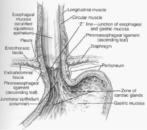
ing surgery. Three layers compose the esophageal mucosa: a stratified, nonkeratinizing squamous epithelial lining; the lamina propria (a matrix of collagen and elastic fibers); and the muscularis mucosae. The squamous epithelium of the esophagus meets the junctional columnar epithelium of the gastric cardia in a sharp transition called the Z-line, typically located at or near the lower esophageal sphincter (Fig. 12.2).
Although the upper third of esophageal muscle is skeletal and the distal portion is smooth, the entire esophagus functions as one coordinated structure. Contraction of the longitudinal muscle fibers of the esophageal body produces esophageal shortening. The inner circular muscle is arranged in incomplete rings, producing a helical pattern that, on contraction, produces a corkscrew-type propulsion. Muscle layers are of uniform thickness until the distal 3 to 4 cm, where the inner circular layer thickens and divides into incomplete horizontal muscular bands on the lesser gastric curve and oblique fibers that become the gastric sling fibers on the greater curve. Although no complete circular band exists as an anatomic lower esophageal sphincter (LES), it is the area of rearranged distal circular fibers that corresponds to the high-pressure zone of the LES. In an adult, the cricopharyngeal muscle is located approximately 15 cm from the incisors, and the gastroesophageal junction is located approximately 45 cm from the incisors.
The esophagus has abundant lymphatic drainage within a dense submucosal plexus. Because the lymphatic system is not segmental, lymph can travel a long distance in the plexus before traversing the muscle layer and entering regional lymph nodes. Tumor cells of the
Figure 12.2. Anatomic relationships of the distal esophagus and phrenoesophageal ligament. (Reprinted from Gray SW, Skandalakis JE, McClusky DA. Atlas of Surgical Anatomy for General Surgeons. Baltimore: Williams & Wilkins, 1985, with permission.)
12. Swallowing Difficulty and Pain 203
upper esophagus can metastasize to superior gastric nodes, or a cancer of the lower esophagus can metastasize to superior mediastinal nodes. More commonly, the lymphatic drainage from the upper esophagus courses into the cervical and peritracheal lymph nodes, while that from the lower thoracic and abdominal esophagus drains into the retrocardiac and celiac nodes.
The esophagus has both sympathetic and parasympathetic innervation. The sympathetic supply is through the cervical and thoracic sympathetic chains as well as through the splanchnic nerves derived from the celiac plexus and ganglia. Parasympathetic innervation of the pharynx and esophagus is primarily through the vagus nerve. The vagal trunks contribute to the anterior and posterior esophageal plexi. At the diaphragmatic hiatus, these plexi fuse to form the anterior and posterior vagus nerves. A rich intrinsic nervous supply called the myenteric plexus exists between the longitudinal and circular muscle layers (Auerbach’s plexus) and in the submucosa (Meissner’s plexus).
Physiology of Swallowing
Passage of food from mouth to stomach requires a well-coordinated series of neurologic and muscular events. The mechanism of swallowing is analogous to a mechanical system consisting of a piston pump (tongue) filling and pressurizing a cylinder (hypopharynx) connected to a three-valve (soft palate, epiglottis, and cricopharyngeus) system that propels material into a worm drive (esophagus) with a single distal valve (LES). Failure of the pump, valves, or worm drive leads to abnormalities in swallowing such as difficulty in propelling food from mouth to stomach or regurgitation of food into the oral pharynx, nasopharynx, or esophagus.
The LES acts as the valve at the end of the esophageal worm drive and provides a pressure barrier between the esophagus and stomach.
Although a precise anatomic LES does not exist, muscle fiber architecture at the esophagogastric junction explains some of the sphincterlike activity of the LES. The resting tone of the LES is approximately 20 mm Hg. With initiation of a swallow, LES pressure decreases, allowing the primary peristaltic wave to propel food into the stomach. A pharyngeal swallow that does not initiate peristaltic contraction also leads to LES relaxation, permitting gastric juice to reflux into the distal esophagus. The coordinated activity of the pharyngeal swallow and LES relaxation appears to be in part vagally mediated. The intrinsic tone of the LES can be affected by diet and medications as well as by neural and hormonal mechanisms (Table 12.1).
History and Physical Examination
A precise medical history is essential to obtaining an accurate diagnosis of swallowing difficulties. Does the patient suffer from difficulty in swallowing (dysphagia) alone, or is pain with swallowing (odynophagia) a primary or associated complaint? If pain is the

204 J.P. Sutyak
primary complaint, elucidate its nature (squeezing, burning, pressure), aggravating factors (temperature and type of food, liquids and/or solids, medications, caffeine, alcohol, position, size or time of meals), relieving factors (medications, position, eructation, emesis), time course (lifelong, several years, slow progression, worsening, stable, episodic, constant), and associated factors (patient age, weight gain or loss, presence of a mass in the neck, preexisting disease processes, chronic cough, asthma, recurrent pneumonia, tobacco and alcohol use). When dysphagia is not associated with pain or with pain as a minor complaint, questioning should still follow the pattern above (nature, aggravating factors/relieving factors/time course/associated factors) and include questions focusing on disease progression (difficulty with solids at first, then difficulty with liquids, or difficulty with both solids and liquids).
Appropriate identification and evaluation of esophageal abnormalities rely on a thorough understanding of the patient’s symptoms and of how these symptoms relate to various disorders. Table 12.2 lists symptoms that may be attributable to esophageal disorders. Occasional symptoms are common and of no pathologic significance. However, frequent and persistent symptoms should prompt further investigation. A useful method is to determine how much the symptoms have affected the patient’s lifestyle in terms of activity, types of food eaten, interruption of employment, and effects on family life.
A precise relationship of symptoms to diagnosis is essential in order to avoid inappropriate and dangerous treatment.
Table 12.1. Neural, hormonal, and dietary factors thought to affect lower esophageal sphincter (LES).
Increase LES pressure Cholinergics Prokinetics a-Agonists b-Blockers
Gastrin
Motilin Bombesin Substance p
Decrease LES Pressure a-Blockers b-Blockers
Calcium channel blockers Cholecystokinin Estrogen
Progesterone
Somatostatin Secretin
Caffeine (chocolate, coffee) Fats
Source: Reprinted from Smith CD. Esophagus. In: Norton JA, Bollinger RR, Chang AE, et al, eds. Surgery: Basic Science and Clinical Evidence. New York: Springer-Verlag, 2001, with permission.

12. Swallowing Difficulty and Pain 205
Table 12.2. Patient symptoms and likely etiologies.
Symptom |
Definition |
Likely etiology |
Heartburn |
Burning discomfort behind breast bone |
Gastroesophageal reflux |
|
Bitter acidic fluid in mouth |
(GER) |
|
Sudden filling of mouth with clear/salty |
|
|
fluid |
|
Dysphagia |
Sensation of food being hindered in |
Motor disorders |
|
passage from mouth to stomach |
Inflammatory process |
|
|
Diverticula |
|
|
Tumors |
Odynophagia |
Pain with swallowing |
Severe inflammatory process |
Globus sensation |
Lump in throat unrelated to swallowing |
|
Chest pain |
Mimics angina pectoris |
GER |
|
|
Motor disorders |
|
|
Tumors |
Respiratory symptoms |
Asthma/wheezing, bronchitis, hemoptysis, |
GER |
|
stridor |
Diverticula |
|
|
Tumors |
ENT symptoms |
Chronic sore throat, laryngitis, halitosis, |
GER |
|
chronic cough |
Diverticula |
Rumination |
Regurgitation of recently ingested food |
Achalasia |
|
into mouth |
Inflammatory process |
|
|
Diverticula |
|
|
Tumors |
Source: Reprinted from Smith CD. Esophagus. In: Norton JA, Bollinger RR, Chang AE, et al, eds. Surgery: Basic Science and Clinical Evidence. New York: Springer-Verlag, 2001, with permission.
Although the majority of preliminary diagnostic information is obtained through a focused history, physical examination can add important clues to the diagnosis, particularly when malignancy is of concern. Signs of chronic or acute weight loss, lymphadenopathy, tobacco abuse, ethanol abuse, portal hypertension, and any abnormal neck or abdominal masses should be noted on physical examination. Further history and examination findings are covered under the specific diagnoses that follow later in this chapter.
Diagnostic Tests for Evaluation of Esophageal and
Swallowing Disorders
Several diagnostic tests are available to evaluate patients with dysphagia/odynophagia. These tests, listed in Table 12.3, can be divided into tests to assess structural abnormalities, tests to assess functional abnormalities, tests to assess esophageal exposure to gastric content, and tests to provoke esophageal symptoms. Gastric motility and biliary disease may need to be evaluated as well to rule out gastroparesis or gallbladder disease. See Algorithm 12.1 for swallowing evaluation.
Assessment of Structural Abnormalities
Radiographic Studies
Plain chest x-ray films may reveal changes in cardiac silhouette or tracheobronchial location, suggesting esophageal disorders. Herniation of

206 J.P. Sutyak
Table 12.3. Assessment of esophageal function.
Condition |
Diagnostic test |
Structural abnormalities |
Barium swallow |
|
Endoscopy |
|
Chest x-ray |
|
CT scan |
|
Cinefluoroscopy |
|
Endoscopic ultrasound |
Functional abnormalities |
Manometry (stationary and 24 hour) |
|
Transit studies |
Esophageal exposure to |
24-hour pH monitoring |
gastric content |
|
Provoke esophageal |
Acid perfusion (Bernstein) |
symptoms |
Edrophonium (Tensilon) |
|
Balloon distention |
Others |
Gastric analysis |
|
Gastric emptying study |
|
Gallbladder ultrasound |
Source: Reprinted from Smith CD. Esophagus. In: Norton JA, Bollinger RR, Chang AE, et al, eds. Surgery: Basic Science and Clinical Evidence. New York: Springer-Verlag, 2001, with permission.
the stomach and other structures above the diaphragm often is identified by abnormal gas patterns on chest radiographs. A simple and often specific diagnostic test for esophageal disease is a contrast esophagogram or barium swallow. Structural abnormalities, including diverticula, narrowing or stricture, ulcers, and hiatal or paraesophageal hernias, all can be demonstrated. Use of fluoroscopy with videotaped recordings of both a liquid and solid contrast swallow increases accuracy in identifying subtle abnormalities. Abnormalities of esophageal motility or gastroesophageal reflux can be seen during a barium swallow, but these disorders are more appropriately diagnosed using other tests. The value of attempting to elicit reflux is questionable because 20% of normal individuals have radiologic reflux. The timed barium esophagogram is a simple test of esophageal function. After ingestion of a premeasured amount of barium, spot films are taken at 1-, 2-, 5-, 10-, and 20-minute intervals. This test allows quantification of esophageal emptying and is useful for the evaluation of motility disorders. Computed tomography (CT) scan of the chest also may be useful in assessing lesions identified with barium swallow or endoscopy thought to be malignancies and in assessing the presence of complex paraesophageal hernias. A modified fluoroscopic barium study in the lateral projection may be useful in identifying mechanical disorders of the pharyngeal swallowing mechanism.
Endoscopy
Most patients with swallowing disorders or pain should undergo esophagoscopy. Patients with dysphagia should undergo esophagoscopy, even in the presence of a normal barium swallow. A barium swallow performed before esophagoscopy helps the endoscopist to focus on any subtle radiographic findings and helps to prevent

12. Swallowing Difficulty and Pain 207
endoscopic misadventures with anatomic abnormalities such as esophageal diverticula.
For the initial assessment, the flexible esophagoscope allows a safe, thorough assessment that can be performed quickly in an outpatient setting with high patient tolerance and acceptance. The mucosa of the entire esophagus, stomach, and duodenum should be inspected carefully. Any areas of mucosal irregularity or abnormality should be photodocumented and biopsied. Retroflex views within the stomach of the gastroesophageal junction should note the presence of hiatal hernia. The location of the transition from squamous mucosa to columnar gastric mucosa (Z-line) should be noted as the distance from the incisors to this point of transition. Known esophageal diverticula can be investigated endoscopically; however, great care should be taken because diverticula can be perforated easily.
Identify if oropharyngeal dysphagia is likely
History
Symptoms
Metabolic disease
Medications (anticholinergics, phenothiazines, etc.)
Physical exam
Identify features of underlying systemic or metabolic disorders
Determine neurologic status
Identify pulmonary and nutritional sequelae of dysphagia
No |
Yes |
Identify structural etiologies: Zenker’s diverticulum, neoplasm, infection, etc.
Endoscopic examination
Radiologic examination
Videofluoroscopy
Cineradiography
Yes
Determine if dysphagia is amenable to therapy
Surgical correction
Swallowing modification techniques
Diet modification
Algorithm 12.1. Algorithm for swallowing evaluation (dysphagia).
208 J.P. Sutyak
Rigid esophagoscopy rarely is indicated and remains a tool used primarily in the operating room when cricopharyngeal or cervical esophageal lesions prevent passage of a flexible scope, when biopsies deeper than those obtainable with flexible endoscopy are needed to stage disease and plan resective therapy, and for the removal of foreign bodies.
Endoscopic ultrasound (EUS) is a newer technique. It allows characterization and staging of esophageal lesions by imaging the layers of the esophageal wall and surrounding structures in order to identify depth of tumor invasion and periesophageal lymphadenopathy, and it allows EUS-guided fine-needle aspiration of lymph nodes.
Assessment of Functional Abnormalities
Esophageal Manometry
Esophageal manometry has become widely available to examine the motor function of the esophagus and the LES (Fig. 12.3). Manometry is indicated when a motor abnormality is suspected on the basis of symptoms of dysphagia or odynophagia and when the barium swallow and esophagoscopy do not show an obvious structural
mm Hg
Figure 12.3. Esophageal manometry. Swallowing requires coordinated relaxation and contraction of the upper and lower esophageal sphincters (UES, LES) and adequate peristalsis of the esophageal body. (Reprinted from Rice TW. Esophagus. In: Norton JA, Bollinger RR, Chang AE, et al, eds. Surgery: Basic Science and Clinical Evidence. New York: Springer-Verlag, 2001, with permission.)
12. Swallowing Difficulty and Pain 209
abnormality. Manometry also is indicated when esophageal dysmotility or LES dysfunction is either associated with or a consequence of other structural abnormalities such as hiatal and paraesophageal hernias. Manometry is essential to confirm diagnosis of primary esophageal motility disorders such as achalasia, diffuse esophageal spasm, nutcracker esophagus, and hypertensive lower esophageal sphincter. It may be useful in identifying nonspecific esophageal motility disorders and motility abnormalities secondary to systemic diseases of scleroderma, dermatomyositis, polymyositis, or mixed connective tissue disease. In patients with symptomatic gastroesophageal reflux, manometry is useful particularly in confirming inadequate function of the LES, assessing preoperative esophageal clearance mechanisms, and determining operative or nonoperative therapy. Esophageal manometry is performed by passing a catheter nasally into the stomach while measuring pressure through a pressuresensitive transducer. Pharyngeal, esophageal body, and LES function can be recorded and assessed using computer-based analysis software.
Assessment of Esophageal Exposure to Gastric Content
Ambulatory 24-hour esophageal pH monitoring has become the standard for quantitating esophageal exposure to acidic content and relating symptoms to esophageal pH. A multichannel pH electrode is positioned with at least two sensors proximal to the manometrically identified LES. Gastric pH can be measured along with esophageal pH. While the patient continues a normal routine, including eating and the usual activities, the pH is recorded throughout a 24-hour cycle. The patient maintains a diary, recording body positions, meals, and symptoms, so that esophageal pH can be correlated with symptoms. At the completion of the test, the results are tallied and compared to normal values for esophageal acid exposure. The study can be performed in the presence or absence of acid-reducing medications in order to determine the effectiveness of the medication.
Twenty-four-hour pH monitoring is indicated for patients who have typical symptoms of gastroesophageal reflux, for patients for whom other diagnostic tests are equivocal, for patients with atypical symptoms of gastroesophageal reflux such as noncardiac chest pain, persistent cough, wheezing, and unexplained laryngitis, or for patients with previously failed esophageal or gastric surgery with recurrent symptoms.
Provocation of Esophageal Symptoms
Three tests previously were used to identify a relationship between symptoms and esophageal exposure to acid or motor abnormalities: the acid perfusion test (Bernstein, 0.1 N HCl infusion), edrophonium (Tensilon, acetylcholinase inhibitor precipitates contractions), and balloon distention tests. Perfusion tests are placebo controlled. Ambulatory pH testing and esophageal manometry have made these tests primarily of historical and academic interest.
210 J.P. Sutyak
Evaluation of Gastric Motility and Biliary Disease
In evaluating a patient with esophageal symptoms, it also is important to consider the impact of gastroduodenal dysfunction on lower esophageal function as well as other common gastrointestinal problems that can mimic esophageal disease. A gastric emptying study and/or right upper quadrant ultrasound may be indicated in patients with symptomatology suggestive of esophageal disorders in order to rule out gastroparesis or gallbladder disease.
Specific Conditions
Tumors
Malignant Esophageal Tumors
Overview: The majority of esophageal neoplasms are malignant. Less than 1% of tumors are benign. Esophageal cancer is among the top 10 leading causes of cancer deaths in the United States and is increasing in incidence. Late presentation and early spread lead to a poor prognosis. The overall 5-year survival rate is 8% to 12%. Although squamous cell carcinoma previously accounted for 90% to 95% of reported esophageal malignancies, the incidence of adenocarcinoma has increased dramatically in the past two decades and now accounts for at least 40% of all malignancies. This relative change may reflect the increased use of flexible endoscopy and closer surveillance of asymptomatic patients who are at risk of developing esophageal carcinoma. Other cell types of esophageal malignancy are extremely rare.
Squamous cell carcinomas are distributed equally among the upper, middle, and lower thirds of the esophagus. Adenocarcinomas most commonly are found in the lower third of the esophagus. Most adenocarcinomas are thought to arise in columnar cell Barrett’s mucosa. Alcohol consumption and tobacco use are well-established factors for the development of esophageal carcinoma. Other risk factors for esophageal cancer include achalasia, radiation esophagitis, caustic esophageal injury, infection (human papilloma virus), Plummer– Vinson syndrome, leukoplakia, esophageal diverticula, ectopic gastric mucosa, and the inherited condition of familial keratosis palmaris et plantaris (tylosis).
Diagnosis: The vast majority of esophageal carcinomas are clinically occult and present well after disease progression prevents cure. As in Case 1, the classic history is a patient who presents with dysphagia and weight loss; chest pain, abdominal pain, and gastrointestinal (GI) blood loss are described less frequently (Table 12.4). Most patients experience dysphagia an average of 2 to 4 months before presentation. Unfortunately, dysphagia almost uniformly indicates extensive disease and incurability.
The initial study should be a barium swallow; this most frequently reveals distinct mucosal irregularity, stricture, a shelf in the lower esophagus, or rigidity. Upper esophageal endoscopy allows visualization of the affected area and biopsy to confirm the diagnosis.

12. Swallowing Difficulty and Pain 211
Table 12.4. Presenting symptoms of esophageal carcinoma.
Symptom |
Incidence (%) |
Dysphagia |
87 |
Weight loss |
71 |
Substernal or epigastric pain/ |
46 |
burning |
|
Vomiting or regurgitation |
28 |
Aspiration pneumonia |
14 |
Palpable cervical nodes |
14 |
Hoarseness |
7 |
Coughing and choking |
3 |
Source: Reprinted from Smith CD. Esophagus. In: Norton JA, Bollinger RR, Chang AE, et al, eds. Surgery: Basic Science and Clinical Evidence. New York: Springer-Verlag, 2001, with permission.
Staging: The stage of esophageal cancer is determined by the depth of penetration of the primary tumor (T) and the presence of lymph node (N0, N1) and distant organ metastasis (M0, M1). The TNM descriptors can be grouped into stages with similar behavior and prognosis. Clinical (cTNM) preoperative staging determines the intent and extent of subsequent treatment, either curative or palliative. Initial staging includes a careful physical examination for the sequelae of esophageal cancer (weight loss, supraclavicular adenopathy, pleural effusion), routine blood tests, and CT scan of the chest and abdomen.
Bronchoscopy is indicated for midesophageal tumors because of their propensity to invade the trachea and left mainstem bronchus.
The recent development of EUS has improved staging by allowing the depth of invasion into the esophageal wall to be determined accurately and by allowing the surrounding lymph node involvement to be identified and biopsied.
Weight loss greater than 10% has been shown to be associated with a significantly poorer outcome in patients with operable esophageal cancer. Clinical staging categorizes patients into two groups: those with potentially curable disease and those with metastatic disease (disease outside of the local or regional area) in whom palliation is currently the only treatment option.
Treatment: The overall prognosis for esophageal carcinoma is bleak. An overall 5-year survival for esophageal cancer patients was reported in only 4% after surgical resection (surgical mortality, 29%) and in only 6% after radiation therapy. The treatment of esophageal cancer is generally a palliative practice, and cure is a chance occurrence. However, precise clinical staging allows treatment modification of patients with carcinoma of the esophagus. Surgical, radiation, and chemotherapy therapies are possible, with optimal outcomes often utilizing a combination approach.
Based on reviews of current literature available on the multimodality management of patients with esophageal carcinoma, treatment pro-

212 J.P. Sutyak
Technically resectable esophageal cancer (Stage 0–III)
If available, entry into clinical trial
Surgery: esophagectomy
Curative resection
Yes
(negative margins, |
|
No |
|
(positive margins, |
|
no distant mets) |
|
|
|
distant mets) |
|
|
|
|
|
|
|
Adjuvant therapy: |
|
Palliative therapy: |
Radiotherapy (5000 – 6000 cGy+ |
|
Radiotherapy (4500 cGyt) |
external boost) |
|
+/– |
+ |
|
Chemotherapy (2 cycles |
Chemotherapy (2 cycles platinum/5-FU) |
|
platinum/5-FU) |
|
|
|
Algorithm 12.2. Management of technically resectable esophageal cancer, 5-Fu, 5-fluorouracil; mets, metastases. (Reprinted from Smith CD. Esophagus. In: Norton JA, Bollinger RR, Chang AE, et al, eds. Surgery: Basic Science and Clinical Evidence. New York: Springer-Verlag, 2001, with permission.)
tocols have been developed and are the basis of published practice guidelines for esophageal cancer (see Algorithms 12.2 and 12.3).1
Surgery: Esophagectomy with gastric or colon replacement is the treatment of choice for small tumors confined to the esophageal mucosa or submucosa. Surgical treatment for resectable esophageal cancers results in 5-year survival rates of 5% to 20% and an operative
1 DeMeester TR, Bonavina L, Albertucci M. Evaluation of primary repair in 100 consecutive patients. Ann Surg 1986;204:9–20. Donohue PE, Samelson S, Nyhus LM, et al. The Nissen fundoplication. Effective long-term control of pathologic reflux. Arch Surg 1985;120:663–667. Ellis FH. The Nissen fundoplication. Ann Thorac Surg 1992;54:1231–1235. Grande L, Toledo-Pimentel V, Manterola C, et al. Value of Nissen fundoplication in patients with gastro-oesophageal reflux judged by long-term symptom control. Br J Surg 1994;81:548–550. Johansson J, Johnsson F, Joelsson B, et al. Outcome 5 years after 360 degree fundoplication for gastro-oesophageal reflux disease. Br J Surg 1993;80:46–49. Luostarinen M, Isolauri J, Laitinen J, et al. Fate of Nissen fundoplication after 20 years. A clinical, endoscopical, and functional analysis. Gut 1993;34:1015–1020. Macintyre IM, Goulbourne IA. Long-term results after Nissen fundoplications: a 5–15- year review. J R Coll Surg Edinb 1990; 35:159–162. Martin CJ, Cox MR, Cade RJ. CollisNissen gastrooplasty fundoplication for complicated gastrooesophageal reflux disease. Aust N Z J Surg 1992;62:126–129. Mira-Navarro J, Bayle-Bastos F, Frieyro-Segui M, et al. Long-term follow-up of Nissen fundoplication. Eur J Pediatr Surg 1994;4:7–10.

12. Swallowing Difficulty and Pain 213
mortality of 2% to 7%. Once symptoms appear, most esophageal cancers have invaded adjacent structures or have spread to distant organs. In those cases in which significant obstructive symptoms exist, operative management often is the most effective means of relieving dysphagia and providing long-term palliation. In general, because esophageal cancer can have extensive and unpredictable spread longitudinally, it seems prudent to perform total esophagectomy, especially for those proximaland middle-third lesions. Distal small lesions may be approached through the abdomen only, or resection for palliation alone can avoid total esophagectomy and its associated morbidity.
Nonsurgical Therapy: A randomized phase III study of chemotherapy with cisplatin, fluorouracil, and radiotherapy versus radiation therapy alone in patients with T1–3, N0–1, M0 esophageal carcinoma has demonstrated a survival advantage for patients receiving combined therapy (Table 12.5). Long-term follow-up of these patients reported a 5-year survival of 26% for combined therapy, while no patient receiving radiation alone survived 5 years. A 5-year survival of 14% was seen
Unresectable esophageal cancer (Stage IV)
If available, entry into clinical trial
Palliative strategies
Chemotherapy if : |
|
|
|
|
|
|
a) no dysphagia |
|
|
|
|
|
|
b) poor operative candidate |
|
|
|
Endoscopic stent |
||
c) locally advanced |
|
|
|
Laser ablation |
||
d) metastatic disease |
|
|
|
Photodynamic therapy |
||
e) good performance status |
|
|
|
|
|
|
|
|
|
|
|||
|
|
|
|
|
|
|
|
|
|
|
|
|
|
|
|
|
|
Radiotherapy: |
|
|
|
Palliative esophagectomy if: |
|
3000 cGy +/–5-FU |
|
||
|
|
radiosensitizer |
|
|||
|
a) dysphagia |
|
|
|||
|
|
|
|
|
||
|
b) good performance status |
|
|
|
|
|
|
c) locally resectable |
|
|
|
|
|
|
|
|
|
|
|
|
Algorithm 12.3. Management of stage IV esophageal cancer. (Reprinted from Smith CD. Esophagus. In: Norton JA, Bollinger RR, Chang AE, et al, eds. Surgery: Basic Science and Clinical Evidence. New York: Springer-Verlag, 2001, with permission.)

214 J.P. Sutyak
Table 12.5. Phase III treatment trials for esophageal carcinoma.
Author |
Cell type |
R1 |
R2 |
Survival |
Positive findings |
|
Cooper et ala |
Both |
Rad |
Che/Rad |
0% vs. 26% |
≠ Toxicity, R2 |
|
Law et alb |
|
|
|
(5 years) |
≠ Downstaging, R2 |
|
Squ |
Surg |
Che/Surg |
Same |
|||
Kelsen et alc |
Both |
Surg |
Che/Surg |
Same |
≠ Curative resection, R2 |
|
Ø N1 and M1 R2 |
||||||
Walsh et ald |
Adeno |
Surg |
Che/Rad/Surg |
6% vs. 32% |
||
Bosset et ale |
|
|
|
(3 years) |
≠ DF survival, R2 |
|
Squ |
Surg |
Che/Rad/Surg |
Same |
|||
Ando et alf |
Squ |
Surg |
Surg/Che |
Same |
≠ Curative resection, R2 |
|
|
Adeno, adenocarcinoma; Che, chemotherapy; Rad, radiation therapy; Squ, squamous cell carcinoma; Surg, surgery. a Cooper JS. Guo MD. Herskovic A, et al. Chemoradiotherapy of locally advanced esophageal cancer. Long-term follow-up of a prospective randomized trial (RTOG 85-01). JAMA 1999;281:1623–1627.
b Law S, Fok M, Chow S, et al. Preoperative chemotherapy versus surgery alone for squamous cell carcinoma of the esophagus: a prospective randomized trial. J Thorac Cardiovasc Surg 1997;114:210–217.
c Kelsen DP, Ginsberg R, Pajak TF, et al. Chemotherapy followed by surgery compared to surgery alone for localized esophageal cancer. N Engl J Med 1998;339:1979–1984.
d Walsh TN, Noonan N, Hollywood D, et al. A comparison of multimodality therapy and surgery for esophageal adenocarcinoma. N Engl J Med 1996;335:462–467.
e Bosset JF, Gignoux M, Triboulet JP, et al. Chemoradiotherapy followed by surgery compared with surgery alone in squamous cell cancer of the esophagus. N Engl J Med 1997;337:161–167.
f Ando N, Iizuka T, Kakegawa T, et al. A randomized trial of surgery with and without chemotherapy for localized squamous cell carcinoma of the thoracic esophagus. J Thorac Cardiovasc Surg 1997;114:205–209.
Source: Reprinted from Rice TW. Esophagus. In: Norton JA, Bollinger RR, Chang AE, et al, eds. Surgery: Basic Science and Clinical Evidence. New York: Springer-Verlag, 2001, with permission.
in patients receiving combined therapy after randomization was closed.2
Palliation: Patients with distant metastases or contraindications to chemoradiotherapy or surgery should be considered for palliative interventions. Local and regional treatment modalities are the cornerstones of symptomatic control. Palliative radiation therapy is a key component and is associated with significant, albeit short-term, success in maintaining adequate swallowing. The value of systemic chemotherapy is limited. Concurrent chemotherapy and radiation have been used in the palliation of patients with metastatic tumors.
2 Cooper JS, Guo MD, Herskovic A, et al. Chemoradiotherapy of locally advanced esophageal cancer. Long-term follow-up of a prospective randomized trial (RTOG 85-01). JAMA 1999;281:1623–1627. Law S, Fok M, Chow S, et al. Preoperative chemotherapy versus surgery alone for squamous cell carcinoma of the esophagus: a prospective randomized trial. J Thorac Cardiovasc Surg 1997;114:210–217. Kelsen DP, Ginsberg R, Pajak TF, et al. Chemotherapy followed by surgery compared to surgery alone for localized esophageal cancer. N Engl J Med 1998;339:1979–1984. Walsh TN, Noonan N, Hollywood D, et al. A comparison of multimodality therapy and surgery for esophageal adenocarcinoma. N Engl J Med 1996;335:462–467. Bosset JF, Gignoux M, Triboulet JP, et al. Chemoradiotherapy followed by surgery compared with surgery alone in squamous cell cancer of the esophagus. N Engl J Med 1997;337:161–167. Ando N, Iizuka T, Kakegawa T, et al. A randomized trial of surgery with and without chemotherapy for localized squamous cell carcinoma of the thoracic esophagus. J Thorac Cardiovasc Surg 1997;114:205–209.
12. Swallowing Difficulty and Pain 215
While efficacious in improving local and regional control, this treatment comes with a significantly increased risk of toxicity and may not be appropriate in most patients.
A number of local measures can preserve swallowing and avoid the toxicity of chemotherapy and radiotherapy. Dilation of malignant strictures with bougies or endoscopic balloon dilators temporarily can relieve dysphagia. Dilation is typically performed not as a sole therapy but as a prelude to other, more definitive measures. Injection with alcohol causes tumor necrosis and a decrease in the exophytic portion of the tumor. However, dysphagia usually recurs within 30 days, necessitating retreatment.
Laser therapy is reserved for patients with severe obstruction of the esophagus requiring palliation until chemotherapy and radiotherapy take effect. It also is used in patients who are not candidates for prosthesis placement because of an anticipated short life expectancy. Photodynamic therapy provides longer relief of dysphagia than neodymium:yttrium-aluminum-garnet (Nd:YAG) laser therapy, but patients must avoid sunlight up to 30 days after injection because of dermal photosensitivity. This is not a desirable method of palliation for patients whose life expectancy is measured in weeks or months.
Newer, self-expanding metal stents are easier to place and require much less tumor dilation before placement. Varied lengths and configurations are available. Silicone-covered stents prevent tumor ingrowth but are more apt to migrate than noncovered stents; they are the prostheses of choice in the treatment of malignant fistula between the airway and esophagus. Stent placement after chemotherapy or radiotherapy may be associated with increased complications. Modern stents provide effective, long-lasting palliation with little morbidity and are the first mode of palliation considered for patients with esophageal carcinoma.
Benign Esophageal Tumors
Leiomyomas, Cysts, and Polyps: Benign tumors of the esophagus are uncommon, with three histologic types accounting for 87% of benign esophageal tumors: leiomyomas, cysts, and polyps. These three tumors have distinct locations in the esophagus that reflect their cells of origin.
Polyps occur almost exclusively in the cervical esophagus, while leiomyomas and cysts tend to occur in the distal two thirds.
Leiomyomas constitute 50% of benign tumors of the esophagus, with an average patient age at presentation of 38 years, in contrast to esophageal malignancy, which typically presents at a more advanced age.
Esophageal cysts are commonly congenital and are lined by columnar epithelium of the respiratory type, glandular epithelium of the gastric type, squamous epithelium, or transitional epithelium. Enteric and bronchogenic cysts are the most common. Treatment is similar to that for leiomyoma, with resection for large or symptomatic lesions. The cyst wall should be removed completely. Search for a fistulous tract
216 J.P. Sutyak
to the respiratory tract should be carried out, especially in patients who have had recurrent respiratory tract infections.
Gastroesophageal Reflux Disease (GERD)
Overview
Gastroesophageal reflux (GER) is defined as failure of the antireflux barrier, allowing abnormal reflux of gastric contents into the esophagus. It is a mechanical disorder that is caused by an inadequate LES, which may be associated with a gastric emptying disorder or failed esophageal peristalsis. These abnormalities result in a spectrum of symptoms and diseases ranging from “heartburn” to esophageal tissue damage with subsequent complications of ulceration and stricture formation. A host of extraesophageal manifestations of GER, such as asthma, laryngitis, and dental breakdown, also are being increasingly identified. Gastroesophageal reflux is an extremely common condition, accounting for nearly 75% of all esophageal pathology. Nearly 44% of Americans experience monthly heartburn, and 18% of these individuals use nonprescription medication directed against GER.
Pathophysiology
Antireflux Mechanism: Although our understanding of the antireflux barrier is incomplete and has evolved over many years, a current view is that the LES, diaphragmatic crura, and phrenoesophageal ligament are key components, and LES dysfunction is the most common cause of GER. Three factors determine the competence of the LES: resting LES pressure, resting LES length, and abdominal length of the LES. Lower esophageal sphincter dysfunction may be either physiologic and transient or pathologic and permanent. Nearly everyone experiences physiologic reflux, most commonly related to gastric distention following a meal. Postprandial gastric distention results in pressure against the LES, which stretches and pulls the sphincter open while shortening the LES length. The resulting incompetence of the LES leads to transient periods of reflux. These transient episodes of reflux are relieved with gastric venting (belching) or when the stomach empties normally. Overeating exacerbates these episodes, and a high-fat Western diet may delay gastric emptying, thereby extending the duration of these transient episodes. Evidence is accumulating that chronic, gastric-related, transient physiologic reflux leads to sufficient esophageal injury to cause dysfunction of the antireflux barrier; this then progresses to more permanent and pathologic reflux.
Consequences of Reflux: Gastroesophageal reflux may lead to symptoms related to the reflux of gastric content into the esophagus, lungs, or oropharynx, or to damage to the esophageal mucosa and respiratory epithelium with subsequent changes related to repair, fibrosis, and reinjury. Manifestations of GER typically are classified as esophageal and extraesophageal. Esophageal symptoms of GER include heartburn, chest pain, water brash, or dysphagia. Dysphagia often implicates complicated GER with esophagitis and ulceration, stricture, or Barrett’s metaplastic changes. Extraesophageal manifestations gener-
12. Swallowing Difficulty and Pain 217
ally are pulmonary, resulting from pulmonary aspiration or bronchospasm induced when reflux stimulates a distal esophageal vagal reflex. Extraesophageal symptoms and signs include chronic cough, laryngitis, dental damage, and chronic sinusitis. With a better understanding of GER and new therapies for eliminating symptoms, fewer patients are presenting with severe complications of GER. However, those with complicated GER (high-grade esophagitis, stricture, or Barrett’s mucosa) have more severe reflux, suggesting a mechanically defective LES as a major etiologic factor. Reconstruction of the defective LES has been the basis of offering operative therapy for complicated GER.
Diagnosis
The clinical diagnosis of GER is fairly straightforward if the patient reports the classic symptom of heartburn that is readily relieved after ingesting antacids. As in Case 2, many patients with this classic presentation will have been treated with an empiric trial of H2 blockers or proton pump inhibitors (PPIs). Other typical symptoms of GER include regurgitation or dysphagia. Chest pain, asthma, laryngitis, recurrent pulmonary infections, chronic cough, and hoarseness may be associated with reflux. Increasing numbers of patients with these atypical GER symptoms are being evaluated for reflux.
A careful history should confirm both typical and atypical symptoms of GER and any response to medical therapy. Atypical symptoms, no response to high-dose medication, dysphagia, odynophagia, GI bleeding, or weight loss suggests complications of GER or another disease process entirely and should prompt a more thorough evaluation. In a patient with typical symptoms, endoscopic findings of esophageal erosions, ulcers, or columnar-lined esophagus are fairly specific for GER. During esophagogastroduodenoscopy (EGD), an esophageal mucosal biopsy should be obtained to confirm esophagitis, and esophageal length and the presence of a hiatal hernia or stricture can be assessed. This eliminates the need for a confirmatory barium swallow. With typical findings, no other tests beyond EGD are necessary to diagnose GER. However, in many patients, the EGD will be normal due to empiric treatment of symptoms. In this setting, 24-hour pH testing is necessary to objectively establish the diagnosis of GER.
Ambulatory 24-hour pH monitoring has been regarded as the gold standard in diagnosing GER and is of unquestionable benefit in patients where the diagnosis is unclear or in those with nonerosive esophagitis on EGD. Ambulatory pH monitoring is not mandatory in patients with typical reflux symptoms and erosive esophagitis on EGD.
Barium swallow is the test of choice in evaluating the patient with dysphagia, suspected stricture, paraesophageal hernia, or shortened esophagus. Other studies may be helpful in difficult cases, such as gastric emptying studies in patients with significant bloating, nausea, or vomiting.
Treatment
There is considerable debate regarding optimal treatment of GER. With many Americans experiencing daily heartburn and the established
218 J.P. Sutyak
impact this condition has on an individual’s quality of life, there is tremendous amount of interest and effort devoted to understanding this condition and establishing treatment algorithms that are effective and cost efficient. See Algorithms 12.4 and 12.5 for treatment algorithms for uncomplicated and complicated GER.
Medical: The principles of nonoperative management of GER include lifestyle modifications, medical therapy for symptomatic control, and identification of those who would be best served with an antireflux operation. Although lifestyle modifications always have been the initial step in therapy, only those patients with mild and intermittent symptoms seem to benefit from lifestyle changes alone. Most patients who seek medical advice are best treated with either medication or an operation. Selection of a particular medical regimen depends on the
|
|
|
|
Trial of H2RA |
||
Responds to H2RA |
|
Suboptimal response to H2RA |
||||
|
|
|
|
|
|
|
Maintenance therapy |
|
|
||||
|
vs. |
|
|
|
||
Treat on demand |
|
|
|
|||
|
|
|
|
Titrate H2RA dose |
||
|
|
|
|
|
or |
|
|
|
|
|
|
Begin PPI |
|
Suboptimal response |
Responds |
|||||
|
|
|
|
|||
|
|
|
|
|
||
|
Confirm diagnosis |
Maintenance therapy |
||||
No GER |
GER |
|
|
|||
|
|
|
|
|||
|
|
|
|
|
||
Further workup |
|
Refer for surgery |
||||
Algorithm 12.4. Management algorithm for treatment of uncomplicated gastroesophageal reflux (based on endoscopic findings). H2RA, H2 receptor antagonist; PPI, proton pump inhibitor; GER, gastroesophageal reflux. (After Fennerty MB, Castell D, Fendrick AM, et al. The diagnosis and treatment of gastroesophageal reflux disease in a managed care environment. Suggested disease management guidelines. Arch Intern Med 1996;156:477–484, with permission. Copyright © 1996 American Medical Association. All Rights Reserved. Reprinted from Smith CD. Esophagus. In: Norton JA, Bollinger RR, Chang AE, et al, eds. Surgery: Basic Science and Clinical Evidence. New York: Springer-Verlag, 2001, with permission.)

12. Swallowing Difficulty and Pain 219
Trial of PPI
Responds |
|
Suboptimal response |
|||
|
|
|
|
||
|
|
|
|
|
|
Maintenance therapy |
|
|
|||
|
|
|
Increase PPI |
|
|
Suboptimal response |
Responds |
||||
|
|
|
|||
|
|
|
|
|
|
Confirm diagnosis |
Maintenance therapy |
||||
No GER |
GER |
|
|
||
Refer for surgery
Further workup
Algorithm 12.5. Management algorithm for treatment of complicated gastroesophageal reflux (based on endoscopic findings). PPI, proton pump inhibitor; GER, gastroesophageal reflux. (After Fennerty MB, Castell D, Fendrick AM, et al. The diagnosis and treatment of gastroesophageal reflux disease in a managed care environment. Suggested disease management guidelines. Arch Intern Med 1996;156:477–484, with permission. Copyright © 1996 American Medical Association. All Rights Reserved. Reprinted from Smith CD. Esophagus. In: Norton JA, Bollinger RR, Chang AE, et al, eds. Surgery: Basic Science and Clinical Evidence. New York: Springer-Verlag, 2001, with permission.)
severity of GER, effectiveness of the proposed therapy, cost, and convenience of the regimen.
Numerous trials have shown that short-term treatment of GER with acid-suppression regimens can effectively relieve symptomatic GER and heal reflux esophagitis in approximately 90% of cases with intensive therapy. Levels of success depend on the type, duration, and dosage of antisecretory therapy. The PPIs have profoundly changed the medical treatment of GER. Rates of healing of esophagitis have improved dramatically with PPIs when compared to H2 receptor antagonists. The cost of PPIs has led many to recommend their use in only complicated or refractory GER. It appears that reflux symptoms permanently disappear only in a minority of patients with GER when they are taken off medications. Recurrence of symptoms and esophagitis is observed frequently, and thus treatment strategies based on effectiveness and outcome must be based on long-term follow-up. In fact, reflux disease must be considered a lifelong disease that requires a lifelong treatment strategy.
Surgical: Treatment algorithms for both uncomplicated (see Algorithm 12.4) and complicated (see Algorithm 12.5) reflux esophagitis begin

220 J.P. Sutyak
with medical therapy. The expense and psychological burden of a lifetime of medication dependence, undesirable lifestyle changes, the uncertainty as to the long-term effects of some newer medications, and the potential for persistent mucosal changes despite symptomatic control all make surgical treatment of GER an attractive option. Surgical therapy, which addresses the mechanical nature of this condition, is curative in 85% to 93% of patients. Chronic medical management may be most appropriate for patients with limited life expectancy or comorbid conditions that would prohibit safe surgical intervention.
Historically, antireflux surgery was recommended only for patients with refractory or complicated gastroesophageal reflux. Recent developments have affected the long-term management of patients with GER. The rapid postoperative recovery seen with laparoscopic surgery is now feasible following antireflux procedures. The widespread availability and use of ambulatory pH monitoring has improved recognition of true GER and selection of patients for long-term therapy. Physicians now recognize that patients with GER have a greatly impaired quality of life, which normalizes with successful treatment.
The management goals of GER have changed. Rather than focusing therapy only on controlling symptoms, modern treatment aims to eliminate symptoms, improve a patient’s quality of life, and institute a lifelong plan for management. Controlled trials that compared medical and surgical therapy of GER have favored surgical therapy.
Surgical treatment was significantly more effective in improving symptoms and endoscopic signs of esophagitis for as long as 2 years. Other longitudinal studies report good to excellent long-term results in 80% to 93% of surgically treated patients (Table 12.6).
Table 12.6. Medical versus surgical treatment of Barrett’s esophagus.
|
|
No. patients |
|
Symptom control |
|
Stricture/esophagitis |
|||
Author |
Medical |
Surgical |
|
Medical |
Surgical |
|
Medical |
Surgical |
|
|
|
|
|
|
|
|
|||
Attwood 1992b (p)a |
26 |
19 |
22% |
81% |
38% |
21% |
|||
Oritz 1996c (pr) |
27 |
32 |
85% |
89% |
53%/45% |
5%/15% |
|||
Sampliner 1994d (p) |
27 |
— |
70% |
— |
50% |
— |
|||
Csendes 1998e (pr) |
— |
152 |
|
— |
46% |
|
— |
64% |
|
pr, prospective randomized; p, prospective; ret, retrospective. |
|
|
|
|
|||||
a |
Before availability of proton pump inhibitors. |
|
|
|
|
|
|
||
b |
Attwood SE, Barlow AP, Norris TL, et al. Barrett’s oesophagus: effect of antireflux surgery on symptom control |
||||||||
and development of complications. Br J Surg 1992;79:1050–1053. |
|
|
|
|
|||||
c |
Ortiz A, Martinez de Haro LF, Parrilla P, et al. Conservative treatment versus antireflux surgery in Barrett’s oesoph- |
||||||||
agus: long-term results of a prospective study. Br J Surg 1996;83:274–278. |
|
|
|
|
|||||
d Sampliner RE. Effect of up to 3 years of high-dose lansoprazole on Barrett’s esophagus. Am J Gastroenterol 1994;89:1844–1848.
e Csendes A, Braghetto I, Burdiles P, et al. Long-term results of classic antireflux surgery in 152 patients with Barrett’s esophagus: clinical, radiologic endoscopic, manometric, and acid reflux test analysis before and late after operation. Surgery (St. Louis) 1998;126:645–657.
Source: Reprinted from Smith CD. Esophagus. In: Norton JA, Bollinger RR, Chang AE, et al, eds. Surgery: Basic Science and Clinical Evidence. New York: Springer-Verlag, 2001, with permission.
12. Swallowing Difficulty and Pain 221
Indications: Antireflux surgery should be considered in patients in whom intensive medical therapy has failed. With the advent of proton pump inhibitors, true medical failures are unusual. Antireflux surgery also should be offered to patients whose symptoms recur immediately after stopping medications and who require long-term daily medication. Many patients want to avoid the cost, inconvenience, and side effects of long-term medication and want to preserve their quality of life. Complications of GER, such as Barrett’s esophagus and esophageal stricture, should not alter the approach to long-term management. However, patients with these complications usually have more severe disease, require more intensive medical therapy, and are referred for surgical evaluation. Occasionally, GER presents atypically with chest pain, asthma, chronic cough, or hoarseness. Patients with these atypical symptoms usually improve with surgery. However, appropriate patient selection can be very difficult. Ambulatory pH monitoring has been thought to provide the most objective way to select these patients for surgery, but an abnormal pH study does not correlate well with symptom relief following antireflux surgery. Therefore, a trial of medical therapy with resolution of symptoms remains the best way to prove an association between GER and an individual’s atypical symptoms. When such an association exists, antireflux surgery is indicated.
Preoperative Evaluation: The preoperative evaluation should both justify the need for surgery and direct the operative technique to optimize outcome. At a minimum, all patients being considered for surgery should undergo a thorough history and a physical exam, EGD, and esophageal manometry. Esophageal manometry allows evaluation of the lower esophageal sphincter and is diagnostic in differentiating GER from achalasia. Equally important is its use in assessing esophageal body pressures and identifying individuals with impaired esophageal clearance who may not do as well with a 360-degree fundoplication.
Procedures: To establish an effective antireflux barrier, operative procedures for GER are designed to restore adequate LES pressure, position the LES within the abdomen where it is under positive (intraabdominal) pressure, and to close any associated hiatal defect.
Advances in laparoscopic technology and technique allow the reproduction of “open” procedures while eliminating the morbidity of an upper midline incision. Open antireflux operations remain indicated when the laparoscopic technique is not available or is contraindicated. Contraindications to laparoscopic antireflux surgery include uncorrectable coagulopathy, severe chronic obstructive pulmonary disease (COPD) such that CO2 elimination is impeded during laparoscopy, and advanced pregnancy. Only a very experienced laparoscopic surgeon should attempt the minimally invasive approach in the presence of previous upper abdominal operation or prior antireflux surgery.
In patients with normal esophageal body peristalsis, laparoscopic Nissen fundoplication (Fig. 12.4) has emerged as the most widely accepted and applied antireflux operation. Thousands of laparoscopic Nissen fundoplication patients have been reported in the world litera-

222 J.P. Sutyak
Figure 12.4. Depiction of Nissen 360degree fundoplication. (Reprinted from Smith CD. Esophagus. In: Norton JA, Bollinger RR, Chang AE, et al, eds. Surgery: Basic Science and Clinical Evidence. New York: Springer-Verlag, 2001, with permission.)
ture, with 93% of patients symptom free at 1 year postoperatively. Only 3% require some medical therapy for symptom control. Overall, 97% of patients are satisfied with their results. Transient dysphagia occurs in nearly 50% and resolves within 3 weeks of surgery.
Summary: Antireflux surgery is indicated in any patient with GER refractory to medical management or in any patient who has symptom recurrence when medicine is withdrawn. In many patients with classic symptoms, an EGD and esophageal manometry are all the preoperative testing necessary. Additional tests are confirmatory in difficult cases. The laparoscopic Nissen fundoplication is both safe and effective in the long-term management of nearly all patients with chronic GER. The Toupet fundoplication may be best used in patients with impaired esophageal body peristalsis.
Hiatal Hernias: Sliding and Paraesophageal Hernias
Overview
The majority of patients with hiatal hernia are asymptomatic, and the diagnosis often is made incidentally during investigation of other gastrointestinal problems. The underlying etiology of hiatal hernias remains unclear.
Hiatal hernias can be classified into four types. A type I hiatal hernia (Fig. 12.5) also is known as a sliding hiatal hernia. It consists of a simple herniation of the gastroesophageal junction into the chest. The phrenoesophageal ligament is attenuated, and there is no true hernia sac. This is the most common hiatal hernia and is frequently diagnosed in women and in the fifth and sixth decades of life. Type II hiatal hernias (Fig. 12.5) are commonly referred to as paraesophageal hernias. The gastroesophageal junction remains at the esophageal

12. Swallowing Difficulty and Pain 223
hiatus while the gastric fundus herniates alongside the esophagus, through the hiatus, and into the chest. Type III hiatal hernias are a combination of type I and type II hernias, with the esophagogastric junction being displaced into the chest along with the gastric fundus and body. Paraesophageal hernias (types II and III) have a true hernia sac accompanying the herniated stomach. Type IV hernias are an advanced stage of paraesophageal hernia in which the entire stomach and other intraabdominal contents (colon, spleen) are herniated into the chest. As in Case 3, paraesophageal hernias are found predominantly in older individuals.
Diagnosis
When symptoms are present, sliding hernias have a different presentation from paraesophageal hernias. Paraesophageal hernias tend to produce more dysphagia, chest pain, bloating, and respiratory problems than do sliding hernias. Symptoms associated with a sliding hernia more often are related to LES dysfunction and include classic reflux symptoms, heartburn, regurgitation, and dysphagia.
|
|
|
|
Esophageal mucosa |
|
|
|
|
||||||||
|
|
|
|
|
|
|
|
|
|
|
|
|
|
|
|
|
|
|
|
|
Esophageal muscle |
|
|||||||||||
Squamo- |
|
|||||||||||||||
|
|
|
|
|
|
|
|
|
|
|
|
|
||||
columnar |
|
|
|
|
|
|
|
|
|
|
|
|
|
|
|
|
|
|
|
|
|
|
|
|
|
|
|
|
|
|
|
|
|
|
|
|
Phrenoesophageal |
|
|
|
||||||||||
junction |
|
|
|
|
|
|
||||||||||
|
|
|
membrane |
|
|
|
||||||||||
Stomach |
|
|
|
|
|
|||||||||||
|
|
|
|
|
|
|
|
|
|
|
|
|
|
|
||
|
|
|
|
|
Endothoracic fascia |
|
||||||||||
|
|
|
|
|
|
|
|
|||||||||
|
|
|
|
|
|
|
|
|
|
|
|
|
||||
|
|
|
|
|
|
|
|
|
Diaphragm |
|
|
|||||
|
|
|
|
|
|
|
|
|
|
|
|
|||||
|
|
|
|
|
|
|
|
Endoabdominal fascia |
|
|||||||
|
|
|
|
|
|
|
|
|
|
|
|
|||||
|
|
|
|
|
|
|
|
Peritoneum |
|
|||||||
|
|
|
|
|
|
|
|
|||||||||
|
|
|
|
|
|
Type I |
|
|
||||||||
|
|
|
|
|
|
hiatal hernia |
|
|
||||||||
|
|
|
|
|
|
|
|
|
|
Stomach |
|
|
|
|
|
|
|
|
Esophageal muscle |
||||||||||||
|
|
|
|
|
|
|
|
|
|
|
|
|
|||
|
|
|
|
|
|
|
|
|
|
|
|
|
|
|
|
|
|
|
Phrenoesophageal |
|
|
|
|
|
|
|
|
|
|
||
|
|
|
membrane |
|
|
|
|
|
|
|
|
|
|
|
|
|
|
|
|
|
|
|
|
|
|
|
|
|
|
|
|
|
|
|
|
|
|
|
|
|
|
Peritoneal sac |
|
||||
|
|
|
|
|
|
|
|
|
|
|
|
||||
Squamocolumnar |
|
|
|||||||||||||
|
|
|
|
|
|
|
|
|
|
|
|||||
|
|
|
|
|
Endothoracic fascia |
||||||||||
junction |
|
|
|
|
|
||||||||||
|
|
|
|
|
|
|
|
||||||||
|
|
|
|
|
|
|
|
|
|
|
Diaphragm |
||||
|
|
|
|
|
|
|
|
|
|
|
|
|
|||
|
|
|
|
|
|
|
|
|
|
Endoabdominal fascia |
|||||
|
|
|
|
|
|
|
|
|
|
|
|
||||
|
|
|
|
|
|
|
|
|
Peritoneum |
||||||
|
|
|
|
|
|
|
|
|
|||||||
|
|
|
|
|
|
|
|
Type II |
|
||||||
|
|
|
|
|
|
|
|
hiatal hernia |
|
||||||
Figure 12.5. Classification of hiatal hernia. (Reprinted from Smith CD. Esophagus. In: Norton JA, Bollinger RR, Chang AE, et al, eds. Surgery: Basic Science and Clinical Evidence. New York: Springer-Verlag, 2001, with permission.)
224 J.P. Sutyak
Treatment
Because a hiatal hernia is a purely mechanical abnormality, nonoperative treatment does not exist. The risk of bleeding, incarceration, strangulation, perforation, and death with paraesophageal hernias is such that when a type II or greater hernia is identified, operative repair should be performed. In contrast, a significant number of patients with type I hiatal hernias are asymptomatic and remain so throughout the remainder of their life. Therefore, the presence of a sliding (type I) hiatal hernia alone does not mandate intervention. However, patients with a type I hernia and gastroesophageal reflux, chest pain, dysphagia, regurgitation, or other symptoms referable to their hernias should undergo symptom-specific workup and may be best treated with an operative repair. Occult gastrointestinal bleeding is a complication of hiatal hernia thought to result from the mechanical trauma of the stomach moving into and out of the chest, causing subtle erosions in the stomach that slowly bleed and lead to anemia.
The operation can be performed through the chest or abdomen and via “open” or minimally invasive techniques. Most sliding type I hernias are repaired on the basis of GER symptoms, and an antireflux procedure is critical to successful treatment. Routine addition of a fundoplication to the repair of the other three types of hiatal hernia is controversial. Most patients with type II hernias do not have reflux symptoms, and an antireflux operation for these patients may add little benefit. With careful questioning, patients with type II hernias may give a history of GER symptoms that spontaneously abated, suggesting an anatomic change (perhaps hernia development) leading to symptom resolution.
Barrett’s Esophagus
Overview
Barrett’s esophagus is a condition in which the normal squamous epithelium of the esophagus is partially replaced by metaplastic columnar epithelium, placing patients at risk for developing adenocarcinoma. Intestinal metaplasia (not gastric-type columnar changes) constitutes true Barrett’s esophagus, with a risk of progression to dysplasia and adenocarcinoma. Barrett’s esophagus occurs in 7% to 10% of people with GER and may represent the end stage of the natural history of GER. Barrett’s esophagus is associated with a more profound mechanical deficiency of the LES, severe impairment of esophageal body function, and marked esophageal acid exposure.
Only a small number of patients with Barrett’s esophagus develop carcinoma. The estimated incidence of adenocarcinoma in patients with Barrett’s esophagus is 0.2% to 2.1% per year. Only patients with specialized columnar epithelium are at an increased risk of developing Barrett’s adenocarcinoma. The presence of epithelial dysplasia, particularly high-grade dysplasia, is a risk factor for adenocarcinoma, and the progression of specialized columnar epithelium to dysplasia and invasive carcinoma is well documented.

12. Swallowing Difficulty and Pain 225
Diagnosis
Heartburn, regurgitation, and—with stricture formation—dysphagia are the most common symptoms. Heartburn is milder than in the absence of Barrett’s changes, presumably because the metaplastic epithelium is less sensitive than squamous epithelium. The diagnosis often is suggested by the esophagoscopic finding of a pink epithelium in the lower esophagus instead of the shiny gray-pink squamous mucosa, but every case should be verified by biopsy. Radiographic findings consist of hiatal hernia, stricture, ulcer, or a reticular pattern to the mucosa—changes of low sensitivity and specificity.
Treatment
Treatment goals for patients with Barrett’s esophagus are relief of symptoms and arrest of ongoing reflux-mediated epithelial damage.
Patients with Barrett’s have more severe esophagitis and frequently require more intensive therapy for control of reflux. Regardless of medical versus surgical treatment, patients with Barrett’s esophagus require long-term endoscopic surveillance with biopsy of columnar segments for progressive metaplastic changes or progression to dysplasia. Esophagectomy, if performed with a low operative mortality, is indicated in patients with a diagnosis of high-grade dysplasia. Several studies have compared medical and surgical therapy in patients with Barrett’s esophagus.
Current evidence suggests that neither medical nor surgical therapy result in regression of Barrett’s epithelium. There is evidence suggesting that antireflux surgery may prevent progression of Barrett’s changes and protect against dysplasia and malignancy. These are very strong data in support of the favorable impact of operative therapy on the natural history of Barrett’s esophagus.3
Achalasia
Overview
This idiopathic degenerative disorder results in aperistalsis of the esophageal body and abnormal to absent LES relaxation. It has been described in all age groups. The majority of patients present at between 20 and 40 years of age. There is no gender partiality. Achalasia is a risk factor for esophageal malignancy. Squamous cell carcinoma is estimated to develop in approximately 5% of patients at an average of 20 years after initial diagnosis. Carcinoma presents approximately 10
3 Ortiz A, Martinez de Haro LF, Parrilla P, et al. Conservative treatment versus antireflux surgery in Barrett’s oesophagus: long-term results of a prospective study. Br J Surg 1996;83:274–278. Martinez de Haro LF, Ortiz A, Parrilla P, et al. Long-term results of Nissen fundoplication in reflux esophagitis without strictures. Clinical, endoscopic, and pH-metric evaluation. Dig Dis Sci 1992;37:523–527. Sagar PM, Ackroyd R, Hosie KB, et al. Regression and progression of Barrett’s oesophagus after antireflux surgery. Br J Surg 1995;82:806–810. Putnam JBJ, Suell DA, Natarajan G. A comparison of three techniques of esophagectomy for carcinoma of the esophagus from one institution with a residency training program. Ann Thorac Surg 1994;57:319–325.

226 J.P. Sutyak
years earlier than in the general population and is associated with a worse prognosis, possibly due to delayed diagnosis.
Diagnosis
Patients typically describe dysphagia for solids and, to varying degrees, for liquids. Exacerbation of dysphagia may occur with ingestion of cold liquids or during emotional stress. Symptoms onset is gradual. The average duration of dysphagia before presentation is 2 years. Regurgitation is reported in 60% to 90% of patients and chest pain in about 50%. Recurrent respiratory infections, aspiration pneumonia, and lung abscess also may be initial presentations.
Barium swallow reveals the typical distal esophageal bird’s- beak deformity and proximal esophageal dilatation in 90% of patients (Fig. 12.6). This typical esophagogram also may be found with “pseudoachalasia,” typically seen with gastroesophageal malignancies or as part of a paraneoplastic syndrome. Vigorous achalasia, a very early stage of achalasia, may present with strong tertiary esophageal contractions resulting in a radiographic appearance similar to diffuse esophageal spasm. Even with a typical presentation, esophagoscopy is essential to investigate the esophageal mucosa and exclude a malignancy. Esophageal manometry is diagnostic and demonstrates absent peristalsis in the distal esophagus with incomplete or failed LES relax-
Figure 12.6. Barium esophagogram. Contour: multiple rapid swallows of lowdensity barium provide a full-column technique that demonstrates esophageal contour in a patient with achalasia. (Reprinted from Rice TW. Esophagus. In: Norton JA, Bollinger RR, Chang AE, et al, eds. Surgery: Basic Science and Clinical Evidence. New York: Springer-Verlag, 2001, with permission.)
12. Swallowing Difficulty and Pain 227
ation. Normal esophageal motility should prompt an aggressive search for a tumor causing pseudoachalasia. A CT scan of the chest or endoscopic ultrasound usually identifies the cause of pseudoachalasia.
Treatment
The treatment of achalasia is palliative and directed at decreasing LES resistance and increasing esophageal emptying.
Pharmacotherapy: Calcium channel blockers (nifedipine, verapamil), opioids (loperamide), nitrates (isorsorbide dinitrate), and anticholinergics (cimetropium bromide) relax smooth muscle and have been used to treat achalasia. They provide transient and incomplete relief of symptoms and may produce unpleasant side effects. With the excellent results obtained by other modes of therapy, pharmacotherapy is best reserved as an adjunct or for patients who are not candidates for more effective treatments.
Endoscopic Botulinum Toxin Injection: Botulinum toxin (BoTox) is a potent inhibitor of acetylcholine release from presynaptic nerve terminals. It has been used with minimal side effects in the management of skeletal disorders such as blepharospasm and dystonias. Endoscopically injected into the LES, BoTox has been used in the management of achalasia by decreasing the resting LES tone. It appears to be a fairly effective short-term therapy, with results lasting at least 6 months in less than 50% of patients. Advantages include safety, ease of administration, and minimal side effects. BoTox injection may be useful in patients who are not candidates for other more efficacious therapies.
Esophageal Dilation: Pneumatic dilation with modern instruments is highly successful in controlling symptoms. The objective is to break muscle fibers of the LES and decrease LES tone. Dilatation carries a risk of esophageal perforation. Decreasing LES pressure does not always correspond to symptom improvement. Up to 50% of patients with initial good response to dilation have recurrence of their symptoms within 5 years. Fortunately, patients who respond to dilatation appear to respond equally well to a second session.
Surgical Myotomy: Operative esophageal myotomy divides the muscle layers of the LES without entering the esophageal mucosa. From the available data, it appears that the long-term effectiveness of operative myotomy is better than that seen with dilatation.
Esophageal Diverticula
Defined as an epithelial-lined mucosal pouch that protrudes from the esophageal lumen, esophageal diverticula are an acquired disorder, with most occurring in adults. Most are pulsion diverticula, the consequence of elevated intraluminal pressure causing mucosal and submucosal herniation through the musculature. Traction diverticula occur as a periesophageal inflammatory process adheres, scars, and retracts, pulling the esophageal wall. Diverticula found in the pharyngoesophageal and epiphrenic locations are pulsion diverticula associ-
228 J.P. Sutyak
ated with abnormal esophageal motility. Midesophageal diverticula usually are traction diverticula resulting from mediastinal lymph node inflammation.
Motor Disorders
Disordered motor function of either the pharyngeal or esophageal phase of swallowing leads to a variety of swallowing disorders, with the primary clinical manifestation being dysphagia. The development and widespread use of esophageal manometry has allowed the characterization of both normal and abnormal motor function of the esophagus.
Disordered Pharyngeal Swallowing
Diseases affecting pharyngoesophageal function produce a characteristic type of dysphagia. Patients experience the more universally understood symptom of “difficulty in swallowing.” Neurologic and muscular disorders such as stroke, amyotrophic lateral sclerosis, and muscular dystrophies may present with dysphagia and esophageal dysfunction. Table 12.7 lists conditions that can disrupt the carefully coordinated steps in the pharyngeal phase of swallowing. Aspiration or nasopharyngeal regurgitation can occur frequently.
Primary Esophageal Motor Disorders
Overview: Spastic disorders of the esophagus are primarily disorders defined by manometric abnormalities in the smooth muscle segment of the esophagus. These smooth muscle “spasms” typically consist of tertiary contractions that are simultaneous, repetitive, nonperistaltic, and often of prolonged duration and increased power. Spastic disorders of the esophagus classically are discussed as four distinct entities: diffuse esophageal spasm, nutcracker esophagus, hypertensive LES, and nonspecific esophageal motility dysfunction (Table 12.8). Reports of evolution of one motility pattern into another suggest that these separate disorders may be within a single spectrum of motor dysfunction.
Diagnosis and Treatment Overview: Dysphagia and chest pain are the dominant presenting symptoms, with chest pain occurring in 80% to 90% of patients and dysphagia in 30% to 60%. Symptoms often are
Table 12.7. Classification of disordered pharyngeal phase of swallowing.
Muscular diseases (dermatomyositis, polymyositis, etc.)
Central nervous system disease (CVA, MS, AMLS, brainstem tumor, etc.)
Miscellaneous
Structural lesions
Cricopharyngeus dysfunction
AMLS, amyotrophic lateral sclerosis; CVA, cardiovascular accident; MS, multiple sclerosis.
Source: Reprinted from Smith CD. Esophagus. In: Norton JA, Bollinger RR, Chang AE, et al, eds. Surgery: Basic Science and Clinical Evidence. New York: Springer-Verlag, 2001, with permission.

12. Swallowing Difficulty and Pain 229
Table 12.8. Manometric criteria for spastic motor disorders of the esophagus.
Diffuse esophageal |
Simultaneous contractions (>10% of wet swallows) |
spasm |
Intermittent normal peristalsis |
Nutcracker esophagus |
High-amplitude contraction (>180 mm Hg) |
|
Normal peristalsis |
Hypertensive LES |
High resting LES pressure (>45 mm Hg) |
|
Normal LES relaxation |
|
Normal peristalsis |
Nonspecific motor |
Frequent nonpropagated or retrograde contractions |
dysfunction |
Low-amplitude contractions (<30 mm Hg) |
|
Abnormal waveforms |
|
Body aperistalsis with normal LES |
LES, lower esophageal sphincter.
Source: Reprinted from Smith CD. Esophagus. In: Norton JA, Bollinger RR, Chang AE, et al, eds. Surgery: Basic Science and Clinical Evidence. New York: Springer-Verlag, 2001, with permission.
brought on by psychological or emotional stress. Before the widespread availability of esophageal motility testing, many patients carried psychiatric diagnoses before their esophageal condition was identified. Often, the diagnosis of a spastic esophageal disorder becomes one of exclusion as cardiac causes or acid reflux explanations for the symptom complex are ruled out. Esophageal manometry remains the gold standard for diagnosing spastic esophageal disorders.
Approaches to the treatment of esophageal spastic disorders are aimed at ameliorating symptoms. After a thorough workup and exclusion of other conditions, a trial of pharmacotherapy with smooth muscle relaxants (calcium channel blockers, nitrates, and anticholinergics) is reasonable. Favorable responses to dilation and BoTox have been reported in patients with diffuse esophageal spasm and hypertensive LES. Operative myotomy may be particularly effective in those with hypertensive LES and less so in patients with segmental spasm and nutcracker esophagus.
Diffuse Esophageal Spasm: Diffuse esophageal spasm (DES) is characterized by simultaneous, nonperistaltic, repetitive high-amplitude contractions of the esophageal body. These contractions may occur spontaneously or with swallowing. The cause is unknown. Patients complain of chest pain and dysphagia and may have had extensive cardiac evaluation. Barium esophagogram demonstrates a normal upper esophagus with a corkscrew pattern in the distal esophagus (Fig. 12.7). Esophageal manometry is diagnostic, but the classic pattern of diffuse spasm is uncommon in patients presenting with chest pain.
Diffuse esophageal spasm has similarities to spastic bowel, and psychiatric abnormalities are present in up to 80% of patients. The use of mild sedatives is currently the first treatment. Calcium channel blockers and nitrates have been used to control symptoms.
Nutcracker Esophagus: Nutcracker esophagus is characterized by hyperperistalsis of the distal esophagus with esophageal contraction pressures at least 2 standard deviations above normal. Psychiatric disturbances also are present. Chest pain and dysphagia are managed

230 J.P. Sutyak
Figure 12.7. Barium esophagogram. Function: single swallows of low-density barium every 20 to 30 seconds to assess esophageal function in a patient with diffuse esophageal spasm and an epiphrenic diverticulum (arrowhead). (Reprinted from Rice TW. Esophagus. In: Norton JA, Bollinger RR, Chang AE, et al, eds. Surgery: Basic Science and Clinical Evidence. New York: Springer-Verlag, 2001, with permission.)
with medical therapy. Myotomy has unpredictable results, and surgical treatment should be avoided.
Hypertensive LES: Manometry characterizes the disorder of hypertensive lower esophageal sphincter. Resting pressure is more than 45 mm Hg. Lower esophageal sphincter relaxation and esophageal peristalsis are normal. Medical therapy and bougie dilation are the treatments of choice. Myotomy rarely is required.
Nonspecific Motor Disorder: Despite all attempts to classify motility disorders, many patients present with functional abnormalities of the esophageal body or sphincters that do not follow patterns. These are classified as nonspecific motility disorders and should not be assumed to be part of a named abnormality in order to avoid a false diagnosis and incorrect therapy. Diagnosis is by exclusion of the named motility disorders following manometry. Therapy must be individualized.
Esophageal Manifestations of Scleroderma
Scleroderma, a systemic collagen-vascular disease, impinges upon esophageal function in approximately 80% of patients. Fibrosis, collagen deposition, and patchy smooth muscle atrophy can be identified. Skeletal muscle is unaffected. Symptoms of GER are common. Esophageal smooth muscle destruction produces diminished esophageal peristalsis and a hypotensive LES. Esophageal stricture and shortening can be significant problems.
12. Swallowing Difficulty and Pain 231
Esophageal Strictures
Diagnosis
Injury or destruction of the esophagus can result in narrowing that restricts swallowing and produces dysphagia. The patient may not perceive difficulty swallowing until the esophageal lumen is one-half the normal 20 to 25 mm diameter. Because the obstruction is structural, dysphagia associated with esophageal stricture is constant, reproducible, and predictable.
Barium esophagogram is the initial investigative tool in the evaluation of dysphagia and suspected esophageal stricture. Barium esophagogram provides a guide for esophagoscopy, which is the crucial invasive investigation in the diagnosis of esophageal strictures.
Endoscopy, biopsy, and dilatation can be performed safely as one procedure.
For benign dilatable strictures, the injuring agent must be removed and the stricture treated by dilatation as necessary. Nondilatable benign strictures and resectable malignant strictures are treated by resection and reconstruction.
Esophageal stricture is a late complication of severe, uncontrolled GER. Most peptic strictures are located in the distal esophagus above a hiatal hernia and are smooth, tapered areas of concentric narrowing. Occurrence of the stricture well above the esophagogastric junction is predictive of Barrett’s mucosa. Barrett’s mucosa has been reported in 44% of patients with peptic esophageal strictures.
Aggressive control of acid reflux and dilatation are applied for longterm control of peptic strictures. Both medical and surgical options are available for reflux control. Medical management should be started in all patients after dilatation. The most potent acid suppression medications (proton pump inhibitors) also are the most successful and provide the best results in the medical treatment of peptic strictures. Surgery should be considered in young patients who will require lifelong medication and in patients who cannot tolerate medication.
Summary
The patient presenting with swallowing problems represents a significant challenge to the clinician. The complex physiology and diverse etiologies of swallowing disorders require a thorough history and a physical examination, as well as physiologically based investigations of the esophageal and upper gastrointestinal tract function. Thorough investigation should provide information sufficient to make a decision about the initiation and/or continuation of medical therapy or the need for surgical intervention.
Selected Readings
Attwood SE, Barlow AP, Norris TL, et al. Barrett’s oesophagus: effect of antireflux surgery on symptom control and development of complications. Br J Surg 1992;79:1050–1053.
232 J.P. Sutyak
Csendes A, Braghetto I, Burdiles P, et al. Long-term results of classic antireflux surgery in 152 patients with Barrett’s esophagus: clinical, radiologic, endoscopic, manometric, and acid reflux test analysis before and late after operation. Surgery (St. Louis) 1998;126:645–657.
DeMeester TR, Bonavina L, Albertucci M. Evaluation of primary repair in 100 consecutive patients. Ann Surg 1986; 204:9–20.
Donohue PE, Samelson S, Nyhus LM, et al. The Nissen fundoplication. Effective long-term control of pathologic reflux. Arch Surg 1985;120:663–667.
Ellis FH. The Nissen fundoplication. Ann Thorac Surg 1992;54:1231–1235. Grande L, Toledo-Pimentel V, Manterola C, et al. Value of Nissen fundoplica-
tion in patients with gastro-oesophageal reflux judged by long-term symptom control. Br J Surg 1994;81:548–550.
Johansson J, Johnsson F, Joelsson B, et al. Outcome 5 years after 360 degree fundoplication for gastro-oesophageal reflux disease. Br J Surg 1993;80:46–49.
Lairmore C, Multiple endocrine neoplasia. In: Norton JA, Bollinger RR, Chang AE, et al, eds. Surgery: Basic Science and Clinical Evidence. New York: Springer-Verlag, 2001.
Lau CL, Harpole DH Jr. Lung neoplasms. In: Norton JA, Bollinger RR, Chang AE, et al, eds. Surgery: Basic Science and Clinical Evidence. New York: Springer-Verlag, 2001.
Luostarinen M, Isolauri J, Laitenen J, et al. Fate of Nissen fundoplication after 20 years. A clinical, endoscopical and functional analysis. Gut 1993;34:1015–1020.
Macintyre IM, Goulbourne IA. Long-term results after Nissen fundoplications: a 5–15-year review. J R Coll Surg Edinb 1990; 35:159–162.
Martin CJ, Cox MR, Cade RJ. Collis-Nissen gastroplasty fundoplication for complicated gastroesophageal reflux disease. Aust N Z J Surg 1992;62: 126–129.
Martinez de Haro LF, Ortiz A, Parrilla P, et al. Long-term results of Nissen fundoplication in reflux esophagitis without strictures. Clinical, endoscopic, and pH-metric evaluation. Dig Dis Sci 1992;37:523–527.
Mira-Navarro J, Bayle-Bastos F, Frieyro-Segui M, et al. Long-term follow-up of Nissen fundoplication. Eur J Pediatr Surg 1994;4:7–10.
Ortiz A, Martinez de-Haro LF, Parrilla P, et al. Conservative treatment versus antireflux surgery in Barrett’s oesophagus: long-term results of a prospective study. Br J Surg 1996;83:274–278.
Putnam JBJ, Suell DA, Natarajan G. A comparison of three techniques of esophagectomy for carcinoma of the esophagus from one institution with a residency training program. Ann Thorac Surg 1994;57:319–325.
Rice TW. Esophagus. In: Norton JA, Bollinger RR, Chang AE, et al, eds. Surgery: Basic Science and Clinical Evidence. New York: Springer-Verlag, 2001.
Sagar PM, Ackroyd R, Hosie KB, et al. Regression and progression of Barrett’s oesophagus after antireflux surgery. Br J Surg 1995;82:806–810.
Sampliner RE. Effect of up to 3 years of high-dose lansoprazole on Barrett’s esophagus. Am J Gastroenterol 1994;89:1844–1848.
Smith CD. Esophagus. In: Norton JA, Bollinger RR, Chang AE, et al, eds. Surgery: Basic Science and Clinical Evidence. New York: Springer-Verlag, 2001.
Way LW. Current Surgical Diagnosis and Treatment, 10th ed. New York: Appleton and Lang, 1994:411–440.

13
Hemoptysis, Cough, and
Pulmonary Lesions
John E. Langenfeld
Objectives
Hemoptysis
1.To know how to assess whether a patient has lifethreatening hemoptysis.
2.To list the differential diagnosis of a patient with hemoptysis.
3.To discuss the initial stabilization of a patient presenting with hemoptysis.
4.To know the different diagnostic modalities available in the assessment of pulmonary bleeding.
5.To understand the risk and benefits of surgery versus pulmonary embolization in the treatment of hemoptysis.
Pulmonary Nodule
1.To discuss the differential diagnosis of nodules presenting in the lung and mediastinum.
2.To describe the common risk factors for lung cancer and the presenting symptoms.
3.To know the algorithm for the evaluation of a patient with a lung nodule.
4.To be able to discuss the prognosis of patients with different stages of lung cancer and how surgical and medical therapies affect on survival.
5.To understand which patients do not benefit from a surgical resection.
6.To know how to evaluate a patient’s risk when considering a pulmonary resection.
7.To discuss the surgical management of metastatic tumors to the lung.
233

234 J.E. Langenfeld
Cases
Case 1
A 57-year-old man presents to the emergency room with the complaint of hemoptysis. What is the initial workup of this patient and how should he be treated?
Case 2
A 62-year-old man is referred to you because a routine chest x-ray demonstrated a 1.2-cm asymptomatic nodule in the right upper lobe. How should this patient be evaluated?
Hemoptysis
Hemoptysis most often is caused by bronchogenic carcinomas and inflammatory diseases of the lung. Hemoptysis also can be caused by interstitial lung disease, pulmonary embolism, cardiac disease, coagulopathy, trauma, and iatrogenic causes. The most commonly associated cardiac disease to cause hemoptysis is mitral stenosis. The Swan-Ganz catheter is the most common iatrogenic cause of massive hemoptysis in the hospital. See Table 13.1 for a thorough listing of causes of hemoptysis.
Immediate Evaluation
The assessment of stability is the most important determination in the initial evaluation of a patient who presents with hemoptysis. Massive hemoptysis generally is defined as more than 250 mL of expectorated blood within 24 hours and is associated with higher mortality rates. Patients rarely exsanguinate from hemoptysis, but rather they asphyxiate from aspirated blood. Aspiration of even a small amount of blood into the airways can lead to asphyxiation. See Case 13.1.
Table 13.1. Causes of hemoptysis.
Bronchogenic carcinoma |
Iatrogenic |
Inflammatory diseases |
Swan-Ganz catheter |
Tuberculosis |
Bronchoscopy |
Aspergillosis |
Pulmonary embolism |
Cystic fibrosis |
Arteriovenous fistula (rare) |
Lung abscess |
Chest trauma |
Pneumonia |
Pulmonary contusion |
Bronchiectasis |
Gunshot wound |
Bronchitis |
Stab wound |
Cardiovascular |
Transected bronchus |
Mitral stenosis |
Miscellaneous |
Congestive heart failure |
Coagulopathy |
Congenital heart disease |
Epistaxis |
Interstitial lung disease |
Broncholithiasis |
Goodpasture’s syndrome |
|
Wegener’s granulomatosis |
|
|
|
13. Hemoptysis, Cough, and Pulmonary Lesions 235
The initial immediate assessment should determine quickly whether the patient has life-threatening hemoptysis. Patients should be considered to have potentially life-threatening hemoptysis if they have an altered mental status, diminished blood pressure, rapid or slow pulse, or labored breathing; give a history of aspiration or massive hemoptysis; or have a room air O2 saturation below 90%. These patients should be evaluated and treated emergently.
Fortunately, most patients do not present with massive hemoptysis or with evidence of aspiration of blood. Most patients can be worked up on a more elective basis, but they should be admitted to the hospital for close observation. Consultants, who typically consist of a pulmonologist and a thoracic surgeon, should be called upon early in the patient’s evaluation.
Evaluation of a Stable Patient
The initial evaluation of a stable patient with hemoptysis consists of a good history and physical. It is important when taking a history to establish clearly that the bleeding is occurring from the lungs. Bleeding from the nose or upper gastrointestinal tract at times can be confused with hemoptysis. A good history usually can distinguish whether the blood was coughed up from the lungs or whether it was regurgitated or vomited from the gastrointestinal tract.
History
The following information, obtained from a good history, can help determine the etiology of the hemoptysis, help guide the diagnostic evaluation, and help direct therapy:
1.The amount of bleeding (greater than 250 mL/24 hours is massive)
2.Smoking or other risk factors for cancer
3.Fever, chills, productive cough (infectious)
4.Acute onset of shortness of breath prior to hemoptysis (pulmonary embolism)
5.History of previous hemoptysis and pulmonary diseases
6.Cardiac history
7.Medications: Coumadin and platelet inhibitors; patients taking immunosuppressive drugs (e.g., steroids, chemotherapy) are at risk of developing fungal opportunistic infections
8.Alcohol use (patient at risk for aspiration pneumonia)
9.Prior history or exposure to tuberculosis
10.Travel history: (coccidioidomycosis in the Southwest, tuberculosis, common in many countries, histoplasmosis in Mississippi, Missouri, Ohio River Valley)
11.Trauma history
Physical Examination
Vital Signs
Heart rate, blood pressure, temperature, and respiratory rate should be determined immediately. The oxygen saturation also should be

236 J.E. Langenfeld
determined using a pulse oxymeter (90% or below demonstrates severe hypoxia).
Head, Eyes, Ears, Nose, Throat
Assess the presence of enlarged lymph nodes, which may signify metastatic lung carcinoma. A carotid bruit suggests the presence of cardiovascular disease. Examine the nose for evidence of bleeding.
Chest/Lung
Assess whether breathing is labored, which may indicate pneumonia, presence of blood in the tracheobronchial tree, or pulmonary embolus. The presence of diminished breath sounds and vocal fremitus suggests consolidation of the lung. The presence of rales suggests congestive heart failure.
Cardiovascular
The rhythm, presence of a cardiac murmur or a jugular venous distention, and the point of maximal impact should be determined.
Abdomen
Examine for the presence of an enlarged liver, which can occur in rightsided heart failure.
Extremities/Skin
Assess for the presence of a coagulopathy (petechia, bruises).
Unilateral leg swelling suggests deep venous thromboses. Bilateral leg edema is more consistent with lymph edema or congestive heart failure.
Diagnostic Evaluation (Table 13.2)
The history and physical help determine which diagnostic tests are needed and the urgency with which you need to proceed. In general,
Table 13.2. Diagnostic evaluation for hemoptysis.
Vital signs
Arterial blood gas
Portable chest radiograph
Blood work
CBC
Electrolytes
BUN/creatinine
Liver function
PT/PTT
Type and crossmatch
Sputum cultures
Pulmonary function test
Computed tomography (CT) of the chest
Bronchoscopy
Flexible
Rigid (massive hemoptysis)
Echocardiogram
IF heart disease is suspected

13. Hemoptysis, Cough, and Pulmonary Lesions 237
Figure 13.1. Chest radiograph of a patient with non–small-cell lung cancer discloses right hilar enlargement.
all patients should receive a chest x-ray, an electrocardiogram (ECG), arterial blood gases, and blood work consisting of a complete blood count (CBC), electrolytes, blood ureanitrogen (BUN)/creatinine, liver function tests, prothrombin time/partial thromboplastin time (PT/PTT), and a type and crossmatch. Sputum cultures for bacteria and fungus should be obtained on all patients.
A chest radiograph is the first diagnostic test that should be done (Fig. 13.1). A chest x-ray may demonstrate the presence of an abscess, lung nodule, consolidation, or atelectasis representing the possible source of bleeding. It also can suggest the presence of heart disease, showing enlargement of the ventricle or atrium and the presence of Kerley B lines. Massive pulmonary hemorrhage may occur from an area that appears normal on routine chest radiograph.
Computed tomography (CT) of the chest helps to delineate the cause of hemoptysis (Fig. 13.2). In patients with bronchiogenic carcinoma, a CT scan may demonstrate the presence of a pulmonary mass, determine the extent of local invasion, and assess metastatic spread to the mediastinal lymph nodes, liver, or adrenal glands. A CT scan also can identify a lung abscess, bronchiectasis, lung consolidation, and an arteriovenous malformation.
A flexible bronchoscopy frequently is used in the evaluation of a patient with hemoptysis. A flexible bronchoscopy can identify the site of the bleeding, which is critical if surgery is contemplated as a means of controlling the bleeding. A flexible bronchoscope can detect the presence of a tumor obstructing a lobar bronchus. Bronchial washings should be sent for cultures, and a cytology specimen should be examined for the presence of cancer cells. A rigid bronchoscopy most often
238 J.E. Langenfeld
* *
*
Figure 13.2. Accompanying computed tomography (CT) scan confirms the presence of extensive hilar and mediastinal adenopathy. In addition, a focal peripheral lung opacity is present: the primary lung neoplasm. Atelecfic changes also are seen in the right upper lung (**). The tumor invades the main pulmonary artery (*). A right pleural effusion is seen. This is too small to be visible on the posteroanterior (PA) chest radiograph.
is used in patients with massive hemoptysis. A rigid bronchoscope basically is a hollow metal tube with a light source and a side port for anesthesia. A rigid bronchoscopy is performed most frequently in the operating room under general anesthesia. The larger size of the rigid bronchoscope allows for better suctioning and control of the airway than a flexible bronchoscope.
Management (Table 13.3)
The treatment options for controlling bleeding originating from the lung include medical management, bronchial lavage, embolization of bronchial arteries, and surgery. The management of patients with hemoptysis is dependent on the amount of hemoptysis, the etiology of the hemoptysis, the number of recurrent bleeding episodes, and the general medical condition of the patient. The initial goal is to control bleeding so the workup can proceed in an organized manner.
Table 13.3. Management of patients with hemoptysis.
Medical management
Bed rest
Antitussive agent
Postural drainage and antimicrobials
Correct coagulopathies
Prevent aspiration
Ice-cold bronchial lavage
Bronchial artery embolization
Surgical resection of the bleeding lung
13. Hemoptysis, Cough, and Pulmonary Lesions 239
Fortunately, bleeding in most patients is not massive and can be controlled with conservative measures, which include bed rest and controlling the cough. Patients with pulmonary infections should be treated with postural drainage and started on the appropriate antimicrobial agent. Any coagulopathies should be corrected. A flexible bronchoscopy should be performed to assess for the presence of a bronchogenic carcinoma. Surgery should be considered in patients who present with recurring hemoptysis.
The objectives in treating patient with massive hemoptysis are to prevent asphyxiation, localize the site of bleeding, and arrest the hemorrhage. Medical therapy should be initiated promptly. Patients are placed with the bleeding side down to prevent aspiration of blood into the contralateral lung. A large-bore intravenous (IV) line is secured; typed and crossmatched blood is made available. Prophylactic antibiotics have been recommended, but this remains controversial. Patients with tuberculosis are treated with antituberculosis therapy. Effective antituberculin therapy can control hemoptysis, and, if surgery becomes necessary, the complication rate is reduced. Bronchodilators are not used because they may cause vasodilatation. Patients with a violent cough are treated with antitussives (codeine). However, the cough should not be suppressed completely, because this may lead to accumulation of blood in the airways.
The treatment options for a patient presenting with massive hemoptysis include continued medical management, embolization of the bleeding bronchial arteries, and a surgical resection of the lung. There are no controlled studies demonstrating superiority of one modality over another; however, the literature does support the recommendations discussed below.
Bronchial lavage has been reported to temporarily control massive hemoptysis in 97% of patients. The procedure is performed by irrigating the major bronchi of the bleeding lung with ice-cold saline using a rigid bronchoscope. Following a brief period of lavage, the nonbleeding lung is ventilated using the rigid bronchoscope. Bronchial lavage is considered a temporizing measure, and definitive therapy should not be delayed if required. The bleeding segmental bronchus can be controlled by passing a Fogarty catheter (bronchial blocker) into the appropriate segmental bronchus using a flexible bronchoscope. The bronchial blocker can tamponade the bleeding and prevent blood from accumulating into the nonbleeding lung.
Embolization of a bleeding bronchial artery is being used more frequently as the initial treatment to control massive hemoptysis. The morbidity and mortality of bronchial artery embolization is significantly less than that of an emergent pulmonary resection. Arterial embolization is 87% to 94% successful in achieving effective homeostasis. A major criticism of systemic embolization is the rate of rebleeding. Some consider embolization only a temporizing measure. Control of hemoptysis following bronchial artery embolization is 77% at 1 year and 50% to 60% at 5 years. Patients presenting with massive hemoptysis from a fungal infection or abscess are thought to be at highest risk of rebleeding. While the rate of rebleeding at 5 years is relatively high,
240 J.E. Langenfeld
bronchial artery embolization may represent the preferred method to control bleeding in a critically ill patient. After controlling the hemoptysis by embolization, a decision can be made as to whether a lung resection should be performed as a more elective procedure.
A surgical resection of the involved lung also has been used to control massive hemoptysis. The most commonly performed operation is a lobectomy. The site of bleeding must be localized, which usually can be achieved by bronchoscopy. Pulmonary resection has been shown to be an effective method to control and prevent recurrent bleeding. Emergency pulmonary resection has substantial mortality and morbidity rates. Spillage of blood or pus into the dependent lung contributes to the morbidity and mortality. Major complications, which include respiratory failure, bronchopleural fistula, empyema, pulmonary edema, and pneumonia, occur in up to 60% of patients. Adequate pulmonary reserve, which can be determined by a bedside spirometer, must be assessed prior to surgery. The morbidity and mortality rates following surgical resection are significantly lower when pulmonary resection is performed as an elective procedure.
Conservative measures using medical treatment and/or bronchial artery embolization should be used initially to control bleeding. An elective pulmonary resection then can be performed on medically fit patients in order to prevent recurrent hemoptysis. Patients who are typically considered for surgery are those with resectable lung carcinomas and patients with recurrent bleeding from benign disease.
Solitary Pulmonary Nodules
Evaluation of a Solitary Pulmonary Nodule (Table 13.4)
Solitary pulmonary nodules typically are found as an incidental finding on a routine chest radiograph. See Case 13.2 and Algorithm 13.1. The incidence of a solitary pulmonary nodule being malignant varies, ranging from 3% to 50%. The most common benign lesions are hamartomas and granulomas. Although metastatic tumors to the lung are frequently multiple, they can present as solitary lesions, representing 5% to 10% of resected nodules. The noninfectious granulomatous diseases sarcoidosis and Wegner’s granulomatosis typically present with multiple pulmonary lesions but occasionally can present as solitary pulmonary nodules.
If the nodule contains a central nidus of calcification, diffuse calcification, or ring-like calcification, it is most likely a granuloma. Lumps of calcification throughout the lesion (popcorn calcification) suggest a hamartoma. All old chest radiographs should be reviewed if available. If the nodule had increased in size compared to a previous radiograph, this is strongly suggestive of a malignancy. Typically, if no growth is observed for 2 years, it is considered benign. However, some tumors, especially bronchioloalveolar cell carcinoma, exhibit no growth for over 2 years.

13. Hemoptysis, Cough, and Pulmonary Lesions 241
Table 13.4. Solitary pulmonary nodules.
Neoplasms Malignant
Bronchial carcinoma Carcinoid tumor Metastasis
Other rare primary lung tumor: sarcomas, lymphoma, melanoma, plasmacytoma
Benign Hamartoma
Other benign lung tumors (uncommon)
Inflammatory
Infectious granulomas Histoplasmosis Tuberculosis Aspergillosis Cryptococcosis Blastomycosis Coccidioidomycosis
Noninfectious (usually multiple) Sarcoidosis
Embolus Rheumatoid nodule
Wegener’s granulomatosis
Bronchiolitis obliterans-organizing pneumonia (BOOP)
Mucoid impaction
Granuloma
Benign lung tumor (hamartoma)
Metastasis
Arteriovenous malformation
History
When evaluating a patient for possible lung cancer the following factors are of particular importance:
1.Symptoms
2.Prior history of cancer
3.Smoking history
4.Family history of cancer
5.Prior medical history
Physical Examination
Since a solitary pulmonary nodule may represent a metastatic nodule, a complete physical should be performed. Female patients should be asked whether a recent mammogram and Pap smear have been performed. Patients should be assessed for the presence of metastatic disease.
Head and Neck
Lymph nodes should be carefully examined for metastatic disease.

242 J.E. Langenfeld
|
|
|
|
|
|
|
|
History and Physical |
|
|
|||
|
|
|
|
|
|
|
|
|
+ family history |
|
|
||
|
|
|
|
|
|
|
|
|
+ smoking |
|
|
||
|
|
|
|
|
|
|
|
|
|
|
|
|
|
|
|
|
|
|
|
|
|
|
Risk of Cancer |
|
|
||
|
|
|
|
|
|
|
|
|
|
CT Chest |
|
|
|
|
|
|
|
|
|
|
|
|
|
|
|
|
|
|
|
|
1 cm or greater |
|
|
|
|
|
|
<1 cm |
|||
|
|
|
|
|
|
|
|
|
|
|
|
|
|
|
PET Scan |
|
|
|
Fine-Needle |
|
|
Surgical |
• May observe |
||||
|
|
|
|
Aspiration (FNA) |
|
|
|||||||
|
|
|
|
|
|
|
|
Biopsy |
|||||
|
|
|
|
|
|
|
|
|
|
• F/U 3 months |
|||
|
|
|
|
|
|
|
|
|
|
|
|
||
|
|
|
|
|
– |
|
|
|
|
|
|
If grows or |
|
+ |
|
|
|
|
|
+ |
|
|
if spiculated, |
||||
|
|
|
|
|
|
|
|
|
|
then remove |
|||
|
|
|
|
|
|
|
|
|
|
|
|
||
|
|
|
Possible observation; |
|
|
|
|
|
|
||||
|
|
|
Consider surgical biopsy |
|
|
|
|
|
|
||||
Surgical |
|
|
|
|
Cancer: |
|
|
|
|
|
|
||
+ |
|
Non–Small-Cell Lung Carcinoma |
|
|
|||||||||
Biopsy |
|
|
|
||||||||||
(NSCLC)
Surgery
Lobectomy/Pneumonectomy |
Wedge resection if poor |
|
pulmonary function |
||
|
Algorithm 13.1. Algorithm for the evaluation of a solitary pulmonary module.
Cardiac
Assess risk of cardiac disease, murmurs, enlarged heart, and jugular venous distention.
Lungs
Examine for accessory muscle use, wheezing, chest wall tenderness, pleural effusion, consolidation, and presence of a barreled chest from severe chronic obstructive pulmonary disease (COPD).
Extremities
Determine the presence of clubbing, peripheral vascular disease, and bone pain.
Neurologic
Assess focal neurologic deficits from metastatic disease.
Imaging Studies
All patients with pulmonary nodules should have a CT scan of the chest (Fig. 13.2). A more recently introduced imaging modality to

13. Hemoptysis, Cough, and Pulmonary Lesions 243
assess a patient’s risk of having lung cancer is a positron emission tomography (PET) scan (Fig. 13.3); see Lung Neoplasms, below. Since diagnostic studies cannot predict absolutely which pulmonary nodules are cancerous, a histologic evaluation is required. The radiographic studies can guide the physician in determining which patients should have more invasive procedures to confirm a diagnosis. Small nodules (less than 1 cm) are observed frequently with serial CT scans. Larger lesions should have a confirmatory histologic diagnosis.
Histologic Diagnosis of a Solitary Lung Nodule
Although not commonly used today, a diagnosis of lung cancer can be made by examining the sputum cytologically for the presence of cancer cells. Bronchoscopy can make the diagnosis of lung cancer for central lesion by direct biopsy, brushing and washings. A transthoracic
fine-needle aspiration (FNA) can yield a diagnosis in 80% to 95% of patients. Patients with intermediate FNA results have a 20% to 30% chance of having cancer. Therefore, patients with a nondiagnostic FNA should have a lung biopsy. The main complication with FNA is a pneumothorax, which occurs in 25% of patients. More invasive methods include a wedge resection via video-assisted thoracoscopic surgery (VATS) or a thoracotomy. Surgery is being used more frequently to make the diagnosis of a solitary pulmonary nodule since the VATS procedure is less invasive than a thoracotomy incision, which had been used previously.
Figure 13.3. Positron emission tomography (PET) scan of patient with lung lesion.
244 J.E. Langenfeld
The decision to proceed with an FNA or a lung biopsy is largely based on physician preference. There are no studies proving one evaluation strategy is better than another. The proponents of early surgery argue that VATS can be performed with a low morbidity and a short hospital stay, and will always obtain a diagnosis. An FNA does carry a lower risk and usually does not require hospitalization, but it is not always diagnostic. An FNA should be encouraged strongly in patients with multiple medical problems. Another important consideration is patients with central tumors. Due to their location to hilar vessels, central tumors may require a lobectomy instead of wedge resection in order to obtain a diagnosis.
Lung Neoplasms
Lung cancer is the leading cause of cancer deaths in the United States among both men and women. Approximately 180,000 patients are diagnosed with lung cancer per year in the United States. The average age at the time of diagnosis is 60 years. The long-term survival for lung cancer remains poor, with only a 14% overall 5-year survival. High cure rates following resection can be expected with patients who present with early disease. It is imperative for physicians treating lung cancer to properly evaluate their patients to determine which patients may benefit from surgical resection and which may benefit from multimodality therapy.
Epidemiology
Studies have confirmed that 80% to 90% of lung cancers are caused by tobacco use. Risk factors include the age at which smoking is started, the frequency, and the duration. The duration of smoking is the most important risk factor. Studies have demonstrated an increased risk of lung cancer in people exposed to secondhand smoke. Several studies also have demonstrated an increased risk of lung cancer in uranium miners. Occupational exposure linked to lung cancer includes inhalation of asbestos and polycyclic aromatic hydrocarbons, and exposure to arsenic, chromates, and bis(chloromethyl)ether.
Cancer Prevention
Studies have linked diets high in fruits and vegetables to lower incidence of lung cancer, suggesting that vitamin A may help prevent lung cancer. However, the vitamin A derivative beta-carotene did not show an improvement in survival or in the prevention of lung cancer in prospective randomized studies. Ongoing studies are evaluating other agents that may reduce the risks of lung cancers.
Malignant Tumors
The majority of lung tumors that are malignant consist of adenocarcinoma, squamous cell carcinoma, large-cell carcinoma, or small-cell lung carcinoma (Table 13.5). The first three are referred to as

13. Hemoptysis, Cough, and Pulmonary Lesions 245
Table 13.5. Histologic classification of malignant epithelial lung tumors.
Squamous cell carcinoma
Variant:
Papillary
Clear cell
Small cell
Basaloid
Small-cell carcinoma
Variant:
Combined small-cell carcinoma
Adenocarcinoma
Acinar
Papillary
Bronchioloalveolar carcinoma
Solid adenocarcinoma with mucin
Large cell carcinoma
Source: Reprinted from World Health Organization. Histological typing of lung and pleural tumours, 3rd ed., 1999:21–22, with permission.
non–small-cell lung carcinoma (NSCLC) and present 80% of all lung carcinomas. The best chance for cure for NSCLC remains surgical resection. Adenocarcinoma is the most common cause of lung cancer, accounting for 46% of the cases. Adenocarcinoma most frequently presents as a peripheral lung nodule and has the tendency to spread via the lymphatics and hematogenously. Bronchoalveolar carcinoma, an adenocarcinoma subtype, may present as a discrete nodule, as multifocal nodules, or as a diffuse infiltrating tumor. Squamous cell carcinoma is the second most common cause of lung cancer. Squamous cell carcinoma typically presents as a central tumor, which can occlude a proximal bronchus.
Small-cell lung carcinomas also are known as “oat cell” carcinomas. At times, the differentiation among carcinoids, lymphocytic tumors, and poorly differentiated NSCLC may be difficult. Immunohistochemical staining should allow for proper identification of these tumors. Small-cell lung carcinomas have a propensity for early spread and are typically treated by chemotherapy. However, patients with small-cell lung carcinomas can present with a small tumor (less than 3-cm) without metastasis and have 5-year survivals of 50% following surgical resection.
Benign Tumors
Benign tumors of the lung represent only 5% of lung tumors. The most common cause of benign tumor is a hamartoma. Hamartoma represents 75% of all benign tumors of the lung and accounts for 8% of pulmonary neoplasm. Other causes of benign lung tumor occur only rarely.
246 J.E. Langenfeld
Bronchial Gland Tumors
Five tumors comprise bronchial gland tumors: bronchial carcinoid, adenoid cystic carcinoma, mucoepidermoid carcinoma, bronchial mucous gland adenoma, and pleomorphic mixed tumors. Bronchial carcinoid constitutes 85% of this group of tumors. Ninety percent of carcinoid tumors present in the main stem or lobar bronchi, with less than 10% presenting as a solitary nodule. Because carcinoid tumors frequently present in major bronchi, patients may present with symptoms secondary to an obstructed airway.
Bronchial carcinoids are classified as neuroendocrine tumors, the amine precursor uptake decarboxylase (APUD) tumors that arise from Kulchitsky cells in the respiratory epithelium. The more common typical carcinoid tumor only metastasizes in 5% to 6% of patients. The more aggressive atypical variant metastasizes more frequently. Carcinoid tumors are a common cause of lung tumors presenting in young patients. Ninety percent of patients with typical carcinoids are cured with a surgical resection. Atypical carcinoids occur in 15% of patients and have a higher incidence of metastasis when compared to typical carcinoids.
Metastatic Tumors
Sarcomas and carcinomas arising from the breast, kidney, and colon have a propensity to metastasize to the lung. The lung may be the only site of distant spread. Although never studied in a prospective randomized trial, several studies have suggested a survival advantage in patients whose lung metastases are surgically resected. The recommendation for surgical resection results from the following criteria: the lung is the only site of metastatic spread, the primary tumor is controlled, there is no effective medical treatment available, and the metastatic tumor can be resected completely. Survival appears to be dependent on the tumor type, number of metastasis, and the latency between the diagnosis of the primary tumor and the development of metastatic spread to the lung. The best survival has been in patients with solitary tumors and a latency over 1 year. However, 5-year survivals up to 20% are reported with multiple pulmonary metastasis presenting as synchronous disease.
Tumors of the Mediastinum
Tumors presenting in the chest cavity that are not originating from the lung or pleural surface are classified according to their location within the mediastinum (Table 13.6). The mediastinum anatomic alley is divided into the anterior, middle, and posterior compartments. The anterior compartment extends from the undersurface of the sternum to the anterior borders of the heart and great vessels. The posterior compartment extends from the anterior border of the vertebral bodies to the ribs posteriorly. The middle mediastinum includes all structures between the anterior and posterior mediastinum. These anatomic boundaries serve as an excellent means to develop an accurate differ-

13. Hemoptysis, Cough, and Pulmonary Lesions 247
Table 13.6. Tumors of the mediastinum.
Anterior
Thymus
Thymoma
Thymic carcinoma
Carcinoid
Lymphoma
Germ cell tumor
Thyroid
Most commonly a benign goiter
Parathyroid adenoma
Posterior mediastinum
Neurogenic tumors
Benign
Schwannoma (neurilemmoma)
Neurofibroma
Ganglioneuroma
Pheochromocytoma
Paraganglioma
Malignant
Malignant schwannoma
Neuroblastoma
Malignant paraganglioma
ential diagnosis. The anterior mediastinum consists mainly of tumors of the thymus (predominantly thymomas), lymphomas, and germ cell tumors. The majority of posterior mediastinal tumors are neuroendocrine in origin and usually are benign in adults.
Clinical Presentations
Primary Tumor
Patients with lung cancer typically present with symptoms (Table 13.7). Symptoms at presentation are from the primary tumor in 27% of patients and are dependent on the location of the tumor. Central tumors may cause a cough, hemoptysis, obstructive pneumonia, or wheezing. Peripheral tumors are more likely to present with chest pain, dyspnea, or pleural effusion. The primary tumor may cause symptoms by direct extension into mediastinal structures. Patients may present with pain from direct rib involvement, or a tumor in the superior sulcus (Pancoast) can cause radicular arm pain and weakness from invasion of the brachial plexus. Invasion of the superior vena cava (superior vena cava syndrome) may cause facial and upper torso venous engorgement. Hoarseness from recurrent laryngeal nerve invasion also may occur.
Metastatic Tumor
Metastatic disease from mediastinal tumors is the presenting symptom in 32% of the cases. The most common sites of distant lung metastasis are the adrenal gland, lung, bone, liver, and brain. Bone pain from metastatic spread occurs in 20% to 25% of patients. Ten percent of
248 J.E. Langenfeld
Table 13.7. Initial symptoms at presentation of lung cancer.
Symptoms |
Percentage (%) |
Cough |
74 |
Weight loss |
68 |
Dyspnea |
58 |
Chest pain |
49 |
Hemoptysis |
29 |
Lymphadenopathy |
23 |
Bone pain |
25 |
Hepatomegaly |
21 |
Clubbing |
20 |
Intracranial |
12 |
Superior vena cava syndrome |
4 |
Hoarseness |
18 |
Source: Reprinted from Hyde L, Hyde CI. Clinical manifestations of lung cancer. Chest 1974;65:300.
patients present with brain metastasis; however, 25% of patients eventually develop brain metastasis.
Paraneoplastic Syndromes
Paraneoplastic syndromes occur most commonly with small-cell carcinomas and squamous cell carcinomas (Table 13.8). Paraneoplastic syndromes include hypertrophic pulmonary osteoarthropathy, inappropriate secretion of antidiuretic hormone (ADH), hypercalcemia, Cushing’s syndrome, and neurologic and myopathic syndromes. Inappropriate secretion of ADH and Cushing’s syndrome most commonly are related to small-cell carcinomas. Hypercalcemia from the production of either a parathyroid hormone or a parathyroidlike substance is associated most commonly with squamous cell carcinomas.
Superior Sulcus Tumor
Superior sulcus tumors arise from the apex of the lung and can invade the upper ribs or brachial plexus. Patients frequently complain of arm or shoulder pain and may have T1 nerve root weakness or present with Horner’s syndrome. All patients presenting with a superior sulcus tumors should have their mediastinal lymph nodes evaluated by mediastinoscopy. The survival is extremely poor when this group of patients presents with mediastinal lymph metastasis. Patients are treated with radiotherapy (30 to 45 Gy), followed by en bloc resection in 4 weeks. Recent studies suggest a benefit to neoadjuvant chemotherapy, in addition to radiotherapy.
Diagnosis and Staging
Patients who are being considered for a potentially curative resection must be properly staged clinically. Diagnostic and staging modalities include chest radiograph, CT scan of the chest and brain, PET scan, bone scan, and lymph node sampling by mediastinoscopy or anterior thoracotomy. The extent of the workup often is dependent on

13. Hemoptysis, Cough, and Pulmonary Lesions 249
Table 13.8. Paraneoplastic syndromes in lung cancer patients.
Metabolic Hypercalcemia Cushing’s syndrome
Inappropriate antidiuretic hormone production Carcinoid syndrome
Gynecomastia Hypercalcitonemia
Elevated growth hormone level
Elevated prolactin, follicle-stimulating hormone, luteinizing hormone levels
Hypoglycemia Hyperthyroidism
Neurologic
Encephalopathy
Subacute cerebellar degeneration
Peripheral neuropathy
Polymyositis
Autonomic neuropathy
Lambert–Eaton syndrome
Opsoclonus and myoclonus
Skeletal
Clubbing
Pulmonary hypertrophic osteoarthropathy
Hematologic
Anemia
Leukemoid reactions
Thrombocytosis
Thrombocytopenia
Eosinophilia
Pure red cell aplasia
Leukoerythroblastosis
Disseminated intravascular coagulation
Cutaneous and muscular
Hyperkeratosis
Dermatomyositis
Acanthosis nigricans
Hyperpigmentation
Erythema gyratum repens
Hypertrichosis lanuaginosa acquisita
Other
Nephrotic syndrome Hypouricemia
Secretion of vasoactive intestinal peptide with diarrhea
Hyperamylasemia
Anorexia-cachexia
Source: Reprinted from Shields TW. Presentation, diagnosis, and staging of bronchial carcinoma and of the asymptomatic solitary pulmonary nodule. In: Shields TW, ed. Thoracic Surgery. Malvern, PA: Williams & Wilkins, 1994. With permission from Lippincott Williams & Wilkins.
250 J.E. Langenfeld
symptoms. All patients should receive a CT of the chest that includes the liver and adrenal gland. Patients with complaints of bone pain or neurologic changes should receive a bone scan or CT of the brain, respectively. A PET scan is being used more often in the evaluation of patients with lung cancer.
Chest Radiograph
A posteroanterior and lateral chest radiograph can determine the size of the tumor, bone metastasis, collapsed lung, and pleural effusion. Mediastinal nodal involvement can be assessed if it is large, but a chest radiograph is not as sensitive as a CT scan.
Computed Tomography
A CT scan determines the size of the primary tumor, tumor growth into the chest wall and mediastinum, enlarged lymph nodes, and liver and adrenal metastasis. A mediastinal lymph node larger than 1 cm is considered suspicious for metastasis; if it is less than 1 cm, it is considered normal. The false-negative rate in assessing mediastinal metastasis is 10% to 20% when using these criteria. The false-negative rate increases the larger and more central the tumor is. The false-positive rate is 25% to 30%. Therefore, a tissue diagnosis is required to confirm the presence or absence of mediastinal metastasis.
Positron Emission Tomography
The PET scan determines the presence of tissue that has increased glucose metabolism when compared to the surrounding normal tissue. Enhanced glucose metabolism occurs with malignant tumors and inflammatory lesions. The PET scan measures the uptake of a positron emission analogue (2–18 F) fluoro-2-deoxy-D-glucose (FDG). The FDGPET has a reported sensitivity of 95% and specificity of 80% in determining whether a solitary pulmonary nodule is benign or malignant. False-positive results can occur with inflammatory conditions. The PET scan also has been shown to be more sensitive and specific than a CT scan in detecting mediastinal metastasis. A whole-body PET scan detects unsuspected distant metastasis in 11% to 14% of patients.
Cervical Mediastinoscopy
Cervical mediastinoscopy is used extensively to examine the presence of metastasis to the mediastinal lymph nodes. (See Algorithm 13.2.) Mediastinoscopy examines the paratracheal, subcarinal, and tracheobronchial lymph nodes. This technique provides histology from N2 and N3 lymph nodes (see section on staging, below), which would dictate treatment. N3 lymph node metastases are considered inoperable, and N2 lymph node metastasis may require preoperative chemotherapy.
Staging of Non–Small-Cell Lung Carcinoma
The staging of lung cancer is based on the TNM classification: (see Algorithm 13.3). The T determines the size of the primary tumor, distance from carina, pleural involvement, and invasion into the chest wall or mediastinum. The presence and location of hilar and mediastinal lymph node metastasis and metastasis outside the involved hemithorax are assessed. The TNM descriptors are shown in Table 13.9.

13. Hemoptysis, Cough, and Pulmonary Lesions 251
INDICATOR FOR MEDIASTINOSCOPY
PET (+) |
CT: Lymph Nodes |
Control Tumor |
Tumor > 2 cm |
Mediastinal Lymph Nodes |
N2 –N 3 > 1 cm |
|
|
N2 –N 3 |
|
|
|
Algorithm 13.2. Algorithm for the use of mediastinoscopy.
Surgical Management of Non–Small-Cell Lung Carcinoma
Surgical resection remains the most effective treatment for NSCLC. Surgery should be performed only in patients in whom complete excision of the tumor can be performed. Patients with N1 disease and selected patients with N2 nodal metastasis are surgical candidates. Patients with contralateral mediastinal lymph node metastasis, malignant pleural effusions, or metastatic spread to other organs are not surgical candidates. Patients with N2 disease are treated with chemotherapy prior to surgery. Two small prospective randomized
Stage
IL
II |
|
|
IIIA |
T3N0–1 |
} |
|
T1–3N2 |
|
IIIB |
T4N0 |
|
|
T4N1–2 |
|
|
Any T, N3 |
|
Treatment
Lobectomy with hilar and mediastinal lymph node dissection; postop. chemotherapy and irradiation (RT) not indicated
Lobectomy with hilar and mediastinal lymph node dissection; postop. chemotherapy/RT does not prolong survival
If clinically stage IIIA, preop. chemotherapy; possible RT followed by surgery*
Chemotherapy/RT (+surgery in highly selected cases)  Chemotherapy/RT
Chemotherapy/RT
IV |
All other M+ |
|
Chemotherapy/RT |
|
|
||||
Special cases |
|
|
|
|
|
Pantumor |
|
|
Preop. chemotherapy/RT followed by surgery |
|
|
|
||
*If stage IIIA disease is diagnosed only after surgery, postoperative irradiation and chemotherapy are given; the combination yields significantly longer disease-free survival than irradiation alone. Postoperative irradiation alone improves local control but has no appreciable effect on survival.
Algorithm 13.3. Algorithm for staging of non–small-cell carcinoma of the lung. (Reprinted from Warren WH, James TW. Non-small cell carcinoma of the lung (continued). Reprinted from Saclarides TJ, Millikan KW, Godellas CV, eds. Surgical Oncology: An Algorithmic Approach. New York: SpringerVerlag, 2002, with permission.)

252 J.E. Langenfeld
Table 13.9. Lung cancer TNM descriptors.
Primary tumor (T)
TX |
Primary tumor cannot be assessed, or tumor proven by the presence of malignant cells in |
|
sputum or bronchial washings but not visualized by imaging or bronchoscopy |
T0 |
No evidence of primary tumor |
Tis |
Carcinoma in situ |
T1 |
Tumor £3 cm in greatest dimension, surrounded by lung or visceral pleura, without |
|
bronchoscopic evidence of invasion more proximal than the lobar bronchus* (i.e., not in |
|
the main bronchus) |
T2 |
Tumor with any of the following features of size or extent: |
|
>3 cm in greatest dimension |
|
Involves main bronchus, ≥2 cm distal to the carina |
|
Invades the visceral pleura |
|
Association with atelectasis or obstructive pneumonitis that extends to the hilar |
|
region but does not involve the entire lung |
T3 |
Tumor of any size that directly invades any of the following: chest wall (including |
|
superior sulcus tumors), diaphragm, mediastinal pleura, parietal pericardium; or tumor |
|
in the main bronchus <2 cm distal to the carina, but without involvement of the carina; |
|
or associated atelectasis or obstructive pneumonitis of the entire lung |
T4 |
Tumor of any size that invades any of the following: mediastinum, heart, great vessels, |
|
trachea, esophagus, vertebral body, carina; or tumor with a malignant pleural or |
pericardial effusion,† or with satellite tumor nodule(s) within the ipsilateral primary tumor lobe of the lung
Regional lymph nodes (N)
NX Regional lymph nodes cannot be assessed N0 No regional lymph node metastasis
N1 Metastasis to ipsilateral peribronchial and/or ipsilateral hilar lymph nodes, and intrapulmonary nodes involved by direct extension of the primary tumor
N2 Metastasis to ipsilateral mediastinal and/or subcarinal lymph node(s)
N3 Metastasis to contralateral mediastinal, contralateral hilar, ipsilateral or contralateral scalene, or supraclavicular lymph node(s)
Distant metastasis (M)
MX Presence of distant metastasis cannot be assessed
M0 No distant metastasis
M1 Distant metastasis present‡
* The uncommon superficial tumor of any size with its invasive component limited to the bronchial wall, which may extend proximal to the main bronchus, is also classified T1.
† Most pleural effusions associated with lung cancer are due to tumor. However, there are a few patients in whom multiple cytopathologic examinations of pleural fluid show no tumor. In these cases, the fluid is nonbloody and is not an exudate. When these elements and clinical judgment dictate that the effusion is not related to the tumor, the effusion should be excluded as a staging element and the patient’s disease should be staged T1, T2, or T3. Pericardial effusion is classified according to the same rules.
‡ Separate metastatic tumor nodule(s) in the ipsilateral nonprimary tumor lobe(s) of the lung also are classified M1. Source: Reprinted from Mountain CF. Revisions in the international system for staging lung cancer. Chest 1997;111:1711, with permission.
studies have shown that patients with N2 disease who receive preoperative chemotherapy (neoadjuvant) have a survival advantage over those who do not.1 It remains to be determined whether neoadjuvant chemotherapy is beneficial for only selected patients with N2 disease.
1 Rosell R, Gomez-Codina J, Camps C, et al. A randomized trial comparing preoperative chemotherapy plus surgery with surgery alone in patients with non-small cell lung cancer. N Engl J Med 1994;330:153–158. Roth JA, Fossella F, Romaki R, et al. A randomized trial comparing perioperative chemotherapy and surgery with surgery alone in resectable stage IIIA non-small cell lung cancer. J Natl Cancer Inst 1994;86:673–680.

13. Hemoptysis, Cough, and Pulmonary Lesions 253
The primary tumor and surrounding intrapulmonary lymphatics must be removed. Lobectomy is considered the operation of choice, but a pneumonectomy may be required to obtain negative margins. Wedge resection has a higher incidence of local recurrence and is not recommended unless the patient cannot tolerate a lobectomy. Patients who are not considered surgical candidates because of extensive disease or general medical condition are treated with chemotherapy and/or radiation.
Preoperative Pulmonary Evaluation
An assessment should be made to determine whether the patient can tolerate surgery. Most patients should have a pulmonary function test prior to surgery. (See Algorithm 13.4.) Patients with a forced expira-
Preop. FEV1 > 2 L |
|
|
|
Pneumonectomy or |
|
|
|
|
|
|
|
|
|
Peak O2 consumption > 15 mL/kg |
|||||||
|
|
|
|
|
|
|
|
|
|
|
|
||||||||||
Good exercise tolerance |
lobectomy should be |
|
|
|
|
|
|
|
Fall in SaO1 of <2% with exercise |
||||||||||||
Normal chest radiography |
tolerated |
|
|
|
|
|
|
|
Predicted DLCO of >40% |
||||||||||||
|
|
|
|
|
|
|
|||||||||||||||
|
|
|
|
|
|
Calculated |
|
postoperative |
|
|
|
|
|
|
|
|
|
|
|
||
|
|
|
|
|
|
|
|
|
|
|
|
|
|
|
|
|
|
||||
|
|
|
|
|
|
|
|
|
|
|
|
|
|
|
|
|
|
||||
|
|
|
|
|
|
FEV1 > 40% |
|
|
|
|
|
|
|
|
|
|
|
||||
Preop. FEV1 < 2 L |
|
|
|
|
|
|
|
|
|
|
|
|
|
|
|
|
|
||||
|
|
|
|
|
|
Calculated postoperative |
|
|
Measure gas |
|
|
|
Peak O2 consumption 10–5 mL |
||||||||
|
|
|
|
|
|
|
|
|
|
|
|||||||||||
|
|
|
|
|
|
|
|
||||||||||||||
|
|
|
|
|
|
|
|
|
|
||||||||||||
|
|
|
|
|
|
FEV1 30–40% |
|
|
exchange at rest |
|
Fall in Sa |
O1 |
>2% |
|
|||||||
|
|
|
|
|
|
|
|
|
|
|
|
|
and during |
|
|
|
|
|
|||
|
|
|
|
|
|
|
|
|
|
|
|
|
|
Predicted DLCO <40% |
|||||||
Measure quantitative |
|
|
|
|
|
|
|
|
|
exercise (VO1 max) |
|
|
|||||||||
|
|
|
|
|
|
|
|
|
If these conditions are satisfied, |
||||||||||||
VQ to predict |
|
|
|
|
|
|
|
|
|
|
|
|
|
|
|||||||
|
|
|
|
|
|
|
|
|
|
|
|
|
|
||||||||
|
|
|
|
|
|
|
|
|
|
|
|
|
|
|
|||||||
postop. FEV1 |
|
|
|
|
|
|
|
|
|
|
|
|
|
consider limited resection |
|||||||
|
|
|
|
|
|
Calculated postoperative |
|
|
|
Avoid |
|
|
|
|
|
||||||
|
|
|
|
|
|
|
|
|
|
|
|
|
|||||||||
|
|
|
|
|
|
FEV1 < 30% |
|
|
pneumonectomy, |
|
|
|
|
|
|||||||
|
|
|
|
|
|
|
|
|
|
|
|
consider lobectomy |
|
|
|
|
|
||||
FVC, forced vital capacity |
|
|
|
|
|
|
|
|
|
|
|
|
Peak O2 consumption <10 mL/kg |
||||||||
|
|
|
|
|
|
|
|
|
|
|
|
||||||||||
|
|
|
|
|
|
|
|
|
|
|
|
|
|
|
|
||||||
|
|
|
|
|
|
|
|
|
|
|
|
Pulmonary resection |
|||||||||
FEV1, forced expiratory volume (I second) |
|
|
|
|
|
|
|
||||||||||||||
|
|
|
|
|
|
|
|
not indicated |
|||||||||||||
DLCO, diffusing capacity for carbon monoxide |
|
|
|
|
|
|
|
|
|||||||||||||
|
|
|
|
|
|
|
|
|
|
|
|||||||||||
Vo1 max, maximum oxygen consumption |
|
|
|
|
|
|
|
|
|
|
|
||||||||||
VQ, ventilation/perfusion scan |
|
|
|
|
|
|
|
|
|
|
|
||||||||||
SaO , saturation of oxygen in arterial blood |
|
|
|
|
|
|
|
|
|
|
|
||||||||||
1 |
|
|
|
|
|
|
|
|
|
|
|
|
|
|
|
|
|
|
|
|
|
Algorithm 13.4. Evaluation of pulmonary function prior to surgery for non–small-cell carcinoma of the lung. (Reprinted from Warren WH, Jomes TW. Non-small cell carcinoma of the lung (continued). Reprinted from Saclarides TJ, Millikan KW, Godellas CV, eds. Surgical Oncology: An Algorithmic Approach. New York: Springer-Verlag, 2002, with permission.)

254 J.E. Langenfeld
tory volume in 1 second (FEV1) of greater than 2.00, or greater than 60% of predicted normal value, are thought to have sufficient pulmonary reserves to tolerate a pneumonectomy. Patients with a predicted postoperative FEV1 of greater than 1.0, or greater than 40% of predicted, should have sufficient pulmonary reserve to tolerate a lobectomy. A quantitative ventilation perfusion scan also may be helpful to predict postoperative FEV1. Arterial blood gases should be drawn to assess for arterial hypoxia and hypercapnia. The pulmonary status also can be assessed clinically by ambulating the patient. Patients who are short of breath at rest or upon minimal activity are considered poor surgical candidates. Patients who can walk a flight of stairs typically can tolerate a lobectomy.
Survival
The 5-year survival following resection for a stage I NSCLC is between 38% and 85% (Fig. 13.4). A significantly worse survival occurs in patients with tumors greater than 3 cm (stage IB) than in those patients presenting with tumors less than 3 cm (stage IA). Surgical resection of stage II tumors results in survival rates between 22% and 55%. Prior to neoadjuvant chemotherapy, patients with N2 mediastinal lymph node metastasis (stage IIIA) had a 5-year survival of only 7%. With the advent of multimodality treatment, an improvement in the 5-year survival to 25% has been reported in completely resected disease stage IIIA patients. Studies currently are evaluating which patients with mediastinal (N2) lymph node metastasis will benefit from surgical resection. Patients with clinical stage IIIB or stage IV disease are not considered surgical candidates. The 1-year survival for these patients is 20% to 37% and 5-year survival is 1% to 7%.
Figure 13.4. Duke University Medical Center data, 1980–1992. Survival curves for stage I NSCLC based on tumor size. Graphs truncated at 72 months with 148 patients alive. (Reprinted from Lau CL, Harpole DH, Jr. Lung neoplasms. In: Norton JA, Bollinger RR, Chang AE, et al., eds. Surgery: Basic Science and Clinical Evidence. New York: Springer-Verlag, 2001, with permission.)

13. Hemoptysis, Cough, and Pulmonary Lesions 255
Neoadjuvant and Adjuvant Therapy
Prospective randomized studies demonstrate there is no advantage of adjuvant (postoperative) chemotherapy for stage I NSCLC patients. Four prospective randomized studies have analyzed the effects of postoperative chemotherapy and/or radiotherapy for patients with stage II to III NSCLC.2 These studies all showed that there was no survival benefit for adjuvant therapy.
Two phase III neoadjuvant trials in stage IIIA lung cancer patients demonstrated a significant improvement in survival in those patients receiving chemotherapy prior surgery.3 The M.D. Anderson trial randomized 60 patients to preoperative and postoperative chemotherapy and surgery versus surgery alone. The 3-year survival was 56% in the neoadjuvant group compared to 15% in the control group. The Spanish trial randomized 60 patients to preoperative chemotherapy followed by surgery and postoperative radiation, or surgery followed by radiation. This study also demonstrated a significant improvement in survival in the chemotherapy-treated group.
Management of Small-Cell Lung Carcinoma
Most patients with small-cell lung carcinoma (SCLC) present with advanced disease, with 70% of patients presenting with extrathoracic metastasis. Therefore, the majority of patients with SCLC infrequently are treated with surgery. Chemotherapy is the main therapeutic modality used to treat SCLC, although thoracic irradiation may be valuable. Rarely, patients with SCLC present with a solitary nodule. Surgery alone provides a curative therapy in 25% of patients. With the addition of postoperative chemotherapy, 5-year survival rates up to 80% have been reported in patients with T1, N0, M0 disease. However, at present, surgery is not recommended even in patients with very limited disease.
Surveillance Following Surgical Resection
There has been no proven benefit to routine chest radiographs following a surgical resection of lung cancer. However, patients frequently are followed with chest radiographs every 4 months for the first 2 years, followed by chest radiographs every 6 months. Whether routine CT scans after surgical resection add a benefit to the patient survival remains to be determined.
2 Lau CL, Harpole DH Jr. In: Norton JA, Bollinger RR, Chang AE, Lowry SF, et al. Surgery: Basic Science and Clinical Evidence, New York: Springer-Verlag, 2001.
3 Rosell R, Gomez-Codina J, Camps C, et al. A randomized trial comparing preoperative chemotherapy plus surgery with surgery alone in patients with non-small cell lung cancer. N Engl J Med 1994;330:153–158. Roth JA, Fossella F, Romaki R, et al. A randomized trial comparing perioperative chemotherapy and surgery with surgery alone in resectable stage IIIA non-small cell lung cancer. J Natl Cancer Inst 1994;86:673–680.
256 J.E. Langenfeld
Summary
When evaluating patients with hemoptysis, it is important to determine whether the bleeding is massive and if the airway is secure. The treatment options used to control bleeding originating from the lung include medical management, bronchial lavage, embolization of bronchial arteries, and surgery.
Critical in treating patients with lung cancer is determining the clinical stage. Patients with stage I and stage II non–small-cell carcinoma are best treated with surgical resection, while patients with stage IIIA non–small-cell carcinoma should receive chemotherapy prior to surgical resection. More advanced lung cancer is treated with chemotherapy with or without radiotherapy.
Basic guidelines for the evaluation, staging, and treatment of lung cancer are highlighted (see Algorithm 13.1).
Selected Readings
International Registry of Lung Metastases, Ginsberg RJ, et al. Long-term results of lung metastasectomy: prognostic analyses based on 5,206 cases. J Thorac Cardiovasc Surg 1997;37–49.
Kato A, Kudo S, et al. Bronchial artery embolization for hemoptysis due to begin immediate and long-term results. Cardiovasc Intervent Radiol 2000; 23(5):1–7.
Lau CL, Harpole DH Jr. In: Norton JA, Bollinger RR, Chang AE, Lowry SF, et al. Surgery: Basic Science and Clinical Evidence, New York: SpringerVerlag, 2001.
Lee TW, Wan S, et al. Management of massive hemoptysis: a single institution experiment. Cardiovasc Surg 2000;6(4):232–235.
Mal H, Rullon I, et al. Immediate and long-term results of bronchial artery embolization and life threatening hemoptysis. Chest 1999;115(4):996–1001.
Pierson FG, Deslauries J, Ginsberg RJ, et al. Thorac Surg 1995.
Rosell R, Gomez-Codina J, Camps C, et al. A randomized trial comparing preoperative chemotherapy plus surgery with surgery alone in patients with non-small cell lung cancer. N Engl J Med 1994;330:153–158.
Roth JA, Fossella F, Romaki R, et al. A randomized trial comparing perioperative chemotherapy and surgery with surgery alone in resectable stage IIIA non-small cell lung cancer. J Natl Cancer Inst 1994;86:673–680.
Rusch VW, Giroux DJ, et al. Induction chemoradiation and surgical resection for non-small cell lung carcinomas of the superior sulcus: initial results of S. Oncology Group Trial 9416 (Intergroup Trial 0160). J Thorac Cardiovasc Surg 2001;121(3):472–483.
Shields TW, LoCicero J, Ponn RB. General Thoracic Surgery, 5th ed, vol 1. 2000.

14
Heart Murmurs: Congenital
Heart Disease
Alan J. Spotnitz
Objectives
1.To understand the significance of a heart murmur in an infant.
2.To understand the classification of congenital heart disease.
3.To understand the difference between palliative and corrective surgery for congenital heart disease.
Case
A 6-month-old baby is brought to your office by his mother. He has been having frequent upper respiratory infections. The mother says she thinks he is short of breath at times and does not eat as well as his older brother did at the same age.
Introduction
The identification of a heart murmur early in life may be indicative of a significant congenital malformation of the heart. Such malformations may be present in 0.5% to 0.8% of all live births. It is important to be able to differentiate potentially life-threatening lesions from benign processes. To do this, a basic understanding of these potentially complex lesions is necessary. When the diagnosis of a significant heart murmur seriously is considered, these infants must be referred to a pediatric cardiologist and pediatric cardiac surgeon for appropriate diagnosis and corrective or palliative procedures.
A relatively simple way to classify these potentially confusing lesions is according to categories based on the major presenting symptom: congestive heart failure or cyanosis (Table 14.1). Diagnosis of these lesions frequently can be made on the basis of the history and physical examination as well as with some basic noninterventional testing,
257
258 A.J. Spotnitz
Table 14.1. Presentation and classification of congenital heart disease.
Congestive heart failure |
|
Left-to-right shunt |
Obstructive lesions |
(increased pulmonary blood flow) |
|
Patent ductus arteriosus |
Aortic stenosis |
Atrial septal defect |
Mitral stenosis |
Ventricular septal defect |
Pulmonic stenosis |
Atrioventricular canal |
Coarctation of the aorta |
Truncus arteriosus |
Interrupted aortic arch |
Aortopulmonary window |
|
Cyanosis |
|
Right-to-left shunt |
Complex lesions |
(decreased pulmonary blood flow) |
|
Tetralogy of Fallot |
Transposition of the great arteries |
|
With intact ventricular septum |
|
With ventricular septal defect |
Tricuspid atresia |
Total anomalous pulmonary venous |
|
connection |
Pulmonary atresia |
Cor triatriatum |
With intact ventricular septum |
|
With ventricular septal defect |
Hypoplastic left heart syndrome |
|
Miscellaneous
Anomalous origin of the left coronary artery from the pulmonary artery Corrected transposition of the great arteries
Ebstein’s anomaly Vascular rings
Source: Reprinted from Backer CL, Mavroudis C. Congenital heart disease. In: Norton JA, Bollinger RR, Chang AE, et al, eds. Surgery: Basic Science and Clinical Evidence. New York: Springer-Verlag, 2001, with permission.
including chest x-ray, electrocardiogram, and echocardiogram.
Cardiac catheterization in the diagnosis of these patients is required in fewer than 20% of all cases. (See Algorithm 14.1.)
Congestive Heart Failure
The infant described in the case presented above is likely to be having signs of congestive heart failure. Infants and children with congestive heart failure are symptomatic for either of two reasons: obstructing lesions or overcirculation of the lungs.
Obstructive lesions leading to signs and symptoms of congestive heart failure involve the heart valves or the aorta. These include aortic stenosis, mitral stenosis, and various degrees of narrowing of the thoracic aorta between the aortic valve and the level of the ductus arteriosus. Initial presentation can range from a benign sounding heart murmur to life-threatening congestive heart failure. The symptoms caused by the obstructive lesion are attributed to blood backing up into the pulmonary circulation, causing pulmonary edema or congestion.
Congestive heart failure also can be caused by left to right shunting of arterial blood, leading to overcirculation of the lungs. This can occur
14. Heart Murmurs: Congenital Heart Disease 259
at several levels of the heart. Abnormal communication can exist at the level of the atria (atrial septal defect), ventricles (ventricular septal defect), or in an extracardiac location (aortopulmonary window or patent ductus arteriosus signs). The most common symptoms that occur in this setting include recurrent upper respiratory infection, tachypnea, tachycardia, and failure to thrive. Oxygenated blood flows from the left side to the right side of the circulation because of the lower resistance and pressures in the right side of the heart. Excessive flow of blood through the pulmonary vasculature results in congestive heart failure and pulmonary hypertension. Enlargement of the right atrium and ventricle will occur. Pulmonary vascular resistance gradually increases due to this overcirculation from a complex interaction of factors. Ultimately, the pulmonary resistance becomes high and irreversible. If the resistance becomes high enough, flow reversal may occur, with right to left shunting and cyanosis (Eisenmenger’s syn-
Heart murmur
CHF |
Cyanotic |
No symptoms |
|
|
|
Echo |
Complex CHD |
|
Echo, probable |
|
|
|
Echo |
|
follow |
|
|
|
||
|
|
|
|
|
|
|
|
|
|
|
Anatomy |
Yes |
OR for palliation |
|
|
defined |
|
or correction, depending |
|
|
|
|||
|
|
|
|
on diagnosis |
No
Yes (PDA in neonate)
Cardiac catheter
Closed with indomethacin |
No |
|
Yes
No further treatment
Algorithm 14.1. Algorithm for the diagnosis and treatment of a child with a heart murmur. CHD, congestive heart disease; CHF, congestive heart failure; PDA, patent ductus arteriosus.
260 A.J. Spotnitz
drome). The goal of surgical therapy is to correct these lesions. Frequently, this can be accomplished with a low operative mortality. As pulmonary resistance increases, so does the operative risk. Even before Eisenmenger’s syndrome occurs, a high fixed resistance may preclude surgical correction.
The infant in the case presented above is consistent with an infant who has either an obstructive lesion or a shunting lesion. The presence of congestive heart failure and the absence of cyanosis places the infant in this category.
Cyanosis
The cyanosis related to cyanotic congenital heart disease is due to the significant mixing of oxygenated and nonoxygenated blood within the heart and the output of this blood to the systemic circulation. For this to occur, either an intracardiac defect with pulmonary outflow obstruction (forcing blood to shunt right to left) or a complex congenital anomaly must exist. When the absolute level of desaturated blood in the systemic circulation exceeds 5 g/mL, cyanosis appears. As noted, two basic categories exist. In the first, septal defects similar to those that occur in left to right shunting are present, but these are associated with some form of pulmonary outflow obstruction (subvalvular, supravalvular, or atresia of the pulmonary arteries). The result is right-to-left shunting and cyanosis. The classic lesion is known as tetralogy of Fallot (ventricular septal defect, overriding aorta, pulmonary arterial obstruction, and right ventricular hypertrophy).
The other lesions causing cyanosis, in which markedly abnormal anatomy exists, such as transposition of the great vessels and total anomalous pulmonary venous return, are referred to as “complex lesions.”
History and Physical Examination
The history is obtained from the parent or from observations of the infant at the time of delivery. The parent usually is most observant of abnormalities in the child’s behavior, especially if there is an older sibling with whom to compare the child’s behavior, as in the case presented above. Family history is relevant, as there may be as much as a threefold increase in the incidence of congenital disease when a prior sibling has been born with a congenital defect. Signs and symptoms of congestive heart failure should be sought from the parent, especially recurrent respiratory infections or difficulties feeding (shortness of breath, sweating). Cyanosis may appear early in neonates born with transposition of the great vessels or some other complex lesion. Perfusion of the pulmonary circulation may have been dependent on a patent ductus arteriosus communicating between the descending thoracic aorta and the pulmonary artery. As the ductus begins to close in the first hours and days of life, decreased pulmonary blood flow and cyanosis, either from hypoxia or new right to left flow, occurs.
14. Heart Murmurs: Congenital Heart Disease 261
Prostaglandin may be necessary to maintain this fetal circulation (patent ductus) until diagnostic studies can be completed. Other infants do not develop signs of cyanosis until they are a few months of age. There may be a history of cyanosis related to crying. Signs of cyanosis related to tetralogy of Fallot may not appear until several months of life as pulmonary outflow obstruction (and right-to-left shunting) increases.
The physical examination is directed to a systematic evaluation of the infant or child. Findings consistent with congestive heart failure or chronic hypoxemia are sought. Low weight and poor nutrition are not uncommon. The pulmonary exam may reveal fine rales and rhonchi. The cardiac exam usually reveals the presence of a heart murmur. With obstructive lesions, this usually is consistent with the murmurs of aortic or mitral stenosis. Ventricular septal defects usually have a continuous “machinery-type” murmur over the anterior chest. The murmur of an atrial septal defect is related to increased blood flow across the pulmonic valve and not to the flow across the atrial septum. This murmur is thus loudest over the pulmonary outflow tract to the left side of the sternum. A systolic murmur heard loudest in the back is suggestive of coarctation of the aorta, especially if lower extremity pulses are decreased. It is likely to be continuous. Hepatomegaly may be a consistent finding in the presence of congestive heart failure. Examination of the periphery is crucial in looking for signs of cyanosis, clubbing, or microemboli, which may be present in right-to-left shunting.
Diagnostic Studies
Routine chest x-ray may be diagnostic, especially to a well-trained pediatric radiologist. Overor undercirculation of the lungs may be present along with cardiomegaly and other deformities of the base of the heart. The classic “figure of eight” appearance of the heart is associated with transposition of the great vessels. When the cardiac silhouette has the appearance of a boot and the infant is cyanotic, tetralogy of Fallot will be suspected. The electrocardiogram can reveal left or right ventricular hypertrophy as well as conduction abnormalities associated with some complex congenital deformities. Echocardiography is an accurate diagnostic tool and can be used for definitive diagnosis and planning for surgical correction in the majority of infants and children requiring surgical intervention. Cardiac catheterization and angiography may be required to confirm the diagnosis and aid in planning surgical correction in more complex situations.
Treatment
The ultimate goal of therapy is to reverse symptoms or, alternatively, restore as normal an anatomy as possible. In the emergency setting, palliation may be all that is possible by surgical intervention. Defin-
262 A.J. Spotnitz
itive correction is best performed on an elective basis. Pharmacologic methods used to maintain the patency of a patent ductus or to enhance its closure have made many surgical interventions less of an emergency. In many situations, early corrective surgery is possible. The infant is maintained by medical treatment of congestive heart failure until the proper time for surgery arrives.
In contrast to surgery in the adult, stenotic lesions in infants and children can be quite challenging due to the absence of a suitable valve substitute. Pulmonic stenosis usually is corrected transvenously by balloon dilatation in the catheterization laboratory. Any resulting pulmonic insufficiency, if the stenosis is the only lesion, is not of concern. Mitral stenosis may be amenable to open commissurotomy, but some form of shunting and correction to bypass the stenotic lesion may be necessary. Aortic stenosis, if the annulus is of adequate size, may be susceptible to open commissurotomy. Otherwise, the Ross procedure, in which the patient’s own pulmonic valve is transplanted to the aortic position, seems to be the best option since there is the likelihood that the valve will continue to grow as the child grows.
Severely cyanotic infants or those in profound heart failure may require immediate diagnosis and surgical intervention. Especially in complex situations or when the remainder of the heart has not developed, palliative procedures are performed. When this is necessary, the goal is to establish sufficient blood flow to maintain life. Emergent atrial septostomy may be required for a neonate with transposition of the great vessels. Profoundly cyanotic infants may require the creation of adequate blood supply to the pulmonary circulation. This is done by the creation of a left-to-right shunt. A modification of the classic Blalock-Taussig shunt (subclavian artery to pulmonary artery) is performed and can be closed when the definitive procedure is performed. The presence of profound pulmonary overcirculation, which may occur with a large ventricular septal defect or aortopulmonary window, may require pulmonary artery banding to restrict pulmonary blood flow.
The dominant approach to many of these lesions now is one of total correction in infancy rather than palliation with later correction.
Lesions that lead to overcirculation of the pulmonary vasculature must be corrected early in life or palliated before irreversible pulmonary hypertension develops. Repairs of atrial septal defects usually can be delayed until a child reaches 3 or 4 years of age and can be corrected before he/she begins school. The risk of endocarditis is increased significantly in these patients as well as in older patients with a patent ductus.
Results
With increasing refinements in the techniques of pediatric cardiac surgery, the operative mortality for many of these procedures has dropped dramatically with improved long-term survival. It is no longer uncommon to see adults who have undergone corrective surgery as children parenting their own children.
14. Heart Murmurs: Congenital Heart Disease 263
Summary
A heart murmur present in a child or an infant with signs and symptoms of congestive heart failure or cyanosis is indicative of a significant mechanical lesion within the heart. A relatively simple method of classification of these potentially complex lesions is based on the presenting symptom of the patient, either congestive heart failure or cyanosis. Prompt referrals to experts in this area result in the best outcomes possible.
Selected Readings
Backer CL, Marroudis C. Congenital heart disease. In: Norton JA, Bollinger RR, Chang AE, et al, eds. Surgery: Basic Science and Clinical Evidence. New York: Springer-Verlag, 2001.
Castaneda AR, Jonas RA, Mayer JE, Handley FL. Cardiac Surgery of the Neonate and Infant. Philadelphia: WB Saunders, 1994.
Townsend CM, Beauchamp DR, Evers MB, Mattox KL, Sabiston DC. Sabiston Textbook of Surgery, 15th ed. Chapter 54 “The Heart,” Sections 3 through 13 and 15 and 16. Philadelphia: Saunders, 1997: 1961–2082, 2118–2135.

15
Heart Murmurs: Acquired
Heart Disease
Alan J. Spotnitz
Objectives
1.To understand the potential significance of a heart murmur in the absence of symptoms.
2.To understand the factors relevant to the selection of a heart valve.
3.To recognize the need for anticoagulants in patients following valvular heart surgery.
4.To understand the risks of valvular heart surgery and its indications.
Cases
Case 1
A 55-year-old man presents to your office complaining of fatigue and shortness of breath after playing one set of tennis. Up until a year ago, he played three sets without difficulty. He was refused induction into the Marines because of a heart murmur. He denies chest pain and is otherwise asymptomatic.
Case 2
A 70-year-old woman presents to your emergency room. She is acutely short of breath and unable to lie flat. She is cold and diaphoretic. The symptoms began a few hours ago following some “indigestion.” Her blood pressure is 80/50. She had the same feeling of “indigestion” a few days ago that lasted 3 to 4 hours. She has been in excellent health prior to this time and denies any prior cardiac or respiratory problems.
264

15. Heart Murmurs: Acquired Heart Disease 265
Introduction
Heart murmurs can be found at any age. They are caused by turbulent or abnormal flow in the heart. A murmur may or may not represent a critical structural abnormality. Chapter 14 described lesions that are congenital in nature and likely to cause murmurs in the neonate or child. This chapter discusses heart murmurs related to acquired heart disease that become apparent in the adult population.
Acquired disease of the heart valves can be a major clinical problem frequently requiring surgical correction. Despite the near elimination of rheumatic fever and rheumatic heart disease (historically, the major cause of acquired valvular heart disease in this country), valve surgery represented 15% of the cases reported in the Society of Thoracic Surgeons (STS) database from 1990 to 1999.1 Of the four cardiac valves, the aortic and mitral valves most commonly are involved. Structural changes in the tricuspid valve can occur, but the leading causes of tricuspid valvular disease are changes secondary to left-sided heart failure and pulmonary hypertension secondary to valvular disease of the aortic or mitral valve. The pulmonic valve rarely is involved.
Onset of symptoms can be quite sudden (Case 2) when attributable to acute changes in structural anatomy of the valve (endocarditis, aortic dissection, and ruptured papillary muscle or chordae tendinae). More often, patients present with progressive symptoms, although an acute episode of heart failure or pulmonary edema may draw attention to the disease process. In either situation, proper workup and appropriate medical and surgical therapy are crucial to the longand short-term well-being of the patient. Symptoms are classified I to IV similar to The American Heart Association classification used for angina (see Table 16.1).
Anatomy of the Valves
Each heart valve is made of similar tissue components. The leaflets consist of endothelial cells on a thin, delicate, fibrous skeleton. Each leaflet is attached to the thicker fibrous skeleton of the valve annulus. Figure 15.1 shows the anatomic relation of the four heart valves in a cross section taken through the base of the heart. The aortic and mitral valves share a common fibrous skeleton. They come within greatest approximation at the noncoronary sinus of the aortic valve: the anterior leaflet of the mitral valve can be viewed, at the time of aortic valve surgery, as lying just below the noncoronary sinus.
The normal aortic valve is a three-leaflet structure consisting of the left, right, and noncoronary leaflets. It usually is 2.5 to 3.5 cm2 in area. Each leaflet is associated with its respective coronary sinus. Although variations can occur, the right coronary artery arises from the right
1 The Society of Thoracic Surgeons National Adult Cardiac Surgery Database, 1999. Voluntary registry of results from more than 500 participating cardiac surgery programs nationwide. Data available at www.sts.org.

266 A.J. Spotnitz
Figure 15.1. Anatomy of the cardiac valves, viewed as transverse section at the level of the base of the heart. (Reprinted with permission from Hollinshead WH. The Heart and Great Vessels. In Hollinshead WH: Anatomy for Surgeons, Volume Two, The Thorax, Abdomen and Pelvis, second edition. New York: Harper and Row: 1971:129.)
coronary sinus, which lies anatomically anterior in the aortic root. The left coronary artery arises from the left sinus and is located relatively posterior. The noncoronary sinus is toward the right side of the aortic root and lies closest to the surgeon when viewed in the operating room. The bundle of His lies just below the aortic annulus in the right coronary sinus adjacent to its junction with the noncoronary sinus. This relationship explains the potential for the development of heart block related to aortic valvular disease or to complications of aortic valve replacement. Often, increasing heart block is an indication of a progressive aortic root abscess in the presence of endocarditis, even if the patient appears to be improving otherwise, and is an indication for urgent surgery.
The mitral valve is anatomically more complex than the aortic valve. The normal valve area is 4.0 to 6.0 cm2. In cross section, it looks like a parachute with the larger anterior leaflet and smaller posterior leaflets tethered to the papillary muscles and mitral valve annulus by the chordae tendinae. Disruption or stretching of the chordae or papillary muscle results in mitral insufficiency due to the loss of the tethering mechanisms, which then permits prolapse of the valve leaflet back into the atrium.
The right-sided heart valves are comparable to those on the left side but less prone to isolated structural problems. The pulmonic valve is a trileaflet valve similar in appearance to the aortic valve. It does have sinuses but no coronary ostia. The tricuspid valve has three leaflets of unequal size with a supporting apparatus similar to the mitral valve.
Significant pulmonary hypertension can lead to secondary dilatation of the tricuspid annulus and result in tricuspid insufficiency.
Differential Diagnosis
In any adult patient presenting with new-onset congestive heart failure, exercise intolerance (Case 1), cardiogenic shock (Case 2), increasing fatigue, or angina, a significant valve problem must be considered. The differential diagnosis of an adult patient with a heart murmur can be

15. Heart Murmurs: Acquired Heart Disease 267
approached in a relatively simple way. (See Algorithm 15.1.) The first step is to determine if the murmur represents a significant pathologic problem. A murmur can be totally benign and of no clinical significance. Next, it must be determined if a heart valve abnormality is, in fact, the cause of the murmur. Other causes do exist, such as congenital heart disease that was not recognized during childhood or an acquired ventricular defect following a myocardial infarction. The presence of a heart murmur can signify a benign or malignant tumor of the heart. Careful history and physical examination will determine the clinical significance of the murmur. Finally, the valve involved and the cause of the murmur must be defined. Table 15.1 lists the numerous etiologies of disease of each of the heart valves.
|
|
|
|
|
|
Heart murmur |
|
|
|
|
|
|
|
Follow |
||||
|
|
|
|
|
|
|
|
|
|
|
|
|
|
|||||
|
|
|
|
|
|
CHF |
No CHF |
|
|
Normal |
||||||||
|
|
|
|
|
ECG, CXR |
Symptoms |
No |
|
Echo, TEE |
|||||||||
|
|
|
|
|
|
|
||||||||||||
|
|
|
|
|
|
|
||||||||||||
|
|
|
|
NSR |
|
Afib |
|
|
|
Yes |
|
|
|
|
|
|
||
|
|
|
|
|
|
|
|
|
|
|
|
|
|
|||||
|
|
|
|
|
|
|
|
|
|
Enlarged LV |
|
|
|
|||||
Depending on |
|
|
Admit |
Echo, TEE |
|
|
|
|
|
|
||||||||
|
|
|
|
|
|
|
|
|||||||||||
severity of symptoms |
|
|
|
|
|
|
|
|||||||||||
Acute or |
|
|
|
|
|
|
|
|
|
|
|
|||||||
admit or not |
|
Rate control |
|
|
|
Normal |
|
AS |
|
|
Cath, OR |
|||||||
|
|
|
|
|
|
|||||||||||||
|
begin medical |
chronic |
Heparin + Coumadin |
|
AI |
|
|
Cath? OR |
||||||||||
|
|
|
|
|||||||||||||||
|
treatment |
|
anticoag |
Echo |
Normal LV |
|
MS |
|
|
Cath? OR |
||||||||
|
|
|
||||||||||||||||
|
|
|
|
|
|
|
|
|
|
|
|
|
|
|||||
|
|
|
|
Echo/TEE |
|
|
History Afib |
|
Medical treatment |
|||||||||
|
|
|
|
|
|
|
||||||||||||
|
|
|
|
|
|
Consider cardioversion |
|
|
|
|||||||||
|
|
|
|
|
|
|
|
|
Follow |
|||||||||
Enlarged |
|
|
Normal |
|
|
|
||||||||||||
|
|
if no thrombus |
|
|
|
|
|
|
|
|
Enlarged LV |
|||||||
Left ventricle |
|
|
|
|
|
|
Consider catheter |
|
|
|
||||||||
|
|
|
|
|
|
|
|
|
|
|
|
|||||||
|
|
|
|
|
|
|
|
|
|
|
|
|
|
|||||
|
|
|
Medical treatment |
|
|
or surgical treatment |
|
|
|
|
|
|
||||||
|
|
|
|
|
|
|
|
|
|
|||||||||
Cardiac catheter |
|
|
class II |
|
|
|
|
|
|
|
|
|
|
|
|
Catheter |
||
|
|
|
|
|
|
|
Yes |
No or unsuccessful: |
|
|
|
|
|
OR |
||||
Elective surgery |
Further evaluation |
|
|
catheter |
|
|
|
|
|
|
||||||||
|
|
|
|
|
|
|
|
|
|
|
|
|
|
|
||||
|
|
Medical treatment |
|
|
|
|
|
|
|
|
|
|
|
|
|
|||
|
|
Possible early surgery |
|
|
|
|
|
|
|
|
|
|
|
|
|
|||
|
|
if mitral stenosis |
|
|
|
|
|
|
|
|
|
|
|
|
|
|||
|
|
or mitral regurgitation |
TEE |
|
|
|
|
|
|
|
|
|||||||
|
|
|
|
|
|
|
|
If symptoms persist |
|
|
|
|
|
|
||||
|
|
|
|
|
|
|
|
|
|
|
|
|
|
|
||||
|
|
|
|
|
|
|
|
|
|
|
|
|
|
|
|
|||
|
|
|
|
|
|
|
|
Catheter |
|
|
|
|
|
|
||||
|
|
|
|
|
|
|
|
OR |
Medical treatment |
|
|
|
||||||
|
|
|
|
|
|
|
Coumadin |
Coumadin |
|
|
|
|
|
|
||||
Algorithm 15.1. Algorithm for diagnosis and treatment of adults with heart murmurs. CHF, congestive heart failure; ECG, electrocardiogram; NSR, normal sinus rhythm; TEE, transesophageal echocardiography.
Table 15.1. Prevalent etiologies of valvular heart disease.
Mitral stenosis
Valvular
Rheumatic disease
Nonrheumatic disease
Infective endocarditis
Congenital mitral stenosis
Single papillary muscle (parachute valve)
Mitral annual calcification
Supravalvular
Myxoma
Left atrial thrombus
Mitral insufficiency
Valvular
Rheumatic fever
Endocarditis
Systemic lupus erythematosis
Congenital
Cleft leaflet (isolated)
Endocardial cushion defect
Connective tissue disorders
Annular
Degeneration
Dilation
Subvalvular
Chordae tendinae
Endocarditis
Myocardial infarction
Connective tissue disorder
Rheumatic disease
Papillary muscle
Dysfunction or rupture
Ischemia or infarction
Endocarditis
Inflammatory disorder
Malalignment
Left ventricular dilation
Cardiomyopathy
Aortic stenosisa
Acquired
Rheumatic disease
Degenerative (fibrocalcific) disease
Tricuspid valve
Congenital bicuspid valve
Infective endocarditis
Congenital
Tricuspid valve with commissural fusion
Unicuspid unicommissural valve
Hypoplastic annulus
Aortic insufficiency
Valvular
Rheumatic disease
Congenital
Endocarditis
Connective tissue disorder (Marfan’s)
Annular
Connective tissue disorders (Marfan’s)
Aortic dissection
Hypertension
Inflammatory disease (e.g., ankylosing spondylitis)
a Excludes subvalvular and supravalvular processes.
Source: Reprinted from Rosengart TK, de Bois W, Francalancia NA. Adult heart disease. In: Norton JA, Bollinger RR, Chang AE, et al, eds. Surgery: Basic Science and Clinical Evidence. New York: Springer-Verlag, 2001, with permission.
15. Heart Murmurs: Acquired Heart Disease 269
Aortic Stenosis
Surgery involving the aortic valve is the second most common procedure performed in adults (isolated coronary artery bypass graft is number one) and represents approximately 60% of the valve cases reported in the STS database. The etiology of aortic stenosis is multifactorial and often can be inferred by the age of onset of symptoms. The primary causes are a congenitally deformed bicuspid valve, rheumatic valvular disease, and degenerative disease of a threeleaflet valve. Patients who present in the fourth or fifth decade of life often have a congenital bicuspid aortic valve that becomes progressively stenotic. The etiology is referred to as congenital in nature.
Those developing symptoms at a later age are likely to have had rheumatic heart disease and often have combined aortic stenosis and regurgitation. Patients presenting in the eighth or ninth decade usually have had a normal three-leaflet valve that has become calcified, and this etiology is referred to as “senile degenerative disease.”
Aortic stenosis has a well-recognized triad of symptoms that develop progressively as the area of the aortic valve drops below 1.0 cm2: angina, heart failure, and syncope. The obstruction to outflow from the left ventricle results in significant pressure loading and the development of ventricular hypertrophy. Intracavitary systolic pressures can reach 300 mm Hg or more. Onset of one or all of these symptoms usually occurs after many years of an increasingly stenotic valve and is a poor prognostic sign. Angina may be one of the first symptoms to develop. Symptoms of shortness of breath or angina are precipitated by exercise when the fixed area of the valve prevents an increase in forward cardiac output. Once frank ventricular failure occurs with increasing diastolic volumes, rapid deterioration of left ventricular function can occur, and the prognosis for the patient worsens. Decrease in the function of the left ventricle is the leading indicator of increased operative mortality and decreased long-term survival in all patients undergoing cardiac surgery. The exact cause of syncopal episodes remains unclear. It has been attributed to, but not proven to be related to, arrhythmias, sudden lack of ejection, or unexplained low cardiac output. The presence of angina, heart failure, and syncope in a patient with aortic stenosis should be considered life threatening, and urgent surgical correction should be performed.
The natural history of aortic stenosis is well recognized, with almost 100% mortality within 5 years of symptom onset without surgical valve replacement. Once symptoms occur, they should be treated appropriately with diuretics and antianginal medications while assessment of the patient progresses. Care must be taken, however, to avoid excessive use of nitrates and diuretics, since the loss of preload can lead to hypotension and death.
Aortic Insufficiency
Aortic insufficiency can cause symptoms of heart failure and cardiac enlargement, but the process is quite different from the process leading to aortic stenosis. Whereas pressure overload is the inciting
270 A.J. Spotnitz
factor in aortic stenosis, volume overload is the culprit in aortic insufficiency. Leakage through the valve results from one of many causes that affect the leaflets directly (rheumatic disease, endocarditis, and connective tissue disorders) or the annulus of the valve (connective tissue disorders, especially Marfan’s syndrome, hypertension, and inflammatory diseases). This volume overloading results in dilation of the ventricle followed by thickening of the ventricular wall. This compensation can be quite effective and result in a massively enlarged heart and progression of ventricular enlargement without significant symptoms. The natural history of aortic insufficiency is less clear than that of aortic stenosis, and patients may survive many years with significant regurgitation without symptoms until late in the natural course of the disease.
The scenario described in Case 1 would not be unusual for a patient with aortic insufficiency, especially at a younger age. Symptoms usually are related to onset of congestive heart failure. In some patients, angina may be present due to reversal of flow in the coronary arteries secondary to a very low aortic diastolic pressure that may occur.
Mitral Stenosis
Mitral stenosis most commonly is caused by rheumatic valvular disease. Scarring due to endocarditis can occur. An atrial myxoma prolapsing into the mitral annulus can mimic the signs of valvular stenosis. The major symptoms are those of congestive heart failure, but left ventricular failure per se does not occur. Symptoms may develop when the valve area drops below 2 cm2 and is related to increasing pressure in the left atrium. Pressure gradients across the mitral valve in excess of 20 mm Hg can occur. Pulmonary congestion occurs when this pressure is transmitted back into the pulmonary circulation, especially when exercise is attempted, and the fixed output by the valve results in dramatic increases in the pulmonary artery pressure. Classically, hemoptysis would develop in late stages of the disease. Atrial dilatation is likely to occur, and subsequent atrial fibrillation will develop.
New-onset atrial fibrillation is not an uncommon presentation for mitral stenosis. Occasionally, embolization of left atrial thrombus that develops secondary to the atrial fibrillation can be the presenting sign. Medical therapy is directed to the treatment of congestive heart failure and atrial fibrillation while the diagnostic workup progresses and a decision regarding surgery is reached.
Mitral Insufficiency
Because of the complex structure of the mitral valve, the causes of mitral insufficiency are numerous, affecting the valve leaflets, the supporting structure, or the annulus, or a combination thereof.
Rheumatic valvular disease and endocarditis tend to affect the leaflet directly, but they also can affect the valve’s supporting structures. Ruptured chordae tendinae or papillary muscle results in significant regurgitation, as may myocardial infarction affecting the ventricular wall at the base of the papillary muscle. Myxomatous degeneration of the
15. Heart Murmurs: Acquired Heart Disease 271
valve can result as a sequela of mitral valve prolapse. Significant ventricular dilation that affects the annulus of the valve can lead to profound symptoms. As with aortic insufficiency, significant leakage can occur through the valve without significant symptoms if onset is gradual. Eventually, excessive volume overload affects both the left ventricle and the left atrium. Thinning of the left atrium occurs and can result in atrial fibrillation. Severe pulmonary hypertension may develop from volume and pressure overload of the pulmonary circulation. When patients reach the later stages of this disease, operative mortalities become extremely high, and the chance for recovery of substantial ventricular function or relief of symptoms is less likely, especially in the presence of associated coronary artery disease.
Tricuspid Regurgitation
Right-sided valvular disease, for the most part, is confined to the tricuspid valve. The typical lesion is tricuspid regurgitation secondary to pulmonary hypertension and annular dilatation. Rheumatic disease or endocarditis can affect the valve. Traumatic rupture of the supporting structures can occur, especially following blunt trauma.
Other Differential Diagnoses
The remaining causes of heart murmurs are infrequent. Congenital anomalies missed in childhood can prompt the need for evaluation. Atrial septal defects may well be missed and not become apparent until signs of congestive failure develop or a stenotic murmur (related to increased flow but no structural abnormality) occurs in the pulmonic area. The murmur of a postinfarction ventricular septal defect may not be recognized until a patient is in the recovery phase of a myocardial infarction (MI). Finally, the intermittent mitral stenosis murmur related to an atrial myxoma that intermittently obstructs diastolic flow across the mitral valve should not be missed.
Acute Changes in Valve Competency
As opposed to the gradual changes and onset of symptoms with chronic valve disease, acute changes in valve competency are not handled well by the heart. Amounts of insufficiency tolerated in the chronic situation where the heart has been able to gradually compensate over time are not tolerated in the acute situation. Acute aortic regurgitation associated with bacterial endocarditis or aortic dissection and acute mitral regurgitation that accompanies a ruptured papillary muscle may lead to the acute onset of severe symptoms of heart failure and shock.
Case 2 describes a patient developing acute mitral regurgitation several days after an MI. This must be differentiated from a post-MI ventricular septal defect by echocardiography, measurements of oxygen saturation in the right heart chambers (a step up from the right atrium to the right ventricle), or left ventriculogram (or all of the above). Emergency surgery may provide the only option despite the high risk (30–75%) in these acute situations.
272 A.J. Spotnitz
Diagnostic Methods
History and Physical Examination
Evaluation of a patient with a heart murmur requires a complete but focused history and physical examination. The present illness should be detailed, including a search for the onset of symptoms (if any). Subtle changes in exercise tolerance need to be explored. Factors that bring on the symptoms or relieve them should be sought. Specifics related to the etiology of the valvular disease should be sought: a history of rheumatic fever, familial history of connective tissue disease, history of endocarditis, history of heart murmur, etc. As in
Case 1, a history of heart murmur described as nonsignificant in the past may be present. A careful review of systems, past medical history, and social history is crucial to help make decisions regarding future therapy.
The physical exam is directed toward the heart and systems that reflect signs of valvular heart disease or secondary congestive heart failure as well as findings that might increase surgical risk. Initial observation of the patient for presence or absence of muscle wasting is important. Many patients report weight loss in later stages of the disease because of an inability to eat related to respiratory symptoms. Examination of the head and neck for venous distention, carotid bruits, delayed carotid upstroke (aortic stenosis), water-hammer pulse (aortic insufficiency), and thyromegaly (as source of atrial fibrillation) is important.
The dentition of the patient needs to be checked. If valve surgery is contemplated, all dental work should be done prior to the implantation of a new valve to minimize the risk of prosthetic valve endocarditis. Pulmonary exam tries to elicit the rales and rhonchi frequently associated with congestive heart failure. Abdominal and peripheral exams are intended to find signs related to right-sided heart failure, including hepatosplenomegaly and peripheral edema. Peripheral pulses are evaluated, and the presence or absence of varicose veins should be noted in case bypass surgery is required.
The cardiac exam should note any cardiac enlargement. The presence or absence of a gallop rhythm indicative of heart failure is listened for. Last, heart murmurs are listened for and described. Murmurs are rated on a scale of I to VI, where I is barely perceptible with a stethoscope and VI describes a thrill (palpable murmur). The typical aortic stenosis murmur is heard loudest over the second intercostal space to the right of the sternum and may radiate to the neck. It usually is a crescendo/ decrescendo murmur that may range from midto holosystolic. Systole may be quite prolonged. An aortic insufficiency murmur usually is loudest in the fourth intercostal space to the left of the sternum, and is a diastolic decrescendo murmur that can be heard best with the patient leaning forward, and may be associated with a widened pulse pressure. Mitral stenosis is heard loudest at the apex of the heart, which usually is not displaced, since left ventricular enlargement is unusual. The murmur is a low-pitched, rumbling diastolic murmur
15. Heart Murmurs: Acquired Heart Disease 273
that may be accentuated by expiration. An opening “snap” may be present. A mitral insufficiency murmur is holosystolic, blowing, loudest at the apex, and may radiate to the axilla.
Chest X-Ray
Frequently, the history and physical give an accurate picture by which the diagnosis can be made. The chest x-ray can be helpful for con-
firming signs of cardiomegaly, chamber enlargement, pulmonary congestion, etc. An associated aortic dilatation of an ascending aortic aneurysm associated with aortic insufficiency may be present.
Electrocardiogram
An electrocardiogram clarifies any cardiac rhythm abnormalities.
Conduction defects, especially in the presence of active endocarditis, should be sought. Left and right ventricular or atrial enlargement may be suggested. Other changes are suggestive of associated coronary artery disease that also must be addressed.
Echocardiogram
The easiest and currently most accurate noninvasive test used in evaluating valvular heart disease is the echocardiogram, more specifically the transesophageal echocardiogram. These studies permit a simple screening for the presence and severity of a valvular lesion. At the same time, the presence of chamber enlargement or dysfunction can be determined. A simple method thus exists to permit the ongoing evaluation of patients not yet deemed candidates for surgery. The presence or absence of calcification that might increase the complexity of surgery can be identified, and information can be provided on the suitability of a patient for mitral valve repair. If these studies indicate the need, cardiac catheterization usually is recommended. If surgery is not needed at the time of initial evaluation, echocardiogram provides a simple method for ongoing evaluation.
Cardiac Catheterization
Both left and right heart catheterizations are performed on most patients being evaluated for valve surgery. Right heart catheterization usually employs a Swan-Ganz catheter inserted via a large vein into the right heart. Measurements of right-sided chamber pressures, the pulmonary artery pressure, and the pulmonary capillary wedge pressure (which reflects the left atrial pressure) are made. Often, oxygen saturation in each location also is measured. A thermodilution cardiac output is determined. In a left heart catheterization, a catheter is passed from the femoral or brachial artery back though the aorta to the heart. It is used to measure pressures in the aortic root and left ventricular chamber. Any gradient indicative of stenosis across the aortic valve is measured. The gradient across the mitral valve is the difference between simultaneous measurements of pulmonary capillary wedge pressure (the equivalent of left atrial pressure) and left ventric-
274 A.J. Spotnitz
ular end-diastolic pressure. Across the aortic valve, a pullback reading is obtained on several occasions. The valve areas then can be calculated using the Gorlin formula that relates the area of the valve to the pressure gradient across the valve and the cardiac output. Coronary angiography is performed to look for any associated coronary disease that could be repaired simultaneously during surgery. In some younger patients and in some emergency situations, the information provided by the echocardiogram may be sufficient and heart catheterization may not be required.
Therapeutic Intervention
Indication for Surgery
Decisions regarding the management of patients with valvular heart disease are based on the recognized progression of the various lesions and the risk versus benefit of surgical intervention. Until the ideal replacement valve is developed, the inherent risks associated with prosthetic valves (limited durability, need for anticoagulation, propensity for infection, sound) must be considered along with the risk of the operation itself. One pathologic situation (the deformed valve) is being substituted with another (the prosthetic valve when needed), although with a different array of potential problems. The surgical risk is associated with age (increases significantly by decade over 70 years of age), ventricular function (ejection fraction <40%), diabetes, renal failure or insufficiency, peripheral vascular disease, chronic obstructive pulmonary disease (COPD), etc. Associated coronary artery disease, especially in the presence of mitral regurgitation, significantly increases operative mortality. Thus, the decision is one of the benefits of preventing further deterioration in ventricular function, death, or other complications related to the valve disease versus the risk of surgery, the patient’s likelihood to regain or maintain an acceptable lifestyle, and the risks inherent in the new valve substituted.
Patients with new-onset symptoms are treated medically to relieve symptoms of congestive heart failure or angina. Congestive heart failure is treated with diuretics, digoxin, and afterload reduction when it can be tolerated. Angina is treated appropriately. Great care must be taken in patients with aortic stenosis to avoid overdiureseis or too much preload reduction (with nitroglycerine and diuretics), which can result in inadequate filling of the left ventricle and subsequent syncope or low output. Heart rate must be controlled with beta-blockers digoxin or calcium channel blockers to permit adequate chamber filling, especially when stenotic lesions are present. Anticoagulants are needed for patients in atrial fibrillation to prevent systemic embolization. There is some evidence that the use of the calcium channel blocker Procardia in asymptomatic patients with aortic insufficiency may delay their need for surgery.
Once diagnostic studies have been completed, recommendations for chronic medical therapy or surgery are made. These decisions

15. Heart Murmurs: Acquired Heart Disease 275
must be made on an individual basis and must involve an informed consent from the patient and family. Medical therapy is used for those patients when it is believed the surgical risk is too high or their long-term benefit is not sufficient for surgery. Others who are not yet ready for surgery receive medical therapy but are followed closely until indications for surgery become manifest.
As noted, the surgical management of valvular heart disease is dependent on the risk-benefit ratio for the patient. Unfortunately, this is not always so clear when the risk of the operation is high and the benefit to an individual patient not clear. However, generalized indications for surgery have evolved based on shortand long-term outcome studies. Detailed diagnostic and therapeutic guidelines are well summarized in “Consensus Statement on Management of Patients with Valvular Heart Disease,” developed by a combined task force of the American Heart Association and the American College of Cardiology.2
Aortic Stenosis and Aortic Insufficiency
In aortic stenosis, the rapidity with which patients deteriorate and die suddenly after the onset of symptoms has made the decision making relatively easy. Any patient with symptomatic aortic stenosis should undergo valve replacement unless there are significant contraindications or the patient’s life expectancy is otherwise severely limited. Even those patients with significant organ dysfunction secondary to the low output state may be considered. In the past, it also was believed those asymptomatic patients with aortic stenosis and a valve area of less than 1 cm2 or a gradient >60 mm Hg also should undergo valve replacement. More recently, with the ability to follow patients closely with echocardiography, surgery may be delayed until symptoms develop without increased risk to the patient as long as surgery occurs rapidly following the onset of symptoms.
In aortic insufficiency, the decision making is not so clear. Studies have shown that a patient with aortic insufficiency and a normal ventricle can undergo replacement with little surgical risk. On the other hand, once the ventricle begins to fail, the risk increases dramatically. Even in the absence of symptoms, increased operative mortality occurs in the presence of indicators of deteriorating ventricular function. As a result, valve surgery is recommended for all class III and IV symptomatic patients and all patients with one of the following signs even in the absence of symptoms: increasing size of the heart, decreasing ejection fraction under observation, ejection fraction of less than 40%; or an end-systolic diameter of greater than 55 mm by echocardiography.
At the present time, valve replacement is the recommended treatment for surgical correction of aortic valvular diseases. There are a few patients with aortic insufficiency in whom valvuloplasty has been successful, although replacement remains the standard.
2 American Heart Disease/American College of Cardiology. Consensus Statement on Management of Patients with Valvular Heart Disease. Circulation 1998;98:1949–1984. Also available at www.americanheart.org.

276 A.J. Spotnitz
Mitral Stenosis and Mitral Insufficiency
Mitral valve disease is different from aortic valvular disease in that reconstructive surgery often can be done instead of replacement of the valve. The operative mortality has been less with a repair when the long-term risks of a prosthetic valve are avoided. Mitral stenosis was the first valve problem approached surgically and was performed successfully in the late 1940s several years before the first successful use of the heart lung machine (by Gibbon3 in 1953). In any case, either direct commissurotomy and reconstruction, if needed, of the subvalvular apparatus are performed, or valve replacement is done. Because of the success of mitral valvuloplasty for mitral stenosis and the detailed diagnostic images of the valves now obtainable by echocardiography, certain patients with mitral stenosis are treated using percutaneous methods in the catheterization laboratory using balloon dilators (larger balloons but similar technique to angioplasty) with good success. The indications for surgery in the presence of mitral stenosis are class II symptoms if a commissurotomy is possible either surgically or using a balloon or all class III and IV patients even if valve replacement is likely to be needed.
Surgical treatment of mitral insufficiency is the most difficult condition about which to make decisions. Many patients are without symptoms despite large amounts of regurgitation and decreased left ventricular function. Unlike other situations, the operative risk in patients with mitral regurgitation is related to the underlying cause of the disease and may be two to three times greater when the etiology is ischemic in nature. Ultimately, at later stages of the disease, the operative risk and the likely lack of prolongation of life or relief of symptoms make surgery inappropriate for some of these patients, although some recent investigational studies suggest certain methods of valvuloplasty may be applicable in this patient population despite the high risk. Early surgery is indicated in patients with American
Heart Association class II symptoms if repair of the valve seems likely. In all others, the recommendation is to await class III symptoms. All class III and IV patients should be considered for surgery. On the other hand, increasing ventricular chamber size or end systolic diameter >55 mm in the absence of symptoms is an indication for surgical correction, similar to the decision making for aortic insufficiency.
Repair of the mitral valve has been shown to carry a lower operative mortality compared to replacement. It is the preferred operation when it can be done expeditiously. If not, valve replacement should be carried out. If replacement is performed, many surgeons recommend that as much of the subvalvular apparatus is retained at the time of valve replacement (especially if a tissue valve is used) in order to maintain the normal architecture of the ventricle following surgery. This is believed to lead to improved shortand long-term success.
3 Gibbon JH Jr. Application of a mechanical heart and lung apparatus to cardiac surgery. Minn Med 1954;37:171.
15. Heart Murmurs: Acquired Heart Disease 277
Selection of Valve Prosthesis
Guidelines for the selection of prosthetic valves have been generalized but should be discussed carefully with each patient before surgery and be part of the informed consent. In general, there are two types of prosthetic valves available: mechanical and tissue. The advantages of the former include longer durability and perhaps lower residual gradient size for size compared to stented tissue valves. The disadvantage of the mechanical valve is the requirement for lifelong anticoagulation to prevent valve thrombosis or embolization of thrombus from the valve. In addition, the closing click of the valve may be audible and objectionable to certain patients or their partners. Tissue valves do not require anticoagulation (after the first 3 months of implantation) if a patient remains in sinus rhythm. They are silent. Their durability, however, is limited. Definitive information on durability is available only for the original first generation porcine valves and is related to the patient’s age at valve implantation. In patients older than 70 years of age, a tissue valve failure is likely less than 10% of the time in the first 10 years. On the other hand, in patients younger than 35 years of age, more than 50% require replacement at a second operation within 5 years. Second-generation tissue valves have shown less of a propensity for deterioration, especially in elderly patients, and frequently outlast the patient’s lifetime. The decision making, however, also is now complicated by the extended lifetime of many elderly patients. In general, the recommendations are that a mechanical valve be used on all patients younger than 65 years of age, unless anticoagulation is contraindicated. In most patients older than 65 or 70 years of age, tissue valves are recommended, unless anticoagulation for other problems (such as chronic atrial fibrillation) is required or unless it is likely the patient will outlive a tissue valve.
Results
For isolated aortic valve replacement, operative mortality ranges from 2% to 5.5%. For isolated mitral valve replacement, the range is 3.5% to 7.5%. Isolated mitral valvuloplasty has even better results. The exception is patients in later stages of mitral regurgitation, especially if ischemic in origin, in whom the 5-year survival is as low as 20%. See
Tables 15.2, 15.3, and 15.4.
Long-Term Care
The goals of long-term care and follow-up in these patients are aimed at minimizing those risks associated with a prosthetic valve or valve repair. In the first 6 months following surgery, the risk of prosthetic valve endocarditis is significantly higher than later time frames and carries a grave prognosis (mortality 50% to 80%). Beyond this time, the risks of endocarditis and methods of treatment are the same as for any deformed native valve. Antibiotic prophylaxis is an absolute must for these patients when any dental work is performed. The same is true for any invasive procedure that might be associated with an episode
Spotnitz .J.A 278
Table 15.2. Selected series of aortic valve replacement.
|
|
|
|
|
Operative |
|
|
Freedom from |
|
|
|
|
|
|
Years of |
n |
mortality |
Actuarial |
valve-related |
Freedom from |
|
|
|
Valve type |
enrollment |
(%) |
survival |
|
complications |
reoperation |
Source |
||||
|
|
|
|
|
|
|
|
|
|
|
|
Mechanical |
|
|
|
|
|
|
|
Fernandeza |
|||
|
|
St. Jude |
1982–1991 |
611 |
5.4 |
5 yr, 78% |
|
— |
— |
||
|
|
St. Jude |
1979–1990 |
254 |
3.9 |
5 yr, 80% ± |
3% |
10 yr, 35% ± 8% |
10 yr, 92% ± 2% |
Kratzb |
|
|
|
|
|
|
|
10 yr, 47% ± |
9% |
5 yr, 69% ± 4% |
5 yr, 96% ± 1% |
|
|
|
|
Carbomedics |
1989–1994 |
349 |
3.4 |
5 yr, 77% ± |
4% |
Bernalc |
|||
Bioprosthetic |
|
|
|
10 yr, 64% ± |
|
|
15 yr, 55% ± 4% |
|
|
||
|
|
Porcine |
1979–1995 |
578 |
5 |
22% |
— |
Jamieson |
|||
|
|
(Carpentier-Edwards) |
|
|
|
15 yr, 39% ± |
3% |
8 yr, 93% ± 3%d |
8 yr, 91% ± 4% |
|
|
|
|
Porcine |
1982–1990 |
376 |
4 |
8 yr, 79% ± |
3% |
Davide |
|||
|
|
(Hancock) |
|
|
|
10 yr, 71% ± |
|
10 yr, 84% ± 6% |
10 yr, 76% ± 2% |
Auportf |
|
|
|
Bovine pericardial |
1984–1993 |
589 |
2.3 |
7% |
|||||
|
|
(Carpentier-Edwards) |
|
|
|
|
|
|
15 yr, 53% ± 4% |
|
|
|
|
||||||||||
|
a |
Purcaro A, Costantini C, Ciampani N, et al. Diagnostic criteria and management of subacute ventricular free wall rupture complicating myocardial infarction. Am J |
|
||||||||
Cardiol 1997;80:397–405. |
|
|
|
|
|
|
|
|
|
||
b |
Yeo TC, Malouf JF, Oh JK, et al. Clinical profile and outcome in 52 patients with cardiac pseudoaneurysm. Ann Intern Med 1998;128:299–305. |
|
|
||||||||
c |
Schwarz CD, Punzengruber C, Ng CK, et al. Clinical presentation of rupture of the left ventricular free wall after myocardial infarction: report of five cases with suc- |
||||||||||
cessful surgical repair. Thorac Cardiovasc Surg 1996;44:71–75. |
|
|
|
|
|
|
|
||||
d |
Freedom from thromboembolism. |
|
|
|
|
|
|
|
|
|
|
e |
Komeda M, David TE. Surgical treatment of postinfarction false aneurysm of the left ventricle. J Thorac Cardiovasc Surg 1993;106(6):1189–1191. |
|
|
||||||||
f |
Auport MR, Sirinelli AL, Diermont FF, et al. The last generation of pericardial valves in the aortic position: ten year follow-up in 589 patients. Ann Thorac Surg |
||||||||||
1996;61:615–620.
Source: Reprinted from Rosengart TK, de Bois W, Francalancia NA. Adult heart disease. In: Norton JA, Bollinger RR, Chang AE, et al, eds. Surgery: Basic Science and Clinical Evidence. New York: Springer-Verlag, 2001, with permission.

Table 15.3. Selected series of mitral valve replacement.
|
|
|
|
|
Operative |
|
|
Freedom from |
|
|
|
|
|
|
Years of |
n |
mortality |
Actuarial |
valve-related |
Freedom from |
|
|
|
Valve type |
enrollment |
(%) |
survival |
|
complications |
reoperation |
Source |
||||
|
|
|
|
|
|
|
|
|
|
|
|
|
Mechanical |
|
|
|
|
|
|
|
Grossia |
||
|
|
St. Jude |
1980–1996 |
514 |
7.2 |
8 yr, 89% |
|
— |
— |
||
|
|
St. Jude |
1979–1990 |
397 |
3.5 |
10 yr, 73% ± |
6% |
3.7% (pt-yr) |
— |
Jegadenb |
|
|
|
Carbomedics |
1989–1994 |
330 |
6.9 |
5 yr, 77% ± |
4% |
10 yr, 69% ± 4% |
— |
Bernalc |
|
Bioprosthetic |
|
|
|
10 yr, 52% ± |
|
|
15 yr, 20% ± 4% |
|
|
||
|
|
Porcine |
1975–1995 |
512 |
9 |
2% |
— |
Jamieson |
|||
|
|
(Carpentier-Edwards) |
|
|
|
15 yr, 24% ± |
3% |
8 yr, 83% ± 5%e |
8 yr, 92% ± 5% |
|
|
|
|
Porcine |
1982–1990 |
195 |
6 |
8 yr, 68% ± |
4%d |
Davidf |
|||
|
|
(Hancock) |
|
|
|
|
|
|
|
|
|
|
|
||||||||||
|
a |
Grossi EA, Galloway AC, Miller JS, et al. Valve repair versus replacement for mitral insufficiency: when is a mechanical valve still indicated? J Thorac Cardiovasc Surg |
|
||||||||
1998;115:389–396. |
|
|
|
|
|
|
|
|
|
||
b |
Lopez-Sendon J, Gonzalez A, Lopez de Sa E, et al. Diagnosis of subacute ventricular wall rupture after acute myocardial infarction: sensitivity and specificity of |
||||||||||
clinical, hemodynamic and echocardiographic criteria. J Am Coll Cardiol 1992;19:1145–1153. |
|
|
|
|
|
||||||
c |
Schwarz CD, Punzengruber C, Ng CK, et al. Clinical presentation of rupture of the left ventricular free wall after myocardial infarction: report of five cases with suc- |
||||||||||
cessful surgical repair. Thorac Cardiovasc Surg 1996;44:71–75. |
|
|
|
|
|
|
|
||||
d |
Freedom from late cardiac death. |
|
|
|
|
|
|
|
|
|
|
e |
Freedom from thromboembolic complications. |
|
|
|
|
|
|
|
|
||
f |
Csapo K, Voith L, Szuk T, et al. Postinfarction left ventricular pseudoaneurysm. Clin Cardiol 1997;20:898–903. |
|
|
|
|||||||
Source: Reprinted from Rosengart TK, de Bois W, Francalancia NA. Adult heart disease. In: Norton JA, Bollinger RR, Chang AE, et al, eds. Surgery: Basic Science and Clinical Evidence. New York: Springer-Verlag, 2001, with permission.
Disease Heart Acquired Murmurs: Heart .15
279

Spotnitz .J.A 280
Table 15.4. Mitral valve repair.
|
|
|
|
|
Operative |
Ten-year |
|
|
|
|
Primary repair |
Year of |
n |
mortality |
actuarial |
Freedom from |
|
|
|
technique |
enrollment |
(%) |
survival |
reoperation |
Comments |
Study |
|||
Annuloplasty/ |
1972–1979 |
206 |
5.5 |
72% ± 4% |
15 yr, 87% ± 3% |
Freedom from reoperation, |
Deloche et al.a |
||
|
|
valvuloplasty |
|
|
|
|
|
93% for degenerative |
|
|
|
|
|
|
|
|
|
disease/76% for rheumatic |
|
|
|
|
|
|
|
84%e,f |
76%b |
disease (p < 0.01) |
Grossi et al.b |
Annuloplasty |
1980–1996 |
725 |
5.4 |
Increased complication/ |
|||||
|
|
|
|
|
|
|
|
failure rate with rheumatic |
|
|
|
|
|
|
|
88% ± 4%e |
95% ± 2% |
or multivalve disease |
David et al.c |
Annuloplasty/ |
1981–1992 |
184 |
0.5 |
Failure risk directly related |
|||||
|
|
valvuloplasty |
|
|
|
75% ± 5% |
96% ± 1% |
to degree of disease |
David et al.d |
Chordal replacement |
1981–1995 |
324 |
0.6 |
Chordal replacement for |
|||||
|
|
with e-PTFEg |
|
|
|
|
|
anterior leaflet prolapse |
|
|
a |
Deloche A, Jebara VA, Relland JYM, et al. Valve repair with Carpentier techniques: the second decade. J Thorac Cardiovasc Surg 1990;99:990–1002. |
|
||||||
b |
Grossi EA, Galloway AC, Miller JS, et al. Valve repair versus replacement for mitral insufficiency: when is a mechanical valve still indicated? J Thorac Cardiovasc Surg |
||||||||
1998;115:389–396. |
|
|
|
|
|
|
|
||
c |
Csapo K, Voith L, Szuk T, et al. Postinfarction left ventricular pseudoaneurysm. Clin Cardiol 1997;20:898–903. |
|
|||||||
d David TE, Omran A, Armstrong, et al. Long-term results of mitral valve repair for myxomatous disease with and without chordal replacement with expanded polytetrafluoroethylene. J Thorac Cardiovasc Surg 1998;115:1279–1286.
e Eight-year data.
f Freedom from late cardiac death.
g Expanded polytetrafluoroethylene.
Source: Reprinted from Rosengart TK, de Bois W, Francalancia NA. Adult heart disease. In: Norton JA, Bollinger RR, Chang AE, et al, eds. Surgery: Basic Science and Clinical Evidence. New York: Springer-Verlag, 2001, with permission.

Table 15.5. Summary of diagnosis and treatment of valvular heart disease in the adult.
|
Aortic stenosis |
Aortic insufficiency |
Mitral stenosis |
Mitral insufficiency |
Etiology |
Congenital bicuspid |
Rheumatic, congenital, |
Rheumatic, endocarditis, |
Rheumatic, ruptured chordae |
|
Rheumatic |
endocarditis, connective |
pseudostenosis due to |
tendinae, papillary muscle |
|
Degenerative |
tissue disease (Marfan’s), |
myxoma |
dysfunction or rupture. LV |
|
|
aortic dissection, |
|
dilation, ischemic |
|
|
hypertension, |
|
cardiomyopathy |
|
|
inflammatory disease |
|
|
Signs and |
Angina |
Indolent onset CHF |
Dyspnea on exertion, CHF, |
CHF, atrial fibrillation, |
symptoms |
DOE followed by |
symptoms; decreased |
atrial fibrillation, |
embolization, right-sided |
|
congestive heart failure |
exercise tolerance |
embolization, hemoptysis |
CHF, hepatomegaly |
|
Syncope |
|
(late) |
|
Physical findings |
Cardiomegaly; crescendo- |
Cardiomegaly; water- |
Possible RVH |
Possible RVH |
|
decrescendo mid- |
hammer pulse; |
Diastolic rumble, loudest |
Holosystolic blowing |
|
systolic murmur, loudest |
decrescendo diastolic |
at apex |
murmur loudest at apex, |
|
2nd right intercostal |
murmur loudest 4th left |
|
radiating to axilla |
|
space radiating to neck |
intercostal space |
|
|
Diagnostic studies |
CXR, ECG, |
CXR, ECG, |
CXR, ECG, |
CXR, ECG, |
|
echocardiography |
echocardiography |
echocardiography |
echocardiography |
Cardiac |
Left and right; left |
Left and right; left |
Left and right; left |
Left and right; left |
catheterization |
ventriculogram, aortic |
ventriculogram, aortic |
ventriculogram, coronary |
ventriculogram, coronary |
and findings |
root injection, coronary |
root injection, coronary |
angiography age >40 or |
angiography age >40 or |
|
angiography age >40 or |
angiography age >40 or |
family history CAD; |
family history CAD; 1+ to |
|
family history CAD; |
family history CAD; 1+ |
simultaneous PCW and |
4+ regurgitation on |
|
gradient >60 mm Hg; |
to 4+ regurgitation |
LVEDP for mitral gradient; |
ventriculogram, ventricular |
|
valve area <1.0 cm2 |
|
valve area < 2 cm2 |
and/or atrial enlargement |
Medical |
If “asymptotic” close |
If asymptotic and normal |
Coumadin and |
Coumadin and |
management |
follow (every 3 months) |
LV, follow medically |
antiarrhythmics for atrial |
antiarrhythmics for atrial |
|
Urgent surgery if |
(Procardia) |
fibrillation, treat early |
fibrillation, treat early |
|
symptoms develop |
|
CHF |
CHF |
Operative |
Any symptomatic patient |
Any symptomatic patient, |
Class II if commissurotomy |
Class II if valvuloplasty |
indications |
|
all patients ESD >55 mm, |
likely |
likely, all class III or IV, |
|
|
EF <40%, decreasing EF |
Class III or IV |
ESD >55 mm regardless of |
|
|
or increasing heart size |
|
symptoms |
|
|
under treatment |
|
|
Operative |
Valve replacement |
Valve replacement, rare |
Balloon commissurotomy, |
Valvuloplasty (may be |
procedure |
|
valvuloplasty |
commissurotomy (open) or |
complex) or valve |
|
|
|
valve replacement |
replacement |
Disease Heart Acquired Murmurs: Heart .15
CAD, coronary artery disease; CHF, congestive heart failure; CXR, chest x-ray; DOE, dyspnea on exertion; ECG, electrocardiogram; EF, ejection fraction; ESD, end systolic diameter; LVEDP, left ventricular end-diastolic pressure; PCW, pulmonary capillary wedge; RVH, right ventricular hypertrophy.
281
282 A.J. Spotnitz
of bacteremia. Patients with mechanical valves must be maintained on proper levels of Coumadin to maintain the international normalized ratio (INR) at a proper range. The addition of aspirin in certain patients also may be warranted. The risk of valve thrombosis or embolization is a real potential for these patients, approaching 1% per patient year. In addition, the risk of anticoagulation-associated death or significant bleeding (requiring transfusion) is 1% to 2% per year. Patients who have had tissue valve replacement or annuloplasty rings inserted should receive anticoagulants for 3 months and can have it discontinued after that time.
Summary
Valvular heart disease was one of the first problems addressed by cardiac surgeons. Valve repair and replacement have become a “routine” method of treatment for symptomatic patients, relieving symptoms and prolonging life. Table 15.5 provides a summary of much of what is discussed in this chapter.
Selected Readings
American Heart Association/American College of Cardiology. Consensus Statement on Management of Patients with Valvular Heart Disease. Circulation 1998;98:1949–1984.
Auport MR, Sirinelli AL, Diermont FF, et al. The last generation of pericardial valves in the aortic position: ten year follow-up in 589 patients. Ann Thoracic Surg 1996;61:615–620.
Csapo K, Voith L, Szuk T, et al. Postinfarction left ventricular pseudoaneurysm. Clin Cardiol 1997;20:898–903.
David TE, Omran A, Armstrong, et al. Long-term results of mitral valve repair for myxomatous disease with and without chordal replacement with expanded polytetrafluoroethylene. J Thorac Cardiovasc Surg 1998;115: 1279–1286.
Deloche A, Jebara VA, Relland JYM, et al. Valve repair with Carpentier techniques: the second decade. J Thorac Cardiovasc Surg 1990;99:990–1002.
Grossi EA, Galloway AC, Miller JS, et al. Valve repair versus replacement for mitral insufficiency: when is a mechanical valve still indicated? J Thorac Cardiovasc Surg 1998;115:389–396.
Komeda M, David TE. Surgical treatment of postinfarction false aneurysm of the left ventricle. J Thorac Cardiovasc Surg 1993;106(60):1189–1191.
Lopez-Sendon J, Gonzalez A, Lopez de Sa E, et al. Diagnosis of subacute ventricular wall rupture after acute myocardial infarction: sensitivity and specificity of clinical, hemodynamic and echocardiographic criteria. J Am Coll Cardiol 1992;19:1145–1153.
Purcaro A, Costantini C, Ciampani N, et al. Diagnostic criteria and management of subacute ventricular free wall rupture complicating myocardial infarction. Am J Cardiol 1997;80:397–405.
Rosengart TK, de Bois W, Francalancia NA: Adult heart disease. In: Norton JA, Bollinger RR, Chang AE, et al, eds. Surgery: Basic Science and Clinical Evidence. New York: Springer-Verlag, 2001.
15. Heart Murmurs: Acquired Heart Disease 283
Schwarz CD, Penzengruber C, Ng CK, et al. Clinical presentation of rupture of the left ventricular free wall after myocardial infarction: report of five cases with successful surgical repair. Thorac Cardiovasc Surg 1996;44:71–75.
Society of Thoracic Surgeons National Adult Cardiac Surgery Database, 1999. Voluntary registry of results from more than 500 participating cardiac surgery programs nationwide. Circulation 1998;98:1949–1984.
Web Sites
American Heart Association: www.americanheart.org.
Society of Thoracic Surgeons: www.sts.org.