

33. Musculoskeletal Injuries 589
Introduction
The musculoskeletal system consists of the bony skeleton, ligaments, joint capsules, and muscle tendon units. Each individual segment of the skeleton is connected to adjacent segments by ligaments and joint capsules. The ligaments and capsules are considered static restraints, and they have no contractile ability. Consequently, they cannot generate motion between adjacent segments. However, as static restraints, the ligaments and capsules control motions between adjacent skeletal segments. The muscle tendon units derive their structural support from the underlying skeleton. The muscle tendon units, having the ability to contract, generate motion between skeletal segments. Thus, the musculoskeletal system consists of three general components that rely on each other in order to function properly. Injury to one component may lead to dysfunction of and ultimately to deterioration of the other two components. In addition, the musculoskeletal system relies on and supports the circulatory system and the nervous system. Musculoskeletal injuries can result in damage to either of these two systems, and damage to the circulatory and or nervous system can result in dysfunction or deterioration of the musculoskeletal system.
This chapter focuses on musculoskeletal injuries. First, basic principles of musculoskeletal injuries and disorders are reviewed. Then, common musculoskeletal injuries are highlighted by body region. Upon completion of the chapter, the reader should have a familiarity with basic principles of musculoskeletal injuries as well as a general knowledge base of specific musculoskeletal injuries.
Muscles: Contusions, Lacerations, and Strains
A muscle contusion occurs when muscular tissue sustains a direct blow. This can be a highor low-energy injury. As a result of the trauma, intramuscular capillaries are injured. Bleeding and a hematoma can form deep within the muscle tissue, and this usually results in surrounding edema. Since muscle tissue is surrounded by a layer of fibrous tissue, or compartment, that has limited expansile ability, pressure can build up within the muscle compartment, leading to pain and sometimes to neurovascular compromise, resulting in a compartment syndrome. Compartment syndrome is discussed in detail later in the chapter.
Muscle lacerations result in damage to myofibers, nerves, and blood vessels. Lacerations heal with formation of scar tissue, and, consequently, the continuity of muscle fibers is disrupted permanently. In addition, neurologic damage at the site of the laceration results in denervation of the muscle fibers distal to the site of the laceration.
Muscle strain injuries occur at the musculotendinous junction.
They usually occur during an eccentric contraction of a muscle (i.e., the muscle is contracting while elongating). This injury results in localized inflammation at the musculotendinous junction, with the
590 C.J. Gatt Jr.
result being pain. In the vast majority of these cases, the injuries heal spontaneously and result in minimal, if any, permanent dysfunction.
Tendon Injuries and Tendonopathies
Tendon avulsions occur as a result of trauma as well as a result of attritional failure. Avulsions almost always occur at the tendon–bone interface. Traumatic tendon avulsions usually are associated with an eccentric muscular contraction. The muscle tendon unit is being elongated usually by an external force, while the muscle tendon unit is contracting to oppose the external force. Since the anchor of the tendon to the bone is lost, the muscle continues to contract and pulls the tendon end further away from its normal site of attachment. Consequently, the opportunity for spontaneous healing is minimal, and these injuries often require surgical repair. Attritional or atraumatic tendon avulsions also occur, but usually they are the consequence of age-related degeneration or a consequence of systemic disorders. Even in the atraumatic tendon avulsions, the muscular component continues to contract, thus pulling the tendon further away from its site of attachment to the bone. If the tendon avulsion results in dysfunction, this also requires surgical repair to restore normal function.
Tendon laceration can occur as a result of penetrating trauma, such as a knife wound. If a tendon is lacerated only partially, the appropriate positioning of adjacent joints can minimize tension on the partially lacerated tendon and allow for spontaneous healing. However, if a tendon is lacerated completely, the proximal aspect of the tendon usually retracts as a result of muscular contraction, producing a significant gap between the two tendon ends and making spontaneous healing unlikely. These injuries usually require surgical repair to restore normal function.
Tendinopathies are very common. They can occur spontaneously, as a result of repetitive use or as a result of repetitive trauma. The patients usually present complaining of pain and sometimes swelling around the tendon. Although tendonopathies often are described as tendonitis, histologic evaluation in many cases demonstrates no evidence of an inflammatory response. These cases usually demonstrate mucoid degeneration of tendinous tissue, and it is more appropriate to refer to tendonopathy as tendonosis. In cases in which inflammation is present, tendonitis is an inappropriate description. These injuries are treated with rest and temporary immobilization, and they usually resolve spontaneously.
Joint Injuries and Arthropathies
Adjacent segments of the skeleton are connected by ligament and joint capsules. In some cases, the ligaments are distinct, identifiable structures, and, in other cases, the ligaments may be only a thickening of the joint capsule. A sprain is a traumatic injury to the joint capsule

592 C.J. Gatt Jr.
inflammation within the joint. This is a consequence of extrinsic and intrinsic factors. Some patients have a genetic predisposition to the development of osteoarthritis, and, in these cases, multiple joints usually are involved. Inflammatory arthropathy, such as rheumatoid arthritis and psoriatic arthritis, is due to autoimmune disorders. Crystalline arthropathy, such as gout or pseudogout, which involve deposition of uric acid crystals and calcium pyrophosphate crystals, respectively, also can lead to joint deterioration over time. Septic arthritis can be due to penetrating trauma that introduces foreign material into the joint or can be due to seeding of the joint from hematogenous spread from a nonsterile site, such as the mouth. Septic arthritis, once diagnosed, requires urgent treatment, which includes aspiration, cultures, and formal irrigation of the joint.
Fractures
Bone is strongest in compression and weakest in tension and torsion. As a consequence, the majority of fractures are due to bending and torsion. In the case of bending, the bone fails on the tension side, and then it finally fails on the compression side. However, in many cases, bending is combined with torsion, resulting in a combination of these forces, leading to failure of the osseous structure. Thus, the external forces that lead to the fracture can be determined by the proper evaluation of the fracture pattern (Fig. 33.2). A simple transverse fracture is evidence of a fracture caused by axial tension, such as in an avulsion fracture at a tendon or ligament attachment site. Transverse fractures also can result from repetitive axial loading, such as in a stress fracture. A spiral fracture is a result of a torsional stress to the bone. This occurs as a result of highor low-energy trauma. In some pediatric cases, spiral fractures of long bones should be a warning sign of abuse. Oblique fractures are a consequence of bending, as the bone initially fails on its tension side and then fails on its compression side. In some cases, failure on the compression side of the bone results in multiple fracture lines, referred to as a comminution fracture. Spiral oblique fractures are the consequence of bending and torsional forces and, in some cases, have comminution on the compression side of the bone. Finally, in cases due to high-energy trauma, the fracture pattern is one of extensive comminution with multiple fragments. This fracture pattern is demonstrated in the case presentation. Compression fractures usually occur in vertebral bodies and can be the consequence of high-energy trauma, or they can occur in pathologic bone in cases of tumors, metabolic disorders, or osteoporosis.
Anatomy of the Bone
Understanding of the gross anatomy of the bone is helpful in fracture evaluation (Fig. 33.3). First, a distinction must be made between the skeletally mature bone and the skeletally immature bone. The description of the bone regions is the same for the two subsets, with the exception that the skeletally immature bone usually has two active

33. Musculoskeletal Injuries 593
Figure 33.2. Common fracture patterns. (Reprinted from Netter PH. The Ciba Collection of Medical Illustrations, Volume 8: Musculoskeletal System. Part III: Trauma, Evaluation, and Management. West Caldwell, NJ: Ciba-Geigy Corporation, 1993. Netter illustrations used with permission from Icon Learning Systems, a division of MediMedia USA, Inc. All rights reserved.)
growth centers. The two ends of the long bone are referred to as the epiphysis. The end of the epiphyseal region usually is covered with articular cartilage. In the skeletally immature bone, the boundary of the epiphysis is the growth plate or physeal plate, commonly referred to as the physis. The physis is where longitude bone growth occurs. In skeletally mature bone, a remnant of the physis exists, referred to as a physeal scar, and usually is visible radiographically and represents a delineating line between bone regions. On the other side of the physis is the metaphysis. This region of the bone has a relatively thin cortex with a relatively large circumference that tapers down to a smaller

594 C.J. Gatt Jr.
Articular cartilage
Cancellous bone
Cortical bone
Medullary canal
Metaphysis
Epiphyseal plate
Epiphysis
Figure 33.3. Gross anatomy of a long bone.
circumference with a thicker cortex. The central portion of the bone is long and tubular with a thick cortex and is referred to as a diaphysis. The cancellous bone in this region is relatively sparse compared to the density of the cancellous bone in the metaphyseal region.
Patient Evaluation
Care for the patient with a suspected fracture begins with the usual history and physical examination.
When the patient is awake and alert, a history should be obtained. The mechanism of injury should be determined. If the injuries were sustained as a result of high-energy trauma, such as motor vehicle accident or a fall from a height, loss of consciousness, mental status change, cardiopulmonary symptoms, and abdominal symptoms should be addressed. In the case of isolated musculoskeletal trauma, patients usually are distressed and unwilling to give a detailed history. However, from a brief history, mechanism of injury can be determined, as can locations of maximum pain and discomfort.
The physical examination in the patient who sustained musculoskeletal trauma can be performed based on the history obtained. If
33. Musculoskeletal Injuries 595
the patient has sustained high-energy trauma, a complete musculoskeletal physical examination should be performed after breathing and circulation have been evaluated. The much more common patient is the one who sustained isolated musculoskeletal trauma to one extremity. In this case, the examination focuses on the involved extremity. Initial inspection of the extremity reveals obvious deformities and breaks in the skin. Peripheral pulses should be evaluated, as well as capillary refill. Motor and sensory examinations, especially distal to the site of injury, should be performed. When complete evaluation of the extremity has been performed, the injured extremity should be splinted. Splinting of the injured extremity makes the patient more comfortable and also minimizes further trauma to surrounding soft tissues as a result of a fracture or dislocation. However, if there is an obvious deformity that is grossly malaligned, longitudinal traction on the affected extremity often provides improvement of the position of the extremity, alleviates pressure on neurovascular structures, and makes the patient more comfortable. Once improved position has been obtained, a splint can be applied. In the upper extremities and the lower extremities below the knee, it is easy to apply a padded plaster splint. In general, the splint should incorporate the joint above and below the site of injury. For femur fractures, skeletal traction or temporary traction applied to the soft tissues may be necessary.
See Algorithm 33.1 for general fracture care.
Open Fractures
If evaluation of the injured extremity demonstrates a break in the skin, an open fracture should be suspected. Open fractures, commonly referred to as compound fractures, occur when the injured bone communicates with the outside world through a skin wound. This can be the case even when the skin has only a very small, pinhole-sized wound. At the time the injury occurs, the displacement of the fracture ends may be much greater than the displacement at the time of evaluation. Therefore, bone fragments that penetrate the skin and retract back into the wound can pull outside debris deep beneath the skin. If the soft tissue injury is significant enough, examination of the soft tissues can reveal a path directly down to the fracture site. In the occurrence of soft tissue injury around a joint, it may be difficult to evaluate whether the soft tissue injury path is in continuity with the joint. In these cases, injecting the joint with an appropriate volume of sterile fluid and observing for evidence of fluid extravasation from the nearby soft tissue wound will confirm continuity between the soft tissue trauma and the articular environment.
Open fractures can be classified into three grades (Table 33.1). Grade 1 open fractures have a relatively small associated soft tissue injury, usually less than 1 cm in length. Grade 2 open fractures have larger wounds, with the length of the skin damage 1 cm or longer and no significant soft tissue loss. In the case presented at the beginning of the chapter, the patient sustained a grade 2 open fracture of the tibia. Grade


33. Musculoskeletal Injuries 597
Table 33.1. Gustilo and Anderson classification of open fractures.
Open fracture |
|
grade |
Description |
|
|
1 |
Low-energy injury |
|
Wound <1 cm in length |
2 |
Moderate-energy injury |
|
Wound 1–10 cm in length |
3A |
High-energy injury |
|
Wound <10 cm in length; requires no major soft tissue |
|
procedure for wound closure |
3B |
As above, but more extensive soft tissue injury with |
|
wound requiring major soft tissue procedure for closure |
3C |
Like grade 3B injury, but in addition with major arterial |
|
injury requiring revascularization for limb salvage |
Source: Reprinted from Gustilo RB, Anderson JT. Prevention of infection in the treatment of 1025 open fractures of long bones. J Bone Joint Surg 1976;58(A);453. Lowenberg DW, Fang A. Orthopaedic surgery. In: Norton JA, Bollinger RR, Chang AE, et al, eds. Surgery: Basic Science and Clinical Evidence. New York: Springer-Verlag, 2001.
requiring a separate procedure for coverage; grade 3C has an associated arterial injury that requires repair.
Open fractures are given extra consideration due to the risk of infection of the fracture site. Once evaluated thoroughly, an open fracture should be treated urgently in the operating room with formal irrigation and debridement. The routine treatment is direct exposure of the fracture site, debridement and removal of any debris that may have entered the fracture site, irrigation with 10 L of pulsatile lavage, stabilization of the fracture, and appropriate treatment of soft tissues. In most cases, primary closure is not performed at the time of initial fracture management. Multiple irrigations and debridements may be required to remove all debris and minimize the risk of infection, and, ultimately, if soft tissue injury is significant enough, coverage procedures, such as rotational or free flaps, may be necessary. Tetanus prophylaxis should be administered if appropriate, and intravenous antibiotics also should be administered for at least 24 and as long as 48 hours.
Compartment Syndrome
Even in low-energy, isolated musculoskeletal trauma, compartment syndrome can occur. This can be one of the more serious complications of extremity trauma. In general, compartment syndrome is an increase in muscular compartment pressure that ultimately prevents or inhibits perfusion of muscular and neural tissue. Classic signs of pain, pallor, pulselessness, and paresthesias are not always present.
In general, an extremity that appears massively swollen with tense skin, diminished distal sensation, and potentially diminished peripheral pulses should be inspected for compartment syndrome. Compartment syndrome is an evolving process and should be monitored very carefully. Repeat clinical examinations should be the hallmark of management. Measuring intracompartmental pressures with
598 C.J. Gatt Jr.
a monitor provides documentation and quantitative data regarding compartment syndrome. Pressure measurement techniques that demonstrate true intracompartmental pressure within 20 mm Hg of the diastolic pressure indicate the presence of compartment syndrome. However, it can be difficult to perform compartment pressure measurement accurately, and equipment often is unavailable. Therefore, it is emphasized that repeat clinical examinations remain the hallmark of management. If it is determined that a patient does have a compartment syndrome based on clinical examination or compartment pressure measurements, the patient should be treated urgently with fasciotomies. At that time, stabilization (provisional or definitive) of the fracture should be performed to minimize further damage to soft tissues.
Radiographic Evaluation
Once an appropriate history has been obtained, a physical examination has been performed, and initial fracture management has been instituted, radiographic evaluation provides definitive information regarding the fracture. All fractures should be evaluated with orthogonal radiographs. This means at least two radiographic views should be obtained from two different angles. This allows for an estimation of the three-dimensional deformity resulting from the injury. In some cases, such as fractures of the vertebral column or the acetabulum, a computed tomography (CT) scan may prove beneficial in evaluating the fracture completely.
Injury Descriptions
After the history, physical examination, and radiographic evaluation are completed, a description of the injury can be formulated. For some reason, fracture description often proves difficult and leads to confusion in the relaying of information from one practitioner to another. However, following simple guidelines should allow for a clear and concise description of the injury and fracture with no confusion. This description should address:
1.Age
2.Sex
3.Whether the injury is isolated or one of multiple injuries in a traumatized patient
4.Location of injury(ies), presence of deformity
5.Presence and description of soft tissue wound
6.Radiographic description
a.Radiographic views obtained of the affected bone
b.Whether the patient is skeletally mature or skeletally immature
c.Location of the fracture with mention of intraarticular extension
d.Fracture pattern


600 C.J. Gatt Jr.
Figure 33.6. Salter-Harris fracture classification. (A) Salter-Harris type I— usually a nondisplaced fracture. (B) Salter-Harris type II. (C) Salter-Harris type III. (D) Salter-Harris type IV. (E) Salter-Harris type V—a crush injury to the physeal plate. (Reprinted from Evarts CM. Surgery of the Musculoskeletal System. New York: Churchill Livingstone, 1983. Copyright © 1983 Elsevier Inc. With permission from Elsevier.)
Pediatric Fractures
Fractures are common in children. Their bones are not as strong as those of adults. Their bones are more plastic and have the ability to bend without breaking. This can result in a torus fracture, which commonly is referred to as a buckle fracture. This usually occurs in a metaphyseal region of the bone and has the appearance of a minimally angulated fracture with a buckling of one cortex. In high-energy injuries, the bone may compress at one side and fail in tension on the other cortex, leading to a greenstick fracture, in which the bone appears bent, as would occur when trying to break a live branch from a tree.
Another important aspect of pediatric fractures is the involvement of the growth plate. The Salter-Harris classification describes the injury through the growth plate of long bones (Fig. 33.6). In a SalterHarris type I fracture, the injury occurs through the growth plate without radiographic evidence of damage to the metaphyseal or epiphyseal bone. In the type II injury, the fracture line extends through the growth plate with a metaphyseal fragment of bone remaining with the epiphyseal fragment. In the type III injury, the fracture line goes through the growth plate and then through the epiphysis of the bone. Consequently, this is an intraarticular injury. In a type IV injury, the fracture line extends through the metaphysis, the growth plate, and the epiphysis, and, again, this is an intraarticular injury. The type V injuries are difficult to recognize. They involve compression of the growth plate with no obvious fractures in the metaphyseal or epiphyseal region.
Type III and IV injuries pose an increased risk for growth disturbance and usually require anatomic reduction to minimize the risk of disturbance of longitudinal growth of the bone. Type V injuries also carry an increased risk of growth disturbance, although it is difficult to change the clinical outcome.
33. Musculoskeletal Injuries 601
Common Musculoskeletal Injuries by Body Region
Shoulder
The skeletal anatomy of the shoulder consists of the humerus, the scapula, and the clavicle. The lone skeletal connection of the upper extremity to the axial skeleton consists of the articulation between the proximal clavicle and the sternum. The scapula articulates with the distal end of the clavicle at the acromioclavicular joint, but the body of the scapula has no true skeletal articulation with the rib cage. The humerus articulates with the scapula at the glenoid, forming the glenohumeral joint. Although the body of the scapula does not form a true joint with the axial skeleton, it is fastened securely to the chest wall by surrounding musculature. The glenohumeral joint does not have tremendous osseous stability, since the articular surface area of the humeral head is much greater than that of the glenoid, yet, the radius of curvatures of both articular surfaces is identical.
The external musculature of the shoulder certainly is prone to muscle strain injury. Since the external muscles, such as the deltoid, pectoralis major, trapezius, and latissimus dorsi, are used to position the arm in space, injuries from lifting heavy objects and protecting oneself from a fall are quite common. A tendon injury that commonly occurs around the shoulder is a rupture of the proximal biceps tendon. The biceps muscle actually has two origins, one from the superior aspect of the glenoid and one from the coracoid process. Rupture of the long head, the tendon that attaches to the superior glenoid tubercle, is common in the older population. This usually occurs with routine daily activities and generally is the result of attritional tearing of the biceps tendon. The result is a biceps muscle belly that retracts distally. This injury does not require surgical treatment. There is essentially no effect on elbow supination strength, and elbow flexion strength also is maintained by the brachialis muscle. Rupture of the biceps proximally usually is indicative of preexisting rotator cuff pathology.
The rotator cuff is formed by four muscles: the supraspinatus, the infraspinatus, the subscapularis, and the teres minor. These four muscles form a conjoined tendinous cuff that attaches to the proximal humerus. Rotator cuff muscles take their origin from the scapula and essentially pull the humeral head into the glenoid. Rotator cuff strains can occur as a result of lifting relatively light as well as heavy objects. Repetitive use of the upper extremity also can lead to inflammation of the rotator cuff. In these cases, patients will note pain with forward elevation or abduction of the upper arm. Most strains and tendinopathy resolve with antiinflammatories and rehabilitative exercises. In some cases, the rotator cuff can tear away from the attachment on the proximal humerus. Rotator cuff tears most commonly involve the supraspinatus tendon. In these cases, patients notice pain and weakness with forward elevation and abduction of the shoulder. Small tears of the rotator cuff can be managed conservatively, using nonsteroidal antiinflammatory drugs and rehabilitative exercises. In a patient who

602 C.J. Gatt Jr.
does not respond to conservative measures or has evidence of a large tear of the rotator cuff, surgical intervention usually is indicated. The usual treatment involves removal of the anterior portion of the acromion, release of the coracoacromial ligament, and repair of the torn rotator cuff to its humeral attachment.
As stated earlier, the glenohumeral joint does not have tremendous osseous stability. Consequently, the glenohumeral capsule and ligaments provide an important role as static stabilizers to the glenohumeral joint. Dislocation of the glenohumeral joint usually results in detachment of the capsule and ligaments from the rim of the glenoid. In the most common dislocation, the humeral head dislocates in an anterior and inferior direction in relation to the glenoid face. However, isolated inferior dislocations and posterior dislocations of the glenohumeral joint also occur. Inferior glenohumeral dislocations are referred to as luxatio erecta, and the patient presents with the arm fully abducted. Posterior glenohumeral dislocations usually result from a seizure, an electrical shock, or a fall onto the anterior aspect of the shoulder. Anterior dislocations of the glenohumeral joint are significantly more common than the other two types (Fig. 33.7). These usually occur as a result of trauma to the shoulder while the arm is held in an abducted and externally rotated position or as a result of a direct blow to the posterior aspect of the shoulder.
When evaluating a patient with a shoulder dislocation, the axillary nerve function should be evaluated prior to reduction, since this can be injured at the time of the dislocation. Injury to the axillary nerve would result in decreased sensation near the distal insertion of the deltoid muscle as well as diminished deltoid function. In the elderly population, recurrence of glenohumeral dislocation is quite rare, although attention should be paid to the function of the rotator cuff during early recovery after the dislocation. If there is evidence of rotator cuff tear early after a glenohumeral dislocation, consideration should be given to surgical repair. In the younger, more athletic population, glenohumeral dislocation frequently can lead to glenohumeral
Figure 33.7. Radiographs of an anterior glenohumeral dislocation: anteroposterior (AP) and axillary views.
33. Musculoskeletal Injuries 603
instability. The risk of redislocation of the glenohumeral joint after a primary traumatic dislocation ranges from 70% to 90%. The subsequent dislocations usually require less trauma than the index dislocation. In many cases, recurrent instability of glenohumeral joint requires surgical intervention. The treatment usually consists of repairing the anterior inferior capsule and the inferior glenohumeral ligament complex to the rim of the glenoid.
The acromioclavicular (AC) joint commonly is injured by a direct blow to the lateral aspect of the shoulder. This occurs when patients fall on the lateral side of their upper arm or run into a hard object, such as an outfield wall in baseball. It commonly is referred to as a shoulder separation. In the clinical evaluation of the AC joint separation injury, it appears as if the clavicle has migrated superiorly. However, the clavicle is fixed rigidly by the sternoclavicular joint and really does not rotate significantly. In the AC joint separation, the ligaments between the distal clavicle and the acromion are disrupted, and the scapula rotates. In grade I and grade II AC joint separations, the AC ligaments are disrupted. In grade III separations, the AC ligaments are disrupted, as are the coracoclavicular ligaments. In the vast majority of cases, surgical intervention rarely is indicated. Patients do quite well with AC joint separations. However, in more significant injuries, disruption of the ligaments is accompanied by penetration of the deltotrapezial fascia by the distal clavicle. In this case, the distal clavicle is in a subcutaneous position and will lead to chronic pain. In these cases, surgical intervention is indicated. The usual treatment is for repair or reconstruction of the coracoclavicular ligaments and repair of the deltotrapezial fascia.
Fractures to the scapula are of little consequence. The scapula body is encased completely by muscle and heals very rapidly. Although there is initial dysfunction of the shoulder, the scapula body heals, and good return of function is expected. The only fracture of the scapula that may require surgical intervention is an intraarticular fracture of the glenoid that involves greater than 30% to 40% of the articular surface with a significant step-off on the joint surface, or a fracture of the glenoid neck in conjunction with a fracture of the clavicular shaft that allows for medial migration of the shoulder.
Clavicular fractures are quite common. They are the result of a fall onto the lateral aspect of the upper arm. The majority of clavicle fractures occur in the midshaft region. Since this portion of the bone is in a relatively subcutaneous position, deformities are visually obvious. Healing usually occurs without surgical intervention. Improvement in the position of fracture fragments usually is not required. This injury can be treated with a sling or a figure-of-eight strap, which attempts to retract the shoulders and to help align the fracture fragments. However, the figure-of-eight strap is difficult to wear and often not well tolerated by patients. Restoration of essentially normal shoulder function is expected as clavicle fractures heal.
Fractures of the proximal humerus are very common in the elderly population. Due to the osteopenic condition of their bone, the proximal humerus is susceptible to fracture as the result of a fall onto the
604 C.J. Gatt Jr.
lateral side of the arm or onto an outstretched hand. In the majority of these injuries, the fractures are displaced minimally and often heal uneventfully, although shoulder stiffness or adhesive capsulitis can be a complicating factor during the recovery. Usually, in high-energy injuries to the proximal humerus, displacement occurs as a result of the fracture. In these cases, the fracture fragments usually are determined by muscular attachment. The greater tuberosity becomes one fracture fragment. The lesser tuberosity, which serves as a subscapularis attachment, becomes another. The articular surface of the humeral head is an individual fracture fragment and can be dislocated in an anterior or posterior direction.
The junction between the humeral shaft and the humeral neck is another commonly involved fracture site. In cases in which there is significant displacement of the tuberosity fragments, surgical intervention is indicated to restore glenohumeral function. In cases in which the articular surface of the humeral head is displaced in conjunction with the tuberosity and shaft fractures or in the case where the articular surface is dislocated, it is difficult to obtain adequate healing even with surgical intervention. Usually, a shoulder hemiarthroplasty is the treatment of choice.
Fracture of the humeral shaft usually occurs as a result of a twisting injury to the upper arm. Spiral fractures of the humeral shaft in the pediatric population can be a sign of physical abuse. In an isolated injury, this fracture rarely requires surgical treatment. It often heals in acceptable alignment with splint immobilization. It is important to assess the integrity of the radial nerve in conjunction with humeral shaft fractures, especially if the fracture has occurred between the junction of the middle and distal thirds of the humeral shaft. In the majority of cases of humeral shaft fracture with radial nerve injury, the radial nerve function returns spontaneously, and surgical exploration of the nerve rarely is indicated unless the fracture is an open fracture and surgical treatment is instituted for the prevention of infection. At that time, the radial nerve can be explored.
Elbow and Forearm
The skeletal anatomy of the elbow consists of the humerus, ulna, and radius. There are three articulations at the elbow joint. The proximal ulna forms the hinge joint with the distal humerus. The head of the radius articulates with the distal humerus at the capitellum. There also is a joint between the radial head and the proximal ulna. The joint between the radial head and the capitellum allows for rotation about the long axis of the radius, thus allowing the pronation and supination of the forearm.
Muscle strain injuries around the elbow are common, since the muscles around the elbow tend to set the wrist and forearm into a position of power for daily activities. Avulsion of the biceps tendon from its attachment on the radial tuberosity has a dramatic presentation. The injury usually occurs during eccentric contraction of the biceps. The patient presents with the biceps musculature migrated proximally in
33. Musculoskeletal Injuries 605
the arm. Although some patients believe that this injury leads to a loss of elbow flexion strength, this is not the case. The strongest flexor of the elbow is the brachialis muscle. Patients who avulse their biceps from its distal insertion actually note a loss in forearm supination strength. In the younger patient, especially in those involved in strenuous manual labor or those concerned with body symmetry, surgical reattachment usually is indicated. In the older patient, nonoperative treatment consisting of range of motion and strengthening usually provides an excellent functional outcome, and surgery can be avoided.
Triceps avulsions from the olecranon are much rarer and almost always require surgical treatment to restore elbow extension strength.
Dislocations of the elbow joint almost always occur as a result of a fall onto an extended arm. As the elbow is forced into hyperextension, the joint dislocates such that the proximal ulna and the radial head lie posterior to the distal humerus. When evaluating a patient with an elbow dislocation, a complete neurovascular examination should be performed prior to reduction, since the brachial artery and the median, ulnar, and radial nerves all pass in close proximity to the elbow joint and can be injured at the time of the dislocation. Associated fractures of the radial head are detected on radiographic evaluation of the dislocation. Once the elbow joint has been reduced, a repeat neurovascular examination should be performed, particularly to ensure no nerve entrapment has occurred during the reduction maneuver. As opposed to the shoulder, the elbow joint tends to be stable after dislocation, and the risk of recurrent dislocation is very low. The primary concern after an elbow dislocation is regaining motion of the elbow. Posttraumatic contractures are not uncommon and can be prevented with early motion and rehabilitation.
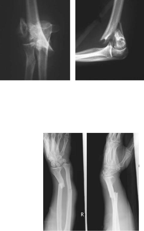
Fractures about the elbow usually are the result of a fall with a direct blow to the elbow or a fall onto the outstretched hand. Fractures of the distal humerus involving the articular surface or the supracondylar region tend to be high-energy injuries with extensive comminution (Fig. 33.8). These usually require open reduction and internal fixation. Fractures of the proximal ulna, referred to as olecranon fractures, result in disruption of the elbow extensor mechanism, and these require surgical internal fixation if displaced. Fractures of the radial head tend to occur as a result of a fall onto on outstretched arm. In general, these tend to be lower energy injuries and often have minimal displacement. The primary treatment of these is early motion to prevent posttraumatic contracture. However, if the radial head fragment is displaced severely and results in mechanical block to full motion, the fragment may need to be reduced and fixed or excised surgically. A Monteggia fracture is a fracture of the proximal ulna with a dislocation of the radial head. This injury requires surgical intervention. Anatomic reduction of the ulnar shaft fracture almost always results in reduction of the radial head with good stability. Fractures of the shaft of the radius and ulna occur as a result of a direct blow to the forearm or a fall onto an outstretched hand. These injuries usually are displaced and angulated. In general, these injuries require open reduction and internal fixation, since healing in a nonanatomic

608 C.J. Gatt Jr.
dislocation often referred to as a perilunate dislocation, the base of the capitate is dislocated from its articulation with the lunate. Either the lunate is dislocated in a volar direction and the capitate appears to articulate with the distal radius, or the lunate maintains its articulation with the distal radius and the capitate and the rest of the carpus have dislocated in a dorsal direction. This injury results in significant pressure on the median nerve as it passes through the carpal tunnel; it requires prompt treatment and almost always requires open reduction and internal fixation due to the multiple ligament injuries that occur between the various carpal bones. More common dislocations involve the metacarpocarpal joints, the metacarpophalangeal joints, and the interphalangeal joints. Many of these dislocations can be treated with closed reduction with longitudinal traction, and maintained with appropriate positioning of the hand. The carpometacarpal dislocations usually require cast treatment to maintain the reduction. Dislocations of the metacarpophalangeal joints and the interphalangeal joints usually require only minimal immobilization, followed by restoration of motion.
Fractures of the distal radius are one of the most commonly encountered injuries. Although a Colles’ fracture describes a comminuted fracture of the distal radius that extends to the articular surface and includes a fracture of the ulnar styloid, the term commonly is used to describe all distal radius fractures. The typical patient with a distal radius fracture is an elderly woman with osteoporosis who has fallen onto her outstretched hand. In these injuries, the distal fragment usually is displaced dorsal relative to the proximal fragment, and the clinical deformity associated with this injury sometimes is referred to as a silver-fork deformity. The majority of these injuries can be treated with a closed reduction and cast immobilization. In the younger patient who sustains a high-energy injury with significant disruption of the articular surface, surgical intervention is required.
Fracture of the scaphoid is another injury that occurs as a result of a fall onto an outstretched hand. Clinically, this injury often does not present without much swelling. However, the patient tends to have tenderness in the anatomic snuffbox to palpation. If a scaphoid fracture is suspected, radiographs should be inspected carefully, since up to 20% of these injuries are not diagnosed at the initial evaluation. If the clinical examination is consistent with a scaphoid fracture and the initial radiographs do not demonstrate a fracture, the patient should be immobilized in a thumb spica splint and follow-up should be arranged, since radiographic evidence of the injury may not be present until 2 to 3 weeks after the injury. This injury does have a high incidence of nonunion, especially if the injury is not immobilized in the early stages or if there is displacement of the fracture. A fracture of the fifth metacarpal neck is referred to as a boxer’s fracture and usually occurs as a result of the patient’s striking a hard object with a clenched fist. This particular injury should be inspected carefully for a laceration over the head of the metacarpal. The laceration can be the result of the clenched fist hitting the tooth of another person. Consequently, this particular injury is at significant risk for infection and requires thorough
33. Musculoskeletal Injuries 609
irrigation in addition to treatment of the fracture. Antibiotics should provide coverage for E. corrodens in addition to routine skin flora.
This injury presents with apex dorsal angulation at the level of the metacarpal neck distally, and this almost always can be successfully reduced and held in good position with cast immobilization.
Pelvis and Hip
The osseous anatomy of the pelvis forms a closed circle. In the posterior aspect, the sacrum, which contains the distal spinal nerve roots, articulates with the ilium on either side. The circle is closed anteriorly at the pubic symphysis. The hip joint is formed by the articulation between the head of the proximal femur and the acetabulum. In contrast to the “ball and socket” joint of the shoulder, the round head of the femur is well contained in the deep socket of the acetabulum. The large muscles around the hip certainly are susceptible to contusion injury. Falls can lead to contusion of the gluteus maximus. In sports events, high-energy direct blows to the anterior thigh can lead to quadriceps contusions and hematomas. This particular injury can be very painful and lead to a very tense-appearing thigh. However, compartment syndrome with this injury is extremely rare. The size of the hematoma formation can be controlled by early splinting of the leg with the knee held in hyperflexion, putting the quadriceps muscle on stretch. Since myositis ossificans at the site of the quadriceps injury is a troublesome sequela, minimizing the size of the hematoma formation is beneficial.
Another sports-related injury that often has a dramatic presentation is avulsion of the sartorius muscle from the anterosuperior iliac spine or avulsion of the rectus femoris from the anteroinferior iliac spine. In either of these injuries, patients report feeling a pop in their hip and present with significant pain with ambulation. Physical examination usually does not demonstrate an obvious muscle deformity. However, palpation over the appropriate iliac spine helps diagnose the site of the injury.
Dislocations of the hip joint usually are caused by high-energy trauma, such as a motor vehicle accident or a fall from a height, although they can occur in sporting injuries. The most common dislocation is a posterior dislocation of the femoral head from the acetabulum. In this case, the patient presents with the hip flexed, adducted, and internally rotated. When the dislocation is anterior, the patient presents with the hip held in abduction, flexion, and external rotation. Prior to reduction, a neurovascular examination should be performed with attention paid to sciatic nerve function, since this nerve can be injured, especially with posterior dislocations. Radiographs should be evaluated for other associated injuries, such as acetabular wall fractures, femoral head fractures, or fractures of the femur. Reduction of hip dislocation usually requires some form of sedation, followed by application of longitudinal traction in line with the deformity. Once reduced, a repeat neurologic examination should be performed, again paying attention to the function of the sciatic nerve. After

33. Musculoskeletal Injuries 611
blood loss. This can be a lifesaving procedure and should not be delayed unless absolutely necessary. High-energy lateral compression injuries to the pelvis also result either in disruption of the pubic symphysis of the pubic rami on the anterior aspect of the pelvis and disruption of the sacroiliac joint or a crush injury of the sacral body on the posterior aspect of the pelvis. Although blood loss is expected with this injury, the pelvic volume is not expanding, and, consequently, urgent stabilization of the pelvis rarely is required.
Fractures of the hip are divided into two categories: intracapsular and extracapsular. Intracapsular fractures are fractures of the femoral neck. In elderly patients, a fracture of the femoral neck can result in an impacted valgus position of the fracture fragments, and this is treated routinely with screw fixation. When a fracture of the femoral neck is displaced, the blood supply to the femoral head usually is disrupted, and there is a significant risk of avascular necrosis of the femoral head. Consequently, many of these injuries are treated with primary hemiarthroplasty in the elderly patient. However, in the younger patient, a displaced femoral neck fracture should be treated with more aggressive attempts to achieve a reduction of the fracture to a near-anatomic position and fixation with screws. Extracapsular or peritrochanteric fractures of the femur can result in significant blood loss into the thigh. This needs to be recognized, especially in the elderly patient with a low cardiac reserve. These injuries generally require surgical treatment with screw and side plate fixation or intermedullary fixation. Fractures in the intertrochanteric region heal readily while fractures to the subtrochanteric region of the femur have a much higher significance of nonunion and hardware failure. In cases of peritrochanteric fractures of the hip, avascular necrosis is not a concern.
Fractures of the femoral shaft are the result of high-energy injuries, such as motor vehicle accidents or falls from a significant height. Physical examination of the thigh should be thorough to be sure that there is not an open wound associated with the femoral shaft fracture. These injuries require surgical fixation, and this usually is done in an intermedullary fashion. Fixation of these fractures within 24 to 48 hours from the time of injury has been shown to decrease the incidence of adult respiration distress syndrome (ARDS), especially in the multitrauma patient population.
Knee and Lower Leg
The osseous anatomy of the knee consists of the distal femur, the proximal tibia, and the proximal fibula. The distal femur articulates only with the proximal tibia. This often is considered a hinged joint, although rotations do occur about the longitudinal axis and in the coronal plane. The proximal fibula articulates with the proximal tibia, but this occurs distal to the femorotibial articulation. The fibula does not articulate with the femur. The patella is a sesamoid bone within the extensor mechanism of the knee. Its deep surface is covered with articular cartilage, and the patella articulates with the femur. The primary role of the patella is to increase the length of the extensor moment arm.
612 C.J. Gatt Jr.
Since very little muscle tissue overlies the knee joint, muscle contusions are not common. In general, significant contusions tend to occur in the posterior aspect of the lower leg as a result of direct blows to the gastrosoleus complex. These injuries can lead to significant swelling, and neurovascular status should be assessed in association with these injuries.
The most dramatic muscle injuries around the knee are disruptions of the extensor mechanism. In the younger population, patellar tendon ruptures can occur while jumping or while landing from a jump. These injuries present with a high-riding patella, referred to as patella alta, and a palpable defect at the inferior pole of the patella. The majority of these injuries are avulsions of the patellar tendon from the distal pole of the patella. Ruptures of the quadriceps tendon tend to occur in the middle-aged and elderly population. These often are lowenergy injuries and can occur with an activity as simple as ascending or descending the stairs. In these cases, the patient presents with a swollen knee, a low-riding patella referred to as patella baja, and a palpable defect at the superior pole of the patella. Similar to patellar tendon ruptures, quadriceps ruptures usually occur as avulsions of the quadriceps tendon from the superior pole of the patella. Both of these injuries usually require primary surgical repair of the tendon avulsion injury in order to restore normal knee function.
Dislocations of the knee joint can occur as a result of high-energy trauma, such as motor vehicle accidents, or as a result of lower energy trauma, such as sporting injuries. In these dislocations, the anterior and posterior cruciate ligaments usually are disrupted, as is the medial and/or lateral collateral ligament. Because the popliteal artery and tibial and peroneal nerves lie close to the posterior knee capsule, this injury does have a high incidence of neurovascular injury. Since the soft tissue envelope around the knee joint is relatively thin, the deformity is obvious to inspection. This injury should be reduced rapidly to minimize the risk of neurovascular damage. However, even after a prompt reduction, a lower extremity angiogram is indicated to evaluate the integrity of the popliteal artery and its intimal layer. Knee dislocations usually are treated with primary repair of injured ligamentous structures. Instability after dislocation usually is less of a problem than posttraumatic stiffness of the knee joint.
Dislocations of the proximal tibiofibular joint are rare. In general, the fibula dislocates in an anterior direction. The most common mechanism is landing from a jump, and this injury is seen in parachute landings. This injury can be difficult to detect both clinically and radiographically, but, once diagnosed, closed reduction usually is achieved easily with direct pressure on the proximal fibula in the appropriate direction.
Fractures of the distal femur usually occur through the supracondylar region of the distal femur with both highand low-energy injuries in this region. It is not uncommon to see a vertical extension of the fracture that splits the medial and lateral femoral condyles. These injuries usually require internal fixation with intermedullary implants or open reduction and internal fixation with plate and screws.

33. Musculoskeletal Injuries 613
Figure 33.12. Radiographs of a tibial plateau fracture: AP and lateral views.
Fractures of the proximal tibia, referred to as tibial plateau fractures, usually involve damage to the lateral compartment of the tibial plateau (Fig. 33.12). At times, these injuries can be difficult to visualize on routine radiographs, and further imaging studies, such as a CT scan, may be necessary to evaluate depressions of the tibial articular surface. When the joint surface has been depressed greater than 1 cm, surgical intervention usually is indicated and involves elevation of the tibial articular surface back to its anatomic position with plate and screw stabilization.
Fractures of the patella result from a fall onto a flexed knee. Many patellar fractures are simple transverse fractures. When the fracture fragments are displaced, there is disruption of the extensor mechanism, and open reduction and internal fixation is indicated to restore normal knee function.
Fractures of the shaft of the tibia and fibula are relatively common. Low-energy injuries can occur from simple falls and sport activities. Even with low-energy injuries, significant swelling in the lower leg can occur. Since there is no significant fascial disruption, the patient should be evaluated and followed for a potential compartment syndrome. The lower energy injuries usually can be reduced into a near-anatomic position with closed manipulation and can be controlled in a long leg cast. If the fracture line has a high degree of obliquity, the fracture should be followed for excessive shortening even in a long leg cast. If this is the case, intermedullary nail fixation is appropriate to maintain length. Higher energy fractures of the tibial and fibular shaft usually result in comminuted fractures that are unstable, and it is difficult to maintain acceptable length even in a long leg cast. These injuries usually are managed with intermedullary nail fixation. However, in these injuries, significant soft tissue swelling can occur, and the patient should be


33. Musculoskeletal Injuries 615
Figure 33.14. AP and lateral radiographs of an open tibia fracture after intramedullary nailing.
operating room for a debridement of his wounds followed by an intramedullary nailing of the tibia (Fig. 33.14).
Foot and Ankle
The ankle joint is formed by the articulation of the distal tibia, fibula, and talus. The body of the talus fits between the medial malleolus, a medial extension of the distal tibia, and the lateral malleolus. This joint acts primarily as a hinge, although rotation in the coronal and transverse planes does occur. In addition, vertical translation occurs between the tibia and fibula. The foot can be divided into three segments: the hindfoot, the midfoot, and the forefoot. The hindfoot consists of the talus and calcaneus. The midfoot is created by the tarsal bones. The forefoot consists of the metatarsals and phalanges.
Muscle strain injuries about the ankle most commonly are associated with sprains of the ankle joint. In general, these sprains of either the peroneal muscles or tibialis posterior muscle are mild sprains and resolve with minimal treatment. The most notable muscle injury is rupture of the Achilles tendon. This injury usually occurs in middleaged individuals during recreational sporting events. The history reveals a sudden pain and sometimes a “pop” in the back of the ankle. There rarely is a history of preexisting Achilles tendonitis. Physical examination demonstrates minimal soft tissue swelling and a palpable defect in the region of the Achilles tendon. The Thompson squeeze test
616 C.J. Gatt Jr.
is performed with the patient prone and the knees flexed. With an
Achilles tendon rupture, manually squeezing the gastrosoleus muscle does not result in plantarflexion of the ankle (a positive test), but it does with an intact Achilles tendon.
Ankle sprains are commonly encountered injuries to the ankle. They usually are the result of an inversion injury or a combination of abduction force and external rotation. The lateral ankle ligaments most commonly are injured. With more substantial force, the deltoid ligament as well as the syndesmosis and interosseous membrane between the tibia and fibula can be injured. Treatment consists of short-term or partial immobilization and rehabilitative exercises. True dislocations of the talus from the ankle mortise are rare. Once reduced, these injuries usually are quite stable, and posttraumatic stiffness is more of a concern than instability.
Sprains of the foot can affect one or several joints of the hindfoot, midfoot, or forefoot. In general, these injuries lead to significant soft tissue swelling at the site of the injury. They usually can be treated with a stiff-soled shoe and progression to full weight bearing as symptoms allow. Dislocations also can affect one or several joints of the foot. Dislocations such as subtalar dislocations and midtarsal dislocations have obvious deformities and can be closed reduced with longitudinal traction and manipulation of the distal segment back to an anatomic position. Since the soft tissue coverage over the dorsum of the foot is thin, these dislocations should be treated promptly to prevent soft tissue loss due to prolonged tension. In rare cases, nearby tendons can block a closed reduction, and these require surgical treatment.
Fractures of the ankle occur as a result of inversion or eversion stress on the ankle combined with axial rotation. They also can result from high-energy axial loads on the lower extremity. Low-energy stable injuries to the ankle result in a fracture of one malleolus and no significant ligamentous injury. On the other hand, unstable fractures of the ankle result in bimalleolar fractures or lateral malleolar fracture with a significant ligamentous injury resulting in translation of the talus from its anatomic position beneath the distal tibia. Radiographs should be carefully scrutinized for evidence of medial clear space widening (Fig. 33.15). Although the unstable injuries can be treated by closed manipulation and casting, open reduction and internal fixation usually are recommended. Fractures of the distal tibia with extension into the ankle joint commonly are referred to as pilon fractures. These usually are high-energy injuries that result in significant soft tissue swelling at the site of the fracture. As a consequence, many of these injuries are treated with a combination of external fixation and limited internal fixation. This technique avoids the soft tissue dissection necessary for open reduction and internal fixation.
Calcaneal fractures usually are the result of a fall from a height, such as a ladder. As with most high-energy injuries, they usually are associated with significant soft tissue swelling. It is important to examine the patient for signs of lumbar spine injury, since 10% of patients with calcaneal fractures have an associated lumbar spine fracture.

33. Musculoskeletal Injuries 617
Figure 33.15. Radiographs of a bimalleolar ankle fracture: AP, lateral, and oblique views.
In the past, surgical treatment for displaced calcaneal fractures was rare. However, surgical intervention is becoming increasingly more common in the hope of improving the long-term outcome of this sometimes devastating injury.
Displaced fractures of the talar neck usually are high-energy injuries. In some cases, the fracture is associated with a dislocation of the talar body from the ankle joint. These injuries have a relatively high incidence of avascular necrosis of the talar body. Open reduction and internal fixation usually are indicated for displaced fractures.
Of the metatarsals, fracture of the fifth metatarsal seems to cause the most confusion. Fractures of the proximal tuberosity of the fifth metatarsal result from inversion injuries to the foot. These are extremely common, and minimal treatment is indicated. On the other hand, a fracture of the proximal metaphyseal-diaphyseal junction, referred to as a Jones’ fracture, can be a troublesome fracture. This injury is treated best by prolonged non–weight bearing and sometimes internal fixation with an intramedullary screw.
Lawn-mower injuries to the foot are worth mentioning. These can be devastating injuries. There usually are multiple fractures and associated soft tissue loss. Prevention of infection is paramount in the treatment. Despite early and aggressive treatment, amputation is not uncommon.
Spine
The spine is a long column of vertebral bodies that serve to protect the spinal cord. There are seven cervical vertebrae, 12 thoracic vertebrae, and five lumbar vertebrae. Below the lumbar spine is the sacrum, consisting of fused vertebrae, and then the coccygeal segments. Rotational motion of the head occurs through rotation in the upper cervical segments. Flexion and extension of the neck occur through motion in the
618 C.J. Gatt Jr.
middle and lower cervical segments. Due to articulation with the ribs, little motion occurs through the thoracic segments. Trunk rotation occurs through the upper lumbar segments, and flexion and extension occur through the lower lumbar segments. Each vertebra articulates with the vertebra above and below via two facet joints and the intervertebral disk.
Muscle strain injuries in the cervical and lumbar spine are common. Symptoms generally are localized to the paraspinal region, and radicular symptoms are rare. The history may reveal minimal to no trauma that results in significant pain. Physical examination demonstrates obvious discomfort and muscle guarding. Signs of neurologic compromise, such as sensory deficits or muscle weakness, are rare and should alert the examiner to potential disk herniation. Radiographic examination usually is normal except for routine age-related degenerative findings at the facet joints or disk spaces. Findings on magnetic resonance imaging (MRI) do not demonstrate soft tissue edema, and the disk bulges commonly detected are not necessarily evidence of disk disease. These muscle strain injuries are self-limiting and resolve with rest and rehabilitative exercises.
Facet joint dislocations occur in the cervical spine as a result of motor vehicle accidents, but they also can occur in sports-related injuries. The patient complains of significant cervical pain. However, radicular complaints are not always present. Physical examination demonstrates muscle guarding, point tenderness to palpation, and limited painful motion of the cervical spine. Neurologic deficits are not always present. Radiographic evaluation should include AP, lateral, and odontoid views that show the cervical spine from C1 to C7. Facet joint dislocations may demonstrate anterior translation of the superior vertebral body in relation to the inferior and acute angulatory change in the longitudinal axis of the cervical spine. However, some facet dislocations can be overlooked on initial review of the radiographs. Enlargement of the soft tissue shadow anterior to the vertebral body may be an indication of ligamentous spine injury. Also, if the patient is awake and alert, flexion-extension lateral radiographs of the cervical spine may be helpful. Otherwise, CT scan usually diagnoses the dislocation of the facet joint. An MRI of the cervical spine prior to treatment helps diagnose associated disk injury. Treatment is by a prompt reduction of the dislocation. A reduction can be performed with the patient awake by applying longitudinal traction with skeletal tongs in the skull. This technique should be done with radiographic and neurologic monitoring and may be safer than open treatment with the patient under anesthesia. Once reduced, these injuries require some form of surgical stabilization.
Fractures of the spinal column without displacement usually are stable and often present with no neurologic compromise. These injuries are amenable to brace treatment. Higher energy injuries can result in fracture fragments encroaching on the neural elements, such as the spinal cord in the cervical and thoracic spine and nerve roots in the lumbar spine (Fig. 33.16). A careful physical examination demonstrates the level and extent of neurologic involvement. In cases of incomplete

33. Musculoskeletal Injuries 619
Figure 33.16. Lateral radiograph and computed tomography (CT) scan of a lumbar burst fracture. Note encroachment of spinal canal by vertebral body fragment.
spinal cord injury and nerve root injury, decompression and stabilization should be performed promptly. When complete loss of function below a spinal cord injury occurs, the prognosis for return is poor, but stabilization usually is required to prevent further deformity and to facilitate care for the patient.
Summary
Proper function of the musculoskeletal system is dependent on the proper form and function of skeletal, musculotendinous, ligamentous, vascular, and neural structures. Injury to even one of these elements usually leads to malfunction and deterioration of one or more of the remaining components. Thus, a thorough understanding of all of these components is necessary to diagnose and treat musculoskeletal injuries.
Assessment of the patient who has sustained musculoskeletal trauma should be systematic. Airway, breathing, and circulation (ABC) are assessed, followed by a comprehensive history. Then a comprehensive or focused physical examination should be performed. The examination should include evaluation of musculoskeletal, vascular, and neural components. Then diagnostics, such as radiographs, should be performed. Urgent treatment should be employed in cases of compartment syndrome. Appropriate traction and immobilization techniques should be employed. With this systematic approach to patient evaluation, communication of injuries and patient status should not be a source of confusion. After an appropriate evaluation and stabilization of the patient, a definitive treatment plan that leads to an optimal outcome can be implemented.
620 C.J. Gatt Jr.
Selected Readings
Bucholz RW. Orthopaedic Decision Making. St. Louis: Mosby, 1996.
Greene WB. Essentials of Musculoskeletal Care, 2nd ed. Rosemont, IL: American Academy of Orthopaedic Surgeons, 2001.
Ivevson LD. Manual of Acute Orthopaedic Therapeutics, 4th ed. Boston: Little, Brown, 1994.
Lowenberg DW, Fang A. Orthopaedic surgery. In: Norton JA, Bollinger RR, Chang AE, et al, eds. Surgery: Basic Science and Clinical Evidence. New York: Springer-Verlag, 2001.
Rizzo M, Levin LS. Hand surgery. In: Norton JA, Bollinger RR, Chang AE, et al, eds. Surgery: Basic Science and Clinical Evidence. New York: Springer-Verlag, 2001.
Rockwood CA Jr, Green DP, Bucholz RW, Heckman JD. Rockwood and Green’s Fractures in Adults, 4th ed. Philadelphia, JB Lippincott, 1996.