

Revision Sinus Surgery
.pdf72
|
|
dent upon the patient’s level of cooperation, which may |
|
|
|
tend to decrease over time. FESS under local anesthesia |
|
|
|
can produce patient anxiety, particularly during cases |
|
|
|
with heavy mucosal bleeding. Rising patient anxiety also |
|
|
|
tends to impact the surgeon and potentially the quality |
|
|
|
of the operation. Some groups use local anesthesia on a |
|
|
|
regular basis for straightforward cases; however, these are |
|
|
|
carefully selected beforehand [4]. In addition, the routine |
|
|
|
use of image-guidance systems makes the local anesthetic |
|
|
|
technique less feasible due to positioning factors and the |
|
|
|
necessity of patient immobility. The author’s group be- |
|
|
|
lieves that these limitations outweigh the theoretical ana- |
|
|
|
tomic advantage of local anesthesia. |
|
|
|
|
General anesthesia consists of the following three |
|
|
phases: induction, maintenance, and emergence. The goal |
|
|
|
of general anesthesia is to safely produce, while maintain- |
|
9 |
|
ing physiologic stability: |
|
|
|
1. |
Hypnosis. |
|
|
||
|
|
2. |
Analgesia. |
|
|
3. Amnesia. |
|
|
|
4. |
Anesthesia. |
|
|
5. |
Areflexia. |
|
|
6. |
Anxiolysis. |
|
|
General anesthesia for FESS is usually either a variant of |
|
|
|
balanced anesthesia, using a combination of sedative/hyp- |
|
|
|
notics, inhaled anesthesia, opioids, and neuromuscular |
|
|
|
blocking agents, or total intravenous anesthesia (TIVA), |
|
|
|
using a combination of propofol and synthetic opioids. |
|
|
|
|
|
|
|
Pharmacology of General Anesthesia |
|
|
|
|
|
|
|
|
|
|
|
Volatile Anesthetics |
|
|
|
|
|
|
|
Inhaled or volatile anesthetics are an important part of |
|
|
|
balanced anesthesia. They have been in use since the mid- |
|
|
|
nineteenth century and represent one of the most impor- |
|
|
|
tant advances in the history of medicine. Although their |
|
|
|
mechanism of action remains unclear, they are known |
|
|
|
to produce profound central nervous system depression |
|
|
|
resulting in unconsciousness and amnesia. They are also |
|
|
|
known to produce respiratory depression, myocardial |
|
|
|
depression, dose-dependent decreases in mean arterial |
|
|
|
blood pressure (MAP), and reflex tachycardia [12]. |
|
|
|
■ Volatile anesthetics are traditionally used in the main- |
|
|
|
|
tenance phase of general anesthesia and to improve the |
|
|
■ |
surgical field by producing “controlled hypotension.” |
|
|
They produce dose-dependent hypotension with re- |
|
flex tachycardia and profound vasodilation, which may worsen the surgical field when used alone.
W. Derek Leight and Brent Senior
Intravenous Anesthesia
Intravenous (IV) anesthesia using sedative/hypnotics such as barbiturates and benzodiazepines are important in the induction phase of general anesthesia. These drugs produce rapid-onset hypnosis and sedation. Since the introduction of propofol in the late 1980s, IV anesthetics have also become popular in the maintenance phase of anesthesia.
Propofol
Propofol, whose chemical name is isopropylphenol, represents a new class of IV anesthetics, the akylphenols. It is the most widely used IV induction agent in anesthetic practice. It is lipophilic, and almost completely insoluble in aqueous solution. It is therefore produced in a milky emulsion with lecithin, glycerol, and soybean oil. This formulation is a potential medium for bacterial growth, and must therefore be used with meticulous sterile technique and should be used within 6 h of opening the vial. Because lecithin comes from egg yolk, patients with egg allergies may be susceptible to allergic reactions. Because of its high lipophilic nature, propofol is rapidly partitioned across the blood-brain barrier upon injection. This accounts for the quick onset of its sedative and hypnotic effects. Plasma clearance and redistribution into skeletal muscle and fat is faster than its hepatic metabolism. The high plasma clearance rate combined with its action as an antiemetic is thought to contribute to its low “hangover effect,” which makes it useful for outpatient surgery.
■ Propofol:
1.Acts through potentiation of GABA receptors in the central nervous system.
2.Produces profound respiratory depression.
3.Produces systemic vasodilation at the level of arteries and veins.
4.Reduces cardiac preload and afterload.
5.Causes systemic hypotension with little or no reflex tachycardia due to inhibition of the baroreceptor reflex and therefore does not increase cardiac output.
6.Produces little or no analgesia [12].
7.Produces a small “hangover effect,” which make it useful for outpatient surgery.
Opioid Agonists
Opioid agonists such as fentanyl produce analgesia without loss of touch, proprioception, or consciousness. Fentanyl, sufentanil, alfentanil, and remifentanyl are syn-

Anesthetic Choices, Techniques, and Injections
thetic opioids that are derivatives of morphine. Fentanyl is approximately 100 times more potent than morphine. It has a more rapid onset and shorter duration of action. It is more lipid soluble than morphine, which determines its potency and onset. It is quickly redistributed into fat and skeletal muscle, with these sites becoming easily saturated. Therefore, during a continuous infusion the plasma clearance decreases significantly, causing a precipitous drop in clearance rates. This can lead to prolonged ventilatory depression and analgesia in cases of infusions longer than 2 h. Remifentanyl is a μ-selective opioid receptor agonist with an analgesic potency similar to that of fentanyl. It is more rapid acting than fentanyl, yet it is susceptible to hydrolysis by plasma and tissue esterases, which gives it a very quick offset. Its clearance is independent of liver and kidney function. The metabolites are inactive, and therefore produce noncumulative effects. In addition, remifentanyl is known to produce hypotension, whereas fentanyl does not [12, 15].
Balanced Anesthesia Versus TIVA
There is controversy regarding the method of choice for anesthetic delivery during FESS. The importance of obtaining the optimal surgical field in FESS has prompted several studies on the topic. In 1993, Blackwell et al. retrospectively showed a decrease in blood loss in patients undergoing ESS using propofol anesthesia compared to those using isoflurane [2]. Eberhart et al. showed an improvement in a visual analog scale assessment of the surgical field in patients treated with propofol and remifentanyl versus those treated with isoflurane and alfentanyl. There was no significant difference between groups in terms of MAP or estimated blood loss. Heart rate was significantly lower in the TIVA group [7]. Wormald et al. showed in a prospective randomized trial that TIVA using propofol and remifentanyl produced superior surgical fields when compared to traditional balanced anesthesia. This study found a positive correlation between surgical grade and MAP for both conditions, as well as an overall positive correlation with heart rate. When compared at specific MAPs, the TIVA group produced superior surgical grades. This suggests that TIVA produces a surgical field that is more sensitive to MAP than balanced anesthesia [15]. A recent study by Beule et al. compared anesthesia using propofol and fentanyl versus sevoflurane and fentanyl. This study found no significant difference in the amount of blood loss or quality of the surgical field as assessed by a visual analog scale. Several features of this study are potentially confounding, including the use of fentanyl instead of remifentanyl, and the overall high volumes of blood loss with large standard deviations
73
reported in each group, which might have potentially confounded any statistical difference. Interestingly, they provide evidence of increased platelet dysfunction after 45 min in both groups, with the propofol group being significantly worse than the sevoflurane group [1].
With balanced anesthesia, the anesthesiologist attempts to improve the surgical field by increasing the concentration of inhaled anesthetic, which causes peripheral vasodilation, thereby lowering systemic vascular resistance and decreasing MAP. Inhaled anesthetics, however, do not decrease cardiac output, and in fact, increasing concentrations of common inhaled anesthetics result in a direct increase in heart rate. It has been postulated that lowering the blood pressure in this manner may actually worsen the surgical field [3, 15]. While propofol administration results in a drop in blood pressure through vasodilation, the venodilator effect produces a decrease in cardiac output by decreasing cardiac preload [9]. In addition, the baroreceptor reflex is blunted and there is no resultant increase in heart rate. Interestingly, these studies point to the potential importance of remifentanyl in improving the surgical field during TIVA. It is known that propofol combined with remifentanyl synergistically produces hypotension and bradycardia [14]. Using modern anesthesia techniques such as target-controlled infusion, TIVA with propofol and remifentanyl most likely produces a superior surgical field for FESS, with the added benefit of superior control of hypotension and heart rate due to the short-acting nature of these drugs. This produces a theoretical safety advantage as controlled hypotension may precipitate ischemic organ failure in rare instances [11].
Anesthesia for Sinus Surgery
Propofol and remifentanil used in combination produce significant hypotension and better blunting of hemodynamic responses to endotracheal intubation than any other combination of sedative/hypnotic and opioid [14].
Propofol and remifentanyl may produce a synergistic drop in MAP and heart rate, thereby significantly lowering cardiac output, which may be responsible for the improved surgical field with TIVA.
Pharmacology of Local Anesthetics
Key points:
1.Amino esters are more likely to produce allergic reactions.
2.Amino esters are metabolized by plasma cholinesterases and so are less likely to produce sustained plasma concentrations.

74
3.Amino amides are metabolized by the liver. Patients with liver disease or with decreased hepatic flow, such as in congestive heart failure, may experience increased plasma levels of anesthetics.
4.Administration of local vasoconstrictors such as epinephrine potentiate the duration of the effect while decreasing the systemic absorption.
The most commonly used local anesthetics are tertiary amines composed of a lipid-soluble benzene ring connected to an amine group by an alkyl group containing either an amide or ester linkage. This linkage divides the tertiary amines into two major groups: amino amides and amino esters. Tertiary amines are weak bases. They can exist in solution in two forms: an unprotonated, neutral (lipid soluble) form or a protonated, charged (water
9 soluble) form. The main mechanism of action of tertiary amine local anesthetics is postulated to occur on the interior of the axon, by reversibly inhibiting voltage-gated sodium channels on the inner surface of the membrane. The lipid-soluble form is able to traverse the cell membrane easily, while the water-soluble form is responsible for binding to the sodium channel and blocking actionpotential propagation. The specific properties of tertiary amines create important clinical differences in efficacy and potency. First, the dissociation constant is a measure of how likely the amine group is to be protonated (hydrophilic) or neutral (lipophilic) at a particular pH, which determines how quickly an anesthetic can traverse a membrane to exert an effect. Second, lipid solubility determines the likelihood of the anesthetic moving through myelin or other supporting cells. Consequently, anesthetics with higher lipid solubility tend to have increased time to onset and offset of effect due to increased sequestration in myelin. Increasing lipid solubility also tends to increase potency. For FESS, lidocaine is the most widely used local anesthetic. Discovered in 1948, it was the first amino amide local anesthetic. Its amide bond makes it more stable and less likely to cause allergic reactions than amino esters. Cocaine is a naturally occurring amino ester that has excellent anesthetic properties as well as vasoconstrictive properties, which make it ideal for FESS. It is the only tertiary amine that acts as a vasoconstrictor; all others are vasodilators. However, the euphoria and highly addictive nature of cocaine have made it one of the most widely abused recreational drugs. Therefore, it is now illegal in most countries and more difficult to use for legitimate medical purposes. Cocaine is also known to cause cardiac arrhythmias and many have recommended its abandonment for the use of safer mixtures [10].
Epinephrine, a human adrenergic catecholamine, is commonly added to local anesthetics at a variety of concentrations. Epinephrine induces peripheral vascular resistance via alpha-receptor-stimulated vasoconstriction.
W. Derek Leight and Brent Senior
The authors commonly use 1% lidocaine with 1:100,000 epinephrine for injections.
Injections
Key points:
1.The greater palatine foramen injection is an effective method for controlling bleeding and providing anesthesia during endoscopic sinus surgery.
2.A combination of the sphenopalatine block with the greater palatine block leads to profound vasoconstriction in the posterior portion of the nasal cavity and significantly reduces bleeding.
The greater palatine foramen is located posteromedially to the third maxillary molar and anteromedially to the maxillary tuberosity and pterygoid hamulus (Fig. 9.1). The foramen opens into the greater palatine canal, which courses superiorly into the pterygopalatine fossa. Here, the third portion of the internal maxillary artery and its multiple branches supply the nose, paranasal sinuses, pharynx, orbit, palate, teeth, and facial skin. The closed space and bony walls of the pterygopalatine fossa make it ideal for local anesthesia. The optimal injection is delivered at the opening of the greater palatine canal, medial to the sphenopalatine foramen, where the terminal portion of the internal maxillary artery lies. Superiorly,
Fig. 9.1 The location and orientation of the greater palatine foramen and canal is clearly seen on this computed tomography scan (arrows)

Anesthetic Choices, Techniques, and Injections |
75 |
Fig. 9.2 The location of the sphenopalatine foramen at the posterior aspect of the middle turbinate is indicated by the asterisk
the pterygopalatine fossa is limited by the anterior basal portion of the greater wing of the sphenoid bone. The inferior orbital fissure is located anterosuperiorly in the pterygopalatine fossa and the superior orbital fissure and optic foramen lie just above it. The foramen rotundum is located posteriorly, superiorly, and laterally to the sphenopalatine foramen. The mean distance of the greater palatine foramen to the sphenopalatine foramen is 28 mm in men and 27 mm in women. The mean distance of the greater palatine foramen to the inferior orbital fissure is 40 mm in men and 37 mm in women [6]. The sphenopalatine foramen can also be injected transnasally. Here, the sphenopalatine artery and several branches of the maxillary nerve and pterygopalatine ganglion enter the nose just posterior to the posterior attachment of the middle turbinate (Fig. 9.2).
A combination of the sphenopalatine block with the greater palatine block leads to profound vasoconstriction in the posterior portion of the nasal cavity and significantly reduces bleeding.
tracheal anesthesia is induced in the operating room, the patient is turned 90 degrees counterclockwise to facilitate the use of stereotactic computed tomography guidance. The patient is then placed in the beach chair position, which elevates the head approximately 10–20 degrees, as the reverse Trendelenburg position has been shown to reduce intracranial MAP without reducing cerebral perfusion pressure [14]. The CT guidance system is then registered and its accuracy confirmed.
Local Anesthesia
■The authors utilize bilateral greater palatine blocks in nearly all cases.
Three milliliters of 1% lidocaine with 1:100,000 of epinephrine are drawn into a Luerlock syringe. Before use, the expiration date and concentration of the lidocaine and epinephrine on the stock container are confirmed by the surgeon. A 25-gauge needle is measured with a ruler and bent to an angle of 60 degrees at a length of 25 mm for all adults (Fig. 9.3).
■The greater palatine foramen is usually located anterior to the junction of the hard and soft palate just medial to the second maxillary molar.
It can often be palpated, or seen as a subtle depression in the hard palate mucosa. The needle is placed into the greater palatine foramen and advanced to the bend of the needle. The needle is then aspirated for blood to prevent an intravascular injection. Then, 1.5 ml of anesthetic is injected. If the needle is properly placed, there is moderate resistance to the injection. If there is minimal resistance, it is likely that the needle is in the nasopharynx, and is
Techniques
General Anesthesia
Prior to the start of each day, the overall anesthetic plans are reviewed with the anesthesiologist. The authors’ preference is TIVA with propofol and remifentanyl when possible. The adjunctive use of beta blockade is discussed, but left up to the discretion of the anesthesiologist. In the preoperative suite, the patient is given oxymetazoline sprays in each nostril 2 h prior to surgery. After general endo-
Fig. 9.3 A 25-gauge needle with a 60 angle at length 25 mm from the tip of the needle

76
not correctly placed in the canal. The same procedure is repeated for the contralateral canal (Video 9.1).
After the patient has been prepped and draped, a 0 degrees Hopkins rod is used to place oxymetazo- line-soaked cotton pledgets in the nasal cavities. This is performed endoscopically to prevent the trauma to the septal mucosa or middle turbinate that can occur with blind placement and to prevent nuisance bleeding that could degrade the surgical field. Next, the remainder of the instrumentation is set up, including the image-guid- ance system, camera, monitors, DVD recording system, and microdebrider. The authors routinely use an endoscope irrigation system to wash away minor bleeding at the scope tip and always sit during the operation with an armrest for the left arm to help stabilize the scope.
9 ■ The sphenopalatine foramen is then injected under endoscopic guidance posterior and superior to the horizontal portion of the basal lamella at the posterior aspect of the middle turbinate (Video 9.2).
A solution of 1% lidocaine with 1:100,000 epinephrine is used. This is a technically difficult injection that is performed by placing a 30 bend in the first centimeter of a spinal needle or by using an angled tonsil needle. The tip of the needle is used to palpate the foramen. The needle is placed in an upward and lateral direction and used to inject the mucosa adjacent to the sphenopalatine foramen. Typically, blanching of the epithelium is already seen after a proper greater palatine block, and the sphenopalatine injection augments this blanching. If the foramen is unable to be reached, then a well-placed injection near the foramen will diffuse to the foramen and cause vasospasm of the sphenopalatine branches. Alternatively, the injection can be placed medially at the rostrum of the septum between the middle turbinate and the inferior turbinate to minimize bleeding from the posterior nasal artery.
■The lateral nasal wall is injected with 1% lidocaine with 1:100,000 of epinephrine.
A 25-gauge needle with a slight bend at the tip is used. The optimal injection is superior and anterior to the anterior attachment of the middle turbinate. The inferior border of the middle turbinate, the septum, the superior turbinate, and other supplemental injections can all be utilized depending on the disease process and type of operation. The uncinate process is then injected in several spots close to its superior attachment.
W. Derek Leight and Brent Senior
Conclusions
A thorough knowledge and understanding of the different options available for anesthesia and local anesthetic injections is critical to consistent operative success in endoscopic sinus surgery. A close, open working relationship with the anesthesia team is a key to safe, effective, and efficient delivery of anesthetic and better surgical outcomes.
References
1.Beule AG, Wilhelmi F, Kuhnel TS, Hansen E, Lackner KJ, Hosemann W (2007) Propofol versus sevoflurane: bleeding in endoscopic sinus surgery. Otolaryngol Head Neck Surg 136:45–50
2.Blackwell KE, Ross DA, Kapur P, Calcaterra TC (1993) Propofol for maintenance of general anesthesia: a technique to limit blood loss during endoscopic sinus surgery. Am J Otolaryngol 14:262–266
3.Boezaart AP, van der Merwe J, Coetzee A (1995) Comparison of sodium nitroprussideand esmolol-induced controlled hypotension for functional endoscopic sinus surgery. Can J Anaesth 42:373–376
4.Danielsen A, Gravningsbraten R, Olofsson J (2003) Anaesthesia in endoscopic sinus surgery. Eur Arch Otorhinolaryngol 260:481–486
5.Danielsen A, Olofsson J (1996) Endoscopic endonasal sinus surgery. A long-term follow-up study. Acta Otolaryngol 116:611–619
6.Das S, Kim D, Cannon TY, Ebert CS Jr, Senior BA (2006) High-resolution computed tomography analysis of the greater palatine canal. Am J Rhinol 20:603–608
7.Eberhart LH, Folz BJ, Wulf H, Geldner G (2003) Intravenous anesthesia provides optimal surgical conditions during microscopic and endoscopic sinus surgery. Laryngoscope 113:1369–1373
8.Fedok FG, Ferraro RE, Kingsley CP, Fornadley JA (2000) Operative times, postanesthesia recovery times, and complications during sinonasal surgery using general anesthesia and local anesthesia with sedation. Otolaryngol Head Neck Surg 122:560–566
9.Goodchild CS, Serrao JM (1989) Cardiovascular effects of propofol in the anaesthetized dog. Br J Anaesth 63:87–92
10.Latorre F, Klimek L (1999) Does cocaine still have a role in nasal surgery? Drug Saf 20:9–13
11.Leigh JM (1975) The history of controlled hypotension. Br J Anaesth 47:745–749
12.Miller RD, Stoelting RK (2007) Basics of Anesthesia. 5th edn. Churchill Livingstone, Philadelphia

Anesthetic Choices, Techniques, and Injections |
77 |
13.Thaler ER, Gottschalk A, Samaranayake R, Lanza DC, Kennedy DW (1997) Anesthesia in endoscopic sinus surgery. Am J Rhinol 11:409–413
14.Wilhelm W, Biedler A, Huppert A, et al. (2002) Comparison of the effects of remifentanil or fentanyl on anaesthetic induction characteristics of propofol, thiopental or etomidate. Eur J Anaesthesiol 19:350–356
15.Wormald PJ, van Renen G, Perks J, Jones JA, LangtonHewer CD (2005) The effect of the total intravenous anesthesia compared with inhalational anesthesia on the surgical field during endoscopic sinus surgery. Am J Rhinol 19:514–520

Chapter 10 |
10 |
Tips and Pearls in Revision |
|
Sinus Surgery |
Alexander G. Chiu and David W. Kennedy
Core Messages
■Successful revision endoscopic sinus surgery starts with proper patient selection and medical management of comorbidities and environmental influences.
■Preoperative planning in at least two and preferably three computed tomography planes is needed in order to plan the surgical approach and identify problem areas.
■A complete sphenoethmoidectomy is the best procedure for long-term success.
■A surgical approach is most safely done when identifying the medial orbital wall, roof of the maxillary sinus, and skull base posteriorly within the sphenoid sinus.
■All bony fragments should be removed from their attachment along the medial orbital wall, skull base, and frontal recess.
■As with primary surgery, the mucoperiosteum should be preserved.
■Nearlyasimportantasagoodtechnicalsurgeryismeticulous long-term postoperative debridements and surveillance to ensure patency and mucosal health.
Introduction
Shortand long-term outcome studies have shown functional endoscopic sinus surgery (FESS) to be successful in over 90% of cases [3, 5]. Those patients who remain symptomatic following surgery often fall into three general categories:
1.Symptomatic due to persistent mucosal inflammation and purulence despite adequate technical surgery.
2.Symptomatic from isolated pathology within a selected sinus (i.e., the frontal sinus) that is secondary to scarring or closure of the individual natural ostium.
Contents |
|
Introduction . . . . . . . . . . . . . . . . . |
. 79 |
Patient Selection . . . . . . . . . . . . . . . . |
80 |
Important Points to Consider when Deciding |
|
to Operate . . . . . . . . . . . . . . . |
. . . 80 |
Nasal Endoscopy . . . . . . . . . . . . . . . |
. 80 |
CT Scan Review . . . . . . . . . . . . . . . . |
82 |
Preoperative Workup . . . . . . . . . . . . . . |
83 |
Surgical Equipment . . . . . . . . . . . . . . |
. 83 |
Surgical Navigation Systems . . . . . . . . . . |
83 |
Angled Endoscopes and Instruments . . . . . . |
84 |
Intraoperative CT Scans . . . . . . . . . . . |
. 85 |
Surgical Technique . . . . . . . . . . . . . . . |
85 |
General Principles . . . . . . . . . . . . . . |
85 |
Maxillary Sinus . . . . . . . . . . . . . . . |
86 |
Inferior Ethmoid Sinus . . . . . . . . . . . . |
86 |
Sphenoid Sinus . . . . . . . . . . . . . . . |
. 87 |
Superior Ethmoid Sinus . . . . . . . . . . . |
. 87 |
Frontal Sinus . . . . . . . . . . . . . . . . |
. 87 |
Packing and Stenting . . . . . . . . . . . . . |
87 |
Postoperative Care . . . . . . . . . . . . . . . |
88 |
Conclusion . . . . . . . . . . . . . . . . . . |
89 |
3.Symptomatic from persistent mucosal inflammation and purulence due to inadequate clearance of ethmoid cells and/or mucosal stripping from previous surgery.
The keys to success in performing a revision FESS are to correctly identify those patients in the latter two categories and address their revision procedure by managing their comorbidities perioperatively, thoroughly examining their preoperative radiography, and utilizing a meticulous mucosa-sparing technique in performing a complete sphenoethmoidectomy.

80
Patient Selection
When evaluating a patient for a revision FESS, it is important to review the patient’s symptoms, associated comorbidities, and radiographic studies. Quality of life questionnaires, such as the Rhinosinusitis Outcome Measure and Sino-Nasal Outcome Test, are helpful in eliciting patient symptoms and importance to their overall quality of life.
|
Tips and Pearls |
|
|
1. |
Managing the expectations of the patient is a criti- |
|
|
cal part in the revision surgery process. |
|
2. |
A patient who complains of clear, postnasal drain- |
|
|
age with a significant history of allergic rhinitis |
|
|
and minimal mucosal disease on computed |
|
|
tomography (CT) scan is not likely to find relief in |
|
|
that symptom from a second surgery. |
10 |
3. |
On the other hand, a patient with a chief com- |
|
plaint of thickened, purulent postnasal drainage |
|
and evidence of mucus recirculation on CT scan is likely to benefit from a revision surgery.
Before deciding on the necessity for any type of revision surgery, it is advisable to review the original CT scan before any surgery is performed. This helps the surgeon to evaluate the indications for the original surgery and is particularly important for the frontal sinus, where the primary surgical indication may be headache. In general, the symptom of headache correlates poorly with chronic rhinosinusitis and it is important to establish the presence or absence of disease in the frontal sinus prior to the first operation. If this remains the primary symptom, a revision surgery on asymptomatic iatrogenic mucosal change may be avoided.
As with the original CT scan, evaluating prior medical therapy, postoperative care, and medical compliance is important in selecting patients who will benefit from a revision surgery. Maximal medical therapy of a prolonged course of systemic oral steroids and culture-directed antibiotics should be performed prior to any surgical procedure. In many cases, a patient may remain symptomatic from an original surgery not for technical reasons, but for antibiotic-resistant bacteria commonly found in postsurgical patients, such as Staphylococcus aureus or Pseudomonas aeruginosa. Endoscopic-directed cultures can guide antimicrobial therapy and often resolve these infections without any additional surgery. Careful consideration should also be given to the environmental and general host factors that predispose to recurrent disease. Underlying factors such as allergic rhinitis, granulomatous disorders, and underlying immune deficiencies should be investigated, and where possible, managed before any revision surgery is undertaken. In general, elective revision sinus surgery should not be performed in patients who
Alexander G. Chiu and David W. Kennedy
have not yet stopped smoking. Smoking postoperatively tends to cause significant scarring, has a major impact on outcomes, and is a major factor in surgical failure.
In order to evaluate whether the initial surgery(s) was technically inadequate, the initial preoperative report should be reviewed along with an examination of the preand post-surgical CT scans. An initial operative report that does not mention the dissection of superior ethmoid cells, agger nasi cells, and/or frontal recess cells may mean that a proper frontal recess dissection was never performed. Other details to look for include the description of the dissection of the skull base and sphenoid sinus. Reviewing postoperative CT scans is an appropriate next step, and is an objective aid in determining a cause for persistent disease. By understanding patient symptoms and expectations and correlating those with areas on nasal endoscopy and CT scan that may explain their symptoms, a calculated decision can be made on whether or not a patient will benefit from revision surgery.
Important Points to Consider when Deciding to Operate
1.Review indications for initial surgery.
2.Have a complete understanding of the patient’s expectations.
3.Review the initial operative report.
4.Make sure maximal medical therapy has been performed.
5.Review initial and postoperative CT scans.
Nasal Endoscopy
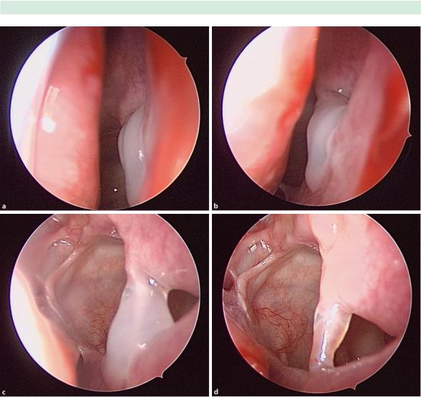
Rigid or flexible nasal endoscopy is the most important part of the physical exam for a sinus surgeon. Using angled endoscopes during the initial patient evaluation will allow inspection of the maxillary sinus and natural ostium. Mucus recirculation in the maxillary sinus is one of the most common causes for revision surgery [4]. This is often secondary to a synechia that has developed between the natural ostium and antrostomy, or to a residual uncinate process. Both of these are difficult to ascertain from a straight-on examination using a 0° endoscope or sometimes even with a 30° telescope. A 45 ° or 70° endoscope is needed to completely eliminate this subtle finding (Fig. 10.1).
Endoscopy of the anterior nasal cavity will determine whether or not there are significant synechiae between the middle turbinate and medial orbital wall. Synechiae between the nasal septum and inferior turbinate can be a significant cause for nasal obstruction. Synechiae between the middle turbinate and lateral nasal wall can prevent examination of the ethmoid cavity and spheno-
Tips and Pearls in Revision Sinus Surgery |
81 |
Fig. 10.1 Endoscopic view of a patient with mucus re-circula- tion following a functional endoscopic sinus surgery (FESS) procedure. A 0° endoscope shows thickened mucus at the antrostomy (a). Closer inspection with a 30° scope (b) and 70°
ethmoid recess. These can often be lysed in clinic after topical analgesia, and a temporary spacer can be placed lateral to the middle turbinate to prevent the formation of additional synechiae.
The degree of mucosal edema and presence of mucopurulence should be determined in the endoscopic examination of the ethmoid cavity. Cultures should be taken of any purulence and examined for the presence of pathogenic aerobic, anaerobic, and fungal organisms. The sphenoid ostium should then be visualized and, if possible, examination of the sinus itself should be performed to determine the presence of mucus within the sinus. Al-
scope (c) shows the scar band between the natural ostium and antrostomy that is creating the recirculation. Once the mucus is cleared, the scar band is clearly visualized (d)
lergic mucin or retained fungal elements in the inferior portion of the sphenoid sinus, as well as within the maxillary sinus, are a common source for persistent purulent rhinorrhea. Thick allergic mucus should also be submitted for pathology for examination with fungal stains and for the presence of Charcot-Leyden crystals.
Finally, the frontal recess should be examined with an angled endoscope. Signs to look for include the presence of polypoid edema, mucopurulence, and residual cells or bony partitions that may obstruct the frontal recess. Endoscopic findings should then be correlated with their radiographic appearance.

82
Fig. 10.2 Coronal computed tomography (CT) scan of a patient with a previous FESS who has a retained uncinate on the left and right sides (red circle)
10
CT Scan Review
Reviewing CT scans are best done in multiple planes. In-office consultation should result in a review of axial and coronal sections, at a maximum of 3-mm sections, through the paranasal sinuses. Many image-guidance companies now offer work-stations that allow for the review of CT scans in the sagittal plane, as well as the coronal and axial views. The coronal view is excellent in determining the presence of a remaining agger nasi, uncinate process, frontal recess, and/or supraorbital ethmoid cells.
Alexander G. Chiu and David W. Kennedy
Sagittal and axial views are important in determining the anterior-to-posterior dimension of the frontal recess, and the identification of a supraorbital ethmoid, frontal recess, and/or interseptal frontal sinus air cell. Sagittal views are also instructive in demonstrating the vertical slope of the skull base and the degree of ethmoid bony partitions attached to the skull base.
The most common anatomical areas identified in revision surgery are the following:
1.Remnant uncinate process with resultant mucus recirculation between the natural maxillary ostium and previously made maxillary antrostomy (Fig. 10.2).
2.Ethmoid bony partitions attached to the medial orbital wall and skull base. In patients with inflammatory polyps or neo-osteogenesis, these bony partitions may be osteitic, serve to increase the surface area where polyps can grow, and as a nidus for persistent mucosal inflammation (Fig. 10.3).
3.Remnant infraorbital ethmoid cells that are obstructing the natural maxillary ostium.
4.Lateralized middle turbinate against the medial orbital wall or scarred to an agger nasi cell and/or ethmoid bulla remnant (Fig. 10.4).
5.Residual sphenoethmoidal (Onodi) cell or posterior ethmoid cell.
6.Remnant supraorbital or frontal recess cells.
7.Synechiae between the middle turbinate and lateral nasal wall and/or medical orbital wall.
8.Neo-osteogenesis of the skull base and frontal recess from previous mucosal stripping and/or prolonged mucosal inflammation
Fig. 10.3 Coronal and sagittal views of patient with nasal polyposis who had undergone a previous FESS. Note the multiple bony ethmoid partitions off the skull base on the sagittal view (red circle). Revision surgery will be aimed at removing these osteitic bony partitions