

Revision Sinus Surgery
.pdf168 |
Sarah K. Wise, Richard J. Harvey, and Rodney J. Schlosser |
approaches for CSF leak repair circumvent the morbidities of intracranial approaches, which include anosmia, cerebral edema, seizures, and memory deficits [25]. The exceptional visualization provided by rigid endoscopes reduced morbidity over craniotomy, and higher success rates as compared to open approaches has made endoscopic repair of CSF leaks and meningoencephaloceles the preferred approach [15, 18, 24].
As surgeons develop increased comfort with endoscopic skull-base repair techniques, more challenging cases are being undertaken. Endoscopic surgeons are increasingly involved in addressing pathology along the skull base and beyond the skull base, often in conjunction with their neurosurgical colleagues. In addition, revision skull-base defect repair is frequently performed endoscopically following failure of primary endoscopic or open procedures. In each case, the endoscopic surgeon should be aware of factors that could lead to failure of skull-base defect repair. By identifying factors that may contribute to failure, surgeons can employ adjunctive perioperative and intraoperative measures that may improve the success of their repair.
Indications for Transnasal Endoscopic Repair of CSF Leaks and Meningoencephaloceles
1.Active CSF leak.
2.Pneumocephalus.
3.Skull-base defect with a history of meningitis.
4.Meningoencephalocele with active CSF leak or history of meningitis.
5.Skull-base defect created during endoscopic sinus surgery or skull-base surgery.
6.Skull-base defect accessible by transnasal endoscopic techniques.
20
Contraindications for Transnasal Endoscopic Repair of CSF Leaks and Meningoencephaloceles
1.Medical issues that render a patient unfit for surgery or general anesthesia.
2.Skull-base defect not accessible by transnasal endoscopic means.
3.Untreated tumor in the area of the skull-base defect. In this case, the tumor should be treated prior to definitive repair of the skull-base defect.
4.Surgeon discomfort or inexperience with endoscopic repair techniques.
5.Equipment or instrumentation unavailable.
6.Active meningitis (relative contraindication).
Preoperative Workup
Diagnosis of CSF Leak – Laboratory
Whether a patient presents with a primary or recurrent CSF leak, confirmation of true CSF rhinorrhea should be made. Studies used in the past, such as fluid analysis for glucose, protein, and electrolytes are no longer encouraged due to high false-positive and false-negative rates. Currently, β2-transferrin testing is advocated due to its high sensitivity and specificity for the detection of CSF [45]. β2-transferrin is found only in the CSF, ocular vitreous humor, and perilymph [45]. While false-positives may occur, these cases are rare, arising in patients with chronic liver disease, genetic variations of the transferrin gene, and inborn errors of glycoprotein metabolism [36, 47]. Analysis for β2-transferrin requires only 0.17 ml of rhinorrhea fluid, and results may be obtained in less than 3 h with immunofixation electrophoresis techniques [33].
■Diagnosis of CSF rhinorrhea by β2-transferrin is the most sensitive, specific, and readily available laboratory technique currently available.
A second protein, β-trace protein, may also be analyzed to detect CSF in rhinorrhea fluid. β-trace protein is an enzyme also known as prostaglandin D synthase, which is produced in the leptomeninges and choroid plexus and subsequently secreted into the CSF [52]. Although β-trace protein is found in fluids throughout the body, its concentration is much higher in the CSF [27], and detection of this protein is also highly sensitive and specific for CSF [3].
Diagnosis of CSF Leak – Radiology
Following confirmation of CSF rhinorrhea by laboratory testing, radiologic evaluation is undertaken to identify the site of the skull-base defect. In cases of recurrent CSF leak, the surgeon should evaluate carefully all areas of the skull base on imaging studies. Certain patients, such as those with traumatic or spontaneous CSF leaks, often have multiple skull-base defects, which may or may not be actively leaking CSF [38]. In addition, patients who have undergone previous skull-base repair may present at a later date with CSF leak from a separate anatomic location [54].
Computed tomography (CT) scan is generally the initial study by which to assess the bony anatomy of the skull base. CT scans are typically acquired in a thin slice (less than 1.5 mm) axial protocol, with coronal and sagittal images created by reformatting the axial data set (Fig. 20.1).
Recurrent Cerebrospinal Fluid Leaks and Meningoencephaloceles |
169 |
Evaluation of images in a bone-window algorithm provides the most accurate assessment of skull-base integrity. The roof of the ethmoid and sphenoid sinuses is best viewed in the coronal plane, while the posterior table of the frontal sinus is visualized in the axial or sagittal plane. Finally, the integrity of the sella and clivus may also be assessed in the sagittal plane.
■Once diagnosis of CSF rhinorrhea is confirmed by laboratory testing, the integrity of the skull base should be assessed radiographically. This evaluation typically begins with a fine-cut axial CT scan, with triplanar image reformations viewed in bone-window algorithm.
When multiple bony skull-base defects are visualized on fine-cut CT scan or additional information is needed to accurately identify the site of active CSF leak, a CT cisternogram may be obtained by giving intrathecal contrast at the time of the CT scan. The nasal cavity and paranasal sinuses are then evaluated for pooling of contrast material (Fig. 20.2). Since the patient must be actively leaking CSF during the study for the best results, this test has sensitivities ranging from 48 to 96% [48]. Low-flow or intermittent CSF leaks have a decreased chance of being detected on a CT cisternogram. Although valuable information may be obtained from a CT cisternogram, this procedure
is invasive and carries the risks of lumbar puncture and intrathecal contrast.
When evaluation of soft tissue along the skull base is necessary, such as in cases of suspected recurrent meningoencephalocele or skull-base tumor, magnetic resonance imaging (MRI) is frequently added to the diagnostic algorithm. Patients with spontaneous CSF leaks have a high rate of meningoencephalocele formation [6, 32, 38] and present with multiple simultaneous meningoencephaloceles in 31% of cases [38]. In such cases, MRI is valuable in determining the presence of meningoencephalocele and the contents of meningoencephalic sacs (Fig. 20.3). In patients with previous treatment for skull-base tumor, MRI may assist in the assessment of possible recurrence prior to attempting closure of a skull-base defect.
Magnetic resonance cisternography is an additional technique by which to localize CSF leaks, which has a reported sensitivity of 85–92% and 100% specificity [46]. The MRI protocol involves a fast spin-echo with fat suppression and image reversal [46], which demonstrates CSF as black against surrounding tissues, which have diminished intensity.
Finally, radionuclide cisternograms may be performed. This technique involves placement of pledgets at specific sites within the nasal cavity (cribriform recess, middle meatus, and sphenoethmoid recess), followed by lumbar
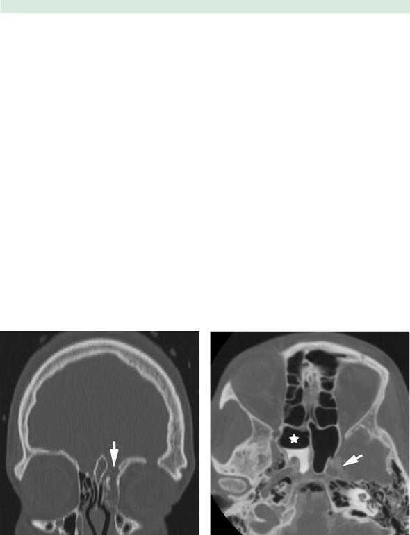
Fig. 20.1 Coronal computed tomography (CT) scan in a bone window algorithm, reconstructed from fine-cut axial data set. A left ethmoid roof defect (arrow) is visible following head trauma
Fig. 20.2 Axial CT cisternogram image in a patient with a spontaneous cerebrospinal fluid (CSF) leak. Contrast enhancement is seen in the intracranial CSF spaces (arrow). In addition, con- trast-enhanced CSF is present in the right sphenoid sinus (star)

170 |
Sarah K. Wise, Richard J. Harvey, and Rodney J. Schlosser |
Fig. 20.3 a Coronal CT scan of a patient with a spontaneous CSF leak. A skull-base defect is noted in the lateral recess of the right sphenoid sinus (arrow). The exact etiology of the sphenoid sinus opacification cannot be determined by CT scan alone.
puncture and injection of intrathecal tracer [41]. Radioactivity is subsequently measured on the pledgets after a period of hours. In comparison to other localization techniques, the sensitivity of radionuclide cisternogram is low (62–76%), with a 33% false-positive rate [12, 16, 48]. However, for low-volume or intermittent CSF leaks, pledgets may be left in the nasal cavity for days to allow for detection of the leak, dependent on the half-life of the tracer used [13].
■Following the assessment of skull-base integrity by fine-cut CT scan, further testing to localize the site of the CSF leak (CT cisternogram, magnetic resonance cisternogram, radionuclide cisternogram) or to assess soft tissue along the skull base (MRI) may be necessary, depending on the individual case.
20
Perioperative Adjuncts
Lumbar Drains
Lumbar drains are often used as an adjunct in the repair of CSF leaks and meningoencephaloceles, although their use is somewhat controversial. A lumbar drain can assist in decreasing intracranial CSF pressure and alleviating potential strain on the skull-base repair. This may be helpful in cases of elevated intracranial pressure, such as in trauma patients with cerebral edema, cases of hydrocephalus, or spontaneous CSF leaks [43, 44]. In addition, the lumbar drain catheter can provide a conduit for injection of intrathecal fluorescein, as described below. Lum-
b T2-weighted magnetic resonance imaging (MRI) scan of the same patient revealing a large meningoencephalocele protruding into the right sphenoid sinus
bar drains, however, have been associated with numerous complications, including the development or worsening of pneumocephalus, headache, nausea and vomiting, radicular pain, meningitis, vocal cord paralysis, and cerebral herniation [7, 11, 20, 37, 50, 51].
When the decision is made to use a lumbar drain, the surgeon should ensure that instructions for operating and monitoring the drain are clear and care of the drain is diligent. In a protocol to prevent the worsening of pneumocephalus with lumbar drains, Chan et al. advocate a passive gravity-dependent drainage system and avoidance of overdrainage [7]. Our protocol is similar, specifying the pressure above which CSF should be drained. This avoids the potential for intracranial “negative” pressures and the development of pneumocephalus, which can be life-threatening. The timing of lumbar drain removal varies depending on surgeon experience and preference, as well as patient-related factors, but in most cases, lumbar drains are removed 2–3 days postoperatively.
■Lumbar drains are a useful adjunct to CSF leak repair in certain cases. The surgeon should be mindful of potential complications associated with lumbar drains, however, and ensure that close monitoring and diligent care of the drain is available.
Intrathecal Fluorescein
Many surgeons use intrathecal fluorescein as an additional measure to localize the site of a skull-base defect intraoperatively. In this instance, injection of fluorescein

Recurrent Cerebrospinal Fluid Leaks and Meningoencephaloceles |
171 |
is often performed at the time of lumbar drain insertion in the operating room. Intrathecal fluorescein is not approved by the United States Food and Drug Administration, and complications such as seizures, extremity weakness, and opisthotonus have been reported [30, 35]. These complications are rare, however, and may be related to high concentration, suboccipital injection, or rapid intrathecal injection [35, 55].
When using intrathecal fluorescein, our protocol involves the slow (over 10 min) intrathecal injection of 0.1 ml of 10% sodium fluorescein diluted in 10 ml of CSF. At the completion of the intrathecal fluorescein injection, the lumbar drain is clamped and the patient is placed into the Trendelenburg position to allow the fluorescein to collect along the skull base. Upon exposure of the skull base intraoperatively, fluorescein is often visible with routine endoscopic lighting. However, visualization of fluorescein may be enhanced with a blue light filter, if necessary.
Although it may be argued that intrathecal fluorescein is not necessary for every CSF leak repair, the challenge of repairing recurrent CSF leaks and meningoencephaloceles may necessitate adjunctive measures such as intrathecal fluorescein. In patients who have undergone previous repair of CSF leaks and meningoencephaloceles, recurrence may be due to failure of repair at the primary site. However, the surgeon should also be mindful that patients may present with new skull-base defects at anatomic sites remote from the original repair. This is especially true in cases of trauma and spontaneous CSF leaks, where multiple bony skull-base defects may be present (Fig. 20.4, Video 20.1) [38]. In such cases, the entire skull base may require intraoperative evaluation, and clear CSF can be difficult to detect. Intrathecal fluorescein provides an additional tool for intraoperative evaluation of the skull base and detection of the skull-base defect in need of repair.
Image-Guided Computer Navigation Systems
Image-guidance systems are commonly used in advanced endoscopic surgery and skull-base surgery. These systems may assist the surgeon intraoperatively in localizing skullbase defects and defining their dimensions. A series of endoscopic nasal CSF leak closures with and without the use of an image-guidance system was reported in 2005 by Tabaee et al. [49]. Although a statistically significant benefit in the success of CSF leak closure was not found with the use of image guidance [49], other authors have reported an increase in surgeon confidence with the use of image-guidance systems [2, 28]. In cases of revision CSF leak and meningoencephalocele repair, image-guid- ance systems may provide an added benefit of precisely localizing the dimensions of a bony skull-base defect in a previously operated and scarred field.
An additional tool available with image-guided surgery systems is the availability of image fusion. With this tool, images obtained by different modalities, such as CT and magnetic resonance, may be fused on a computer workstation to allow visualization of both the bone and soft-tissue components of the skull-base defect. This can be helpful in assessing meningoencephaloceles and bony skull-base defects with surrounding soft tissue and scar, such as in the case of recurrent CSF leaks.
■Intraoperative adjuncts for the localization of skullbase defects include intrathecal fluorescein and im- age-guided navigation systems.
Surgical Techniques
Specific techniques for the endoscopic repair of CSF leaks and meningoencephaloceles continue to evolve. While changes in technique will continue to occur over time, some basic principles are presented here. Surgery is generally performed with the patient under general anesthesia and in the supine position. In patients with known skull-base defects, positive-pressure ventilation with the bag-mask apparatus is avoided during anesthesia induction in order to decrease the risk of pneumocephalus [41, 42]. Once the patient is intubated, a lumbar drain may be inserted if the surgeon prefers, and the patient may be placed in the Trendelenburg position to increase the chance of identifying clear drainage or fluorescein from the skull base. If a lumbar drain is inserted, the drain is typically clamped during the early portions of the procedure to keep CSF pressure high and allow for better detection of the site of CSF leak. The nasal cavities are then decongested topically, and perioperative intravenous antibiotics with intrathecal penetration are administered. Under endoscopic visualization, intranasal structures are injected with local anesthesia according to surgeon preference.
The site of the skull-base defect is then identified, utilizing the principles and techniques of endoscopic sinus surgery. Depending on the specific site of the skull-base defect, the extent of sinus dissection will vary. However, in general, the skull-base defect should be adequately exposed to allow for visualization of the entire defect. This often requires some combination of uncinectomy, maxillary antrostomy, ethmoidectomy, sphenoidotomy, and frontal sinusotomy, depending on the location of the defect. For defects in the lateral recess of the sphenoid sinus, Bolger has described the endoscopic transpterygoid approach, which provides improved access to this difficult area over traditional transseptal and transethmoid approaches [4]. In addition, while this chapter focuses on endoscopic repair of skull-base defects, it should be kept in mind that

172 |
Sarah K. Wise, Richard J. Harvey, and Rodney J. Schlosser |
Fig. 20.4a–c Patient status post traumatic brain injury, craniotomy, and frontal sinus fracture repair referred for evaluation of persistent pneumocephalus 2 months after the initial injury. a A small bony defect
is visualized in the right frontal sinus posterior table with adjacent pneumocephalus (arrow). b A second bony defect is noted in the sphenoid roof (arrowhead). CT cisternogram contrast is also seen along the sphenoid defect, but no contrast is
20 pooling in the sphenoid sinus. c Intrathecal fluorescein highlights a small meningocele identified intraoperatively along the sphenoid roof. Image-guidance pointer rests on the sella, with the meningocele anterior
most of the principles described may also be applied to combined open/endoscopic approaches, as well as purely open approaches via an osteoplastic flap. The specific location of the defect and surgeon comfort and experience will dictate which approach is necessary to provide adequate exposure and visualization of the defect.
As in endoscopic sinus surgery for inflammatory disease, dissection is often facilitated by the use of angled
endoscopes and giraffe instruments along the anterior ethmoid roof and in the frontal recess (Fig. 20.5). Angled endoscopes are also helpful in the lateral recess of the sphenoid sinus, where visualization can be difficult. During surgical dissection and exposure of skull-base defects, frontal and sphenoid sinusotomies should also be considered to prevent mucocele formation and allow for adequate assessment of ostia patency in the postoperative period.

Recurrent Cerebrospinal Fluid Leaks and Meningoencephaloceles |
173 |
Fig. 20.5 a Angled rigid nasal endoscopes improve visualization of the frontal sinus/recess and lateral sphenoid recess for skull-base defect repair. b Giraffe angled instruments allow frontal sinus/recess manipulation
■Dissection of the paranasal sinuses surrounding the skull-base defect should be thorough. This allows for adequate visualization and access of instrumentation to perform the repair.
Once the skull-base defect is identified and exposed, the bone of the defect is denuded of surrounding mucosa for several millimeters (Fig. 20.6, Video 20.2). This pre-
vents secretion of mucus from any mucosa underlying the graft, which may cause the graft to separate from its recipient bed [41, 42]. At this time, any meningoencephaloceles that are present at the site of skull-base defect are reduced to an intracranial position with bipolar cautery. This completes preparation of the recipient bed at the site of the skull-base defect. At this time, if an active CSF leak is present, CSF pressure may be diverted away from the
Fig. 20.6a,b Patient with recurrent CSF rhinorrhea following craniotomy for resection of olfactory meningioma. a Skull-base defect with likely meningocele identified in posterior ethmoid roof (arrow) on coronal CT scan in the bone window algorithm.
b Presence of a posterior ethmoid meningocele (arrowhead) confirmed on sagittal MRI images. Video 20.2 shows endoscopic reduction of this meningocele and repair of the skull-base defect

174 |
Sarah K. Wise, Richard J. Harvey, and Rodney J. Schlosser |
site of the skull-base defect by taking the patient out of the Trendelenburg position and placing him/her in a 30 head-up position. If present, the lumbar drain may also be opened to drain approximately 5–10 ml per hour for the remainder of the procedure.
■Careful removal of mucosa surrounding the bony skull-base defect prevents the secretion of mucus, which may separate the graft from the recipient bed.
Specific techniques for repair will vary based on the site, size, and etiology of the defect. For small (less than 3 mm) defects, simple soft-tissue overlay grafts will typically close the defect successfully. In addition, if there is concern that nerves or vessels may be damaged when dissecting the dura away from the skull base, overlay grafting is recommended [15]. For larger defects, multilayer repairs are advocated. Repair layers may consist of soft tissue only, or a structural layer of bone may be added for support of very large defects or defects in patients with increased intracranial pressure [41, 42].
Various grafting materials have been used, and graft choice will depend on surgeon comfort and preference. Free bone graft choices include septal bone, turbinate bone, and mastoid bone. Free cartilage grafts may also be used. Autologous septal and turbinate mucosa, temporalis fascia, and abdominal fat have also been used, as have alloplastic collagen, and cadaveric dermis, fascia, and pericardium [42]. One may also consider intranasal vascularized pedicle grafts in patients with large defects or in patients with significant scarring, history of skullbase radiation therapy, or decreased blood supply to the recipient bed. Tissue adhesives may be applied following graft placement, if desired.
Whenever possible, placement of a soft-tissue or bone underlay graft in the epidural space, followed by a softtissue overlay graft intranasally, should be considered. A few caveats should be kept in mind, however. First,
20 mucosal grafts should not be placed in an intracranial underlay position due to the risk of intracranial infection and mucocele [42]. Secondly, in patients with spontaneous CSF leaks and increased intracranial pressure, the entire skull base is often attenuated [42]. Significant manipulation of the skull base in these patients may lead to fracture and creation of larger defects. Finally, when placing grafts, the surgeon should remain aware of surrounding sinus ostia and avoid iatrogenic ostia obstruction during graft placement. When working near the frontal sinus outflow tract, short-term (1 week) insertion of a soft silastic stent may be useful in preventing iatrogenic obstruction during placement of the graft and packing material.
Once the graft materials are in place, the nose is packed to support the graft. Packing should begin with absorbable materials against the graft, followed by non-
absorbable materials if desired. Placement of absorbable materials in proximity to the graft allows any nonabsorbable materials to be removed without risking disruption of the graft. Absorbable materials will dissolve naturally over a period of approximately 6 weeks.
Potential Complications
Intracranial Complications
Pneumocephalus
With a known defect in the skull base, the surgeon should be aware of the potential for pneumocephalus, which is reported to carry a 25% risk of meningitis and 16% mortality [31]. Positive pressure from the external environment, such as sneezing, vomiting, continuous positive airway pressure respiratory devices, and application of bag-mask apparatus during general anesthesia induction can force air intracranially, leading to tension pneumocephalus. Patients should be alerted to this potential and instructed accordingly prior to their surgery. In addition, intubation protocols that avoid positive pressure during anesthesia induction are advisable [41, 42].
Intracranial Hemorrhage
When working along the skull base with a meningoencephalocele or skull-base tumor, vessels present in the mass often track intracranially. Traction forces on such masses may lead to rupture of the vessels in the intracranial cavity, leading to dangerous hemorrhage [41, 42]. Bipolar cautery for reduction of meningoencephaloceles and excision of skull-base tumors is typically advocated to safely cauterize these vessels without avulsing them [18].
Creation of New Skull-Base Defects
During the course of preoperative planning for endoscopic skull-base defect repair, the surgeon will carefully assess the skull base for the site of possible defects. As in preparation for endoscopic sinus surgery, sites of potential new complications involving the skull base should also be evaluated, such as asymmetry of the skull base, low-lying skull base, or tall middle-turbinate lateral lamellae. Awareness of such anatomic variants will alert the surgeon to possible sites for creation of new skullbase defects. This is especially important in the case of recurrent CSF leak or meningoencephalocele, given the altered anatomy and likely scarring from previous repairs.

Recurrent Cerebrospinal Fluid Leaks and Meningoencephaloceles |
175 |
Ocular Complications
Ocular complications are generally rare in endoscopic sinus surgery, and the same is true for endoscopic skull-base defect repair. However, the principles that guide the endoscopic surgeon during both types of procedure remain the same. Assessment of the patient’s anatomy on preoperative CT scan will alert the surgeon to any anatomic variants and the position and integrity of the lamina papyracea, optic nerve, and optic nerve bony canal. As many skull-base defects are located in the sphenoid sinus, the surgeon should be keenly aware of the location of the optic nerve and cavernous sinus when working in this area.
Frontal and Sphenoid Ostium Stenosis
In the course of repairing skull-base defects, multiple layers of grafts and packing material are often required to ensure adequate repair. Depending on the site of skullbase defect, the ostia of the frontal or sphenoid sinus may be in the vicinity of the repair if the defect is located in the anterior ethmoid roof or frontal recess. In such cases, application of sufficient packing material may be difficult while maintaining the patency of the sinus ostia. Our typical protocol in such cases is to adequately open the sinus ostia during the course of the repair, leaving as much mucosa intact around the ostium as possible. The repair is then completed in the routine fashion. Upon completion of repairs near the frontal recess, we consider placement of a soft T-shaped silastic stent for 5–7 days. This stent can then be gently removed in the clinic without disturbing the graft or packing material and may increase the chances of maintaining frontal patency long term. In addition, during the course of postoperative care, a probe or very light suction may be carefully passed into the ostium to ensure its patency while avoiding disturbance of the graft site.
Tips and Pearls for Avoiding Complications
1.Carefully evaluate preoperative imaging for anatomic variants of the orbit and skull base, as well as integrity of the carotid canal and optic nerve canal.
2.Avoid the application of positive pressure to decrease the risk of pneumocephalus.
3.In order to reduce the chance of intracranial hemorrhage from torn vessels, use bipolar cautery along the skull base to reduce meningoencephaloceles and resect tumors.
4.For repairs near the frontal sinus ostium, identify and enlarge the ostium intraoperatively, meticulously preserving surrounding mucosa. Postoperatively, gently probe or suction the frontal sinus to ensure its patency while avoiding disturbance of the skull-base graft.
Factors Contributing to Failure in Skull-Base Repair
Factors leading to failure of skull-base defect repair have not been evaluated comprehensively. However, certain factors may alert the surgeon to potential failure and deserve mention. Most frequently, the presence of increased intracranial pressure is cited as placing patients at risk for failure [29, 56]. Various authors have described a clinical association between spontaneous CSF leaks, increased intracranial pressure, obesity, and empty sella syndrome [20, 38–40, 43]. Entities such as spontaneous CSF leaks and meningoencephaloceles, traumatic brain injury, and hydrocephalus may place patients at increased risk of skull-base repair failure due to the concomitant increased intracranial pressure. Often, perioperative adjuncts such as lumbar drains and diuretics, as well as placement of underlay bone grafts are advocated in such patients to decrease their propensity for failure.
Lindstrom et al. [20] also note increased failure rate for skull-base defects located in the lateral sphenoid recess, citing the technical difficulty of visualization and repair in this region. Another surgical factor that may make skull-base repair challenging is large skull-base defect size, especially following significant trauma or extirpation of skull-base tumors [20, 54].
A recent series at our institution designed to assess factors differentiating patients undergoing primary skullbase defect repair as compared to those undergoing revision repair identified several additional factors thought to play a role in skull-base defect repair failure [54]. These included failure to precisely localize the skull-base defect during the initial surgery (Video 20.3) and identification of a new skull-base defect at an anatomic location remote from the original repair. Factors that may influence wound healing were also present in higher percentages in revision patients, such as prior skull-base surgery, prior radiation therapy to the skull base, intracranial infection, and history of a skull-base neoplasm (excluding pituitary neoplasms). This highlights the importance of fully assessing each patient’s clinical picture to ensure that surgical factors as well as patient-related factors are taken into account to provide the best possible repair circumstances.
Postoperative Care
While postoperative care and office debridement is integral to good surgical outcomes in endoscopic sinus surgery for inflammatory disease, office debridement is generally minimal in the setting of endoscopic transnasal CSF leak and meningocele repair. Postoperative protocols will vary depending on the operating surgeon and the individual case. However, some of our general

176 |
Sarah K. Wise, Richard J. Harvey, and Rodney J. Schlosser |
principles following endoscopic CSF leak repair are discussed.
At the time of hospital discharge, patients are instructed to avoid heavy lifting and strenuous activity for approximately 6 weeks. We also recommend stool softeners and laxatives as necessary to avoid straining. In patients with spontaneous CSF leaks and increased intracranial pressure, we add diuretic therapy to decrease CSF production and alleviate stress on the repair site. Acetazolamide decreases CSF production by as much as 48% [5], and a mean reduction in intracranial pressure by 10 cmH2O has been shown with the institution of acetazolamide postoperatively [44].
|
|
During surgery, the nose is typically packed with both |
|
|
absorbable and nonabsorbable packing after placement of |
|
|
graft material over the skull-base defect. In general, non- |
|
|
absorbable packs are removed 5–7 days postoperatively. |
|
|
At that time, we briefly evaluate the nasal cavity with |
|
|
an endoscope to ensure that there is no obvious recur- |
|
|
rence of CSF leak in the early postoperative period, but |
|
|
no significant debridement is performed. Over the next |
|
|
6 weeks, the patient is endoscopically evaluated approxi- |
|
|
mately every 2–3 weeks, and minimal debridements are |
|
|
performed with care not to disturb the graft site. For de- |
|
|
fects in the anterior ethmoid and frontal recess, the area |
|
|
of the frontal sinus ostium may be carefully probed or |
|
|
debrided to ensure that the frontal sinus remains patent. |
|
|
As previously discussed, any frontal recess stents are gen- |
|
|
erally removed within 1 week. At 6 weeks postoperatively, |
|
|
the majority of the absorbable packing has dissolved and |
|
|
the graft site is usually visible. During the postoperative |
|
|
course, the patient does not perform any nasal irrigation, |
|
|
as nasal drainage following irrigations may often be mis- |
|
|
taken for recurrence of CSF leak. |
|
|
|
|
|
Outcomes |
|
|
|
20 |
|
With success rates for endoscopic repair of CSF leaks and |
|
|
meningoencephaloceles greater than 90% in most series |
[6, 8, 15, 17–19, 21–24, 26, 29, 56], transnasal endoscopic repair of skull-base defects has become a preferred approach. However, recurrent CSF leaks and meningoencephaloceles may put forward challenges not present in primary cases. In a recent series at our institution, endoscopic primary skull-base defect repairs were compared to revision repairs. While the success rate for the overall group was high (93%), patients undergoing revision repair had a lower rate of successful closure (85%) than patients undergoing primary repair (97%) [54].
Patients with recurrent CSF leaks and meningoencephaloceles should be carefully assessed for factors that may contribute to failure of their repair. Such factors include spontaneous CSF leaks and meningoencephalo-
celes with increased intracranial pressure [14, 29], location of a skull-base defect in the lateral sphenoid sinus [20], and a large skull-base defect [20]. Other factors that may play a role in predisposing patients to recurrent CSF leaks and meningoencephaloceles include failure to accurately localize the skull-base defect, development of new skull-base defects, history of prior skull-base surgery or craniotomy, history of skull-base radiation, intracranial infections, and skull-base neoplasm (other than pituitary neoplasms) [54]. It is important to recognize these surgical factors and patient-related factors in order to adequately plan for surgery and for the care of the patient in the perioperative period.
References
1.Aarabi B, Leibrock LG (1992) Neurosurgical approaches to cerebrospinal fluid rhinorrhea. Ear Nose Throat J 71:300–305
2.Anon JB (1998) Computer-aided endoscopic sinus surgery. Laryngoscope 108:949–961
3.Arrer E, Meco C, Oberascher G (2002) Beta trace protein as a marker for cerebrospinal fluid rhinorrhea. Clin Chem 48:939–941
4.Bolger WE (2005) Endoscopic transpterygoid approach to the lateral sphenoid recess: surgical approach and clinical experience. Otolaryngol Head Neck Surg 133:20–26
5.Carrion E, Hertzog JH, Medlock MD (2001) Use of acetazolamide to decrease cerebrospinal fluid production in chronically ventilated patients with ventriculoperitoneal shunts. Arch Dis Child 84:68–71
6.Casiano RR, Jassir D (1999) Endoscopic cerebrospinal fluid rhinorrhea repair: is lumbar drain necessary? Otolaryngol Head Neck Surg 121:745–750
7.Chan E, Meiteles L (2007) Otogenic tension pneumocephalus caused by therapeutic lumbar CSF drainage for posttraumatic hydrocephalus: a case report. ENT Ear Nose Throat 86:391–393
8.Chin GY, Rice DH (2003) Transnasal endoscopic closure of cerebrospinal fluid leaks. Laryngoscope 113:136–138
9.Dandy WE (1926) Pneumocephalus: intracranial pneumatocele or aerocele. Arch Surg 12:949–982
10.Dohlman G (1948) Spontaneous cerebrospinal rhinorrhoea: case operated by rhinologic methods. Acta Otol Suppl 67:20–23
11.Dowd G, Molony T, Voorhies R (1998) Spontaneous otogenic pneumocephalus: case report and review of the literature. J Neurosurg 89:1036–1039
12.Eljammel MS, Pidgeon CN, Toland J (1994) MRI cisternography and localization of CSF fistulae. Br J Neurosurg 8:433–437

Recurrent Cerebrospinal Fluid Leaks and Meningoencephaloceles |
177 |
13.Flynn BM, Butler SP, Quinn RJ (1987) Radionuclide cisternography in the diagnosis and management of cerebrospinal fluid leaks: the test of choice. Med J Aust 146:82–84
14.Gendeh BS, Mazita A, Selladural BM, et al. (2005) Endonasal endoscopic repair of anterior skull base fistulas: the Kuala Lumpur experience. J Laryngol Otol 119:866–874
15.Hegazy HM, Carrau RL, Snyderman CH, et al. (2000) Transnasal endoscopic repair of cerebrospinal fluid rhinorrhea: a meta-analysis. Laryngoscope 110:1166–1172
16.Hubbard JL, McDonald TJ, Pearson BW, et al. (1985) Spontaneous cerebrospinal fluid rhinorrhea: evolving concepts in diagnosis and surgical management based on the Mayo Clinic experience from 1970 through 1981. Neurosurgery 16:314–321
17.Kirtane MV, Gauitham K, Upadhyaya SR (2005) Endoscopic CSF rhinorrhea closure: our experience in 267 cases. Otolaryngol Head Neck Surg 132:208–212
18.Lanza DC, O‘Brien DA, Kennedy DW (1996) Endoscopic repair of cerebrospinal fluid fistulae and encephaloceles. Laryngoscope 106:1119–1125
19.Lee TJ, Huang CC, Chauang CC, et al. (2004) Transnasal endoscopic repair of cerebrospinal fluid rhinorrhea and skull base defect: ten-year experience. Laryngoscope 114:1475–1481
20.Lindstrom DR, Toohill RJ, Loerhl TA, et al. (2004) Management of cerebrospinal fluid rhinorrhea: the Medical College of Wisconsin experience. Laryngoscope 114:969–974
21.Locatelli D, Rampa F, Acchiardi I, et al. (2006) Endoscopic endonasal approaches for repair of cerebrospinal fluid leaks: nine year experience. Neurosurgery 58:ONS246–257
22.Lopatin AS, Kapitanov DN, Potapov AA (2003) Endonasal endoscopic repair of spontaneous cerebrospinal fluid leaks. Arch Otolaryngol Head Neck Surg 129:859–863
23.Marshall AH, Jones NS, Robertson IJA (1999) An algorithm for the management of CSF rhinorrhoea illustrated by 36 cases. Rhinology 37:182–185
24.Mattox DE, Kennedy DW (1990) Endoscopic management of CSF leaks and cephaloceles. Laryngoscope 100:857–862
25.McCormack B, Cooper PR, Persky M, et al. (1990) Extracranial repair of cerebrospinal fluid fistulas: techniques and results in 37 patients. Neurosurgery 27:412–417
26.McMains KC, Gross CW, Kountakis SE (2004) Endoscopic management of cerebrospinal fluid rhinorrhea. Laryngoscope 114:1833–1837
27.Melegos DN, Diamandis EP, Oda H, et al. (1996) Immunofluorometric assay of prostaglandin D synthase in human tissue extracts and fluids. Clin Chem 42:1984–1991
28.Metson RB, Cosenza MJ, Cunningham MJ, et al. (2000) Physician experience with an optical based image guided system for sinus surgery. Laryngoscope 110:972–976
29.Mirza S, Tahaper A, McClelland L, et al. (2005) Sinonasal cerebrospinal fluid leaks: management of 97 patients over 10 years. Laryngoscope 115:1774–1777
30.Moseley JI, Carton CA, Stern WE (1978) Spectrum of complications in the use of intrathecal fluorescein. J Neurosurg 48:765–767
31.Noth J (1971) On the importance of intracranial air. Brit J Surg 58:826–829
32.Ommaya AK, DiChiro G, Baldwin M, et al. (1968) Nontraumatic cerebrospinal fluid rhinorrhea. J Neurol Neurosurg Psychiatry 31:214–215
33.Papadea C, Schlosser RJ (2005) Rapid method for beta-2 transferrin in cerebrospinal fluid leakage using an automated immunofixation electrophoresis system. Clin Chem 51:464–470
34.Park JL, Strelzow VV, Friedman WH (1983) Current management of cerebrospinal fluid rhinorrhea. Laryngoscope 93:1294–1300
35.Rainer K, Rainer W, Wolfgang D, et al. (2004) Use of sodium fluorescein solution for detection of cerebrospinal fluid fistulas: an analysis of 420 administrations and reported complications in Europe and the United States. Laryngoscope 114:266–272
36.Roelandse FWC, Van de Zwart AZJ, Didden JH (1998) Detection of CSF leakage by isoelectric focusing on polyacrylamide gel, direct immunofixation of transferrin and silver staining. Clin Chem 44:351–353
37.Roland PS, Marple BF, Meyerhoff WL, et al. (1992) Complications of lumbar spinal fluid drainage. Otolaryngol Head Neck Surg 107:564–569
38.Schlosser RJ, Bolger WE (2002) Management of multiple spontaneous nasal meningoencephaloceles. Laryngoscope 112:980–985
39.Schlosser RJ, Bolger WE (2003) Significance of empty sella in cerebrospinal fluid leaks. Otolaryngol Head Neck Surg 128:32–38
40.Schlosser RJ, Bolger WE (2003) Spontaneous nasal cerebrospinal fluid leaks and empty sella syndrome: a clinical association. Am J Rhinol 17:91–96
41.Schlosser RJ, Bolger WE (2004) Nasal cerebrospinal fluid leaks: critical review and surgical considerations. Laryngoscope 114:255–265
42.Schlosser RJ, Bolger WE (2006) Endoscopic management of cerebrospinal fluid rhinorrhea. Otolaryngol Clin North Am 39:523–538
43.Schlosser RJ, Wilensky EM, Grady MS, et al. (2003) Elevated intracranial pressure in spontaneous CSF leaks. Am J Rhinol 17:191–195
44.Schlosser RJ, Wilensky EM, Grady MS, et al. (2004) Cerebrospinal fluid pressure monitoring after repair of cerebrospinal fluid leaks. Otolaryngol Head Neck Surg 130:443–448
45.Sibler H (1978) The normal cerebrospinal fluid proteins identified by means of thin-layer isoelectric focusing and crossed immunoelectrofocusing. J Neurol Sci 36:273–288