

УЗИ лимфузлов и ПЭТКТ
.pdfORIGINAL ARTICLE |
ACTA RADIOLOGICA |
|
|
The role of ultrasonography and FDG-PET in axillary lymph node staging of
breast cancer
JHII-HYUN AHN1, EUN JU SON1, JEONG-AH KIM1, JI HYUN YOUK1, EUN-KYUNG KIM1, JIN YOUNG KWAK1, YOUNG HOON RYU2 & JOON JEONG3
Departments of 1Radiology, 2Nuclear Medicine, and 3General Surgery, Yonsei University College of Medicine, Research Institute of Radiological Science, Seoul, Korea
Background: The presence of axillary lymph node metastasis is the most important prognostic factor and an essential part of staging and prognosis of breast cancer.
Purpose: To elucidate the usefulness and accuracy of ultrasonography (US), fluorodeoxyglucose positron emission tomography (FDG-PET) scan, and combined analysis for axillary lymph node staging in breast cancer.
Material and Methods: A total of 250 consecutive breast cancer patients who had undergone US, FDG-PET, and sentinel lymph node biopsy (SLNB) before surgery from January 2005 to December 2006 were included in the study. If an axillary lymph node had a length to width ratio 1.5 or cortical thickening 3 mm or compression of the hilum on US, focal hot uptake (maximal standardized uptake value, SUVmax 2.0) in the ipsilateral axilla on FDG-PET, it was considered to be a metastatic lymph node. In combined analysis of US and FDG-PET, the interpretation was considered positive if at least two of any of the criteria were met. Each imaging finding was compared with a pathologic report regarding the presence of axillary lymph node metastasis, the number of metastatic lymph nodes, and the T stage of the breast mass.
Results: Pathologically confirmed axillary lymph node metastasis was noted in 73 cases (29.2%). The mean number of metastatic lymph nodes in pathology was 3.1 3.2, and the size of breast cancer was 2.0 1.04 cm. In the detection of lymph node metastasis, the diagnostic accuracy of US was 78.8% and that of FDG-PET was 76.4%. On combined US and FDG-PET, accuracy was improved (91.6%). The number of metastatic lymph nodes on pathology was correlated with the positivity of US and FDG-PET (P 0.01).
Conclusion: Combined evaluation of US and FDG-PET was a sensitive and accurate method for axillary lymph node staging in breast cancer.
Key words: Staging; metastasis
Eun Ju Son, MD, Department of Radiology, Gangnam Severance Hospital, Yonsei University College of Medicine, 146-92, Dogok-dong, Gangnam-gu, Seoul, 135-270, Korea (tel. 82 2 2019 3510, fax. 82 2 3462 5472, e-mail: ejsonrd@yuhs.ac)
Submitted October 12, 2009; accepted for publication June 7, 2010
The presence of axillary lymph node metastasis is the most important prognostic factor in breast cancer (1, 2). Traditionally, axillary lymph node dissection has been the standard for axillary nodal assessment; however, this procedure can cause lymphedema, restriction of arm and shoulder movement, and numbness of the upper arm (3, 4). This has led to the increased necessity for a less invasive procedure. Therefore, sentinel lymph node biopsy (SLNB) has become the new standard in breast cancer patients for axillary lymph node evaluation as a less invasive alternative (5), but this approach has technical and conceptual limits (6, 7). False-negative results of SLNB can occur in a variable
percentage of patients (from 0% to 15%) (2). Most of these false-negative results occur due to massive lymph node metastasis in the first drainage node.
Axillary ultrasonography (US) and fluorine-18 fluorodeoxyglucose positron emission tomography (FDG-PET) scan have not been systemically performed in evaluation of axillary lymph node staging in breast cancer patients due to their historically low sensitivity. However, many studies have evaluated the role of US or FDG-PET for axillary lymph node staging as non-invasive alternatives (8–15). A previous report described the combined evaluation of axillary lymph node staging of US and 99mTc-sestamibi
DOI 10.3109/02841851.2010.501342 © 2010 Informa Healthcare
860 J.-H. Ahn et al.
scintimammography (16). However, there have not been any reports on the combined use of US and FDGPET for evaluation of axillary lymph node staging. We performed a study to assess the accuracy of axillary US and FDG-PET both independently and in combination, for axillary lymph node staging, especially in stage Tis, T1, and T2 breast cancer.
Material and Methods
The present study study was approved by the institutional review board at our institution.
Inclusion criteria
From January 2005 to December 2006, a total of 320 patients were diagnosed with breast cancer at our institute, and all of these patients underwent breast US, FDG-PET, and SLNB before surgery. Among 320 patients, 63 patients with neoadjuvant chemotherapy were excluded due to difficult evaluation of lymph node status. We also excluded seven cases that failed during SLNB. In total, 250 consecutive patients (median age 48.9 years, range 28–79 years) were enrolled in the study.
Imaging procedure
Axillary US was performed by two radiologists (E.J.S and J.A.K) with over 5 years experience each in breast radiology. US was performed with 10–12 MHz linear transducers (HDI5000, Advanced Technology Laboratories, Bothell, Wash., USA). If indeterminate/ suspicious/metastatic lymph node was detected in the axilla, transverse and longitudinal scans were taken and longitudinal and the transverse diameters and cortical thicknesses of the nodes were measured. If there were multiple lymph nodes in the axilla, once the images of all visible lymph nodes were obtained, the most suspicious lymph node was evaluated with the consensus of two radiologists.
In FDG-PET imaging, patients received an intravenous injection of 370 MBq of FDG in the arm contralateral to the primary tumor. At 60 min after injection of FDG, whole-body emission scans were obtained using a Philips Allegro PET camera (Philips Medical Systems, Cleveland, Ohio, USA). All patients were studied in the supine position with their arms raised. Attenuation-corrected transaxial images were reconstructed with an iterative transmission algorithm called row-action maximum likelihood 3D protocol using a 3D image filter into a 128 128 matrix. Quantitative measurement of the single-pixel maximal standardized uptake value (SUV max) was performed for all breast and axillary areas.
Image interpretation
US scans of axillary lymph nodes were evaluated for shape, cortical thickening, and the morphology of the hilum. A positive finding was given if the lymph node showed: 1) the cortex of the nodes was concentrically or eccentrically thickened more than 3 mm; or 2) compression of the hilum and especially the absence of the fatty hilum; or 3) the length-to-width ratio was less than 1.5. If the axillary lymph nodes exhibited any of the three findings mentioned above, they were defined as suspicious for axillary lymph node metastasis (8, 9). Nodes that exhibited none of the three findings mentioned above were defined as negative for axillary lymph node metastasis. In case of multiple axillary lymph nodes on US, we analyzed the most suspicious lymph node.
On evaluation of FDG-PET images, if the SUVmax was 2.0 or greater in the ipsilateral axillary lymph node-bearing area, it was considered a positive lymph node metastasis, and the SUVmax of the main mass was also measured. FDG-PET scans were interpreted by one specialist who had 10 years of experience in nuclear medicine.
In a combined analysis using US and FDG-PET, the interpretation was considered positive if at least two of any of the criteria included was positive.
Surgery and pathologic review
All 250 patients underwent SLNB. On the morning of the surgery, 1.0 mCi of technetium99 sulfur colloid was injected at the subareolar area. Lymphoscintigraphy was routinely performed. Intraoperatively, the sentinel lymph node was identified with a hand-held gamma probe.
Pathologic evaluation was performed by one dedicated pathologist who had over 10 years of experience of breast pathology. All specimens were submitted in formalin and frozen section for hematoxylin and eosin (HE) and immunohistochemical (cytokeratin) staining. The number and sizes of sentinel lymph nodes were documented. Sentinel lymph nodes 0.5 cm in the maximal dimension were serially sectioned transversely; those 0.5 cm were bisected. After frozen microscopic examinations, the pathologic result was immediately reported to the surgeon. If there was lymph node metastasis in the sentinels, axillary lymph node dissection was performed.
Each imaging finding was compared with the pathologic report on the presence of axillary lymph node metastasis (not including micrometastasis), the number of metastatic lymph nodes, the histologic type of breast cancer, and the T stage of the breast mass.
Statistical analysis
Sensitivity, specificity, positive and negative predictive value (PPV and NPV), false positive and false
Acta Radiol 2010 (8)

US and FDG-PET in axillary lymph node staging of breast cancer 861
negative rate, and accuracy of axillary US, FDG-PET, and SLNB were calculated using SigmaStat 2.03 statistical software (SPSS Inc., Chicago, Ill., USA).
Comparisons between groups were performed using the chi-square (χ2) test when required. Statistical significance was calculated using the t test, and P values0.05 were considered statistically significant.
Results
The histologic diagnoses of breast cancer included invasive ductal carcinoma (n 185), ductal carcinoma in situ (DCIS, n 32), invasive cribriform carcinoma (n 11), invasive lobular carcinoma (n 8), invasive apocrine carcinoma (n 3), invasive micropapillary carcinoma (n 3), medullary carcinoma (n 3), mucinous carcinoma (n 3), and tubular carcinoma (n 2). The average size of the breast masses was 2.01.04cm (range 0.3–5.0 cm). The T stages of breast cancer were: Tis (n 32), T1a (n 7), T1b (n 37), T1c (n 84), and T2 (n 90).
Among 250 patients, 73 (29.2%) were pathologically confirmed with axillary lymph node metastasis. However, there was no lymph node metastasis case in DCIS patients. The pathologic grades of DCIS were: group 1 (n 4, 12.5%), group 2 (n 9, 28.1%), and group 3 (n 19, 59.4%) according to Van-Nuys classification. The mean number of metastatic lymph nodes in pathology was 3.1 3.2 (range 1–15).
Ultrasonography
On US, 96 patients (38.4%) showed positive lymph nodes. Of the patients who showed positive findings
by US, 88 patients showed concentric or eccentric cortical thickening of more than 3 mm, 23 patients had compression of the hilum or absence of the fatty hilum, and 7 patients had a length-to-width ratio 1.5. Sixteen patients showed more than two suspicious findings. Among 96 patients, 39 patients showed multiple lymph nodes on US. Overall sensitivity, specificity, PPV, NPV, and diagnostic accuracy of US were 79.5%, 78.5%, 60.4%, 90.3%, and 78.8%, respectively (Table 1). Among 73 patients who had pathologically confirmed axillary lymph node metastasis, 15 patients were noted to have negative findings. In other words, among 177 patients who were found to be N0 stage, 38 patients had positive findings, respectively. Therefore, the false-negative rate and false-positive rate of US were 20.5% (Fig. 1) and 21.5%, respectively. Among 38 patients who had false-positive findings on US, 34 patients showed cortical thickening of more than 3 mm (mean 4.54 1.66), 5 patients had compression of the hilum, and 2 patients had a length- to-width ratio 1.5. One patient showed all of the three suspicious findings. The overall PPVs of each finding were: 61.4% (54/88) for cortical thickening, 78.3% (18/23) for hilum changes, and 71.4% (5/7) for length-to-width ratio.
FDG-PET
FDG-PET revealed that 48 patients (19.2%) had focal hot uptake (SUVmax 2.0) in the axillary area. The overall sensitivity, specificity, PPV, NPV, and diagnostic accuracy of FDG-PET were 42.5%, 90.4%, 64.6%, 79.2%, and 76.4%, respectively (Table 1). In patients with positive lymph node on FDG-PET, the
Table 1. Overall sensitivity, specificity, PPV, NPV, and accuracy of US, FDG-PET, and SLNB according to T stage of breast cancer.
|
|
Sensitivity |
Specificity |
PPV |
|
NPV |
Accuracy |
||||
|
|
|
|
|
|
|
|
|
|
|
|
US |
Tis |
0 |
(0/0) |
68.8 |
(22/32) |
0 |
(0/10) |
100.0 |
(22/22) |
68.8 |
(22/32) |
|
T1 |
78.6 |
(22/28) |
85.0 |
(85/100) |
59.5 |
(22/37) |
93.4 |
(85/91) |
83.6 |
(107/128) |
|
T2 |
80.0 |
(36/45) |
71.1 |
(32/45) |
73.5 |
(36/49) |
78.0 |
(32/41) |
75.6 |
(68/90) |
|
All |
79.5 |
(58/73) |
78.5 |
(139/177) |
60.4 |
(58/96) |
90.3 |
(139/154) |
78.8 |
(197/250) |
FDG-PET |
Tis |
0 |
(0/0) |
93.8 |
(30/32) |
0 |
(0/2) |
100.0 |
(30/30) |
93.8 |
(30/32) |
|
T1 |
35.7 |
(10/28) |
92.0 |
(92/100) |
55.6 |
(10/18) |
83.6 |
(92/110) |
79.7 |
(102/128) |
|
T2 |
46.7 |
(21/45) |
84.4 |
(38/45) |
75.0 |
(21/28) |
61.3 |
(38/62) |
65.6 |
(59/90) |
|
All |
42.5 |
(31/73) |
90.4 |
(160/177) |
64.6 |
(31/48) |
79.2 |
(160/202) |
76.4 |
(191/250) |
US FDG-PET |
Tis |
0 |
(0/0) |
90.6 |
(29/32) |
0 |
(0/3) |
100.0 |
(29/29) |
90.6 |
(29/32) |
|
T1 |
82.1 |
(23/28) |
98.0 |
(98/100) |
92.0 |
(23/25) |
95.1 |
(98/103) |
94.5 |
(121/128) |
|
T2 |
84.4 |
(38/45) |
91.1 |
(41/45) |
90.5 |
(38/42) |
85.4 |
(41/48) |
87.8 |
(79/90) |
|
All |
83.6 |
(61/73) |
94.9 |
(168/177) |
87.1 |
(61/70) |
93.3 |
(168/180) |
91.6 |
(229/250) |
SLNB |
Tis |
0 |
(0/0) |
100.0 |
(32/32) |
0 |
(0/0) |
100.0 |
(32/32) |
100.0 |
(32/32) |
|
T1 |
75.0 |
(21/28) |
100.0 |
(100/100) |
100.0 |
(21/21) |
93.5 |
(100/107) |
94.5 |
(121/128) |
|
T2 |
97.8 |
(44/45) |
97.8 |
(44/45) |
97.8 |
(44/45) |
97.8 |
(44/45) |
97.8 |
(88/90) |
|
All |
89.0 |
(65/73) |
99.4 |
(176/177) |
98.5 |
(65/66) |
95.7 |
(176/184) |
96.4 |
(241/250) |
|
|
|
|
|
|
|
|
|
|
|
|
PPV, positive predictive value; NPV, negative predictive value; US, ultrasonography; FDG-PET, fluorodeoxyglucose positron emission tomography; SLNB, sentinel lymph node biopsy. Values in parentheses are numbers of cases.
Acta Radiol 2010 (8)
862 J.-H. Ahn et al.
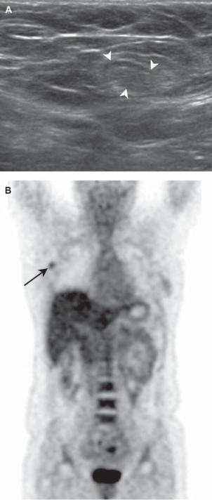
Fig. 1. A 39-year-old woman with infiltrating ductal carcinoma in the right breast. (A) The right axillary lymph node (arrowheads) does not have a length-to-width ratio 1.5, a cortical thickening of 2 mm or the loss of hilum on US, so we concluded that it was a negative lymph node. (B) On FDG-PET, hot uptake lesion (SUVmax 2.0) is noted at the right axillary area (arrow), so we decided that it was positive for lymph node metastasis. A pathologically metastatic lymph node was reported, while there were false-negative findings on US.
mean SUVmax of primary breast cancer was 7.4 5.2 (0.4–43.8). Meanwhile, in patients with negative lymph nodes on FDG-PET, it was 5.4 4.4. There was a significant correlation between SUVmax of primary breast cancer and positivity of axillary lymph nodes on FDG-PET (P 0.033). Among 73 patients who had pathologically confirmed axillary lymph node metastasis, 42 patients were noted to have negative findings on FDG-PET. In other words, among 177 patients who were found to be N0 stage, 17 patients had positive findings on FDG-PET. Therefore, the false-negative rate and false-positive rate of FDG-PET were 57.5% and 9.6%, respectively.
The mean SUV of the main mass in false-negative patients was 6.4. In other words, the mean SUVmax of the main mass in false-positive cases was 6.2. There was no significant correlation for main mass mean SUVmax between these false-negative and falsepositive cases (P 0.82).
US combined FDG-PET
When both imaging modalities were combined, there were 70 (28.0%) cases of positive axillary lymph nodes. Overall sensitivity, specificity, PPV, NPV, and diagnostic accuracy were 83.6%, 94.9%, 87.1%, 93.3%, and 91.6%, respectively, on combined US and FDG-PET (Table 1). Among 73 patients who had pathologically confirmed axillary lymph node metastasis, 12 patients were noted to have negative lymph nodes on FDG-PET and US together, and therefore the false-negative rate of combined examinations was 16.4%. On the other hand, among 177 patients who were pathologically confirmed negative for axillary lymph node metastasis, 9 patients were disclosed as having a positive lymph node on combined FDG-PET and US, and therefore the false-positive rate was 5.1% (Fig. 2).
The number of metastatic lymph nodes on pathology was correlated with the positivity of US and FDG–PET (P 0.01). However, suspicious axillary lymph nodes on FDG-PET and US would have less relevance than the size of the main breast mass and the pathologic type of breast cancer.
Sentinel lymph node biopsy
Among 250 patients, 66 (26.4%) had metastatic axillary lymph nodes by SLNB. Among 73 patients who had pathologically confirmed axillary lymph node metastasis, 8 patients were noted to have negative findings on SLNB. Therefore, the false-negative rate of SLNB was 11.0%. Diagnostic parameters are listed in Table 1.
We also evaluated the overall sensitivity, specificity, PPV, NPV, and diagnostic accuracy according to T stage: Tis and T1 (1a, 1b, and 1c) vs T2 (Table 1).
Acta Radiol 2010 (8)

US and FDG-PET in axillary lymph node staging of breast cancer 863
Fig. 2. A 41-year-old woman with infiltrating ductal carcinoma at the left breast. (A) Left axillary lymph node (arrowheads) shows a globular shape and cortical thickening of 2 mm on US. (B) Hot uptake lesions (SUV max 2.0) are noted at the left axillary area (arrows) on FDGPET. We concluded that the lymph node was positive on both US and FDG-PET. However, a pathologically negative lymph node was reported, with a false-positive finding on both US and FDG-PET.
There was higher sensitivity in diagnosing the axilla lymph node state in T2 stage breast cancer. Otherwise, the specificities were high in Tis and T1 stage breast cancer (Table 1).
Discussion
The reported sensitivity and specificity of axillary US range between 50% and 90%, and between 70% and 100%, respectively (8, 10, 11, 17). LUMACHI et al. (16) reported that the sensitivity and specificity of US alone are 68% and 80%, respectively, in patients with T1–2 stage breast cancer. According to ALVAREZ et al. (18), axillary US is moderately sensitive and quite specific in the diagnosis of axillary metastatic involvement in patients with breast cancer. Therefore, axillary US cannot be used in isolation as a method for deciding whether to perform axillary lymph node dissection. In our study, the sensitivity, specificity, PPV, and NPV of US were 79.5%, 78.5%, 60.4%, and 90.3%, respectively, so metastatic axillary lymph node cannot be determined using axillary US alone, even though US is acceptable for the detection of metastatic axillary lymph node. In addition, PPV of cortical thickening in US findings was lower (61.4%) than other criteria in our study. Even though cortical thickening is an important and widely used criterion for assessment of lymph node status (8, 9, 16, 18, 19), it cannot be used alone for deciding whether the lymph node is metastatic or not.
FDG-PET is increasingly being used to stage patients with breast cancer. Previous studies have revealed the capability to detect axillary metastases in patients with large primary breast tumors, with sensitivities ranging from 90% to 100% and specificities of 75–100%. However, higher false-negative rates were noted in patients with smaller tumors (15–17). Several studies about FDG-PET for axillary metastases have reported a relationship between sensitivity and primary tumor size. In addition, some studies relate the diagnostic accuracy of FDG-PET to primary tumor characteristics. UTECH et al. (13) explored a variety of variables, including tumor size, type, grade, estrogen receptor/progesterone receptor status, and number of dissected nodes. They found weak correlations between axillary uptake and tumor size and the number of cells in S-phase. GRECO et al. (20) reported a higher sensitivity for T2 tumors, but a higher specificity for T1 tumors. CHUNG et al. (21) noted that larger axillary metastatic lymph nodes were more likely to be positive in FDG-PET. The size of the primary tumor may weakly affect the SUVs of axillary lymph nodes, but other tumor characteristics were not correlated with FDG-PET activity of the axillary
Acta Radiol 2010 (8)
864 J.-H. Ahn et al.
lymph node (21). Sensitivity has been reported to vary with tumor type (22) and is in general higher in cases of invasive ductal carcinoma than in invasive lobular carcinoma (23). However, in our study there was no significant correlation between the positivity of FDGPET and the size or tumor type of primary breast cancer. In addition, there was no significant correlation for main mass SUVmax between the false-negative case and true-positive case.
In our study, the SUVmax of primary breast cancer was found to vary, and to reflect the degree of tumor metabolism. In addition, we observed a significant correlation between mean SUVmax of primary breast cancer and positivity of axillary lymph nodes on FDGPET. Therefore, a false-negative axillary lymph node on FDG-PET may result from metastatic axillary lymph nodes derived from primary breast cancer with a low SUVmax.
When US and FDG-PET are combined, the accuracy of detection of axillary lymph node metastasis was improved relative to the accuracy of each modality alone. In addition, the false-negative rate and false-pos- itive rate also decreased with the combined use of US and FDG-PET. Therefore, US and FDG–PET might have complementary roles in the evaluation of axillary lymph node metastasis in breast cancer patients.
SLNB is an invasive modality for axillary assessment, but it is a highly sensitive and specific method for detecting metastatic axillary lymph nodes in breast cancer patients. However, a variable percentage of false-negative axillary results have been reported for SLNB (2). In our study, SLNB also showed a high sensitivity, specificity, and accuracy, but the falsenegative rate of SLNB was 11.0%. In addition, SLNB has a possibility of failure. Therefore, the combined use of US and FDG-PET can improve the detection of axillary lymph node metastasis, especially in SLNB failed cases.
This study has several limitations. First, there is the issue of objectivity in detection of suspicious axillary lymph node on US, since US is an operator-dependent imaging modality. However, axillary lymph nodes were evaluated by two radiologists and we used morphologic criteria to promote objectivity. Second, the SUV is affected by various conditions such as lesion size, acquisition and processing protocol, and the time after fluorine 18-FDG injection (24, 25).
In conclusion, US revealed high sensitivity and acceptable specificity for detecting axillary lymph node metastasis in breast cancer patients. On the other hand, FDG-PET was highly specific in diagnosing lymph node metastasis in breast cancer. However, the combination of axillary US and FDG-PET is a sensitive and accurate procedure for preoperative evaluation
of the axillary lymph node status in breast cancer patients. Therefore, the evaluation of axillary lymph nodes by a combination of US and FDG-PET can be used as a supplement for axillary lymph node staging.
Acknowledgment
This work was supported by Yonsei University College of Medicine Research Fund of 2009 for 6-2009-0061.
Declaration of interest: The authors report no conflicts of interest. The authors alone are responsible for the content and writing of the paper.
References
1.Singletary SE, Connolly JL. Breast cancer staging: working with the sixth edition of the AJCC cancer staging manual. CA Cancer J Clin 2006;56:37–47.
2.Crippa F, Gerali A, Alessi A, Agresti R, Bombardieri E. FDG-PET for axillary lymph node staging in primary breast cancer. Eur J Nucl Med Mol Imaging 2004;31: S97–102.
3.Ivens D, Hoe AL, Podd TJ, Hamilton CR, Taylor I, Royle GT. Assessment of morbidity from complete axillary dissection. Br J Cancer 1992;66:136–8.
4.Shaw JH, Rumball EM. Complications and local recurrence following lymphadenectomy. Br J Surg 1990;77:760–4.
5.Lyman GH, Giuliano AE, Somerfield MR, Benson AB 3rd, Bodurka DC, Burstein HJ, et al. American Society of Clinical Oncology guideline recommendations for sentinel lymph node biopsy in early-stage breast cancer. J Clin Oncol 2005;23:7703–20.
6.Borqstein PJ, Pijpers R, Comans EF, van Diest PJ, Boom RP, Meijer S. Sentinel lymph node biopsy in breast cancer: guidelines and pitfalls of lymphoscintigraphy and gamma probe detection. J Am Coll Surg 1998;186:275–83.
7.Chaqpar AB, Martin RC, Scoqqins CR, Carlson DJ, Laidley AL, El-Eid SE, et al. Factors predicting failure to identify a sentinel lymph node in breast cancer. Surgery 2005;138:56–63.
8.Duchesne N, Jaffey J, Florack P, Duchesne S. Redefining ultrasound appearance criteria of positive axillary lymph nodes. Can Assoc Radiol J 2005;56:289–96.
9.Damera A, Evans AJ, Cornford EJ, Wilson AR, Burrell HC, James JJ, et al. Diagnosis of axillary nodal metastasis by ultrasound-guided core biopsy in primary operable breast cancer. Br J Cancer 2003;89:1310–13.
10.Yang WT, Ahuja A, Tang A, Suen M, King W, Metreweli C. High resolution sonographic detection of axillary lymph node metastases in breast cancer. J Ultrasound Med 1996; 15:241–6.
11.Vaidya JS, Vyas JJ, Thakur MH, Khandelwal KC, Mittra I. Role of ultrasonography to detect axillary node involvement in operable breast cancer. Eur J Surg Oncol 1996; 22:140–3.
12.Adler LP, Crowe JP, al-Kaisi NK, Sunshine JL. Evaluation of breast masses and axillary lymph nodes with [F-18] 2-deoxy-2-fluoro-D-glucose PET. Radiology 1993;187: 743–50.
Acta Radiol 2010 (8)
US and FDG-PET in axillary lymph node staging of breast cancer 865
13.Utech CI, Young CS, Winter PF. Prospective evaluation of fluorine-18 fluorodeoxyglucose positron emission tomography in breast cancer for staging of the axilla related to surgery and immunohistochemistry. Eur J Nucl Med 1996; 23:1588–93.
14.Avril N, Dose J, Jänicke F, Ziegler S, Römer W, Weber W,
et al. Assessment of axillary lymph node involvement in breast cancer patients with positron emission tomography using radiolabeled 2-(fluorine-18)-fluoro-2-deoxy- D-glucose. J Natl Cancer Inst 1996;88:1204–9.
15.Khalkhali I, Diqqles LE, Taillefer R, Vandestreek PR, Peller PJ, Abdel-Nabi HH. Procedure guideline for breast scintigraphy. Society of Nuclear Medicine. J Nucl Med 1999;40:1233–5.
16.Lumachi F, Tregnaghi A, Ferretti G, Povolato M, Marzola MC, Zucchetta P, et al.Accuracy of ultrasonography and 99mTc-sestamibi scintimammography for assessing axillary lymph node status in breast cancer patients. A prospective study. Eur J Surg Oncol 2006;32:933–6.
17.Motomura K, Inaji H, Komoike Y, Kasugai T, Nagumo S, Hasegawa Y, et al. Gamma probe and ultrasonographi- cally-guided fine-needle aspiration biopsy of sentinel lymph nodes in breast cancer patients. Eur J Surg Oncol 2001; 27:141–5.
18.Alvarez S, Añorbe E, Alcorta P, López F, Alonso I, Cortés J. Role of sonography in the diagnosis of axillary lymph node metastases in breast cancer: a systematic review. Am J Roentgenol 2006;186:1342–8.
19.Deurloo EE, Tanis PJ, Gilhuijs KG, Muller SH, Kröger R, Peterse JL, et al. Reduction in the number of sentinel lymph node procedures by preoperative ultrasonography of the axilla in breast cancer. Eur J Cancer 2003;39:1068–73.
20.Greco M, Crippa F, Agresti R, Seregni E, Gerali A, Glovanazzi R, et al. Axillary lymph node staging in breast cancer by 2-fluoro-2-deoxy-D-glucose-positron emission tomography: clinical evaluation and alternative management. J Natl Cancer Inst 2001;93:630–5.
21.Chung A, Liou D, Karlan S, Waxman A, Fujimoto K, Hajiike M, et al. Preoperative FDG-PET for axillary metastases in patients with breast cancer. Arch Surg 2006;141:783–9.
22.Gopalan D, Bomanji JB, Costa DC, Ell PJ. Nuclear medicine in primary breast cancer imaging. Clin Radiol 2002;57:565–74.
23.Avril N, Rosé CA, Schelling M, Dose J, Kuhn W, Bense S, et al. Breast imaging with positron emission tomography and fluorine-18 fluorodeoxyglucose: use and limitations. J Clin Oncol 2000;18:3495–502.
24.Feuardent J, Soret M, de Dreuille O, Foehrenbach H, Buvat I. Reliability of SUV estimates in FDG PET as a function of acquisition and processing protocols. Nuclear Science Symposium Conference Record 2003;4:2877–81.
25.Beaulieu S, Kinahan P, Tseng J, Dunnwald LK, Schubert EK, Pham P, et al. SUV varies with time after injection in (18)F-FDG PET of breast cancer: characterization and method to adjust for time differences. J Nucl Med 2003;44:1044–50.
Acta Radiol 2010 (8)
Copyright of Acta Radiologica is the property of Taylor & Francis Ltd and its content may not be copied or emailed to multiple sites or posted to a listserv without the copyright holder's express written permission. However, users may print, download, or email articles for individual use.