

УЗИ лимфатических узлов (статья. англ.язык)
.pdfReview
Med Ultrason
2012, Vol. 14, no. 4, 294-306
Ultrasonography of superficial lymph nodes: benign vs. malignant
Sorin M. Dudea, Manuela Lenghel, Carolina Botar-Jid, Dan Vasilescu, Magdalena Duma
Radiology Department, “Iuliu Haţieganu” University of Medicine and Pharmacy, Cluj-Napoca, Romania
Abstract
The aim of the paper is to review and illustrate the current status of the knowledge on the applications of ultrasonography in superficial lymph node disease diagnosis. The grey-scale and Doppler ultrasonographic signs pointing to benignity or malignancy are presented, illustrated and their diagnostic usefulness is discussed. Peculiar types of lymphadenopathy such as inflammatory, tuberculous, lymphoma and metastasis of different origins are also discussed. The paper briefly reviews the nodal applications of some recent technical developments such as contrast and sonoelastography.
Keywords: ultrasonography, lymph node, metastasis, Doppler.
Accurate staging is of paramount importance for the workup and prognosis of tumors that may produce superficial lymph node metastasis. In this respect, it has been shown that unilateral nodal metastasis in patients with head and neck malignancies lowers the five years survival rate by 50% and that bilateral malignant nodes will further reduce the survival rate to 25% [1].
Palpation, as a diagnostic means, is unreliable. Small metastatic nodes or even moderate size nodes in thick neck patients may be missed while large inflammatory or specific nodes may be mislabeled. The sensitivity of palpation in detecting superficial nodal metastasis from melanoma was only 41.5% [2].
For more than two decades, ultrasonography (US) has been used as a highly accurate and cost effective diagnos-
Received 15.10.2012Accepted 25.10.2012 Med Ultrason
2012, Vol. 14, No 4, 294-306 Corresponding author: Sorin M. Dudea
Radiology Deaprtment
Emergency Clinical University Hospital
“Iuliu Haţieganu” University of Medicine and Pharmacy, Cluj-Napoca
1-3 Clinicilor str, 400006 Cluj-Napoca, Romania E-mail: dudea@clicknet.ro
tic tool for superficial lymph node assessment. Reports as early as the mid eighties showed the diagnostic potential ofUS[3].Bytheturnofthemillennium,theroleofUSin lymph node staging appeared to be settled [4,5]. In spite of the tremendous progress in image resolution induced by high frequencies and signal processing, in spite of the advent of color and power Doppler and, more recently, elastography and contrast enhancement, some controversies still persist. They are due to the fact that, at times, the “golden standard” is not so golden. Fine needle aspiration biopsy (FNAB) with cytology assessment of the specimen may produce equivocal reports, not contributive to the treatment, in as many as 20% of the patients [6]. Sohn et al [7] reported the presence of thyroid cancer metastasis in one third of the cases of lymph nodes with suspicious ultrasound features having a FNAB report negative for malignancy.
Furthermore, the pool of knowledge gathered in the lastdecadechangedthewaysomeclassicultrasonographic signs are interpreted and valued. Quite often, “classic” signs of benignity or malignancy are misleading.
The aim of the paper is to review and illustrate the current status of the knowledge on the applications of ultrasonography in superficial lymph node disease diagnosis.

|
Med Ultrason 2012; 14(4): 294-306 295 |
||
Technical considerations |
settings and color gain adjusted immediately below the |
|
|
Linear array, high frequency transducers should |
level of nonvascular flickering within tissues. |
||
Diagnostic criteria |
|||
be used for the assessment of superficial lymph nodes. |
|||
Small footprint, large bandwidth transducers with central |
|
|
|
frequency above 10 MHz are ideal. In these circumstanc- |
Normal superficial lymph nodes are not palpable and, |
||
es, standoff pads are not necessary. The highest available |
quite often, they are not seen with US. Inflammatory or |
||
Doppler frequency should be used, with low wall filter |
“reactive” nodes may become apparent on US, still being |
||
Table I. Classic US criteria used in differentiating benign vs. malignant lymph nodes.
Criterion |
Benign |
Malignant |
|
|
|
B scan criteria |
|
|
Size |
small |
large |
shape |
oval |
rounded |
hilum |
present |
absent |
echogenicity |
moderate or low |
marked hypoechoic |
margins |
sharp |
irregular, blurred, angular, invasive |
Structural changes |
absent |
present |
–focal cortical nodules
–intranodal necrosis
–reticulation
–calcification
–matting
Soft tissue edema |
may be present |
absent |
Doppler criteria |
|
|
Flow |
absent |
present |
Vessel location |
central |
peripheral |
Vascular pedicles |
single |
multiple |
Vascular pattern |
regular |
chaotic |
Impedance values |
low |
high |
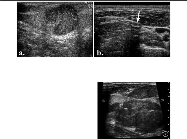
Fig 1. Typical US appearance of lymph nodes: typical reactive node image (a) and schematic drawing (b): the node is elongated, oval, with hypoechoic cortex peripherally (black), paracortex underneath it (dark grey) and echogenic hilum comprising the medulla in the center (light grey); c) typical malignant node: rounded, hypoechoic and inhomogeneous, with no visible hilum and indenting the neighboring jugular vein.

296 Sorin M. Dudea et al |
Ultrasonography of superficial lymph nodes: benign vs. malignant |
impalpable. Palpable and visible nodes may be benign or malignant.
The US diagnostic criteria used to separate benign from malignant lymph nodes are: size, shape, presence or absence of the hilum, echogenicity, margins, structural changes such as focal cortical nodules, intranodal necrosis, reticulation, calcification, matting and soft tissue edema. Doppler criteria include presence of flow, central or peripheral distribution, number of vascular pedicles, vascular pattern, and impedance values (RI, PI). The classic signs used to differentiate between benign and malignant are summarized in table I and illustrated in figure 1.
Although they apply to all superficial lymph nodes, these criteria were developed mainly by studying the nodes of the neck [1,6,8 -14]. In order to appreciate their value, a critical appraisal of these criteria is mandatory.
B scan criteria
Size
The axial, or transverse, diameter of the node was used as a diagnostic criterion. Nodes with diameters less than the cutoff point were considered benign. In the neck, different cutoff points were proposed, according to the anatomic level, as summarized in table II.
Obviously, size alone cannot be relevant, as metastatic nodes may be small and acute inflammatory or specific
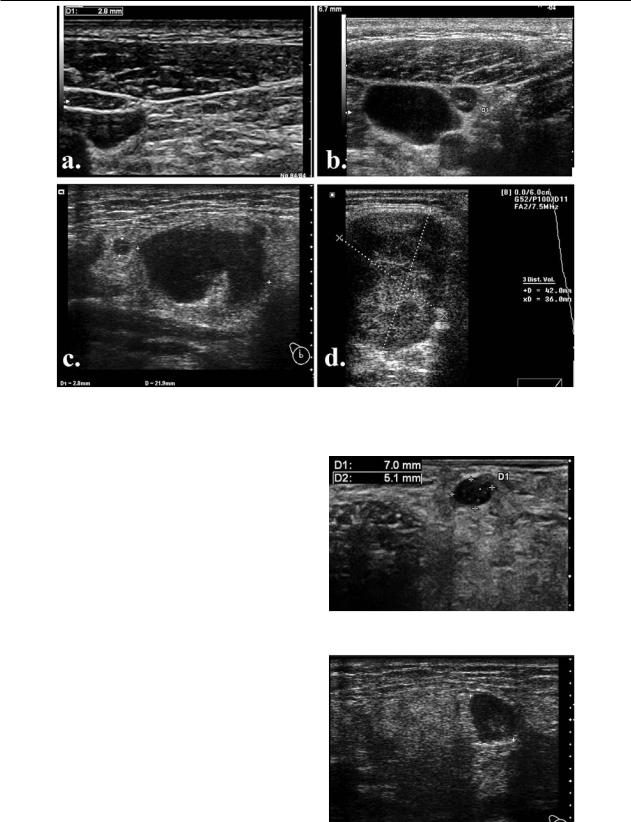
nodes, quite large. It has been shown that the size of the lymph nodes is not an accurate predictor of metastasis, at least in the N0 neck [14]. The smaller the size, the greater is the sensitivity but the worse the specificity and vice-versa (fig 2).
Size was demonstrated to be of value in the follow-up of lymph nodes. The increase in lymph node size on consecutive examinations performed in patients with known carcinoma is highly suspicious for metastatic nodal involvement. In proven metastatic nodes, size reduction on serial examinations is a useful indicator for monitoring patient’s response to treatment [1].
The use of combined criteria of Doppler US and nodal short-axis diameter may facilitate earlier detection of metastatic nodes than does the use of the single criterion of size. They may also increase the ability to predict benign, reactive nodes in cases with equivocal appearance [15].
In a study conducted by Kim HC and coworkers, 3 D ultrasonography was used to measure the volume of cervical lymph nodes, and a cut off volume of 0.7 cm was found to have 80% sensitivity and 90% specificity for differentiating metastatic from reactive lymphadenopathy [16].
Shape
Benign nodes are oval or elongated while malignant nodes are often described as rounded (see figure 1). The
Table II. Cutoff level of axial nodal diameter in the neck, for the diagnosis of malignancy and the respective diagnostic relevance.
Author |
Level |
Cutoff (mm) |
Diagnostic value (%) |
Notes |
|
|
|
|
|
Van den Brekel et al 1998 [14] |
1 |
5 |
SE=77 |
|
|
|
|
SP=72 |
|
|
1 |
10-11 |
|
High SP for malignancy |
|
2 |
8 |
SE=81 |
|
|
|
|
SP=80 |
|
|
2 |
7 |
|
More useful when clinical findings are |
|
|
|
|
negative |
|
3-6 |
6 |
SE=76 |
|
|
|
|
SP=89 |
|
|
2 |
7 |
|
Final suggested cutoff values |
|
1,3-6 |
6 |
|
|
Yonetsu et al 2001 [15] |
1 |
8-9 |
SE>85 |
|
|
2 |
9 |
SP>90 |
|
|
|
|
||
|
3-4 |
7 |
|
|
|
1 |
6 |
SE=89 |
When combining size & Doppler criteria |
|
2 |
7 |
SP= 94 |
|
|
|
|
||
|
3-4 |
5 |
|
|
Ahuja et al 2005 [8] |
1, 2 |
9 |
|
|
|
3-6 |
8 |
|
|
Med Ultrason 2012; 14(4): 294-306 297
Fig 2. Irrelevance of lymph node size: a) small (2,8mm) benign node; b) small (6,8 mm) malignant node; c) large (22mm) inflammatory submandibular node; d) large (42 mm) lymphoma node.
ratio between the longitudinal axis (L) of the node and the nodal transverse or short axis (S), also termed as axial diameter, is used to define the nodal shape. The long axis of an oval benign node will be at least two times greater than the axial diameter, situation that may be described as L/S > 2 or S/L< 0.5 [6,8]. In malignant, rounded nodules, the value of L/S is less that 2 or even < 1.5 or S/L > 0.5 [1,7,8] (fig 3). In another study, the values of sensitivity and specificity for the diagnosis of nodal metastasis, based on the cutoff transverse to longitudinal ratio above 0.65 were 66% and 92%, respectively [14].
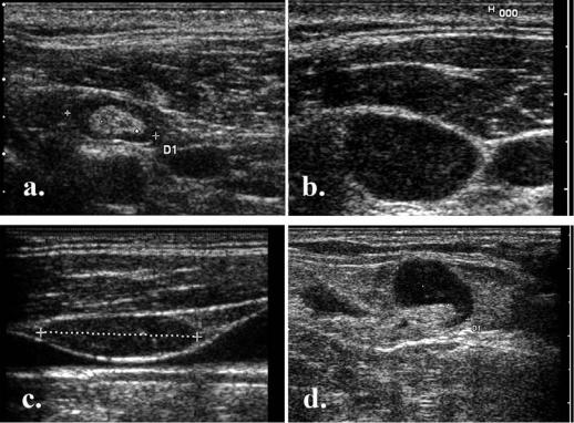
Shape may also be misleading as normal or reactive parotid and submandibular nodes are usually rounded, exhibiting S/L ration grater than 0.5 [1,8] (fig 4) . Not only metastatic but also lymphomatous nodes are rounded. Furthermore, nodes in nonmalignant conditions such as tuberculosis, Kimura or Rosai –Dorfman disease are also described as being rounded [8].
Hilum
Normal and reactive nodes present a central echogenic hilum that interrupts the continuity of the cortical and is continued with the perinodal fat tissue. This appearance is due to the abutment of multiple medullar sinuses acting as interfaces [1,8,11]. It has been shown that about
Fig 3. Small, “wider – than – tall” malignant node with S/L= 1,37.
Fig 4. Rounded inflammatory submandibular node.
298 Sorin M. Dudea et al |
Ultrasonography of superficial lymph nodes: benign vs. malignant |
||
|
|
|
|
Fig 5. Nodalechogenichilum:a)benignnode withechogenic centralhilum;b)malignantnodewith absenthilum; c) benign node displaying no hilum; d) malignant node with visible hilum.
90% of benign cervical nodes with a diameter above 5mm display an echogenic hilum [1] (fig 5a).
Malignant nodes are, traditionally, described as having no visible hilum [1,8,11,17]. The absence of anechogenic hilum due to replacement or effacement is considered to represent diagnostic criteria of abnormality and is significantly greater in malignancies than in benign lesions [18,19]. In a study, absent hilum was found in 83% of metastatic nodes while only 26% of tubercular and 28% of lymphomatous nodes had absent hilum [6] (fig 5b).
However, even from these results it is obvious that benign nodes may also have no visible hilum while some malignant nodes may still exhibit hilar echogenicity (fig 5c,d). It was shown that loss of fatty hilum is not a definite criterion for differentiation between malignant and benign lymph nodes [7]. Absence of hilum was found in as many as 9% of reactive lymph nodes [6].
The association of round shape and absent echogenic hilum, termed as a stringent criteria for malignancy, had high specificity but questionable sensitivity [2].
Echogenicity
Both reactive and malignant lymph nodes are hypoechoic compared to neighboring strap muscles. Lympho-
matous, tuberculous and lymphadenitis nodes are also hypoechoic; therefore hypoechogenicity is not a useful diagnostic sign [1,8]. On contrary, focal or diffuse hyperechogenic nodes (with higher echogenicity than the surrounding muscles), are encountered in papillary or medullary thyroid cancer metastasis, due to intranodal thyroglobulin deposits [1,7,8]. In our experience, metastasis from scuamous cell carcinomas display, quite often, echogenicity comparable to or higher than that of neck muscles, as well (fig 6).
Margins
Benign nodules are characterized by sharp margins. Irregular margins were found in only 7% of reactive lymph nodes [6]. However, malignant nodules often exhibit sharp margins as well. In these cases, tumor infiltration leads to high impedance mismatch [1] (fig 7a).
Blurred margins may be observed in acute inflammatory nodes. In melanoma patients, irregular or angular nodal margins represent a criterion of suspicion for metastasis [2]. In malignancy, irregular and blurred margins usually indicate, just as does frank invasive contour, extracapsular and extranodal spread and bear a severe prognosis [1,8] (fig 7b).
Med Ultrason 2012; 14(4): 294-306 299
Fig 6. Echogenic metastatic nodes: a) heavy echogenic deposits (arrow) in papillary thyroid cancer nodal metastasis; b) relative echogenic metastatic node from a squamous carcinoma of the nasopharinx.
Fig 7. Margins of malignant nodes : a) sharp margins; b) blurred, invasive margins.
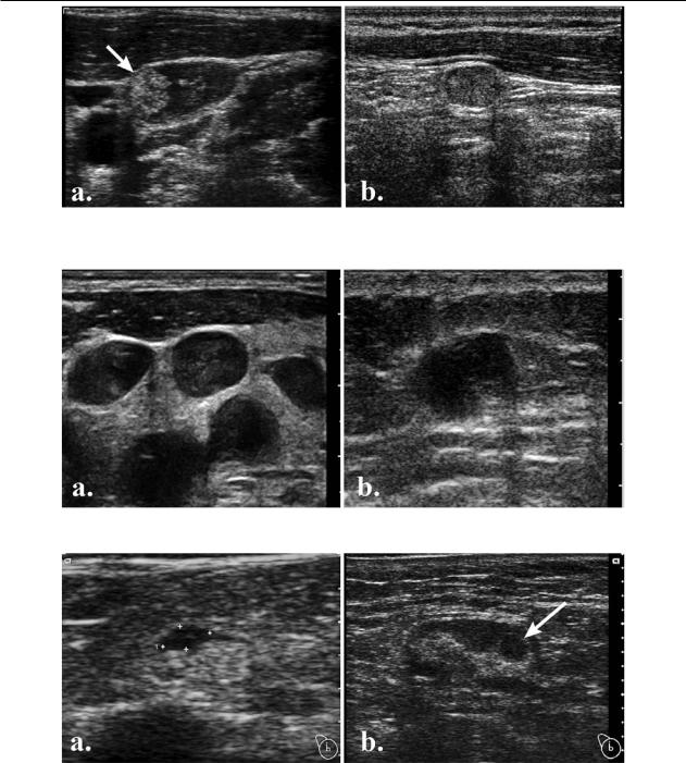
Fig 8. Focal cortical hypertrophy: a) benign nodule (calipers); b) malignant nodule (arrow).
Structural changes
Structural changes are, most often, encountered in malignant nodes and are absent in benign conditions.
Focal cortical nodules – also named focal cortical hyperplasia or eccentric cortical hypertrophy – indicate partial
tumor infiltration and represent a useful, although not very sensitive, sign for identifying metastatic nodes in the neck or in melanoma patients [1,2,8]. However, we have also observedsuchnodesinacuteinflammatorylymphadenopathy; therefore the specificity of this sign is not absolute (fig 8).
300 Sorin M. Dudea et al |
Ultrasonography of superficial lymph nodes: benign vs. malignant |
||
|
|
|
|
Fig 9. Intranodal necrosis: a) coagulation necrosis (arrow); b) liquefaction necrosis.
Fig 10. Intranodal reticulation in a lymphoma patient.
Intranodal necrosis indicates malignancy in most instances.Itencompassesthecoagulationorliquefactiontype.
Coagulation necrosis appears as an echogenic focus that casts no shadow and shows no contact with the hilum or continuity with perinodal fat [1,8] (fig 9a). Although described in metastasis, it was observed in tuberculous nodes, as well. Therefore, this type of necrosis represents a sign of certainty for pathologic changes, without disease specificity.
Cystic or liquefaction necrosis appears as eccentric fluid areas within the structure of the lymph node (fig 9b). It is more frequent in scuamous carcinoma metastases [8] but it may appear in any type of metastasis. In patients with papillary thyroid carcinoma, the presence of a cystic lymph node detected by ultrasonography is highly suggestive of locally metastatic disease [7,20]. When cytological findings are negative, confirmation of meta-
static papillary thyroid carcinoma may be achieved with thyroglobulin aspirate from cystic lymph nodes [20].The pseudocystic appearance of lymphomatous nodes is a matter of low resolution transducers and belongs rather to the history of ultrasonography. True cystic necrosis in lymphomatous nodes is rare, encountered mostly in advanced stages or after radiation therapy [1].
However, tuberculous nodes as well as abscessed lymphadenopathy may present with nonmalignant cystic necrosis.
Reticulation describes the occurrence of thin echogenic lines that septate the hypoechoic solid texture of enlarged nodes, at times producing a micronodular appearance [1,8] (fig 10). This appearance indicates malignancy but is encountered mostly in lymphomas; therefore it has low sensitivity and unexplored specificity.
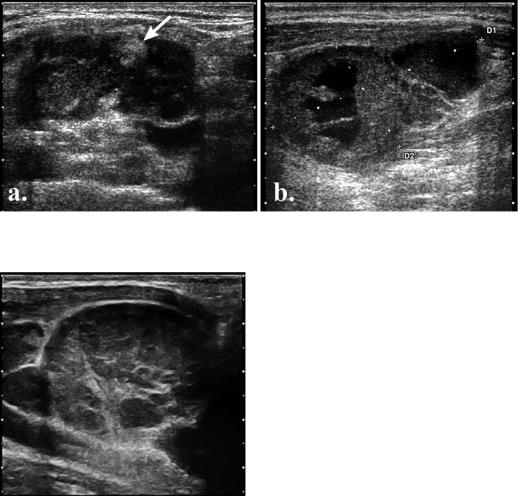
Calcification may indicate malignancy. Punctuate, peripheral microcalcifications, shadowing only at high resolution, are encountered in both papillary and medullary thyroid carcinomas [1,8] (fig 11a). They are due to psammoma bodies, reported to be formed by calcification of infarcted tips of malignant papillae or intravascular tumor thrombi [7,21]. Metastases from other types of tumors are not accompanied by calcification. In lymphoma, calcification is rarely encountered, most often after radiation therapy, is coarse and shadowing [1]. However, we encountered calcification in residual tuberculous adenopathy, as well (fig 11b).
Matting of the lymph nodes is suggestive of malignancy [8] (fig 12). This sign does not appear in reactive nodes [6]. The presence of matting suggests extracapsular spread of malignancy but it may also be observed after radiation therapy [1]. However, nonmalignant conditions such as tuberculosis also show matting [1,11]. In a study, matting was encountered in 83% of cases with
Med Ultrason 2012; 14(4): 294-306 301
Fig 11. Intranodal calcification; a) sparse microcalcification in a thyroid cancer metastasis; b) coarse calcification in a tuberculous node (arrow).
tuberculous nodes, 66% cases with metastases and 14% of lymphoma cases [6].
Peripheral halo and perinodal edema are not encountered in reactive nodes, as well. These signs are suggestive of tuberculosis or malignancy [1,6,8,11].
Doppler criteria
Absence or presence of flow is a low specificity feature. Absence of flow within the lymph node is encountered in small benign nodes. It has been observed that normal or reactive nodes may have an avascular appearance and that, in normal volunteers, roughly 90% of normal lymph nodes with a maximum transverse diameter more than 5 mm show vessels in the hilum [10]. Malignant nodes are vascularized. However, necrotic areas lack Doppler signals. Depiction of nodal vascularization is highly dependent on machine settings and sensitivity.
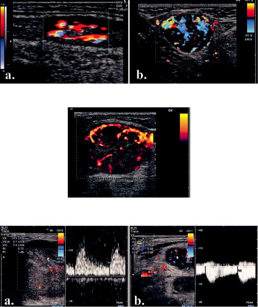
Vessel location and distribution, as assessed with color or power Doppler, has a much higher importance. The vast majority of benign nodes present central hilar vessels [12]. The hilar signal appears as Y-shaped or club-shaped color signals that occupy the central, hilar region of the lymph node [22] (fig 13 a). Hilar vascular signals may be identified with power Doppler even if the hilum is not apparent on the gray scale image [9].
Malignant nodes show vessels in the parenchyma, at the periphery (capsular flow) or display a mixed hilar and peripheral pattern [8]. The parenchymal flow is seen as multiple signals, variably colored and scattered in the nodal cortex and medulla [22] (fig 13 b). When nodal metastasis was diagnosed on the basis of the presence of parenchymal color signal, sensitivity was 83% and specificity 98% [22]. As most of malignant nodes show peripheral of mixed flow, it was stated that the
Fig 12. Lymph node matting. Multiple malignant nodes are fused in a single, ill defined mass.
distribution of intranodal vascularity is more useful than impedance indices in differentiating malignant from benign lymphadenopathy in the neck. It is easier to evaluate the distribution of the vessels and the results are readily applicable in routine clinical practice [12].
Some critical studies have shown that color - flow criteria may alter psychological confidence but may not alter the conclusion reached by gray-scale criteria. The suggestion was that “power Doppler sonographic features could assist observers who are less experienced in sonographic diagnosis of the lymph node by providing information in interpreting gray-scale sonographic findings that are critical for predicting metastatic or benign lymph nodes, such as the presence or absence of hilar echoes “ [23].
A study by Ahuja et al [13] showed that power Doppler sonography contributed to the diagnosis in 5% of patients with metastatic nodes and 17% of nonmetastatic nodes, indicating that power Doppler is not necessary for
302 Sorin M. Dudea et al |
Ultrasonography of superficial lymph nodes: benign vs. malignant |
||
|
|
|
|
Fig 13. Nodal vascular pattern: a) benign, central, hilar vessels; b) malignant, mixed peripheral and central increased vascularization.
Fig 14. Malignant node with multiple feeders and chaotic intranodal vascular branching.
Fig 15. Flow impedance in malignant nodes: a) relatively high impedance; b) very low impedance.
every case, in routine clinical practice. It may be useful especially in patients where grey-scale sonographic features are equivocal.
The number and location of vascular pedicles as well as the distribution of vessels are other noteworthy observations. Benign nodes display a single vascular pedicle, entering the hilum while malignant nodes show multiple pedicles, invading the cortex. Whereas benign nodes show a regular radial distribution of the vessels, starting in the hilum, in malignant nodes the vessel dis-
tribution is chaotic [4,5] (fig 14). In tuberculosis, hilar vessels may be dislodged by necrosis, creating a chaotic pattern, as well [8].
Flow impedance, as expressed by the values of RI and PI, has also been studied as a diagnostic criterion. Theoretically, low impedance, produced by vasodilatation, is encountered in inflammation while vessel compression by tumor cells leads to increased impedance.
In normal or reactive lymph nodes, as the size of the nodes increases, the intranodal blood flow velocity in-
|
|
|
Med Ultrason 2012; 14(4): 294-306 303 |
|
creases significantly, as well, whereas there is no signifi- |
Color or power Doppler is also useful in monitoring re- |
|
||
cant variation in the vascular resistance [10] (fig 15). |
sponse to treatment, as quick reduction of vessel signal |
|||
Clinically, although metastatic nodes tended to have |
indicates not only favorable response but also prolonged |
|||
higher intranodal vascular resistance than reactive nodes, |
remission. On the other hand, impedance indices do not |
|||
there was considerable overlap of the parameters be- |
correlate with response to therapy and are, therefore, of |
|||
tween the two groups [12]. The cut-off value of 0, 7 for |
limited prognostic value [1]. |
|||
RI yielded a sensitivity of 86% and a specificity of 70%. |
Nodal metastases show peculiarities according to |
|||
For PI, the cut-off value of 1, 4 had a sensitivity of 80% |
their source and location. |
|||
and a specificity of 86% [1]. Therefore, the role of in- |
Cervical metastases have various sources. Thyroid |
|||
tranodal vascular resistance in routine clinical practice is |
carcinoma, pharynx, larynx and upper esophageal tumors |
|||
considered to be of limited value. |
|
metastasize in the internal jugular group. Nasopharyngeal |
||
Peculiar types of lymphadenopathy |
carcinomaspreadstotheupperneckandposteriortriangle |
|||
nodes. Oral cavity tumors metastasize to the submandibu- |
||||
Typical normal or reactive nodes are small, oval (L/S |
lar and upper cervical regions. Tumors of infraclavicular |
|||
organs such as breast and lung may produce metastases |
||||
> 2) or elongated, with sharp margins, present an echogen- |
to the supraclavicular fossa and the posterior triangle [1]. |
|||
ic central hilum and a hypoechoic, uniformly thick periph- |
Most epithelial metastatic nodes are rounded (L/S around |
|||
eral parenchyma, with no or central hilar vascularization |
1.2) [6] and display the typical malignant features pre- |
|||
of low impedance. The size and appearance do not change |
sented above. Gray scale US is credited with a sensitivity |
|||
over time or there is progressive shrinking [14]. However, |
of 95% and a specificity of 83% in differentiating meta- |
|||
irregular margins, hypoechoic center or absence of hilum |
static from reactive neck nodes [8]. When the diagnostic |
|||
in were found in 7 – 9% of reactive lymph nodes [6]. |
criteria are both peripheral parenchymal flow on power |
|||
Tuberculous nodes are rounded (L/S = 1.8), hyp- |
Doppler and a transverse to longitudinal ratio greater than |
|||
oechoic with no visible hilum, with blurred margins or |
0.65, the diagnostic accuracy reaches 92% sensitivity and |
|||
matting and perinodal edema [6]. Cystic necrosis and in- |
100% specificity [22]. Sonography performed better than |
|||
ternal echoes are often encountered.The Doppler appear- |
CT for detecting metastatic nodes in patients with head |
|||
ance may mimic malignancy due to vessel dislocation by |
and neck squamous cell carcinoma [17]. |
|||
necrosis [6,8]. We noted that the lymph node capsule, |
Lung cancer metastases to the supraclavicular nodes |
|||
when intact, appears thickened and that microcalcifica- |
(up to 4 cm above the clavicle) were detected by sonogra- |
|||
tions may be observed in tuberculous nodes, as well [24]. |
phywithasensitivityof100%,betterthanCT(83%),notto |
|||
Other nonmalignant inflammatory disease of the |
mention palpation (33%), if the indication for biopsy was |
|||
nodes may present confusing US appearance. |
set at greater than 5 mm nodal transverse diameter [25]. |
|||
Histiocytic necrotizing lymphadenitis (Kikuchi - Fu- |
Detection of supraclavicular malignant nodes leads to up- |
|||
jimoto disease) in localized in the neck. The nodes are |
staging of lung cancer and may prevent adrenal biopsy. |
|||
benign in aspect: hypoechoic, oval, with present hilum |
Thyroid cancer metastases arelocated,inmostcases, |
|||
and hilar vessels [8]. |
|
in the lower neck [18]. Echogenic cortex, microcalcifica- |
||
Eosinophylic |
hyperplastic |
lymphogranuloma |
tions and cystic changes are more often encountered in |
|
(Kimura disease) is an autoimmune condition in which |
this type of nodal metastasis, but all the common features |
|||
the appearance of the nodes is typically benign except for |
of malignant nodes are usually present. Increase of short |
|||
the rounded shape [8]. |
|
axis diameter is the best predictor of metastatic nodes |
||
In sinus histiocytosis (Rosai-Dorfman disease), the |
and the presence of normal hilar blood flow, hilar echoes, |
|||
massive neck lymphadenopathy has an appearance in- |
or both is the best predictor for reactive nodes [17]. |
|||
dicative of malignancy (hypoechoic, rounded, no hilum, |
It has been stated that “clinicians should strongly con- |
|||
peripheral or mixed vascularization). The differential di- |
sider surgical resection of cystic lymph nodes regardless |
|||
agnosis requires pathology [8]. |
|
of the preoperative cytological findings by FNAB” [20]. |
||
Lymphoma,bothHodgkinandnon-Hodgkin,present |
In fact, FNAB is far from being a golden standard since it |
|||
an evocative appearance. Multiple, large, rounded nodes |
may induce false results, as shown by a study of Sohn et |
|||
with L/S around 1.5 [6] with sharp margins and nodal |
al. In this study, FNAB was performed, in thyroid cancer |
|||
reticulation present mixed, peripheral and hilar vessels |
patients, in all nodes with suspicious US features. After |
|||
with impedance greater than in TB but less than in me- |
node resection, pathology was compared with FNAB. It |
|||
tastasis, although no clear limits exist [1,8]. Change of |
was shown that malignancy was present in 87.5% of the |
|||
size over time is |
indicative of response to treatment. |
nodes with smears read as macrophages without malig- |
||