


CHAPTER 4 ■ Connective Tissue |
57 |
|
Figure 4-15A,B |
Dense Regular Connective Tissue, Tendon |
|
Figure 4-15C |
Clinical Correlation: Tendinosis |
|
Figure 4-16A,B |
Loose Connective Tissue |
|
Synopsis 4-2 |
Functions of Connective Tissue |
|
Figure 4-17A,B |
Loose Connective Tissue, Small Intestine |
|
Figure 4-17C |
Clinical Correlation: Whipple Disease |
|
Types of Connective Tissue: Specialized Connective Tissues |
|
|
Figure 4-18A,B |
Adipose Tissue |
|
Figure 4-18C |
Clinical Correlation: Obesity |
|
Figure 4-19A,B |
Reticular Connective Tissue |
|
Figure 4-19C |
Clinical Correlation: Cirrhosis |
|
Figure 4-20A,B |
Elastic Connective Tissue |
|
Figure 4-20C |
Clinical Correlation: Marfan Syndrome—Cystic Medial Degeneration |
|
Types of Connective Tissue: Embryonic Connective Tissues |
|
|
Figure 4-21A |
Mesenchyme, Embryo |
|
Figure 4-21B |
Mucous Connective Tissue |
|
Synopsis 4-3 |
Pathological Terms for Connective Tissue |
|
Table 4-3 |
Connective Tissue Types |
|
Introduction and Key Concepts for Connective Tissue
Connective tissue provides structural support for the body by binding cells and tissues together to form organs. It also provides metabolic support by creating a hydrophilic environment that mediates the exchange of substances between the blood and tissue. Connective tissue is of mesodermal origin and consists of a mixture of cells, fibers, and ground substance. The hydrophilic ground substance occupies the spaces around cells and fibers. Fibers (collagen, elastic, and reticular) and the ground substances constitute the extracellular matrix of connective tissue. The classification and function of connective tissue are based on the differences in the composition and amounts of cells, fibers, and ground substance.
Connective Tissue Cells
A variety of cells are found in connective tissue, which differ according to their origin and function. Some cells differentiate from mesenchymal cells, such as adipocytes and fibroblasts; these cells are formed and reside in the connective tissue and are called fixed cells. Other cells, which arise from hematopoietic stem cells, differentiate in the bone marrow and migrate from the blood circulation into connective tissue where they perform their functions; these mast cells, macrophages, plasma cells, and leukocytes are called wandering cells (Fig. 4-1). Cells found in connective tissue proper include fibroblasts, macrophages, mast cells, plasma cells, and leukocytes (Figs. 4-2 to 4-4). Some cells, such as fibroblasts, are responsible for synthesis and maintenance of the extracellular material. Other cells, such as macrophages, plasma cells, and leukocytes, have defense and immune functions.
FIBROBLASTS are the most common cells in connective tissue. Their nuclei are ovoid or spindle shaped and can be large or small in size depending on their stage of cellular activity. They have pale-staining cytoplasm and contain
well-developed rough endoplasmic reticulum (RER) and rich Golgi complexes. With routine H&E staining, only the very thin, elongated nuclei of the cells are clearly visible. Their thin, pale-staining cytoplasm is usually not obvious. They are responsible for the synthesis of all components of the extracellular matrix (fibers and ground substance) of connective tissue (Figs. 4-2, 4-3, and 4-7).
MACROPHAGES, also called tissue histiocytes, are highly phagocytic cells that are derived from blood monocytes. With conventional staining, macrophages are very difficult to identify unless they show visible ingested material inside their cytoplasm. Macrophages may be named differently in certain organs (Figs. 4-2 and 4-3). For example, they are called Kupffer cells in the liver, osteoclasts in bone, and microglial cells in the central nervous system.
MAST CELLS are of bone marrow origin and are distributed chiefly around small blood vessels. They are oval to round in shape, with a centrally placed nucleus. With toluidine blue stain, large basophilic purple staining granules are visible in their cytoplasm. These granules contain and release heparin, histamines, and various chemotactic mediators, which are involved in inflammatory responses. Mast cells contain Fc membrane receptors, which bind to immunoglobulin (Ig) E antibodies, an important cellular interaction involved in anaphylactic shock (Fig. 4-4A,B).
PLASMA CELLS are derived from B lymphocytes. They are oval shaped and have the ability to secrete antibodies that are antigen specific. Their histological features include an eccentrically placed nucleus, a cartwheel pattern of chromatin in the nucleus, and basophilic-staining cytoplasm due to the presence of abundant RER and a small, clear area near the nucleus. This cytoplasmic clear area (Golgi zone [GZ]) marks the position of the Golgi apparatus (Figs. 4-2 and 4-3).

58 UNIT 2 ■ Basic Tissues
LEUKOCYTES, white blood cells, are considered the transient cells of connective tissue. They migrate from the blood vessels into connective tissue by the process of diapedesis. This process increases greatly during various inflammatory conditions. After entering connective tissue, leukocytes, with the exception of lymphocytes, do not return to the blood. The following leukocytes are commonly found in connective tissue: (1) Lymphocytes: These cells have a round or bean-shaped nucleus and are often located in the subepithelial connective tissue. (2) Neutrophils (polymorphs): Each cell has a multilobed nucleus and functions in the defense against infection. (3) Eosinophils: Each cell has a bilobed nucleus and reddish granules in the cytoplasm (Figs. 4-2 and 4-3). They have antiparasitic activity and moderate the allergic reaction function. (4) Basophils: These cells are not easy to find in normal tissues. Their primary function is similar to that of mast cells. A detailed account of the structure and the function of leukocytes is given in Chapter 8, “Blood and Hemopoiesis.”
ADIPOCYTES (FAT CELLS) arise from undifferentiated mesenchymal cells of connective tissue. They gradually accumulate cytoplasmic fat, which results in a significant flattening of the nucleus in the periphery of the cell. Adipocytes are found throughout the body, particularly in loose connective tissue (Figs. 4-2 and 4-18). Their function is to store energy in the form of triglycerides and to synthesize hormones such as leptin.
Connective Tissue Fibers
Three types of fibers are found in connective tissue: collagen, elastic, and reticular. The amount and type of fibers that dominate a connective tissue are a reflection of the structural support needed to serve the function of that particular tissue. These three fibers all consist of proteins that form elongated structures, which, although produced primarily by fibroblasts, may be produced by other cell types in certain locations. For example, collagen and elastic fibers can be produced by smooth muscle cells in large arteries and chondrocytes in cartilages.
COLLAGEN FIBERS are the most common and widespread fibers in connective tissue and are composed primarily of type I collagen. The collagen molecule (tropocollagen) is a product of the fibroblast. Each collagen molecule is 300 nm in length and consists of three polypeptide amino acid chains (alpha chains) wrapped in a right-handed triple helix. The molecules are arranged head to tail in overlapping parallel, longitudinal rows with a gap between the molecules within each row to form a collagen fibril. The parallel array of fibrils forms crosslinks to one another to form the collagen fiber. Collagen fibers stain readily with acidic and some basic dyes. When stained with H&E and viewed with the light microscope, they appear as pink, wavy fibers of different sizes (Fig. 4-13). When stained with osmium tetroxide for EM study, the fibers have a transverse banded pattern (light–dark) that repeats every 68 μm along the fiber. The banded pattern is a reflection of the arrangement of collagen molecules within the fibrils of the collagen fiber (Figs. 4-5 to 4-7).
ELASTIC FIBERS stain glassy red with H&E but are best demonstrated with a stain specifically for elastic fibers, such
as aldehyde fuchsin. Elastic fibers have a very resilient nature (stretch and recoil), which is important in areas like the lungs, aorta, and skin. They are composed of two proteins, elastin and fibrillin, and do not have a banding pattern. These fibers are primarily produced by the fibroblasts but can also be produced by smooth muscle cells and chondrocytes (Figs. 4-8 and 4-9).
RETICULAR FIBERS are small-diameter fibers that can only be adequately visualized with silver stains; they are called argyrophilic fibers because they appear black after exposure to silver salts (Figs. 4-10 and 4-11). They are produced by modified fibroblasts (reticular cells) and are composed of type III collagen. These small, dark-staining fibers form a supportive, meshlike framework for organs that are composed mostly of cells (such as the liver, spleen, pancreas, lymphatic tissue, etc.).
Ground Substance of Connective Tissue
Ground substance is a clear, viscous substance with a high water content, but with very little morphologic structure. When stained with basic dyes (periodic acid-Schiff [PAS]), it appears amorphous, and with H&E, it appears as a clear space. Its major component is glycosaminoglycans (GAGs), which are long, unbranched chains of polysaccharides with repeating disaccharide units. Most GAGs are covalently bonded to a large central protein to form larger molecules called proteoglycans. Both GAGs and proteoglycans have negative charges and attract water. This semifluid gel allows the diffusion of water-soluble molecules but inhibits movement of large macromolecules and bacteria. This water-attracting ability of ground substance gives us our extracellular body fluids.
Types of Connective Tissues
CONNECTIVE TISSUE PROPER
Dense Connective Tissue can be divided into dense irregular connective tissue and dense regular connective tissue. Dense irregular connective tissue consists of few connective tissue cells and many connective tissue fibers, the majority being type I collagen fibers, interlaced with a few elastic and reticular fibers. These fibers are arranged in bundles without a definite orientation. The dermis of the skin and capsules of many organs are typical examples of dense irregular connective tissue (Figs. 4-13 and 4-14). Dense regular connective tissue also consists of fewer cells and more fibers, with a predominance of type I collagen fibers like the dense irregular connective tissue. Here, the fibers are arranged into a definite linear pattern. Fibroblasts are arranged linearly in the same orientation. Tendons and ligaments are the most common examples of dense regular connective tissue (Fig. 4-15).
Loose Connective Tissue, also called areolar connective tissue, is characterized by abundant ground substance, with numerous connective tissue cells and fewer fibers (more cells and fewer fibers) compared to dense connective tissue. It is richly vascularized, flexible, and not highly resistant to stress. It provides protection, suspension, and support for the tissue. The lamina propria of the digestive tract and the mesentery are good examples of loose connective tissue (Figs. 4-16 and 4-17).

CHAPTER 4 ■ Connective Tissue |
59 |
This tissue also forms conduits through which blood vessels and nerves course.
SPECIALIZED CONNECTIVE TISSUES
Adipose Tissue is a special form of connective tissue, consisting predominantly of adipocytes that are the primary site for fat storage and are specialized for heat production. It has a rich neurovascular supply. Adipose tissue can be divided into white adipose tissue and brown adipose tissue. White adipose tissue is composed of unilocular adipose cells. The typical appearance of cells in white adipose tissue is lipid stored in the form of a single, large droplet in the cytoplasm of the cell. The flattened nucleus of each adipocyte is displaced to the periphery of the cell. White adipose tissue is found throughout the adult human body (Fig. 4-18). Brown adipose tissue, in contrast, is composed of multilocular adipose cells. The lipid is stored in multiple droplets in the cytoplasm. Cells have a central nucleus and a relatively large amount of cytoplasm. Brown adipose tissue is more abundant in hibernating animals and is also found in the human embryo, in infants, and in the perirenal region in adults.
Reticular Tissue is a specialized loose connective tissue that contains a network of branched reticular fibers, reticulocytes (specialized fibroblasts), macrophages, and parenchymal cells, such as pancreatic cells and hepatocytes. Reticular fibers are very fine and much smaller than collagen type 1 and elastic fibers. This tissue provides the architectural framework for parenchymal organs, such as lymphoid nodes, spleen, liver, bone marrow, and endocrine glands (Fig. 4-19).
Elastic Tissue is composed of bundles of thick elastic fibers with a sparse network of collagen fibers and fibroblasts filling the interstitial space. In certain locations, such as in elastic arteries, elastic material and collagen fibers can be produced by smooth muscle cells. This tissue provides flexible support for other tissues and is able to recoil after stretching, which helps to dampen the extremes of pressure associated with some organs,
such as elastic arteries (Fig. 4-20). Elastic tissue is usually found in the vertebral ligaments, lungs, large arteries, and the dermis of the skin.
EMBRYONIC CONNECTIVE TISSUES is a type of loose tissue formed in early embryonic development. Mesenchymal connective tissue and mucous connective tissue also fall under this category.
Mesenchymal Connective Tissue is found in the embryo and fetus and contains considerable ground substance. It contains scattered reticular fibers and star-shaped mesenchymal cells that have pale-staining cytoplasm with small processes (Fig. 4-21A). Mesenchymal connective tissue is capable of differentiating into different types of connective tissues (Fig. 4-1A).
Mucous Connective Tissue exhibits a jellylike matrix with some collagen fibers and stellate-shaped fibroblasts. Mucous tissue is the main constituent of the umbilical cord and is called Wharton jelly (see Fig. 4-21B). This type of tissue does not differentiate beyond this stage. It is mainly found in developing structures, such as the umbilical cord, subdermal connective tissue of the fetus, and dental pulp of the developing teeth. It is also found in the nucleus pulposus of the intervertebral disk in adult tissue.
SUPPORTING CONNECTIVE TISSUE is related to cartilage and bone. Cartilage is composed of chondrocytes and extracellular matrix; bone contains osteoblasts, osteocytes, and osteoclasts and bone matrix. These will be discussed in Chapter 5, “Cartilage and Bone.”
HEMATOPOIETIC TISSUE (BLOOD AND BONE MARROW) is a specialized connective tissue in which cells are suspended in the intercellular fluid, and it will be discussed in Chapter 8, “Blood and Hemopoiesis.”

60 |
UNIT 2 ■ |
Basic Tissues |
|
|
A |
Undifferentiated mesenchymal cells |
Hematopoietic stem cells |
|
|
|
|
|
|
|
|
|
|
|
|
|
|
Chondroblast |
|
|
|
|
|
B lymphocyte |
Mast cell |
Eosinophil |
|
||||
|
|
|
|
|
Osteoblast |
|
|
|
|
|
|
|
|
|
Adipocyte |
Fibroblast |
|
|
|
|
|
|
|
||||
|
|
|
|
|
|
|
|
|
|
|
|
|
|
|
|
|
|
|
|
|
|
Monocyte |
Basophil |
Neutrophil |
|||
|
|
|
|
|
|
|
|
|
|
|
|
|
|
|
|
|
|
|
|
Plasma cell |
|
|
|
|
|
||
Chondrocyte |
|
|
Osteocyte |
|
|
|
|
|
|||||
(cartilage) |
|
|
(bone) |
|
Osteoclast |
Macrophage |
|
|
|
||||
|
|
|
|
|
|
|
|
|
|
|
|||
Figure 4-1A. The origin of connective tissue cells.
The left panel shows cells arising from undifferentiated mesenchymal cells. These cells are formed in, and remain within, the connective tissue and are also called fixed cells. The panel on the right shows cells arising from hematopoietic stem cells. These cells differentiate in the bone marrow, and then must migrate by way of circulation to connective tissue where they perform their various functions. They are also called wandering cells.
B
Fibroblast |
Macrophage |
Adipocyte |
Mast cell |
D. Cui
Plasma cell |
B lymphocyte |
Neutrophil |
Eosinophil |
Basophil |
Figure 4-1B. A representation of the main types of connective tissue cells in connective tissue proper.
The nuclei of these connective tissue cells are indicated in purple. Note: Mast cells, eosinophils, basophils, and neutrophils all contain granules in their cytoplasm. The light yellow circle in the adipocyte (fat cell) represents its lipid droplet. These cells are not drawn to scale; the adipocyte is much larger than the others.
SYNOPSIS 4 - 1 Functions of the Cells in Connective Tissue Proper
■Fibroblasts are responsible for synthesis of various fibers and extracellular matrix components, such as collagen, elastic, and reticular fibers.
■Macrophages contain many lysosomes and are involved in the removal of cell debris and the ingestion of foreign substances; they also aid in antigen presentation to the immune system.
■Adipocytes function to store neutral fats for energy or production of heat and are involved in hormone secretion.
■Mast cells contain many granules, indirectly participate in allergic reactions, and act against microbial invasion.
■Plasma cells are derived from B lymphocytes and are responsible for the production of antibodies in the immune response.
■Lymphocytes participate in the immune response and protect against foreign invasion (see Chapter 10, “Lymphoid System”).
■Neutrophils are the first line of defense against bacterial invasion.
■Eosinophils have antiparasitic activity and moderate allergic reactions.
■Basophils have a (primary) function similar to mast cells; they mediate hypersensitivity reactions (see Chapter 8, “Blood and Hemopoiesis”).

CHAPTER 4 ■ Connective Tissue |
61 |
Connective Tissue Cells
A
Fibroblasts
C
Mast cell
E
Macrophages
A:Nuclei of fibroblasts are elongated and, when inactive, these cells have little cytoplasm. The fibroblasts are formed and reside in the connective tissue; they are also called fixed cells.
B:Plasma cells are characterized by cartwheel (clockface) nuclei showing the alternating distribution of the heterochromatin (dark) and the euchromatin (light). The pale (unstained) area of cytoplasm in each plasma cell is the location of the Golgi complex, which is also called the Golgi zone. (GZ, Golgi zone.)
C:A mast cell has a single, oval-shaped nucleus and granules in its cytoplasm. In paraffin H&E–stained sections, these granules are typically unstained, but they appear red in sections of plastic-embedded tissues stained with a faux H&E set of dyes.
D:An eosinophil has a segmented nucleus (two lobes, usually) and numerous eosinophilic (red) granules filling the cytoplasm. Eosinophils, mast, and plasma cells are wandering cells (Fig. 4-1A).
E:Black particles fill the cytoplasm of these active macrophages; the nuclei are obscured by the phagocytosed materials.
F:Each adipocyte contains a large droplet of lipid, appearing white (clear) here because the fat was removed during tissue preparation. The nucleus of each cell is pushed against the periphery of the cell.
B
GZ
Plasma cell
D
Eosinophil
F
Nuclei of the
adipocytes
Adipocytes
Figure 4-2A–D. Cells in the connective tissue of the small intestine. Modified H&E, 1,429 Figure 4-2E. Macrophages in lung tissue. H&E, 2,025
Figure 4-2F. Adipocytes in connective tissue of the mammary gland. H&E, 373

62 UNIT 2 ■ Basic Tissues
A
Macrophage Fibroblast
Macrophage
Plasma cells
Fibroblast
Eosinophil
Lymphocyte
Plasma cells |
Neutrophil |
Eosinophil |
Mast cell |
GZ
Figure 4-3A. Connective tissue cells in lamina propria.
Modified H&E, ×680; inset approximately 1,200
An example of cells in loose connective tissue is shown. Fibroblasts are the predominant cells in connective tissue, where they produce procollagen and other components of the extracellular matrix (Fig. 4-7A). Plasma cells arise from activated B lymphocytes and are responsible for producing antibodies. Mast cells have small, ovoid nuclei and contain numerous cytoplasmic granules. When stained with toluidine blue, these granules are metachromatically stained and appear purple (Fig. 4-4A). Mast cells are involved in allergic reactions. Eosinophils arise from hematopoietic stem cells and are generally characterized by bilobed nuclei and numerous eosinophilic cytoplasmic granules; they are attracted to sites of inflammation by leukocyte chemotactic factors where they may defend against a parasitic infection or moderate an allergic reaction. Neutrophils are phagocytes of bacteria; each cell has a multilobed nucleus and some granules in its cytoplasm. For more details on leukocytes, see Chapter 8, “Blood and Hemopoiesis.”
B
Eosinophil |
Neutrophil |
|
|
|
B |
Macrophage |
|
|
lymphocyte |
|
|
Plasma cell |
|
|
D. Cui |
Mast cell |
Fibroblast |
Adipocyte |
|
|||
|
|
|
|
Elastic fiber
Collagen fiber
Figure 4-3B. A representation of the cells found in loose connective tissue. (These cells are not drawn to scale.)
(1) Fibroblasts are spindle-shaped cells with ovoid or elliptical nuclei and irregular cytoplasmic extensions. (2) Macrophages have irregular nuclei. The cytoplasm contains many lysosomes; cell size may vary depending on the level of phagocytic activity. (3) Adipocytes contain large lipid droplets, and their nuclei are pushed to the periphery. They are usually present in aggregate (see Fig. 4-18). (4) Mast cells have centrally located ovoid nuclei and numerous granules in their cytoplasm. (5) Plasma cells have eccentric nuclei with peripheral distribution of heterochromatin (clock face) within the nuclei; a clear Golgi area is present within the cytoplasm. (6) Eosinophils have bilobed nuclei and coarse cytoplasmic granules. (7) Neutrophils and lymphocytes are also found in connective tissue, and their numbers may increase in cases of inflammation.
CLINICAL CORRELATION
C |
Dilated blood vessel |
|
Active basophil |
|
Active mast cell |
|
Collagen fiber |


 Dilated capillary
D. Cui
Figure 4-3C. Anaphylaxis.
Anaphylaxis is an allergic reaction that may range from mild to severe and is characterized by increased numbers of basophils and mast cells, dilated capillaries, and exudates in the loose connective tissue. Symptoms include urticaria (hives), pruritus (itching), flushing, shortness of breath, and shock. Anaphylaxis results from the activation and release of histamine and inflammatory mediators from mast cells and basophils. Some drugs can cause IgE-mediated anaphylaxis and non–IgE-mediated anaphylactoid reactions. Previous exposure to a suspect antigen is required for the formation of IgE, but anaphylactoid reactions can occur even upon first contact in rare cases. Some antibiotics, such as penicillin, can cause severe allergic reactions. Immediate administration of epinephrine, antihistamine, and corticosteroids is the first option of emergency treatment, along with endotracheal intubation to prevent the throat from swelling shut, if necessary.

CHAPTER 4 ■ Connective Tissue |
63 |
A |
|
Figure 4-4A. |
Mast cells. EM, 42,000; |
|
|
inset toluidine blue 3,324) |
|
|
|
||
|
Mitochondrion |
||
Granules
Plasma membrane
Outer nuclear membrane
The contents of the granules that fill the cytoplasm of a mast cell are electron dense. Mitochondria are the only other prominent constituent of the cytoplasm. These granules are not the only source of signaling molecules released by activated mast cells. The plasma membrane and outer nuclear membrane are labeled here to highlight their roles in the generation of eicosanoids, such as prostaglandins and leukotrienes. These potent mediators of inflammation are not stored but are synthesized from fatty acids of membranes when the mast cell is stimulated.
The inset shows a mast cell in paraffin section stained with toluidine blue. The purple color of the mast cell granules is an example of metachromatic stains.
B
IgE binds to Fc receptor |
IgE binds to antigen |
Release of histamine |
|
Mast cell degranulates |
|
Fc receptor
D. Cui


IgE |
Fc receptor |
|
IgE |
||
|
Figure 4-4B. A representation of a mast cell in an allergic reaction (anaphylaxis).
Mast cells derive from bone marrow and migrate into connective tissue where they function as mediators of inflammatory reactions to injury and microbial invasion. The cytoplasm of mast cells contains many granules, which contain heparin and histamine and other substances. In most cases, when the body encounters a foreign material (antigen), the result is clonal selection and expansion of those lymphocytes that happen to synthesize an antibody that recognizes the antigen. Some of the stimulated lymphocytes will differentiate into plasma cells that secrete large amounts of soluble antibody, which enter circulation. Those antibodies that are of the
IgE class bind to Fc receptors on mast cells and basophils. The IgE-Fc receptor complexes can act as triggers that activate the mast cell or basophil if the antigen is encountered again. Binding of the antigen leads to cross-linking of the Fc receptors, which initiates a series of reactions culminating in discharge (exocytosis) of the contents of the granules of the mast cell or basophil. The histamine and heparin that are released from the granules contribute to inflammation at the allergic reaction site.
Histamine stimulates many types of cells to produce a variety of responses, depending on where the allergic reaction takes place. Effects on blood vessels include dilation due to relaxation of smooth muscle cells (redness and heat) and fluid leakage from venules (edema) due to loosening of cell-to-cell junctions between endothelial cells. Histamine can stimulate some smooth muscle cells to contract, as occurs with asthma in the respiratory tract, and it can cause excessive secretion in glands. Extremely strong mast cell– mediated allergic reactions (also called allergic or type 1 hypersensitivity reactions) result in anaphylactic shock, which can happen very quickly and often requires emergency attention. It can sometimes be fatal.

64 UNIT 2 ■ Basic Tissues
Connective Tissue Fibers
|
|
B |
|
|
Fibroblast |
A |
Ground |
|
substance |
Elastic fiber |
|
|
Lymphocyte |
|
|
|
|
|
Elastic fiber |
Collagen fiber |
|
Collagen fiber |
|
|
Fibroblast |
|
|
Macrophage cell |
|
D. Cui |
|
|
Figure 4-5A. A representation of collagen fibers in loose connective tissue.
Collagen fibers are flexible but impart strength to the tissue. They are arranged loosely, without a definite orientation in loose connective tissue.
Figure 4-5B. Collagen fibers, mesentery spread. Verhoeff stain, 314
Loose connective tissue, also called areolar connective tissue, is shown in a mesentery spread. In this tissue preparation, both collagen fibers and elastic fibers are visible. The elastic fibers are thin strands stained deep blue, and collagen fibers are thick and stained purple. Fibroblasts are seen among the fibers.
B
A |
|
|
Fibroblast |
|
Elastic fiber |
|
Collagen fiber |
|
(collagen bundle) |
D. Cui |
Ground substance |
 Collagen fiber
Elastic fiber
Figure 4-6A. A representation of collagen fibers in dense connective tissue.
Interwoven bundles of collagen fibers interspersed with elastic fibers are illustrated here. These fibers are tightly packed together in dense connective tissue.
Figure 4-6B. Collagen fibers, skin. Elastic stain, 279
An example of collagen fibers in the dense irregular connective tissue of the dermis of the skin is shown. Both collagen fibers (pink) and elastic fibers (black) are present. Collagen fibers predominate in dense irregular connective tissue. They are arranged in thick bundles tightly packed together in a nonuniform manner.
CHAPTER 4 ■ Connective Tissue |
65 |
Figure 4-7. Collagen fibrils and
fibroblasts. EM, 14,000
Cytoplasmic |
Type I |
|
collagen fibrils |
||
process |
||
|
Heterochromatin
Fibroblast 
Sometimes, the term fibrocyte is used to designate an inactive fibroblast such as the cell seen in this electron micrograph. The quiescent state of the cell can be inferred from the scant cytoplasm and small nucleus in which heterochromatin is the predominant form of chromatin. The small circles that fill the extracellular space are type I collagen fibrils, which are uniformly cut in cross section in this specimen of dura mater, the tough outer layer of the meninges.
There are many types of collagen fibers in humans; types I, II, III, IV, V, and VII are some of the most common types. Collagen fibers are the principal fibers and are most abundant in the connective tissue. Collagen fibers are flexible and have a high tensile strength.
TABLE 4 - 1 |
|
Major Collagen Fibers |
|
|
|
Type of |
|
Synthesizing Cells |
Main Location |
Main Function |
Example of Collagen |
|
|||||
Collagen Fibers |
|
|
|
|
Disorders |
|
|
|
|
|
|
I |
|
Fibroblasts, osteoblasts, |
Skin, ligaments, tendon, |
Resists force and |
Osteopsathyrosis |
|
|
odontoblasts |
bones, dentin |
tension |
(fragilitas ossium or |
|
|
|
|
|
osteogenesis imperfecta); |
|
|
|
|
|
Ehlers-Danlos syndrome |
|
|
|
|
|
|
II |
|
Chondroblasts, |
Hyaline and elastic |
Resists pressure |
Kniest dysplasia; |
|
|
chondrocytes |
cartilages |
|
collagenopathy, type II |
|
|
|
|
|
|
III |
|
Fibroblasts, reticular |
Reticular fibers in organs |
Forms structural |
Ehlers-Danlos syndrome |
|
|
cells, hepatocytes, smooth |
(e.g., spleen, lymph node, |
framework in |
|
|
|
muscle cells |
liver), blood vessels, skin |
expansible organs |
|
|
|
|
|
|
|
IV |
|
Endothelial cells, epithelial |
Basement membrane |
Provides support and |
Alport syndrome |
|
|
cells, lens epithelial cells |
(epithelium), lens capsule |
filtration |
|
|
|
|
(eye), glomerulus (kidney) |
|
|
|
|
|
|
|
|
V |
|
Mesenchymal cells, |
Placenta, dermis, most |
Controls the initiation |
Classical Ehlers-Danlos |
|
|
fibroblasts, osteoblasts, |
interstitial tissues, bones, |
of collagen fibril |
syndrome |
|
|
cementoblasts |
and cementum |
assembly; associated |
|
|
|
|
|
with type I collagen |
|
|
|
|
|
|
|
VII |
|
Keratinocytes |
Basement membrane |
Anchors epidermal |
Epidermolysis bullosa |
|
|
|
|
basal lamina |
|
|
|
|
|
to underlying |
|
|
|
|
|
connective tissue |
|
|
|
|
|
|
|

66 UNIT 2 ■ Basic Tissues
A |
|
|
Fibroblast |
|
Elastic fiber |
|
Collagen fiber |
D. Cui |
Ground substance |
|
B
Elastic fibers |
Collagen fiber |
Figure 4-8A. A representation of the elastic fibers in dense connective tissue.
Elastic fibers are thinner than collagen fibers and are interspersed among collagen fibers. They are composed of elastin and microfibrillar proteins and are specialized for stretch and resilience.
Figure 4-8B. Elastic fibers, skin. Elastic stain, 408
An example of elastic fibers in dense connective tissue of the dermis of the skin is shown. The elastic fibers stain dark with the special stain used in this section. Collagen fibers appear as thick, pink bundles.
|
B |
A |
|
Elastic laminae/ |
|
elastic membranes |
Elastic laminae |
|
|
Smooth muscle |
|
cell |
|
Collagen fiber |
|
Reticular fiber |
|
Ground substance |
Smooth muscle cells |
|
|
D. Cui |
|
Figure 4-9A. |
A representation of elastic laminae in a large |
Figure 4-9B. |
Elastic laminae, elastic artery. H&E, 426 |
artery. |
|
|
|
Shown is an example of another form of elastic fibers in a large artery, called elastic laminae (elastic membranes). Elastic laminae, as well as reticular and collagen fibers, are produced by smooth muscle cells in the walls of the artery. The collagen fibers, reticular fibers, and ground substance lie between the elastic laminae. Smooth muscle cells are interspersed between the fiber layers.
An example of elastic laminae in an elastic artery is shown. The elastic material is arranged in parallel wavy sheets (lamellar form) instead of fibers. The elastin is eosinophilic and appears red with H&E stain. Smooth muscle cells are interspersed between the elastic laminae. Elastic laminae in the large arteries are able to stretch, allowing these vessels to distend and recoil during the cardiac cycle (see Fig. 4-20A).

CHAPTER 4 ■ Connective Tissue |
67 |
A |
Pancreatic cell |
|
|
|
Reticular fiber |
|
Ground substance |
D. Cui |
|
Figure 4-10A. A representation of reticular fibers in the pancreas.
B
Figure 4-10B. Reticular fibers, pancreas. Silver stain, 762
Reticular fibers are composed of type III collagen, have small diameters, and do not form large bundles. They form a delicate, architectural framework in the pancreas, liver, and lymph nodes and can be found in many tissues.
An example of reticular fibers in the exocrine pancreas is shown. The thin, reticular fibers surrounding pancreatic acinar cells form a netlike supporting framework. In most locations, reticular fibers are produced by reticular cells (fibroblasts); in some places, reticular fibers can be secreted by smooth muscle cells (blood vessels) or by Schwann cells (peripheral nerve tissue).
|
B |
Hepatocyte |
|
(liver cell) |
|
A |
|
Reticular |
|
fibers |
|
Sinusoid |
|
Ground substance |
Hepatocytes |
D. Cui |
(liver cells) |
|
Figure 4-11A. A representation of reticular fibers in the liver.
These reticular fibers are arranged in cords (column pattern) to form a fine framework, which holds the hepatocytes in place.
Figure 4-11B. Reticular fibers, liver. PAS/reticular stain, 544
An example of the reticular fibers in the liver is shown. The reticular fibers appear black because of the silver stain. The structure of the hepatocytes is difficult to identify because their cytoplasm does not take up silver. The spaces between the reticular fibers are the lumens of sinusoids running between the plates of the hepatocytes.

68 UNIT 2 ■ Basic Tissues
Types of Connective Tissue: Connective Tissue Proper
Connective Tissue Proper
Dense irregular connective tissue Dense regular connective tissue Loose connective tissue
D. Cui 

 D. Cui


Specialized Connective Tissue
Adipose tissue Reticular connective tissue Elastic connective tissue
D. Cui
Embryonic Connective Tissue
Mesenchyme |
Mucous connective tissue |
Figure 4-12. Overview of connective tissue types.
TABLE 4 - 2 Classification of Connective Tissues
Types of |
Connective Tissue Proper |
Specialized Connective Tissues |
Embryonic Connective |
||||||
Connective |
|
|
|
|
|
|
Tissues |
||
Tissues |
|
|
|
|
|
|
|
|
|
Subtype of |
Dense |
Dense |
Loose |
Adipose |
Reticular |
Elastic |
Mesenchyme |
Mucus |
|
connective |
irregular |
regular |
|
|
|
|
|
|
|
tissue |
|
|
|
|
|
|
|
|
|
Character of |
Fewer cells, |
Fewer |
More |
Adipocytes |
Reticular |
Elastic fibers |
Mesenchymal |
Spindle-shaped |
|
the tissue |
more fibers; |
cells, more |
cells, fewer |
predominant, |
fibers |
predominant |
cells |
fibroblasts, |
|
|
fibers are |
fibers; |
fibers; |
supported |
predominant |
|
predominant; |
jellylike matrix |
|
|
arranged |
fibers are |
fibers are |
by reticular |
|
|
hyaluronic |
(Wharton |
|
|
without |
arranged |
randomly |
fibers |
|
|
acid matrix |
jelly) high in |
|
|
definite |
in uniform |
distributed |
|
|
|
|
heparan sulfate |
|
|
orientation |
orientation |
|
|
|
|
|
proteoglycan |
|
|
|
|
|
|
|
|
|
|
|

CHAPTER 4 ■ Connective Tissue |
69 |
A
Collagen
fibers
Fibroblasts
Blood vessel
Figure 4-13A. Dense irregular connective tissue, mammary gland. H&E, 272
Dense irregular connective tissue can be found in the mammary gland and also in other places such as capsules of organs. In this example, dense irregular connective tissue in the mammary gland is shown, distributed in between glandular tissues. Collagen fibers predominate, appear pink, and are arranged in wavy bundles without a consistent orientation. Fibroblasts are visible among these fibers. Occasionally, blood vessels and glands can be found in dense irregular connective tissue; however, dense irregular connective tissue is generally not a richly vascularized tissue.
B
Collagen fibers
Elastic fibers
Collagen fibers
Glands
Figure 4-13B. Dense irregular connective tissue, dermis of the skin. Elastic stain, 68; inset 151
Collagen fibers are arranged in randomly oriented bundles and appear pink here; they are the principal fibers in dense connective tissue. Collagen fibers are flexible and have a high tensile strength. Elastic fibers are made visible with a special stain and are seen as thin, dark strands scattered among the collagen fibers. They have the ability to stretch and return to their original length. Dense irregular connective tissue is a cushionlike tissue, which provides great strength against pressure-induced stresses on structures or organs.
Elastic fibers
CLINICAL CORRELATION
C
|
Figure 4-13C. |
Actinic Keratosis. H&E, 205 |
|
|
Actinic keratosis, also called solar elastosis, is a degenera- |
||
|
tive skin condition, which mainly affects the collagen and |
||
|
elastic fibers of the sun-exposed areas of the body. Frequent |
||
|
exposure of the skin to sunlight or ultraviolet light causes |
||
|
and accelerates this degenerative process. Actinic keratosis |
||
Loose |
is a premalignant lesion. Signs and symptoms include loose, |
||
wrinkled, dry, and sagging skin. Histologically, the amount |
|||
collagen |
|||
fibers |
of collagen fibers present in the affected skin is decreased, |
||
|
whereas the amount of elastic fibers increases, but they lose |
||
Dermal |
some of their elasticity and flexibility. A bluish discoloration |
||
of the papillary dermis is characteristic of ultraviolet dam- |
|||
atrophy |
|||
|
age to the connective tissue of the dermis. Dermal atrophy |
||
|
and loose collagen fibers are illustrated here. Avoiding |
||
|
unnecessary sun exposure is the most important prevention. |
||
|
Treatment includes liquid nitrogen cryotherapy, surgical |
||
|
curettage, and chemotherapy. |
||

70 UNIT 2 ■ Basic Tissues
A |
|
|
Figure 4-14A. |
Dense irregular connective tis- |
|
Epithelium |
|
sue, thin skin. H&E, 193 |
|
|
|
|||
|
|
This is dense irregular connective tissue in the |
||
|
|
|
||
|
|
|
dermis of thin skin. The epidermis is composed of |
|
|
|
|
epithelial tissue; the dermis is composed of dense |
|
|
|
|
irregular connective tissue and lies beneath the |
|
|
|
|
epidermis. “Dense” refers to the high abundance |
|
|
|
|
of collagen fibers (but fewer cells) compared to |
|
|
|
|
loose connective tissue. “Irregular” indicates that |
|
|
|
|
the orientation of the fiber bundles is in many dif- |
|
|
|
|
ferent directions (or randomly oriented bundles). |
|
|
|
|
This type of connective tissue contains mostly col- |
|
|
|
|
lagen fibers with a lesser number of other fibers |
|
|
|
|
such as elastic fibers. The skin has a thick layer |
|
|
|
Collagen |
of dense irregular connective tissue, with fibers |
|
|
Dense irregular |
fibers |
arranged in various directions to resist stretching |
|
|
connective tissue |
|
forces in any direction. Dense irregular connective |
|
|
|
|
tissue is prominent in the dermis of the skin, mam- |
|
|
|
|
mary glands, and capsules of many organs. |
|
B
 Fibroblast
 Elastic fiber
 Collagen fiber
D. Cui |
Ground substance |
|
Figure 4-14B. A representation of dense irregular connective tissue.
The background represents ground substance. Collagen fibers are represented by randomly arranged, thick, pink bundles, and elastic fibers are indicated by thinner dark lines. A few fibroblasts are scattered sparsely among these fibers. Most collagen and elastic fibers are produced by fibroblasts. Maintaining the normal metabolism of collagen is very important to the body.
Malfunctioning collagen can cause a series of connective tissue diseases such as Ehlers-Danlos syndrome (see Table 4-1). Overproduction of collagen in the dermis of the skin can cause hypertrophic scars or keloids (Fig. 4-14C).
CLINICAL CORRELATION
C
 Thick collagen bands
 Increased
fibroblasts
Figure 4-14C. Hypertrophic Scars and Keloids. H&E, 53 Hypertrophic scars and keloids are disorders caused by accumulation of excessive amounts of collagen deposited in the skin by hyperproliferation of fibroblasts. They often occur after burns, radiation injury, or surgical procedures. Hypertrophic scars appear raised, are characterized by redness, and usually remain within the margins of the original wound. There is a tendency for spontaneous regression over time. If the scar tissue grows beyond the boundaries of the original wound and does not regress, it is called a keloid. A keloid is more severe and more difficult to treat than a hypertrophic scar. Treatments of hypertrophic scars and keloids include cryosurgery (freezing), laser surgery, and steroid injections. This photomicrograph is a keloid on the earlobe; the collagen fibers appear thicker and denser, forming thick bands. The number of fibroblasts is increased.

CHAPTER 4 ■ Connective Tissue |
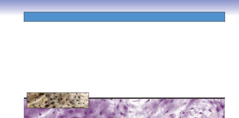
71 |
A |
|
|
Figure 4-15A. |
Dense regular connective tissue, |
|
|
Collagen |
tendon. H&E, |
289; inset 410 |
|
|
This type of tissue is composed of coarse collagen |
||
|
|
fibers |
||
|
Fibroblasts |
bundles that is densely packed and oriented into |
||
|
parallel cylinders. Long, thin fibroblasts are found |
|||
|
|
|
||
|
|
|
among the fiber bundles and are oriented in the |
|
|
|
|
same direction as the fibers. The nuclei of the fibro- |
|
|
|
|
blasts are visible, but the cytoplasm is not easily |
|
|
|
|
seen. The thick bundles of collagen fibers fill the |
|
|
|
|
intercellular spaces. Dense regular connective tis- |
|
|
|
|
sue provides resistance to traction forces in ten- |
|
|
|
|
dons and ligaments. |
|
Fibroblasts |
|
|
|
|
|
Collagen |
|
|
|
|
fibers |
|
|
|
|
|
|
|
|
|
|
|
|
|
B
 Fibroblast
 Collagen fiber
D. Cui |
Ground substance |
Figure 4-15B. A representation of dense regular connective tissue.
Collagen fibers are represented by uniformly arranged thick, pink bundles that are tightly packed in a parallel fashion. Fibroblasts are seen among these fibers. The white background represents the ground substance. This tissue architecture can be found in tendons, ligaments, and aponeuroses. The structure formed by this arrangement is particularly strong and resistant to stress such as the intense forces exerted on ligaments and tendons by athletes.
CLINICAL CORRELATION
C


 Excess fibroblasts
  Collagen microtear
 Increased mucoid ground substance
 Calcification
D. Cui
Proliferated blood vessel
Figure 4-15C. Tendinosis.
Tendinosis is a degenerative disease that occurs within the substance of a tendon. This condition is usually associated with age, overexertion, or both. Histologic examination reveals abnormal fibrotic structure including collagen disorganization, decreased fiber diameter, and increased mucoid ground substance. Additional findings are collagen microtears, focal hypercellularity, vascular proliferation, and focal necrosis with calcification. Tearing of the tendon can occur in severe cases. Treatment includes pain relief, rest, physical therapy, nonsteroidal anti-inflammatory drugs, corticosteroids, and surgical repair, when necessary. The goal is to prevent further degeneration and to preserve function.

72 |
UNIT 2 ■ |
Basic Tissues |
|||
|
|
|
|
|
|
|
A |
|
|
Figure 4-16A. |
Loose connective tissue, mesentery. Verhoeff |
|
|
|
|
stain, 112; inset |
200 |
Collagen
fiber 
|
Elastic fibers |
Loose connective tissue, also called areolar connective tissue, in |
|
|
|
|
|
Fibroblast |
|
|
a mesentery spread preparation is shown. In this tissue prepara- |
|
|
|
tion, both collagen fibers and elastic fibers are visible. The elastic |
|
|
|
fibers stain deep blue as thin strands and collagen fibers appear as |
|
|
|
thick, purple bundles. Fibroblasts are seen among the fibers. This |
|
|
|
type of connective tissue has abundant ground substance, with |
|
|
Elastic fiber |
|
|
|
many connective tissue cells and relatively few fibers. It is richly |
|
|
|
|
|
|
|
|
vascularized, flexible, and not highly resistant to stress. |
Collageno fiber
B
Gut epithelium
Eosinophil
 Plasma
cell
Macrophage
Loose
connective
tissue
Fibroblast
Plasma
cell
Figure 4-16B. Loose connective tissue, large intestine: H&E,
680; inset 1,506
The lamina propria of the digestive tract is an extreme example of loose connective tissue. This tissue lies immediately beneath the thin epithelium of the gut, which is one place where the body’s defense mechanisms initially attack bacteria and pathogens. Therefore, plasma cells, mast cells, leukocytes, and fibroblasts are common in this area. Loose connective tissue is characterized by loosely arranged, woven connective fibers, abundant ground substance, and tissue fluid, which contains the rich array of connective tissue cells.
SYNOPSIS 4 - 2 Functions of Connective Tissue
Connective Tissue Proper
■Dense irregular connective tissue: Provides strong fiber meshwork to resist stress from all directions (e.g., dermis of the skin) and provides protective covering of organs (e.g., capsule of the kidney).
■Dense regular connective tissue: Provides resistance to traction forces in a single specific direction (e.g., tendons, ligaments).
■Loose connective tissue: Provides suspension and support for tissues that are not subjected to strong forces and forms conduits in which vessels and nerves course. Cells in loose connective tissue have defense and immune functions (e.g., lamina propria of the digestive tract).
Specialized Connective Tissues
■Adipose connective tissue: Provides both cushioning for organs and energy storage; some involved in hormone secretion such as leptin (e.g., hypodermis of the skin, mammary glands).
■Reticular connective tissue: Provides supportive framework for hematopoietic and solid (parenchymal) organs (e.g., liver, pancreas).
■Elastic connective tissue: Provides distensible support and accommodates pressure changes on the walls of the arteries closest to the heart (e.g., vertebral ligaments, large arteries).
Embryonic Connective Tissues
■Mesenchymal connective tissue: Gives rise to all types of connective tissues (embryonic mesoderm).
■Mucous connective tissue: Provides cushioning for the nucleus pulposus of the intervertebral disk and helps prevent
kinking in the blood vessels of the umbilical cord.

CHAPTER 4 ■ Connective Tissue |
73 |
A |
|
|
Figure 4-17A. |
Loose connective tissue, small |
|
|
|
intestine. H&E, |
136; inset 384 |
|
|
|
||
|
Epithelial cells |
|
This is an example of the loose connective tissue |
|
|
|
that lies just below the epithelium in the lamina |
||
|
|
|
||
|
|
|
propria of the small intestine. The collagen fibers |
|
|
Loose connective tissue |
|
are loosely arranged and inconspicuous. Many |
|
|
|
cells are tightly packed among the fiber bundles. In |
||
|
|
|
||
|
|
|
comparison, loose connective tissue has more cells |
|
|
|
|
and fewer fibers than dense connective tissue. This |
|
|
|
|
type of tissue is well vascularized, flexible, and not |
|
|
|
|
highly resistant to mechanical stress. |
|
|
|
Collagen |
||
|
|
fibers |
||
Connective tissue cells
B
|
Neutrophil |
|
|
|
Lymphocyte |
|
Fibroblast |
cell |
|
|
|
|
|
Plasma |
Macrophage |
|
Mast cell |
D. Cui
Ground substance
Adipocyte
Elastic fiber
 Collagen fiber
Figure 4-17B. A representation of loose connective tissue.
There are numerous connective tissue cells shown among fibers here. They include fibroblasts, macrophages, adipocytes, mast cells, plasma cells, and leukocytes. If there is a microorganism invasion or mechanical trauma, activation of mast cells and subsequent activation of endothelial cells and vasodilation are among the responses to the tissue injury. Vasodilation promotes delivery of more blood to the local tissue and leads to increased local temperature. Loosening of junctions between endothelial cells enables fluid and serum proteins to leak into the connective tissue. Expression of adhesive molecules (selectins) on endothelial cells increases the chance that leukocytes can migrate into the connective tissue from the blood stream. Mast cells and macrophages also increase in number to participate in the repair of tissue damage.
CLINICAL CORRELATION
C


 Inflammatory
neutrophil


 Lymphocyte
|
Active |
D. Cui |
macrophage |
Figure 4-17C. Whipple Disease.
Whipple disease is a multisystemic disease caused by an infection of the bacillus Tropheryma whippleii. It primarily affects the small intestine. The clinical symptoms include abdominal pain, flatulence, malabsorption, and diarrhea. Symptoms are varied and depend upon the organ infected. The lamina propria (loose connective tissue) of the small intestine reveals an increased number of macrophages. These macrophages contain large numbers of bacteria within their phagosomes, which are clearly stained by the PAS stain (periodic acid combined with Schiff reagent). Treatment for Whipple disease is antibiotic administration, including intravenous penicillin and streptomycin by mouth.

74 UNIT 2 ■ Basic Tissues
Types of Connective Tissue: Specialized Connective Tissues
A
|
Adipose |
tissue |
Adipose cells |
|
|
|
|
Adipose
cells
Dense irregular
Nuclei of connective tissue adipocytes
Figure 4-18A. Adipose tissue, mammary gland.
H&E, 68; inset 178
Adipose tissue is a special form of connective tissue and has a rich neurovascular supply. Adipocytes appear white here, and this tissue is referred to as white adipose tissue. Each adipocyte contains a single, large lipid droplet in its cytoplasm, so the cells are called unilocular adipose cells. Most of the cytoplasm is occupied by the lipid droplet, and the nucleus is displaced to one side. Each adipocyte is surrounded by a basal lamina. This type of adipose tissue is found throughout the adult human body. There is another type of adipose tissue that is highly specialized, called brown adipose tissue. It is composed of multilocular adipocytes; each adipocyte contains multiple lipid droplets in its cytoplasm. This tissue is mainly found in hibernating mammals and newborn infants but can also be found scattered in some areas in adults, such as the esophagus, trachea, posterior neck, and interscapular areas as vestigial remnant tissue. Tumors sometimes arise from the remnant brown adipose tissue and are called hibernomas.
B |
Fibroblast |
|
Adipocyte |
|
Reticular fibers |
|
Collagen fiber |
D. Cui |
Blood vessel |
Figure 4-18B. A representation of adipose tissue.
Adipocytes (fat cells) are scattered within a loose collagenous supporting tissue in this unilocular adipose tissue. Each adipose cell contains a single large drop of lipid; it has a thin rim of cytoplasm around the lipid, and its flattened nucleus is located in the periphery of the cell. Adipocytes are the primary site for storage of energy, and lipid deposition and mobilization are regulated by hormonal factors (steroids, insulin, thyroid hormone, etc.). Adipocytes also play a role in the synthesis of some hormones such as leptin. During childhood, the adipocyte numbers may increase depending on nutrition and other factors, but in adulthood, adipocyte numbers normally remain constant.
CLINICAL CORRELATION
C 


Expanded adipocytes
containing huge lipid droplets


 Collagen bundles
pushed to the side
D. Cui
Figure 4-18C. Obesity.
Hypertrophic obesity is a disorder characterized by an increase in total body fat, particularly by expansion (hypertrophy) of preexisting fat cells. Obesity increases the risk for a number of conditions, including diabetes, hypertension, high cholesterol, stroke, and coronary artery disease. Obesity may also increase the risk for some types of cancer, and it is a risk factor for the development of osteoarthritis, pancreatitis, and sleep apnea. Obesity can result from a sedentary lifestyle and the chronic ingestion of excess calories; genetic predisposition may also play a role in the development of obesity. The possible treatments include exercise, diet, medications, and surgery. By contrast, hyperplastic obesity is excessive weight gain associated with childhood-onset obesity, characterized by the creation of new fat cells.
|
CHAPTER 4 ■ Connective Tissue |
|
|
75 |
||
|
|
|
|
|||
Figure 4-19A. |
Reticular connective tissue, pancreas. Silver |
|||||
|
A |
|
||||
|
|
|
stain, 136 |
|
||
|
|
|
|
|
||
Pancreatic cells
D. Cui |
|
Reticular tissue is a specialized loose connective tissue that |
|
provides a delicate supporting framework for many highly |
|
cellular organs, such as endocrine glands, lymphoid organs, |
|
the spleen, and the liver. Reticular fibers are shown in black |
|
with a silver stain. These fibers are small in diameter and do |
|
not form large bundles. They are arranged in a netlike frame- |
Reticular fibers |
work to support parenchymal cells, in this example, pancre- |
|
atic cells. The inset drawing represents the organization of |
|
reticular fibers and pancreatic cells. |
B |
Figure 4-19B. Reticular connective tissue, liver. Silver |
|
stain, 312 |
|
Reticular fibers |
|
The reticular fibers can be selectively visualized with a silver |
|
stain, that is, they are argyrophilic. These fibers consist of col- |
|
lagen type III, which forms a meshlike network that supports |
|
the liver cells and holds these cells together. The liver cells’ |
|
cytoplasm is unstained in this preparation, and the structure |
|
of the cells is not easy to distinguish here. The inset draw- |
|
ing represents the organization of reticular fibers and hepato- |
|
cytes. There is a sinusoid running between the reticular fibers, |
|
which appears as empty space here. |
|
Hepatocytes |
CLINICAL CORRELATION
C
Nodule of regenerated liver cells
Collagen bundles and elastic fibers have replaced normal reticular fibers
 Damaged and reduced reticular fibers
D. Cui
Figure 4-19C. Cirrhosis.
Cirrhosis is a liver disorder caused by chronic injury to the hepatic parenchyma. The major causes of cirrhosis include alcoholism and chronic infection with hepatitis B or hepatitis C virus. Pathologic changes are characterized by the collapse of the delicate supporting reticular connective tissue with increased numbers of collagen and elastic fibers. There is disruption of the liver architecture and vascular bed. Regenerating hepatocytes form nodules rather than the characteristic columnar plates. Symptoms include jaundice, edema, and coagulopathy (a defect of blood coagulation). The resulting damage to the liver tissue impedes drainage of the portal venous system, a condition known as portal hypertension, which may eventually lead to gastroesophageal varices, splenomegaly, and ascites.

76 UNIT 2 ■ Basic Tissues
A |
|
|
|
Figure 4-20A. |
Elastic connective tissue, carotid |
|
|
Elastic lamellae |
artery. Elastic stain (Verhoeff), 275; inset 516 |
||
|
|
||||
|
|
This is an example of elastic connective tissue in the |
|||
|
|
|
|
||
|
|
|
|
tunica media of a carotid artery. The wavy elastic |
|
|
|
|
|
lamellae are distributed among collagen and smooth |
|
|
|
|
|
muscle cells in the tunica media layer of a large artery. |
|
|
|
|
|
The smooth muscle cells are not visible here because |
|
|
|
|
|
of the type of stain. In general, the elastic material (as |
|
|
|
|
|
either elastic fibers or elastic lamellae) and other con- |
|
|
|
|
|
nective tissue fibers are produced by fibroblasts in the |
|
|
|
|
|
||
|
|
|
|
connective tissue, but in blood vessels, smooth muscle |
|
|
|
|
|
cells are the principal cells that produce elastic material |
|
|
|
|
|
and other connective tissue fibers. Elastic connective |
|
|
Elastic |
|
|
tissue consists predominately of elastic material, and |
|
|
|
|
this allows distension and recoil of the structure. This |
||
|
lamellae |
|
|
||
|
|
|
tissue can be found in some vertebral ligaments, arte- |
||
|
|
|
|
||
|
|
|
|
rial walls, and in the bronchial tree. |
|
|
|
|
|
|
|
|
|
|
|
|
|
B |
|
|
Elastic lamellae |
|
Smooth muscle |
|
cell |
|
Collagen fiber |
|
Reticular fiber |
D. Cui |
Ground substance |
|
Figure 4-20B. A representation of elastic connective tissue in the tunica media of a large artery.
Thick bundles of elastic lamellae are arranged in parallel wavy sheets, with the smooth muscle cells and collagen fibers insinuated between alternating lamellae. The elastic fibers are formed by elastin and fibrillin microfibrils. Elastic connective tissue is able to recoil after stretching. This property in large arteries helps to moderate the extremes of pressure associated with the cardiac cycle. Abnormal expression of the fibrillin (FBN1) gene is associated with abnormal elastic tissue disease.
CLINICAL CORRELATION
C

 Loss of elastic lamellae and increased ground substance
 Cystic space filled with amorphous extracellular matrix
 Fragmentation of elastic lamellae
D. Cui
Figure 4-20C. Marfan Syndrome—Cystic Medial Degeneration.
Marfan syndrome is an autosomal dominant disorder caused by an FBN1 gene mutation, which affects the formation of elastic fibers, particularly those found in the aorta, heart, eye, and skin. Signs and symptoms include tall stature with long limbs and long, thin fingers and enlargement of the base of the aorta accompanied by aortic regurgitation. There is increased probability of dissecting aortic aneurysms and prolapse of the mitral valve. Treatment includes pharmacologic or surgical intervention to prevent potentially fatal or long-term complications, but no permanent cure is yet available. This illustration depicts cystic medial degeneration (cystic medionecrosis) of the aorta, including disruption and fragmentation of elastic lamellae in the tunica media of the aorta, loss of elastic fibers, and increase in ground substance causing formation of cystic space.

CHAPTER 4 ■ Connective Tissue |
77 |
Types of Connective Tissue: Embryonic Connective Tissues
A
Mesenchymal cells
Mesenchymal connective tissue
Cytoplasmic
processes
Embryonic red blood |
Mesenchymal |
cells |
cells |
Figure 4-21A. Mesenchyme, embryo. H&E, 136; inset
408 (left) and 438 (right)
Mesenchyme (mesenchymal connective tissue) is found in the developing structures in the embryo. It contains scattered reticular fibers and mesenchymal cells, which have irregular, star or spindle shapes and pale-stained cytoplasm. These cells exhibit cytoplasmic processes, which often give the cells a stellate appearance. Mesenchymal cells are relatively unspecialized and are capable of differentiating into different cell types in mature tissue cells, such as cartilages, bones, and muscles. Embryonic red blood cells can be seen in this specimen. These blood cells contain a nucleus in each cell; this is characteristic of their immature state (anucleated red blood cells are characteristic of the mature state and are found in adult tissues). Interestingly enough, some vertebrates, such as frogs and chickens, have nucleated red blood cells in the adult state.
B
Mucous connective tissue
Fibroblasts
Smooth muscle
Figure 4-21B. Mucous connective tissue, umbilical cord.
Toluidine blue stain, 68; inset 178
An example of mucous connective tissue that has an abundance of a jellylike matrix with some fine aggregates of collagen fibers and stellate-shaped fibroblasts is shown. It is found in the umbilical cord and subdermal connective tissue of the embryo. Mucous tissue is a major constituent of the umbilical cord, where it is referred to as Wharton jelly. This type of connective tissue does not differentiate beyond this stage. In this example, the viscous ground substance has been stained with a special stain to reveal jellylike mucin, which contains hyaluronic acid and glycoproteins. Collagen fibers and large stellate-shaped fibroblasts (not mesenchymal cells) predominate in the mucous tissue.
SYNOPSIS 4 - 3 Pathological Terms for Connective Tissue
■Urticaria: An itchy skin eruption, also known as hives, characterized by wheals with pale interiors and well-defined red margins, often the result of an allergic response to insect bites, foods, or drugs (Fig. 4-3C).
■Pruritis: Itching of the skin due to a variety of causes including hyperbilirubinemia and allergic and irritant contact conditions (Fig. 4-3C).
■Cirrhosis: An abnormal liver condition characterized by diffuse nodularity, due to fibrosis and regenerative nodules of hepatocytes; frequent causes are alcohol abuse and viral hepatitis (Fig. 4-19C).
■Jaundice: Yellow staining of the skin, mucous membranes, or conjunctiva of the eyes caused by elevated blood levels of the bile pigment bilirubin (Fig. 4-19C).
■Coagulopathy: A disorder that prevents the normal clotting process of blood; causes may be acquired, such as hepatic dysfunction, or congenital, such as decreased clotting factors, as seen in inherited conditions like hemophilia (Fig. 4-19C).
■Necrosis: Irreversible cell changes that occur as a result of cell death (Fig. 4-20C).

78 UNIT 2 ■ Basic Tissues
TABLE 4 - 3 |
|
Connective Tissue Types |
|
|
|
|
Type |
|
Connective Tissue |
Connective |
Organization of |
Main Locations |
Main Functions |
|
||||||
|
|
Cells |
Tissue Fibers |
Fibers and Cells |
|
|
|
|
|
|
|
|
|
Connective Tissue Proper |
|
|
|
|
||
|
|
|
|
|
|
|
Dense irregular |
|
Predominantly |
Collagen fibers, |
Fewer cells and |
Dermis of the skin, |
Resists stress |
connective tissue |
|
fibroblasts; other |
elastic fibers, |
more fibers; fibers |
capsules of many |
from all |
|
|
connective tissue |
reticular fibers |
arranged randomly |
organs |
directions; |
|
|
cells occasionally |
|
without a definite |
|
protects organs |
|
|
present |
|
orientation in |
|
|
|
|
|
|
relatively less ground |
|
|
|
|
|
|
substance |
|
|
|
|
|
|
|
|
|
Dense regular |
|
Predominantly |
Collagen fibers, |
Fewer cells and |
Tendons, ligaments |
Provides |
connective tissue |
|
fibroblasts; other |
elastic fibers, |
more fibers; fibers |
|
resistance to |
|
|
connective tissue |
reticular fibers |
arranged in uniform |
|
traction forces |
|
|
cells occasionally |
|
parallel bundles |
|
|
|
|
present |
|
|
|
|
|
|
|
|
|
|
|
Loose |
|
Fibroblasts, |
Collagen fibers |
More cells and |
Lamina propria |
Provides |
connective tissue |
|
macrophages, |
predominate; |
fewer fibers; fibers |
of gastrointestinal |
protection, |
|
|
adipocytes, mast |
elastic and |
randomly distributed |
tract; around the |
suspension, and |
|
|
cells, plasma cells, |
reticular fibers |
in abundant ground |
nerves and vessels |
support; conduit |
|
|
leukocytes |
also present |
substance |
(in adventitia layer) |
for vessels and |
|
|
|
|
|
|
nerves; environ- |
|
|
|
|
|
|
ment for immune |
|
|
|
|
|
|
defense function |
|
|
|
|
|
|
|
Specialized Connective Tissues |
|
|
|
|
||
|
|
|
|
|
|
|
Adipose |
|
Predominantly |
Collagen fibers |
Fibers form fine |
Hypodermis of the |
Provides |
connective tissue |
|
adipocytes (fat cells); |
and reticular |
meshwork that |
skin, mammary |
energy storage, |
|
|
fibroblasts and other |
fibers |
separates adjacent |
glands, and around |
insulation; |
|
|
connective tissue |
|
adipocytes |
many organs |
cushioning of |
|
|
cells occasionally |
|
|
|
organs; hormone |
|
|
present |
|
|
|
secretion |
|
|
|
|
|
|
|
Reticular |
|
Fibroblasts, |
Reticular fibers |
Fibers form delicate |
Liver, pancreas, |
Provides support- |
connective tissue |
|
reticular cells, |
|
meshlike network; |
lymph nodes, |
ive framework |
|
|
hepatocytes, |
|
cells with process |
spleen, and bone |
for hematopoietic |
|
|
smooth muscle |
|
attached to the fibers |
marrow |
and parenchymal |
|
|
cells, Schwann cells |
|
|
|
organs |
|
|
depending on the |
|
|
|
|
|
|
location |
|
|
|
|
|
|
|
|
|
|
|
Elastic |
|
Predominantly |
Elastic fibers |
Fibers arranged |
Vertebral ligaments, |
Provides flexible |
connective tissue |
|
fibroblasts or |
predominate; |
in parallel wavy |
walls of the large |
support for the |
|
|
smooth muscle cells; |
collagen and |
bundles |
arteries |
tissue; reduces |
|
|
other connective |
reticular fibers |
|
|
pressure on the |
|
|
tissue cells occasion- |
also present |
|
|
walls of the |
|
|
ally present |
|
|
|
arteries |
|
|
|
|
|
|
|
Embryonic Connective Tissues |
|
|
|
|
||
|
|
|
|
|
|
|
Mesenchymal |
|
Mesenchymal cells |
Reticular fibers |
Scattered fibers |
Embryonic |
Gives rise to all |
connective tissue |
|
|
and collagen |
with spindle- |
mesoderm |
connective tissue |
|
|
|
fibers |
shaped cells having |
|
types |
|
|
|
|
long cytoplasmic |
|
|
|
|
|
|
processes; |
|
|
|
|
|
|
mesenchymal cells |
|
|
|
|
|
|
uniformly distributed |
|
|
|
|
|
|
|
|
|
Mucous |
|
Spindle-shaped |
Collagen fibers |
Fibers and |
Umbilical cord, |
Provides cushion |
connective tissue |
|
fibroblasts |
predominate; few |
fibroblasts randomly |
subdermal layer |
to protect the |
|
|
|
elastic and reticu- |
displayed in jellylike |
of the fetus, dental |
blood vessels in |
|
|
|
lar fibers |
matrix (Wharton |
pulp of the devel- |
the umbilical cord |
|
|
|
|
jelly) |
oping teeth, nucleus |
|
|
|
|
|
|
pulposus of the disk |
|
|
|
|
|
|
|
|
5 Cartilage and Bone
Cartilage
Introduction and Key Concepts for Cartilage
Types of Cartilage
Figure 5-1 |
Overview of Cartilage Types |
|
Table 5-1 |
Cartilage |
|
Figure 5-2A |
A Representation of Hyaline Cartilage |
|
Figure 5-2B,C |
Hyaline Cartilage, Bronchus |
|
Figure 5-3A |
Hyaline Cartilage, Trachea |
|
Figure 5-3B |
Hyaline Cartilage, Finger Bone |
|
Figure 5-3C |
Clinical Correlation: Osteoarthritis |
|
Figure 5-4 |
Hyaline Cartilage and Chondrocytes |
|
Figure 5-5A–C |
Elastic Cartilage |
|
Figure 5-6A,B |
Fibrocartilage |
|
Figure 5-6C |
Clinical Correlation: Disk Degeneration and Herniation |
|
Cartilage Growth |
|
|
Figure 5-7 |
A Representation of Cartilage Growth |
|
Synopsis 5-1 |
Functions of Cartilage |
|
Synopsis 5-2 |
Special Features of Cartilage |
|
Bone
Introduction and Key Concepts for Bone
Figure 5-8 |
Overview of Bone Structure, Long Bone |
Synopsis 5-3 |
Functions of Bone |
Types of Bone |
|
Figure 5-9A,B |
Compact Bone |
Figure 5-9C |
A Representation of an Osteon of the Compact Bone |
Figure 5-10A–C |
Compact and Cancellous Bone |
79