156 UNIT 3 ■ Organ Systems
The Capillary System |
|
Figure 9-13A,B |
Continuous Capillaries |
Figure 9-14A,B |
Fenestrated Capillaries |
Figure 9-15A,B |
Discontinuous (Sinusoidal) Capillaries |
The Venous System |
|
Figure 9-16A–C |
Venules and Small Veins |
Figure 9-17A–C |
Medium Veins |
Figure 9-18A–C |
Large Veins |
Table 9-1 |
Blood Vessels |
The Lymphatic Vascular System
Figure 9-19A,B |
Lymphatic Vessels |
Figure 9-19C |
Clinical Correlation: Lymphangioma |
Introduction and Key Concepts for the Circulatory System
The circulatory system includes the cardiovascular system and the lymphatic vascular system. The cardiovascular system includes the heart and the arterial, capillary, and venous systems. Blood is transported from the heart through the arterial system to the capillaries, where exchange of gases, nutrients, and other substances takes place. Blood is carried back to the heart by the venous system. Blood flows through two routes:
(1) The systemic circulation system transports oxygenated blood from the heart to the capillaries in the tissues and organs of the body and then collects and carries the blood back to the heart (Fig. 9-1). (2) The pulmonary circulation system transports deoxygenated blood from the heart to the capillaries of the lungs. After gas exchange, blood is carried back to the heart (see Fig. 9-1). The lymphatic vascular system consists of lymphatic capillaries, lymphatic vessels, and lymphatic ducts. This system collects lymph (excess tissue fluid) from the tissues of all organs (except the nervous system, bone marrow, and hard tissues) by lymphatic capillaries and then transports it through lymphatic vessels to the lymphatic ducts, which eventually empty the lymph into the venous system. The collected lymph passes through lymph organs, where it is filtered, and lymphocytes are exposed to antigens. Lymphopoiesis and the immune response occur here (Fig. 9-19A,B).
The Cardiovascular System
The Heart
The heart contains four chambers: the left and right atria and the left and right ventricles. The atria receive blood flow discharged from the venous system, whereas the ventricles pump blood into the arterial system (Fig. 9-1). The wall of the heart is composed of three layers: endocardium (innermost layer), myocardium (middle layer), and epicardium (outermost layer).
(1) Endocardium consists of endothelium, subendothelial
connective tissue, and subendocardium (Purkinje fibers, small coronary blood vessels, and nerve fibers). (2) Myocardium, the thickest layer of the heart, contains an abundance of cardiac muscle cells (Fig. 9-3A,B). Cardiac muscle contracts producing heart beats, which are generated and regulated by the heart conductive system including the sinoatrial (SA) node, the atrioventricular (AV) node, the AV bundle, and Purkinje fibers
(Fig. 9-2). (3) Epicardium is covered by mesothelium and contains fibrous connective tissue, nerves, coronary vessels, and adipose tissue (Fig. 9-3C).
Types of Blood Vessels
THE ARTERIAL SYSTEM is composed of large (conducting) arteries, medium (distributing) arteries, small arteries, and arterioles. The arterial system conducts blood (under higher pressure than veins) from the ventricles to the capillary networks. The walls of arteries can be generally divided into three layers: tunica intima, tunica media, and tunica adventitia (Figs. 9-5 and 9-6).
Large arteries are also called elastic arteries because of the large quantity of elastic material in their walls (Fig. 9-7A–C). They have a thick tunica media with numerous elastic membranes. The internal and external elastic laminae are hard to distinguish from the nearby elastic membranes. Large arteries conduct blood from the ventricles into the medium arteries. Rich elastic materials in large arteries enable the vessels to recoil to accommodate pressure changes and maintain a continuous flow of blood during ventricular diastole (relaxation).
Medium arteries are also called muscular arteries because of their thick tunica media, which contains circularly arranged multiple layers of smooth muscle cells in a distinct sheath (Figs. 9-8 to 9-9A). Internal and external elastic laminae are easy to distinguish from nearby tissues.
Small arteries and arterioles are smaller diameter vessels. The walls of small arteries contain two to six layers of smooth muscle cells (Figs. 9-9B and 9-10A,B). Arterioles are the smallest components of the arterial system, with only one or two layers of smooth muscle cells (Figs. 9-10A,C, and 9-11). They control the blood flow into the capillaries.
158 UNIT 3 ■ Organ Systems
The Cardiovascular System
Pulmonary circulation system
 veins Medium
|
Pulmonary |
|
|
|
|
|
|
capillaries |
|
|
|
|
|
|
Small arteries |
|
|
Small veins |
|
|
|
|
Arterioles |
|
|
Venules |
circulation |
|
|
|
Mediumarteries |
Lungs |
|
veins |
|
|
|
|
|
|
|
|
|
|
Large veins (vena cava) |
Aorta |
Medium |
Pulmonary |
|
|
|
|
LA |
|
Large (pulmonary) veins |
|
|
|
|
|
|
|
|
|
|
|
RA |
|
|
|
|
|
|
|
|
|
LV |
Large arteries |
|
|
|
|
|
RV |
|
|
|
|
|
|
|
|
|
|
circulation |
|
Large arteries (pulmonary artery) |
|
|
|
|
|
|
|
|
|
|
|
|
|
|
|
|
|
|
edium arteries |
Systemic |
|
Systemic |
|
|
|
M |
|
|
|
|
|
|
capillaries |
|
|
|
|
|
Small veins |
|
|
Small arteries |
|
|
|
Venules |
|
|
Arterioles |
|
|
|
T. Yang |
Organs and |
tissues of the body |
|
|
|
|
Systemic circulation system |
RA: right atrium |
|
|
Deoxygenated blood in systemic circulation system |
LA: left atrium |
|
|
RV: right ventricle |
|
|
|
LV: left ventricle |
|
|
Oxygenated blood in systemic circulation system |
|
|
|
|
|
Deoxygenated blood in pulmonary circulation system |
|
|
|
Oxygenated blood in pulmonary circulation system |
Figure 9-1. Overview of the cardiovascular system.
The cardiovascular system consists of the heart, arterial system, venous system, and capillary system. The heart is composed of two atria and two ventricles. The right atrium receives blood from the body, and the left atrium receives blood from the lungs; the left ventricle pumps blood to the body, and the right ventricle pumps blood to the lungs. The arterial system conducts blood from the heart to capillaries in the body and the lungs. This system includes large arteries (elastic arteries), medium arteries (muscular arteries), small arteries, and arterioles. The venous system carries blood from the capillary system in the body and lungs to the heart. It includes venules, small veins, medium veins, and large veins. The capillary system is located between arterioles and venules and often forms capillary beds where exchange of gases and various substances and movement of blood cells (diapedesis) take place. It includes continuous, fenestrated, and sinusoidal (discontinuous) capillaries. Blood flows through two routes: (1) the systemic circulation system, which supplies oxygenated blood from the heart to the organs and tissues of the body and then carries deoxygenated blood back to the heart and (2) the pulmonary circulation system, which sends deoxygenated blood from the heart to the lungs for gas exchange and then returns oxygenated blood to the heart.
Systemic and Pulmonary Circulation Systems
Systemic circulation system
Left |
Large/elastic arteries |
|
Medium systemic |
|
Small arteries |
Arterioles |
ventricle |
(aorta, etc.) |
arteries (muscular arteries) |
|
|
|
Right |
Large veins |
|
Medium |
Small |
Venules |
Systemic capillaries |
|
atrium |
(vena cava, etc.) |
veins |
veins |
(exchange of gases |
|
|
Pulmonary circulation system |
|
|
|
|
and other substances) |
|
|
|
|
|
Right |
Large/elastic arteries |
|
Medium pulmonary |
Small arteries |
Arterioles |
ventricle |
(pulmonary arteries) |
|
arteries (muscular arteries) |
|
|
Left |
|
Large veins |
|
Medium |
Small |
Venules |
Pulmonary capillaries |
atrium |
(pulmonary veins) |
|
veins |
veins |
|
|
(gas exchange) |
|
|
|
|
|
|
|
|
|
|
|
|
|
|
|
CHAPTER 9 ■ Circulatory System |
159 |
Superior |
|
|
Left pulmonary veins |
vena cava |
|
|
|
Sinoatrial (SA) |
|
Left |
|
node |
|
atrium |
|
|
Right |
|
Atrioventricular |
Atrioventricular |
atrium |
|
(AV) bundle |
|
|
|
(AV) node |
|
|
|
|
|
LV |
Left bundle branch |
Cardiac |
|
|
|
valves |
|
|
|
Papillary |
|
RV |
Purkinje fibers |
|
|
|
muscle |
|
|
|
Inferior |
|
|
Epicardium |
vena cava |
|
|
Myocardium |
|
|
|
|
Right bundle |
Endocardium |
RV: right ventricle |
|
|
LV: left ventricle |
|
branch |
|
|
|
|
Figure 9-2. The heart and its impulse conductive function.
The contractions of the right and left atria and ventricles must be coordinated precisely in order for blood to be pumped efficiently to the lungs for oxygenation and then to the rest of the body. A network of specially modified cardiac muscle fibers generates and conducts electrical impulses that provide the necessary coordination. This network includes the SA node, the AV node, the AV bundle, and Purkinje fibers. (1) The SA node is located in the right atrial wall near the opening of the superior vena cava. The SA node is important for initiating the heartbeat impulse and controlling its frequency; it is also called the “pacemaker” of the heart. (2) The AV node is also located in the right atrial wall, medial to the right AV valve, and along the lower part of the interatrial septum. (3) The AV bundle arises from the AV node and divides into two branches (right and left) to run along the sides of the interventricular septum. (4) Purkinje fibers are the terminal branches of the right and left branches of the AV bundle. Purkinje fibers run along the inferior and lateral wall of the ventricle and extend to the papillary muscles. These fibers are modified large cardiac muscle cells, which contain numerous gap junctions and well-developed intercalated disks. The SA node generates the heartbeat signal, which quickly spreads to adjacent cardiac muscle cells in the myocardium of both atria to cause the atria to contract. Impulses are also picked up by the AV node and travel along the AV bundle. These impulses pass through the right and left bundle branches to Purkinje fibers in the ventricles, where they induce contraction of the cardiac muscles of the ventricles. There is a time delay, which allows the atria to contract first to empty blood into the ventricles, before the ventricles contract. This time delay ensures that blood flows smoothly from the atria to the ventricles and then to the conductive arteries.
SYNOPSIS 9 - 1 Structure and Functions of the Heart
■The heart is composed of two atria (singular: atrium) and two ventricles.
■The wall of the heart consists of endocardium (inner layer), myocardium (cardiac muscle layer), and epicardium (outer layer).
■Endocardium is composed of endothelium, subendothelium (a thin layer of connective tissue), and subendocardium. Purkinje fibers are located in the subendocardium.
■Myocardium is composed of cardiac muscles, the thickest layer of the heart; cardiac muscles are branched and connected to each other end to end by intercalated disks (junction complexes). Cardiac muscle fibers require high oxygen supply.
■Epicardium is composed of mesothelium and a thicker layer of connective tissue, which contains coronary vessels, nerves, and adipose tissue.
■The conductive system of the heart consists of groups of specially modified cardiac muscle fibers, the SA node, AV node,
AV bundle, and Purkinje fibers.
162 UNIT 3 ■ Organ Systems
|
Endothelial cells |
|
|
Endothelial cells |
|
|
Subendothelial |
Tunica |
|
Tunica |
|
intima |
|
intima |
|
layer |
|
|
|
|
|
|
|
Internal elastic |
|
|
Subendothelial |
|
|
lamina (IEL) |
|
|
|
|
|
|
layer |
|
|
|
|
|
|
|
Circular |
|
Tunica |
Circular |
Tunica |
|
smooth muscle |
|
media |
smooth muscle |
media |
 with connective tissue
 External elastic lamina (EEL)
 Connective tissue
Tunica adventitia
 Fibroblast
 Longitudinal smooth muscle
Connective Tunica
tissue adventitia
Fibroblast
Figure 9-5. A representation of the general structure of blood vessel layers (tunicae) and a comparison of the medium artery and the medium vein.
The walls of the blood vessels can be divided into three layers (tunicae). The innermost layer in contact with the blood is called the tunica intima. This layer contains endothelium and subendothelial connective tissue and may contain an internal elastic lamina (IEL) in some vessels, particularly arteries. The endothelium is a layer of simple squamous cells, which forms the smooth surface of the lumen. Subendothelial connective tissue is a thin layer of connective tissue beneath the endothelium. The IEL, if present, is a sheet of elastic material that divides the tunica intima from the tunica media. The middle layer is called the tunica media. It primarily contains circularly arranged smooth muscle cells (except in elastic arteries). Contraction and relaxation of these smooth muscle cells will change the vessel diameter and affect blood pressure. The outermost layer, the tunica adventitia, is a layer of connective tissue dominated by collagenous and elastic fibers. In large and some medium veins, the tunica adventitia may also contain longitudinal smooth muscle bundles. Tunica adventitia surrounds and covers the vessels for protection. Occasionally, small blood vessels called vasa vasorum are found in the tunica adventitia of large vessels. The vasa vasorum provide oxygen and nutrients for the large vessel walls when the distance from the lumen is great and it is difficult to get nutrients from diffusion. Some differences between a medium artery and a medium vein are listed here: (1) the artery has a smaller and more rounded lumen, whereas the vein has a larger and oval or irregular-shaped lumen; (2) the artery has a thicker wall than does the vein; (3) in the artery, the tunica media is much thicker than the tunica adventitia, but in the vein, the tunica adventitia is much thicker than the tunica media; (4) circularly oriented smooth muscle cells form uniform sheets in the tunica media of the artery; however, smooth muscle cells are fewer and do not form a distinct sheet in the vein; (5) There are a few longitudinal smooth muscle bundles in the tunica adventitia of medium veins in some locations, whereas these smooth muscle bundles are more abundant in large veins. This pattern does not occur in arteries. Longitudinal smooth muscle bundles in the tunica adventitia of the vein contract to help push blood back to the heart; and (6) valves are present in many veins, especially in the medium veins of the extremities. Their function is to prevent gravitational backflow of the blood and to help blood return to the heart. Arteries do not have valves (except the aortic and pulmonary valves).
CHAPTER 9 ■ Circulatory System |
|
|
|
|
163 |
Large artery |
Medium artery |
Small artery |
Arteriole |
(elastic artery) |
(muscular artery) |
|
|
|
|
|
|
|
|
|
|
|
|
|
|
|
|
|
|
|
|
|
|
|
|
|
|
|
|
D. Cui /T. Yang
Large vein |
Medium vein |
Small vein |
Venule |
|
|
|
|
|
|
|
|
|
|
|
|
|
|
|
|
|
|
|
|
D. Cui /T. Yang
Continuous capillary |
Fenestrated capillary |
Discontinuous (sinusoidal) |
capillary |
T. Yang
Figure 9-6. A representation of types of blood vessels: arteries, veins, and capillaries.
Blood vessels make up the arterial, the venous, and the capillary systems. In general, arteries have smaller, rounder lumens than veins; their tunica media are thicker than the tunica adventitia, and the IEL are prominent. Veins have larger, flatter lumens than arteries; longitudinal smooth muscle bundles may be present in the tunica adventitia, which is the most dominant layer in large and some medium veins. The tunica adventitia is much thicker in the veins than the tunica media. For details, also see Table 9-1. Exchange of gases, nutrients, and materials occurs in the capillaries. Three types of capillaries are illustrated here. Continuous capillaries have complete endothelial cells and continuous basal laminae. Fenestrated capillaries have continuous basal laminae with fenestrated endothelial cells (perforated by small pores); discontinuous (sinusoidal) capillaries have incomplete endothelial cells perforated by large pores, and part of the cytoplasm may be missing. The basal lamina is discontinuous. Discontinuous capillaries have gaps between endothelial cells, and their lumen sizes are much larger than the other two types. Blood vessel sizes are not drawn to scale.
164 UNIT 3 ■ Organ Systems
The Arterial System
A Tunica
intima


 Connective tissue Tunica
adventitia
 Elastic lamina
Tunica
media
D. Cui /T. Yang
 Vasa vasorum

 Nerve
Nerve
 Fibroblast
Endothelial cell
Figure 9-7A. A representation of a large (elastic) artery.
Large arteries (elastic arteries) are also referred to as conducting arteries, because of their function in conducting blood flow out of the heart. There are abundant elastic laminae (thick bundles of elastic fibers) in the tunica media of large arteries, which enable the walls to recoil to resist pressure as well as to maintain arterial pressure during diastole when the ventricles are relaxed and pressure is not generated by the heart. Small vessels located in the tunica adventitia are called vasa vasorum; they supply the tissue of the artery wall with oxygen and nutrients when they are far from the lumen and diffusion of nutrients is difficult. The aorta, pulmonary arteries, and their main branches are good examples of elastic arteries.
Elastic laminae
 Tunica
adventitia
 Tunica
media
Tunica
intima
Figure 9-7B. Large (elastic) artery, carotid artery. Elastic stain, 68; inset 242
This is an example of a large artery (elastic artery) from a portion of the carotid artery. The carotid artery generally has the characteristics of elastic arteries. The tunica media of the vessel wall has multiple layers of elastic lamina that are well organized in concentric fashion. These elastic laminae are sheets of fenestrated elastic material produced by smooth muscle cells in the tunica media. These cells are interspersed between the elastic laminae (see Fig. 4-9B). The elastic laminae in this specimen are revealed as parallel wavy black sheets by a special elastic stain; smooth muscle cells are not visible here with this type of stain. The tunica adventitia of an elastic artery consists of loosely arranged connective tissue fibers. These fibers are mainly collagen fibers, with a small number of elastic fibers that are produced by fibroblasts.
C
|
|
Tunica |
Tunica |
Tunica |
adventitia |
intima |
|
media |
|
|
|
Elastic laminae
Vasa vasorum
Smooth muscle cells
Figure 9-7C. Large (elastic) artery, aorta. H&E, 68; insets 198
An example of a large artery (aorta) is shown. The elastic laminae are pink and are more difficult to distinguish with routine H&E stain than they are with elastic stains. The dark nuclei in the tunica media belong to smooth muscle cells (left inset), which produce the elastic membrane. There are some vasa vasorum deep in the tunica adventitia (right inset), which provide oxygen and nutrients for the tunica media and tunica adventitia of the large artery wall. Nerves belonging to the autonomic nervous system may occasionally be found in the tunica adventitia (see Fig. 9-7A).
166 UNIT 3 ■ Organ Systems
A
|
Endothelial |
|
Tunica |
|
cell junction |
Internal |
media |
|
|
|
|
|
elastic |
Dense |
|
|
lamina |
|
External |
bodies |
|
|
|
|
|
|
elastic lamina |
|
Basal lamina |
|
|
|
Tunica media
Internal elastic lamina
Endothelium |
Erythrocytesocytes |
|
Lumen |
Collagen
fibers
Figure 9-9A. Medium artery. TEM, 1,000 left; 8,700 above
In the wall of the healthy medium artery on the left, the layer of squamous endothelial cells adheres closely to the prominent, highly corrugated, internal elastic membrane. The external elastic membrane is also corrugated but thinner. The thick tunica media is composed of many layers of closely apposed smooth muscle cells. In the higher magnification view on the right, the dense bodies and basal laminae that are characteristic of smooth muscle cells are readily apparent. The amorphous appearance of the internal elastic membrane is typical of elastic membranes in transmission electron micrographs.
Figure 9-9B. Small artery. SEM,
1,300
In this scanning electron micrograph of a small artery, the endothelial cells are extremely squamous, and they are so closely apposed to the internal elastic membrane that the interface between
the two layers is indistinguishable.
Erythrocytes The smooth muscle cells of the tunica media are also closely apposed to each other so that boundaries between cells are difficult to discern. The ground substance of the tunica adventitia and the surrounding connective tissue has been removed during specimen processing so that only some bundles of collagen fibers remain.
Collagen
fiber Endothelial cells Internal elastic bundle
lamina
168 |
UNIT 3 ■ Organ Systems |
|
|
|
|
Nucleus of |
Internal elastic |
Nucleus of smooth |
|
|
SchwannSchwanncelcells |
lamina |
muscle cell |
|
|
Internal |
|
|
|
|
elastic |
|
|
|
|
lamina |
|
|
|
|
|
|
Nucleus of |
|
|
|
|
endothelial |
|
|
|
|
cell |
|
|
Endothelium–-ssmoothooth muscleuscle |
|
|
|
|
cell junctions |
Nuclei of |
|
|
|
|
endothelial |
|
|
|
|
cells |
|
|
Nucleus of smooth |
|
|
|
muscle cell |
|
|
|
|
Figure 9-11. |
Arteriole. TEM, 5, 800; inset 3,800 |
|
|
The arteriole in the larger image was fixed in a dilated state, whereas the inset shows a slightly smaller arteriole that was fixed in a contracted state. In both vessels, a thin elastic lamina separates the endothelium from the smooth muscle cells of the tunica media. The state of contraction of the smooth muscle cells is regulated largely by signals from the endothelial cells. In addition to chemical messengers, such as nitric oxide and endothelin, signaling may also involve direct contacts between endothelial and smooth muscle cells. The cell in the upper left corner of the larger micrograph is a Schwann cell with several unmyelinated axons embedded in its cytoplasm.
SYNOPSIS 9 - 2 Functions of Endothelium in Blood Vessels
■Endothelium provides a permeability barrier and controls the composition of interstitial tissue fluid.
■Endothelium allows the movement of leukocytes, fluid, and immunoglobulins into tissues.
■Endothelium provides for angiogenesis in the formation and differentiation of new blood vessels.
■Endothelium provides signals that regulate the tone of adjacent vascular smooth muscle cells that control blood pressure by vasoconstriction (endothelin) and vasodilation (nitric oxide, prostacyclin).
■Endothelium provides anticoagulant signals (thrombomodulin, nitrous oxide, and prostacyclin) that inhibit platelet attachment and aggregation to prevent blood clotting and allow unobstructed flow of blood in normal conditions.
When endothelial cells are injured, they reduce the production of platelet inhibitors and increase the release of stimulators of platelet activation (e.g., von Willebrand factor, tissue thromboplastin). Exposure of the underlying basal lamina and connective tissue further stimulates platelets to adhere and aggregate. Damage to the endothelium will slow down oxidation and clearance of low-density lipoprotein, leading to many cardiovascular diseases such as atherosclerosis.
CHAPTER 9 ■ Circulatory System |
169 |
CLINICAL CORRELATIONS
A |
Internal elastic |
|
lamina |
|
Narrow lumen |
|
Fibrous |
|
thickening of the |
|
tunica intima |
|
Central lipid core |
|
(with cholesterol |
|
clefts) |
|
Tunica media |
|
Tunica adventitia |
Figure 9-12A. Coronary Artery Atherosclerosis. H&E, 21 Coronary artery atherosclerosis, also known as CAD, is characterized by the presence of atherosclerotic changes within the walls of the coronary arteries (see Fig. 3-3C). These changes include accumulation of lipid, formation of atherosclerotic plaques, and thickening of arterial tunica intima. Eventually, normal blood flow to the heart muscle is impaired or obstructed. Rupture of an atherosclerotic plaque promotes the formation of a blood clot, or thrombus, which may occlude the lumen of the coronary artery. Angina (cardiac chest pain), even MI (heart muscle death) (see Fig. 9-4C), can develop when blood flow is reduced below a certain level. CAD is a progressive disease process that generally begins in childhood and manifests clinically in mid-to-late adulthood. Lifestyle changes to lower blood cholesterol and control hypertension and diabetes are important in preventing the disease and reducing the severe consequences.
|
B |
|
Fibrous |
|
|
|
|
Inflammatory |
thickening of the |
|
tunica intima |
|
leukocytes |
|
|
Diminished  lumen
lumen
of the small artery
 Disrupted internal elastic lamina
Figure 9-12B. Polyarteritis Nodosa (Vasculitis), Small Artery in Intestine. H&E, 71
Polyarteritis nodosa is a multisystem necrotizing vasculitis of small and medium-sized muscular arteries in which involvement of renal and visceral arteries is characteristic. Classic polyarteritis nodosa spares the pulmonary arteries. The cause of the disease is not clear, but it is associated with hepatitis B virus infection in 20% to 30% of cases. Symptoms may be nonspecific, making it difficult to diagnose. Fever, weight loss, and malaise are present in more than 50% of cases. Patients may present with fever, headache, weakness, abdominal pain, and myalgias. Histologically, affected arteries are characterized by segmental transmural inflammation. Internal and external elastic laminae are disrupted, which may lead to aneurysms and bleeding. Polymorphonuclear leukocytes and mononuclear leukocytes are visible in the cellular infiltrate. Steroids are the major drugs used to control disease progression.
SYNOPSIS 9 - 3 Pathological and Clinical Terms for the Circulatory System
■Vasculitis: Inflammation and damage to blood vessels often resulting in ischemia of the tissue served by the affected vessels; may be related to infection, medications, and various autoimmune disorders.
■Angina: Chest pain associated with ischemia of the myocardium, most often due to severe atherosclerosis of the coronary arteries; the pain of angina, often described as pressure, is typically substernal but may radiate to the arms or neck.
■Myocardial infarction: Necrosis of cardiac muscle due to interruption of its blood supply, most often due to coronary artery atherosclerosis.
■Atherosclerosis: The most clinically significant form of arteriosclerosis (“hardening of the arteries”), characterized by the accumulation of lipid within the intima of arteries producing plaques that cause narrowing, or “stenosis,” of the artery.
■Arteriosclerosis: A general term referring to any hardening (loss of elasticity) of arteries, especially small arteries. High blood pressure is the most common cause. Atherosclerosis is one form of arteriosclerosis.
■Thrombus: A blood clot formed within an artery or vein as a result of endothelial injury, abnormal blood flow, or an increased tendency to form clots (hypercoagulability); rupture of an atherosclerotic plaque in a coronary artery promotes the formation of a thrombus.
170 UNIT 3 ■ Organ Systems
The Capillary System
Muscle fibers
Arteriole
Capillaries
A Endothelial cells
Figure 9-13A. Continuous capillaries, skeletal muscle.
H&E, 272; inset 870
Capillaries, the smallest vessels (5–10 μm in diameter) in the blood circulatory system, connect arterioles to venules. Capillaries contain one layer of endothelial cells with a basal lamina sometimes wrapped by pericytes. According to the structure of the endothelial cells and the continuity of the basal lamina, capillaries can be classified into three types: (1) continuous capillaries, (2) fenestrated capillaries, and (3) discontinuous (sinusoidal) capillaries. Continuous capillaries have complete endothelial cells (no pores), interrupted only by intercellular clefts between the cells, and a continuous basal lamina with no breaks in the endothelial barrier between the vascular compartment and the surrounding tissues. Such structures in the continuous capillaries allow very limited amounts of materials to pass through their walls. Continuous capillaries can be found in muscles, connective tissues, nervous tissues, lungs, exocrine glands, and the thymus.
Basal lamina of |
Basal lamina |
Caveolae |
pericyte |
|
Intercellular
junction
Erythrocyte
Marginal fold
Intercellular |
|
junction |
Pericyte |
|
B
Figure 9-13B. Continuous capillary, loose connective tissue. EM 5,100
The caliber of this continuous capillary can be inferred from the erythrocyte that occupies the lumen. Nuclei of two endothelial cells are prominent, and portions of pericytes can be seen external to the endothelial cells. A continuous layer of endothelial cell cytoplasm forms a barrier between the capillary lumen and the surrounding tissue compartment. Junctions between adjacent endothelial cells can be of both the occludens and the adherens types. Marginal folds, such as the one beneath the erythrocyte, are typical features at the junctions between capillary endothelial cells. Each endothelial cell has numerous caveolae beneath its plasmalemma, although these structures are difficult to discern at this magnification. The caveolae are a manifestation of active transcytosis of materials between the plasma and the tissue space.
CHAPTER 9 ■ Circulatory System |
|
|
171 |
|
|
|
A |
|
Figure 9-14A. |
Fenestrated capillaries, kidney. H&E, |
|
|
680; inset 1,405 |
|
|
Podocyte
Glomerular
capillaries 
|
Endothelial cells |
Lumen of |
|
capillary |
|
|
Fenestrated capillaries have endothelial cells that are perforated by small pores, often bridged by diaphragms (the glomerulus of the kidney is an exception). The basal laminae beneath the endothelial cells are continuous. This type of capillary allows a greater range of substances to pass freely through the capillary wall. Examples of fenestrated capillaries are the capillary beds in the intestine (where nutrients are absorbed), the glomerulus of the kidney (where blood is filtered and urine is formed), and the capillaries in the endocrine organs, such as in the pituitary gland and endocrine pancreas (where hormones are secreted and released into the capillaries). The glomerular capillaries in the kidney are shown here.
Endothelial cell
Secretory granules containing hormones
|
Endothelial cell |
Diaphragm of |
|
cytoplasm |
|
fenestration |
|
|
Lumen of a fenestrated capillary
Collagen
fibers
Figure 9-14B. Fenestrated capillary, pituitary. EM, 14,000
The wall of this fenestrated capillary consists of a thin layer of cytoplasm interrupted at frequent intervals by circular pores, termed fenestrations, of fairly consistent diameter (about 70 nm). This permits freer movement than occurs in continuous capillaries for some molecules such as peptide hormones. In order to pass between the vascular compartment and the interstitial tissue compartment, dissolved molecules still must pass through the diaphragms that bridge the pores, and the endothelial cells still produce a complete basal lamina at the interface with the surrounding tissue. Fenestrated capillaries are found in the gastrointestinal tract, the kidney, and in endocrine glands. The vessel shown here is located in the anterior pituitary.
172 UNIT 3 ■ Organ Systems
A |
|
|
|
Figure 9-15A. |
Discontinuous capillaries, spleen. H&E, |
|
|
|
|
274 |
|
|
|
|
|
|
|
Endothelial |
Erythrocytes |
|
Discontinuous capillaries, also called sinusoidal capillaries |
|
|
|
(or sinuses), are characterized by large pores, which per- |
|
cells |
|
|
|
|
|
forate the cytoplasm of endothelial cells, and incomplete |
|
|
|
|
|
|
|
|
or missing basal laminae. In addition, the sinusoids have |
|
|
|
|
large intercellular clefts (gaps between endothelial cells). |
|
|
|
|
These structures permit free movement of proteins, plasma |
|
|
|
|
constituents, and other materials. In some cases, even cells |
|
|
|
|
can pass through the capillary wall. This type of capillary |
|
|
|
|
has a large diameter (30–60 μm) and may have an irregular |
|
|
Capillaries |
|
shape due to an incomplete basal lamina, spindle-shaped |
|
|
(sinusoids) |
Capillaries |
endothelial cells, and fenestrated structures. Discontinu- |
|
|
|
(sinusoids) |
ous capillaries can be found in the liver, spleen, bone mar- |
|
|
|
|
row, and lymph nodes. |
B |
Gap |
Endothelial |
Incomplete basal lamina |
|
Basement |
cells |
|
|
|
|
|
membrane |
|
Basement |
|
|
|
|
|
|
membrane |
Red pulp cord
Erythrocyte
Erythrocyte Monocyte
|
Gap |
Endothelial cells |
T. Yang |
Endothelial
cells
Lumen
Migrating cell
Basement membrane
Figure 9-15B. Discontinuous (sinusoidal) capillary. EM, 5,200; inset 3,400
Sinusoidal capillaries vary greatly in appearance, but they all have as common features, lumens of large diameter and gaps or potential gaps between and sometimes even within endothelial cells. Objects as large as cells can pass through these discontinuities. The sinusoidal capillary shown here is located in the red pulp of the spleen, and it is termed a red pulp sinusoid. The endothelial cells are elongated with the long axis parallel to the long axis of the vessel. In cross section, as seen here, the endothelial cells are nearly circular in shape. There are gaps between cells, such as the one seen at the top edge of this example, but many neighboring endothelial cells are in close contact. Nevertheless, as seen in the inset, motile blood cells and macrophages can pass readily between adjacent endothelial cells because of sparseness of intercellular junctions and the incomplete basement membrane.
174 UNIT 3 ■ Organ Systems
A |
Tunica |
Tunica |
intima |
media |
Circular smooth |
Tunica |
muscle layer |
|
adventitia |
|
|
Longitudinal smooth |
|
muscle bundle |
|
Endothelium |
D. Cui /T. Yang |
|
|
Connective tissue |
Figure 9-17A. A representation of a medium vein.
In general, the tunica media of veins is thinner than the tunica adventitia, and smooth muscle cells in this layer are not as well organized as in arteries. The tunica adventitia of medium veins is more prominent and much thicker than the tunica media. Medium veins have various sizes and structural features depending on their location. The tunica intima consists of an endothelium and a thin subendothelial layer. The tunica media contains a few layers of circularly arranged smooth muscle cells, which usually do not form a distinct sheet. In some areas, large-sized medium veins have a structure similar to that of large veins. Their tunica intima is thinner, and the tunica adventitia contains fewer bundles of longitudinal smooth muscle than large veins. However, this characteristic is not prominent in small segments of the medium veins. Valves are prominent in medium veins, especially in the extremities of the body.
B |
Circular smooth |
Tunica |
|
muscle layer |
intima |
|
Tunica media
Tunica adventitia
Smooth muscle bundles
Longitudinal smooth muscle bundles
Figure 9-17B. Medium vein, large segment of medium vein. H&E, 68; inset 182
This sample of a large segment of medium vein has a similar structure to a large vein but has fewer longitudinal smooth muscle bundles in the tunica adventitia (Fig. 9-18B). The tunica intima is thinner than in the large veins, and the tunica media contains several layers of circular smooth muscle. The tunica adventitia is the thickest and the most prominent layer in most medium veins. There are some longitudinal smooth muscle bundles (inset) and connective tissue in the tunica adventitia. In the extremities, such as the legs and arms, there are many valves at varying intervals in the innermost layer of veins. They prevent blood backflow against gravity. Veins carry blood toward the heart under lower pressure than arteries and have a great capacity for housing blood (about 65% of blood is in the venous system); for this reason, they are sometimes called capacitance vessels.
C |
Internal |
|
|
elastic |
|
|
lamina |
Lumen of the |
|
|
|
|
medium vein |
Lumen of the medium artery
Nerve
fibers Venule Arteriole
Figure 9-17C. Medium vein, small segment of medium vein. H&E, 68
Veins accompanying companion arteries are often found in the tissues and organs. Structurally, medium arteries have relatively smaller lumens, thicker walls, and more prominent IEL than medium veins. The IEL is usually absent in medium veins. In this example, a small segment of a medium vein with a companion medium artery, the vein has a larger lumen and a thinner wall than a companion artery. This medium vein does not show the prominent tunica adventitia or the longitudinal smooth muscle bundles usually seen in medium veins.
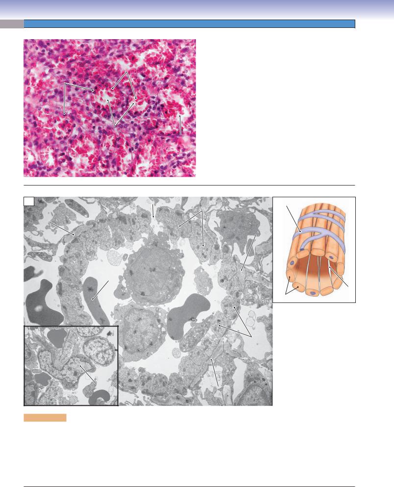
176 UNIT 3 ■ Organ Systems
TABLE 9 - 1 |
Blood Vessels |
|
|
Tunica Intima |
Tunica Media |
Types of |
|
Arteries and |
|
|
|
Veins |
|
|
|
Arterial System |
|
|
Large/elastic |
Endothelium; |
Abundant fenestrated |
(conducting) |
subendothelial |
elastic membranes; |
arteries |
layer (connective |
interspersed smooth |
|
|
tissue); IEL |
muscle cells; EEL |
Medium/ |
Endothelium; |
Abundant, thick layer |
muscular |
subendothelial |
of smooth muscle cells |
(distributing) |
layer (connective |
(6–40 layers of muscle |
arteries |
tissue); prominent |
cells); prominent EEL |
|
complete IEL |
in larger-sized medium |
|
|
arteries |
|
|
3–6 layers of smooth |
Small arteries |
Endothelium; thin |
|
subendothelial |
muscle cells; EEL |
|
layer; thin IEL |
absent |
|
|
1–2 layers of smooth |
Arterioles |
Endothelium; no |
|
subendothelial |
muscle cells; no EEL |
|
layer; IEL not |
|
|
prominent |
|
Venous System |
|
|
|
Endothelium; no |
Very thin; smooth |
Venules |
(postcapillary, |
subendothelial |
muscle cells not promi- |
collecting, |
layer; no valves |
nent |
and muscular |
|
|
venules) |
|
|
|
|
|
Small veins |
Endothelium; |
Thin; 1–3 layers |
|
subendothelial |
isolated smooth muscle |
|
layer; few valves |
cells |
Medium veins |
Endothelium; |
Thicker than small |
|
subendothelial |
vein; a few layers of |
|
layer; increased |
smooth muscle cells |
|
number of valves |
that usually do not |
|
|
form a distinct sheath |
|
|
Thinner than its tunica |
Large veins |
Endothelium; thick |
|
subendothelial |
adventitia layer; several |
|
layer; large valves; |
layers of circularly |
|
IEL may be |
arranged smooth |
|
present |
muscle cells but do not |
|
|
form a distinct sheath; |
|
|
some collagen fibers |
|
|
|
Tunica Adventitia |
Size |
Main Functions |
|
(Diameter) |
|
Thinner than |
>10 mm |
Conducting blood flow |
its tunica media |
|
from the heart to the |
(connective tissue); |
|
arterial system; being |
vasa vasorum present |
|
able to recoil (elastic |
|
|
membranes) to buffer |
|
|
pressure against arte- |
|
|
rial wall and conserve |
|
|
energy to force the blood |
|
|
forward, even while the |
|
|
ventricle is relaxing |
Thinner than |
10–0.5 mm |
Distributing blood |
its tunica media |
|
flow to small arteries |
(connective tissue); |
|
in various parts of |
vasa vasorum not |
|
body; adjusting the |
prominent |
|
rate of blood flow by |
|
|
vasoconstriction and |
|
|
vasodilation |
Thinner than its |
500–100 μm |
Distributing blood |
tunica media (loose |
|
to arterioles and to |
connective tissue); |
|
capillaries; participating |
no vasa vasorum |
|
in vasoconstriction |
|
|
and vasodilation to |
|
|
adjust the blood flow |
Very thin; a sheath of |
100–30 μm |
Leading blood to |
loose connective tissue |
|
the capillary beds; |
|
|
regulating resistance; |
|
|
controlling blood flow |
|
|
to capillaries |
Very thin; layer of |
10–100 μm |
Draining exchanged |
collagen fibers |
|
blood from capillaries |
|
|
to the small veins; |
|
|
promoting leukocyte |
|
|
migration from blood- |
|
|
stream to inflamed |
|
|
tissue; primary sites for |
|
|
inflammatory response |
Thin; connective |
0.1–1 mm |
Collecting blood flow |
tissue layer |
|
from venules to the |
|
|
medium veins |
Thicker than its |
1–10 mm |
Carrying blood to large |
tunica media; variable |
|
veins leading toward |
number and size of |
|
the heart; preventing |
longitudinal bundles of |
|
blood from backflow |
smooth muscle mixed |
|
|
with connective tissue |
|
|
Thickest layer in large |
>10 mm |
Transporting blood |
veins; composed of |
|
from the venous system |
many large bundles of |
|
back to the heart; |
longitudinal smooth |
|
forcing blood toward |
muscle; cardiac |
|
the heart by |
muscle may be present |
|
constriction of circu- |
near the atria |
|
lar and longitudinal |
|
|
smooth muscle |
CHAPTER 9 ■ Circulatory System |
177 |
The Lymphatic Vascular System
Figure 9-19A. Small lymphatic vessels, lymph node. H&E,
|
|
|
|
272; inset 767 |
|
|
|
Small |
The lymphatic vascular system is composed of lymphatic |
|
|
|
artery |
|
|
|
|
capillaries, lymphatic vessels, and lymphatic ducts, which col- |
|
|
|
Valve |
lect and drain interstitial fluid from the tissue into the large veins |
|
Small |
(for subclavian veins, see Chapter 10, “Lymphoid System,” |
|
artery |
|
Fig. 10-6). Lymph (fluid in the lymphatic system) contains lym- |
|
|
|
|
phocytes, immunoglobulins, plasma, foreign antigens, and other |
|
|
|
|
substances. Lymphatic vessels carry lymph through the lymph |
|
|
|
|
nodes along the lymphatic vessels. Lymph nodes filter lymph |
|
|
|
|
and expose lymphocytes to antigens as part of the immune |
Small |
|
Small |
|
response (see Chapter 10, “Lymphoid System”). After filtration, |
|
lymphatic |
|
the lymph is transported via large lymphatic vessels to lymphatic |
lymphatic vessel |
|
|
|
vessel |
|
ducts (the thoracic right lymphatic ducts) and finally enters the |
|
|
|
|
|
|
|
|
|
|
|
subclavian veins and becomes part of the blood plasma. An |
|
|
|
|
example of small lymphatic vessels is shown in the hilus of a |
|
|
|
|
lymph node. Small lymphatic vessels have large lumens and very |
|
|
|
|
thin walls, which are composed of a layer of endothelium and a |
|
|
|
|
little connective tissue with a few smooth muscle cells. |
|
|
|
|
|
Valve
Lymphatic
vessels
Smooth |
|
|
Smooth |
|
Coagulated |
muscle |
Lymphocytes |
muscle cells |
plasma |
cells |
|
Figure 9-19B. Large lymphatic vessels, lymph node. H&E,
136; inset 422
Large lymphatic vessels have thicker walls than small lymphatic vessels. Large lymphatic vessels are composed of connective tissues and multiple layers of smooth muscle cells. Contraction of smooth muscle cells helps to move the lymph forward. Large lymphatic vessels are structurally similar to small veins, except they have larger lumens and prominent valves. Valves are present in all sizes of lymphatic vessels. They prevent lymph from flowing backward. Lymphatic vessels are often distinguished by lumens that contain clusters of lymphocytes and coagulated plasma. Lymphatic vessels can be found in most of the tissues of the body but not in the central nervous system, the bone marrow, or the hard tissues.
CLINICAL CORRELATION
 Lymphatic vessels
 Lymphatic vessel
C
Figure 9-19C. Lymphangioma, Skin. H&E, 44
Lymphangioma is a congenital malformation of the lymphatic system that involves the skin and subcutaneous tissues. It occurs most often in the head and the neck. It is characterized by multiple clusters of translucent vesicles that contain lymph fluid varying from clear to pink to dark red. These vesicles are separated from the normal network of lymphatic vessels but have communications with the superficial lymph vesicles through vertical and dilated lymph channels. Skin lesions range from minute vesicles to cystic and cavernous spaces. Clinically, it appears as a raised, soft, spongy, and pinkish-white lesion. Surgical excision may be chosen if vital structures are involved or for cosmetic correction, but it has a high recurrence rate after surgery.

10 Lymphoid System
Introduction and Key Concepts for the Lymphoid System
Cells in the Lymphoid System
Figure 10-1A,B |
Lymphocytes |
Figure 10-2 |
A Representation of Types of Lymphocytes |
B Lymphocytes |
|
Figure 10-3 |
A Representation of B-Lymphocyte Maturation |
Synopsis 10-1 |
Characteristics of Types of Immunoglobulins |
T Lymphocytes |
|
Figure 10-4A |
A Representation of T-Lymphocyte Maturation |
Figure 10-4B |
A Representation of Helper T-Cell and Cytotoxic T-Cell Maturation Markers |
Figure 10-5A |
A Representation of Helper T-Lymphocyte Activation |
Figure 10-5B |
Clinical Correlation: HIV Infection |
Lymphoid Tissues and Lymphoid Organs
Figure 10-6 |
Overview of the Lymphoid Organs |
Figure 10-7 |
Orientation of Detailed Lymphoid Organ Illustrations |
Mucosa-Associated Lymphoid Tissue |
Figure 10-8A |
Pharyngeal Tonsil, MALT |
Figure 10-8B |
Palatine Tonsil, MALT |
Table 10-1 |
Tonsils |
Figure 10-9A |
Appendix, MALT |
Figure 10-9B |
Clinical Correlation: Diffuse Large B-Cell Lymphoma |
Figure 10-9C |
Clinical Correlation: Lymph Node, HIV Infection |
Lymph Nodes |
|
Figure 10-10 |
Overview of the Lymph Node |
Synopsis 10-2 |
Lymphoid Organs |


 normal cardiac muscle cells
 cardiac muscle


 Internal elastic lamina