


CHAPTER 3 ■ Epithelium and Glands |
27 |
Epithelium
Introduction and Key Concepts for Epithelium
Epithelium covers nearly all body surfaces. The basic functions of epithelial tissue are (1) protection of the body from abrasion and injury (e.g., skin and esophagus); (2) absorption of material from a lumen (e.g., tubules in kidney, small and large intestines); (3) transportation of material along a surface (e.g., cilia-mediated transport in the trachea); (4) secretion of mucus, hormones, and proteins (e.g., glands); (5) gas exchange (e.g., alveoli in the lung); and (6) lubrication between two surfaces (e.g., mesothelium of pleural cavity). Epithelium is an avascular tissue, which lacks a direct blood supply. Nutrients are delivered by diffusion from blood vessels in the neighboring connective tissue. Most epithelial tissues are renewed continuously.
do not form a brush border; these cells can be found in the distal and collecting tubules of the kidney. Simple cuboidal epithelium is mainly found lining most of the tubules in the kidney and in some excretory ducts of glands.
SIMPLE COLUMNAR EPITHELIUM is composed of one layer of columnar cells resting on the basement membrane (Fig. 3-7A,B). The cell’s height is greater than the width. The elongated ovoid nucleus is most often located in the basal region of the cell. The apical surface of this epithelium may reveal microvilli. Microvilli are often densely packed to form a brush border and function to increase the apical surface area of the cell to aid in absorption of fluid and other material from a lumen. Simple columnar epithelium can be found in the digestive tract, oviducts (fallopian tubes) in the female reproductive system, and ductuli efferentes testis of the male reproductive system.
Classification of Epithelial Tissues
Epithelium can be classified as simple or stratified based on the number of layers of cells. If there is a single layer of cells, it is referred to as simple epithelium. If there are two or more layers of cells, it is considered to be stratified epithelium. Epithelium is also classified according to the shape of the cells in the most superficial layer. If the surface cells are flattened in shape, it is called squamous epithelium. If surface cells are cuboidal in shape, it is called cuboidal epithelium. If the surface cells are tall, with their height much greater than their width, it is called columnar epithelium. If the surface cells change shape in response to stretching and relaxing, it is called transitional epithelium (urothelium). As described below, these terms may be variously combined to designate layers of cells and shapes forming the superficial layer of the epithelium. In some cases, the height of an epithelial cell represents the level of metabolic activity. For example, epithelial cells lining the thyroid follicle usually exhibit as simple cuboidal epithelium. However, when the follicle cells are in a high metabolic state, they form a simple columnar epithelium. By contrast, when the follicle cells are in a low metabolic state, they form a simple squamous epithelium.
SIMPLE SQUAMOUS EPITHELIUM is composed of one layer of uniform flat cells, which rest on the basement membrane (Figs. 3-2A to 3-4). Apical surfaces are smooth, and the width of the cells is greater than their height. The nuclei appear flattened and can easily be recognized following hematoxylin and eosin staining (H&E) because of the basophilia (affinity for blue stains) of the nucleic acids in the nuclei. This type of epithelium is found lining the posterior surface of the cornea; lining blood vessels and lymphatic vessels (where it is called endothelium); lining the surface of the body cavities, including the pericardial, pleural, and peritoneal cavities (where it is called mesothelium); and lining the alveoli of the lungs.
SIMPLE CUBOIDAL EPITHELIUM is composed of one layer of uniform cuboidal cells, which rest on the basement membrane (Figs. 3-5A,B and 3-6A,B). The cell’s height, width, and depth are roughly equal. Nuclei are centrally placed and spherical in shape. Some cuboidal cells have long and abundant microvilli, which form a brush border on their apical surfaces. Such cells are found in the proximal tubules of the kidney. Other cuboidal cells have few, short microvilli which
PSEUDOSTRATIFIED COLUMNAR EPITHELIUM is composed of one layer of nonuniform cells that vary in shape and height (Fig. 3-9A,B). Cells appear similar to stratified cells, but all cells are in contact with the basement membrane. In general, most cells are tall columnar cells, but there are also some short basal cells, some of which are stem cells. The most widespread type of pseudostratified columnar epithelium is found in the respiratory tract and has long fingerlike, motile structures called cilia on the apical surface of the cells. Cilia aid in the transport of material across the surface of epithelial cells. Pseudostratified columnar epithelium is often referred to as respiratory epithelium because it is found in the linings of the respiratory tract, including the nasal cavity, trachea, and primary bronchi.
STRATIFIED SQUAMOUS EPITHELIUM contains several layers of cells, with cells in the superficial layer being flattened. Only the deepest layer of cells is in contact with the basement membrane. This type of epithelium protects the body against injury, abrasion, dehydration, and infection. This epithelium may be keratinized or nonkeratinized, depending on functional demands (Figs. 3-13A,B and 3-14A,B). Keratinized stratified squamous epithelium is found in the skin. The top layers consist of either thick or thin keratinized cells (flattened, nonnucleated dead cells) that are filled with tonofilaments. The thickness of the stratified squamous epithelium varies from region to region.
Nonkeratinized stratified squamous epithelium is similar to keratinized squamous epithelium except that surface cells are nucleated instead of nonnucleated. This type of epithelium often covers wet surfaces and is found lining the oral cavity (soft palate, cheeks, and floor of the mouth), esophagus, vagina, and true vocal cords.
STRATIFIED CUBOIDAL EPITHELIUM is composed of two or three layers of cuboidal cells with the basal layer of cells often appearing nonuniform in distribution (Fig. 3-15A,B). It is mainly found lining large ducts of exocrine glands. The cells often have smooth apical surfaces and form barriers and ducts.
STRATIFIED COLUMNAR EPITHELIUM is also composed of two or three layers of cells. The top layer is columnar in shape and the basal layer is usually cuboidal in shape (Figs. 3-1B and 3-16A,B). This is not a common type of epithelium and has

28 UNIT 2 ■ Basic Tissues
a very limited distribution. Occasionally, it can be found in the |
Specializations of the Lateral Surface |
|
conjunctiva of the eye and in some large ducts of the exocrine |
(Lateral Domain) |
|
glands. |
||
The lateral surface of epithelial cells contains cell junctions and |
||
|
||
TRANSITIONAL EPITHELIUM is stratified epithelium, often |
cell adhesion molecules that are responsible for the cohesive |
|
nature of epithelial tissue. Intercellular connections of the epi- |
||
referred to as urothelium, which lines the excretory channels |
||
thelial cells include (1) tight junctions (zonula occludens), that |
||
leading from the kidney (renal calyces, ureters, bladder, and |
||
completely surround the apical cell borders to seal the underlying |
||
proximal segment of the urethra). It may contain four to six cell |
||
intercellular clefts from the outside environment (Fig. 3-8A,B); |
||
layers in the relaxed state. However, the histological appearance |
||
(2) adhering junctions (zonula adherens), found just beneath the |
||
of the epithelium can change when stretched (Fig. 3-17A–C). |
||
tight junction, also forming a bandlike junction surrounding |
||
In the empty bladder, the basal cells are mostly cuboidal and |
||
the entire cell and serving to attach adjacent cells (Fig. 3-8A,B); |
||
the middle layer is polygonal, although surface cells bulge into |
||
(3) desmosomes (macula adherens), located beneath the adher- |
||
the lumen. Surface cells are often described as “dome shaped” |
||
ing junctions, also assist in cell to cell attachment (Fig. 3-8A,B) |
||
and are called dome cells or umbrella cells; they contain extra |
||
(the junctional complex is composed of tight junction, adher- |
||
cell membrane material near the superficial (apical) surface. The |
||
ing junction, and desmosome); and (4) gap junctions, which are |
||
dome cells may contain two nuclei. In the stretched bladder, the |
||
communicating junctions, provide a low-resistance channel to |
||
thickness of the epithelium is much reduced, and surface cells, as |
||
permit passage of ions and small molecules between adjacent |
||
well as the intermediate cells, are extremely flattened. |
||
cells (Fig. 3-8B). Gap junctions are present not only in epithelial |
||
|
||
Specializations of the Apical Surface |
tissues, but they can also be found in many other tissues (smooth |
|
muscle, cardiac muscle, and nerve tissues) in the body. However, |
||
(Apical Domain) |
gap junctions are not present in skeletal muscle, blood cells, and |
|
Apical surfaces of the epithelium may reveal cilia, microvilli, and |
spermatozoa. |
|
|
||
stereocilia, depending on their function and location. (1) Cilia |
|
|
are elongated, motile structures that have a greater diameter |
Specializations of the Basal Surface |
|
and length than microvilli. The core of a cilium is composed |
||
(Basal Domain) |
||
of microtubules arranged in a consistent array of two central |
||
microtubules surrounded by a circle of nine peripheral pairs of |
Epithelial cells rest on a basement membrane, consisting of a |
|
microtubules (Figs. 3-9A to 3-11A). Cilia arise from electron- |
basal lamina and a reticular lamina, which provide an under- |
|
dense, cylindrical structures, called basal bodies, in the apical |
lying foundation for the cells (Figs. 3-2B and 3-8B). The term |
|
cytoplasm just below the cell membrane. There are many mito- |
“basement membrane” is used in light microscopy observation, |
|
chondria at the apical surface of cells with motile cilia. The |
although the basement membrane is often difficult to visualize |
|
function of cilia is to aid in the transport of material along the |
with the light microscope. The terms “basal lamina” and “retic- |
|
surface of epithelial cells. Cilia are present in the pseudostrati- |
ular lamina” are ultrastructural terms and refer to features that |
|
fied ciliated columnar epithelium in the respiratory tract and |
require electron microscopy to be seen (see Fig. 3-4). Epithelial |
|
ciliated simple columnar epithelium in the oviduct (fallopian |
cells produce their own basement membrane. Cells are anchored |
|
tube). (2) Microvilli are smaller than cilia; each has a core that |
to the basement membrane by hemidesmosomes, junctions that |
|
is composed of actin microfilaments. Microvilli are anchored |
connect the cells to the underlying basement membrane (Fig. |
|
to a network structure called the terminal web, which contains |
3-8B). Basal plasma membrane enfolding may also be present |
|
actin filaments to stabilize the microvillus (Figs. 3-7A,B and |
in some epithelial cells (e.g., salivary gland excretory duct epi- |
|
3-11B). These specialized structures increase apical surface |
thelium). This is a corrugation of the cell membrane in the basal |
|
area to aid in absorption. Microvilli are commonly seen in sim- |
(and sometimes lateral) regions of the cell, which increases cell |
|
ple columnar epithelium lining the small intestine and simple |
surface area and is involved in ion and fluid transport. There |
|
cuboidal epithelium lining the proximal tubules in the kidney. |
are many mitochondria in the vicinity of the plasma membrane |
|
(3) Stereocilia are long microvilli, which consist of actin micro- |
enfolding. These produce adenosine triphosphate (ATP) for |
|
filaments and help with absorption (Fig. 3-11C). They can |
active transport. The combination of the plasma membrane |
|
be found in the pseudostratified columnar epithelium in the |
enfolding and the concentration of mitochondria result in a |
|
epididymis and vas deferens of the male reproductive system. |
striated appearance in some of the epithelial cells. |

CHAPTER 3 ■ Epithelium and Glands |
|
|
|
29 |
Squamous |
Cuboidal |
Columnar |
Pseudostratified |
|
epithelium |
epithelium |
epithelium |
columnar epithelium |
|
A
D. Cui
Figure 3-1A. Overview of types of simple epithelia (one layer of epithelial cells).
Squamous |
Squamous |
|
|
Transitional |
epithelium |
epithelium |
Cuboidal |
Columnar |
epithelium |
(keratinized) |
(nonkeratinized) |
epithelium |
epithelium |
(relaxed) |
B
Figure 3-1B. Overview of types of stratified epithelia (two or more layers of epithelial cells).
Epithelium can be classified as simple or stratified based on the number of layers of cells. Epithelium is also classified according to the shape of the cells in the most superficial layer (Table 3-1).
TABLE 3 - 1 Classification of Epithelium
Shape of the Surface |
|
Simple Epithelium |
Stratified Epithelium |
Cells |
|
(Single Cell Layer) |
(Multiple Cell Layers) |
Flat |
|
Simple squamous epithelium |
Stratified squamous epithelium (keratinized) |
|
|
|
Stratified squamous epithelium (nonkeratinized) |
Cuboidal |
|
Simple cuboidal epithelium |
Stratified cuboidal epithelium |
Columnar |
|
Simple columnar epithelium |
Stratified columnar epithelium |
|
|
Pseudostratified columnar epithelium |
|
From dome to flat |
|
|
Transitional epithelium (relaxed) |
|
|
|
Transitional epithelium (distended) |
|
|
|
|

30 UNIT 2 ■ Basic Tissues
Simple Squamous Epithelium
A |
Lumen |
|
Mesothelial cells
 Mesothelium
B


 Mesothelial cell
  Basement membrane

 Connective tissue
D. Cui
Figure 3-2A. Simple squamous epithelium, oviduct.
H&E, 155; inset 310
Mesothelium is a term for the epithelial layer of the serous membranes (peritoneum, pleura, and pericardium) that line the body cavities and cover the organs that project into the cavities. The other component of these membranes is the loose connective tissue layer beneath the mesothelium. The membrane that lines the cavity wall constitutes the parietal layer, and the visceral layer
(serosa) covers the organs located within the cavity.
The mesothelium secretes a slippery lubricating fluid that allows organs to easily slide against and over each other or against the cavity wall (e.g., beating heart, expanding or contracting lungs, peristaltic activity of the intestine) without damage to the mesothelium.
Excessive fluid in a mesothelial-lined cavity is called an “effusion,” for example, a pleural effusion. Inflammation of the pleura is called pleurisy; of the peritoneum, it is called peritonitis; and of the pericardium, pericarditis. These may exist in conjunction with a variety of clinical conditions or diseases.
Figure 3-2B. A representation of a simple squamous epithelium lining a body cavity.
Mesothelial cells are flattened, generally pentagonal in shape, and form irregular borders with one another. The basement membrane is immediately adjacent to the mesothelium, which is barely visible with the light microscope. The basement membrane includes the basal lamina and an additional layer, the reticular lamina. The term “basal lamina” is used at the electron microscope level and includes the lamina densa and lamina lucida. Connective tissue is found beneath the basement membrane. The loose connective tissue beneath the basement membranes of mesothelia is the second component of the serous membranes lining the peritoneal, pleural, and pericardial cavities.
CLINICAL CORRELATION
C
|
Normal mesothelial cell |
|
Basement membrane |
D. Cui |
Connective tissue |
Tumor |
Fragmented |
cells |
basement membrane |
Figure 3-2C. Mesothelioma.
Mesothelioma (cancer of the mesothelium) is a neoplasm that arises from surfaces of the pleural and peritoneal cavities. It is only occasionally found in the pericardial mesothelium. This type of tumor is more commonly seen in individuals who either were exposed to asbestos or who smoke. Mesothelioma cells develop long, slender, and curved microvilli. These tumors may invade nearby tissues and organs. In general, cancer cells are those that can metastasize (spread) from their original site to distant parts of the body, and mesotheliomas are no exception. The lymphatic system is a common route through which metastasis may occur.

CHAPTER 3 ■ Epithelium and Glands
A
Lumen of the artery
Endothelium
Endothelial cells |
Tunica media |
|
|
|
of the artery wall |
Subendothelial
connective Internal tissue elastic
lamina
31
Figure 3-3A. Simple squamous epithelium, blood vessels. H&E, 219; inset 310
Tunica
intima Simple squamous epithelium lining the lumen surface of all types of blood vessels and lymphatic vessels is called an endothelium (sometimes a vascular endothelium). In this example, a single layer of squamous cells lines the inner layer, the tunica intima, of a medium artery. The endothelial cells are flattened and elongated, and they always rest on a thin basement membrane. Electron microscopy is required to see the ultrastructural features of the basement membrane. Endothelial cells of vessels sense changes in blood pressure, oxygen tension, and blood flow and respond to these changes by secreting substances, which have effects on the tone of vascular smooth muscle. Endothelial cells are also important in the control of blood coagulation; the endothelium produces von Willebrand factor that mediates platelet adhesion to collagen in subendothelial connective tissues at an injury site to stop bleeding. They also produce anticoagulant substances that prevent blood clotting and allow unobstructed flow of blood in normal conditions.
B
 Endothelial cell

 Basement membrane
 Subendothelial connective tissue

 Internal elastic lamina
D. Cui
Figure 3-3B. A representation of simple squamous epithelium, artery endothelium.
These endothelial cells are flattened and elongated, oriented parallel to the direction of blood flow, and rest on a basement membrane. The cells and basement membrane are linked by junctions called hemidesmosomes. Beneath the basement membrane is a subendothelial layer of connective tissue. The wavy structure is called the internal elastic lamina. The endothelium, subendothelial connective tissue, and the internal elastic lamina comprise the tunica intima.
CLINICAL CORRELATION
C |
Endothelial cell |
|
Basement membrane |
|
Internal elastic lamina |
D. Cui |
Cholesterol deposits |
|
Foam macrophage (foam cells) |
Fat |
Fragmented internal elastic lamina |
deposits |
|
Figure 3-3C. Atherosclerosis.
Atherosclerosis is the formation of deposits of yellowish plaques that contain cholesterol, lipoid material, and lipophages (macrophages). These deposits form the innermost layers of largeand medium-sized arteries. Of particular clinical significance are those plaques that form at the bifurcation of the common carotid artery into the internal and external carotid arteries and in the cerebral vessels. Although there are many possible causes of plaques, the more common are endothelial dysfunction, dyslipidemia, inflammatory and immunologic factors, and hypertension. As shown in this image, deposits of cholesterol and fatty material accumulate in the inner layers of the vessel resulting in damage to the vessel wall, including disruption of the endothelium. These deposits, when hardened, may occlude blood flow to distant tissues, and blood clots may form on exposed collagen in subendothelial connective tissue. Clot formation or dislodged pieces of plaque may result in vascular occlusion and stroke.
32 UNIT 2 ■ Basic Tissues
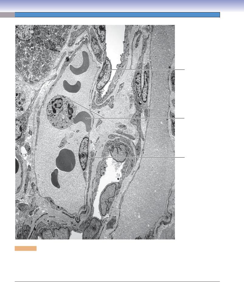
Lumen of thin segment
Simple squamous cell (lining epithelium
of loop of Henle)
Blood cells
Lumen of the blood vessel
 Endothelial cell
Basement membrane
Lumen of thin segment
Lumen of the blood vessel
Figure 3-4. Simple squamous epithelium in the medulla of the kidney. EM, 8,900
This view of the medulla of the kidney provides two examples of simple squamous epithelium. The endothelial cells of the vessels (vasa recta) are extremely thin except at the nucleus. Red blood cells, a neutrophil, and a platelet can be seen in the lumen of the blood vessel. The cells that form the renal tubules shown here are squamous, but they are not as thin as the endothelial cells. These tubules are thin segments of the loop of Henle. There is considerable diffusion of water between the vasa recta vessels and the renal tubules in the medulla.

CHAPTER 3 ■ Epithelium and Glands |
33 |
Simple Cuboidal Epithelium
A
Simple cuboidal epithelium
 Simple
cuboidal
epithelium Colloid
Figure 3-5A. Simple cuboidal epithelium, thyroid gland. H&E, 155; inset 537
Simple cuboidal epithelial cells that line the thyroid follicles of the thyroid gland are called follicular cells. Adjacent follicles are separated by a thin layer of connective tissue, containing occasional fibroblasts. Follicular cells are normally cuboidal in shape but become columnar when stimulated or in a state of hyperfunction (hyperthyroidism—excessively high levels of thyroid hormone secretion). At the other extreme, these cells may become flattened and squamous—like when they are inactive or in a state of hypofunction (hypothyroidism—excessively low levels of thyroid hormone secretion).
Follicular cells synthesize and release a precursor
(thyroglobulin) of thyroid hormones (T3 and T4) into the lumen of the follicle where it forms the colloid. Thyroid hormones are essential for normal brain and body development in infants and for the regulation of the metabolic rate in adults, and they affect the function of every organ system.
B
D. Cui
 Cuboidal cell
 Basement membrane
 Connective tissue
Figure 3-5B. A representation of simple cuboidal epithelium lining the thyroid follicle.
A layer of cuboidal cells rests on a thin basement membrane. The nuclei are round and located in the center of the cell. These cells have a large amount of rough endoplasmic reticulum (RER) and an elaborate Golgi complex in the cytoplasm, which reflects their high metabolic activity and production of secretory granules containing thyroglobulin. The thyroglobulin is secreted via exocytosis into the follicular lumen, where it is stored as a colloid. The apical (luminal) surface of these cuboidal cells is characterized by numerous short microvilli.
CLINICAL CORRELATION
C
 Active columnar cell
Basal membrane
|
Connective |
D. Cui |
tissue |
Hypothyroidism
D. Cui
Figure 3-5C. Hyperthyroidism.
Hyperthyroidism is a condition characterized by the overproduction of thyroid hormone. In this condition, the follicular cells have changed from cuboidal cells to become columnar cells as a result of their high activity. Symptoms include nervousness, irritability, increased heart rate (tachycardia), increased perspiration, difficulty sleeping, muscle weakness, warm moist skin, trembling hands, and hair loss. This disorder is seen most often in women 20 to 40 years age. Graves disease, the most common form of hyperthyroidism, results from antibodies in the blood that mimic thyroid-stimulating hormone, stimulating the thyroid to grow and secrete excessive thyroid hormone. Symptoms of thyroid hyperfunction also can be induced by excessive thyroid hormone medication.
At the other extreme, in hypothyroidism, there are very low levels of thyroid hormone secretion and follicular cells become flat, squamous cells; the patient may experience weight gain, somnolence, fatigue, and depression.

34 |
UNIT 2 ■ |
Basic Tissues |
|
|
|
|
|
|
Proximal tubule |
|
Figure 3-6A. |
Simple |
cuboidal |
epithelium, |
|
|
|
|
|
tubules of kidney. H&E, 155; inset 410 |
|||
|
|
|
Simple |
The appearance of this epithelium varies in different |
|||
|
|
L |
cuboidal |
||||
|
|
epithelium |
segments of the uriniferous tubules. For example, |
||||
|
|
|
|
in the proximal tubules, the cuboidal cells have |
|||
|
|
|
|
pink-stained cytoplasm and display numerous |
|||
|
Simple cuboidal |
L |
long microvilli on the apical surface. The microvilli |
||||
|
generally fill the lumen and may appear disrupted |
||||||
|
|
epithelium |
|||||
|
|
|
|
in histological specimens, but this is an artifact of |
|||
|
|
|
L |
postmortem deterioration. The acidophilia of the |
|||
|
Collecting duct |
cytoplasm of proximal tubule cells results partly |
|||||
|
|
from the numerous mitochondria that provide the |
|||||
|
|
|
|
||||
|
|
|
|
ATP necessary to power the ion pumps in the baso- |
|||
|
|
|
|
lateral membranes of these cells. The cuboidal cells |
|||
|
|
L |
|
of distal tubules have short and scanty microvilli. |
|||
|
|
|
They display less acidophilia than the proximal |
||||
|
|
|
|
tubules, although they do have mitochondria that |
|||
|
Simple cuboidal |
|
support ion pumps. The cuboidal cells forming |
||||
|
|
collecting tubules and ducts have few microvilli, |
|||||
|
A |
epithelium |
|
and the cytoplasm is less stained. A defect in renal |
|||
|
|
|
|
tubules can result in renal tubular acidosis (acido- |
|||
|
|
|
|
sis and electrolyte disturbance). (L, lumen.) |
|||
B
 Microvilli

 Cuboidal cell
 Basement membrane
 Connective tissue
D. Cui
Figure 3-6B. A representation of simple cuboidal epithelium of the proximal tubule in the kidney.
These simple cuboidal epithelial cells have round nuclei that are in the center of the cells. The apical surface of the cell exhibits abundant long microvilli, indicating absorption and secretion functions. Most of the water, sodium, chloride, amino acids, proteins, and glucose in the glomerular filtrate are reabsorbed by the cuboidal cells of the proximal tubules and transported into the underlying connective tissue.
CLINICAL CORRELATION
C |
Loss of microvilli |
|
Thickened/wrinkled |
|
basement membrane |
|
Distorted nucleus of cuboidal cell |

 Connective tissue
D. Cui
Enlarged Distorted squamous cell cuboidal cell
Figure 3-6C. Renal Fanconi Syndrome.
Renal Fanconi syndrome is mainly an impairment of proximal tubular function in the kidney resulting from an abnormality in the epithelial lining. The alteration of the epithelium can be caused by a variety of genetic defects (mostly in children) and by certain environmental factors. The result is that some substances that should be reabsorbed into the bloodstream are instead excreted in the urine. These substances include glucose, amino acids, phosphate, bicarbonate, and calcium. Their loss into the urine may lead to failure to thrive in children and decreased bone mineralization (rickets in children, osteomalacia in adults).
Some histological features typical of renal Fanconi syndrome are illustrated here. Epithelial cells become more squamous rather than cuboidal, nuclei are distorted, the basement membrane becomes wrinkled and thickened, and microvilli are reduced in number and length.
CHAPTER 3 ■ Epithelium and Glands |
35 |
Simple Columnar Epithelium
A
Simple |
Goblet |
columnar |
|
epithelium |
cell |
Brush border
Brush border
Simple columnar epithelium
Figure 3-7A. Simple columnar epithelium, small intestine. H&E, 155; inset 408
This is a section taken from the ileum of the small intestine. The apical surface of the columnar epithelium reveals a brush border, consisting of microvilli with a glycocalyx coating. Microvilli are fingerlike structures that increase the surface area of the apical membrane where absorption of nutrients occurs. The cells with seemingly empty cytoplasm are goblet cells, which are mucus-secreting cells interspersed among the simple columnar absorptive cells (enterocytes). The nuclei of the epithelial cells are elongated, “hot-dog” shaped, and located toward the basal end of the cells. Sometimes, the simple columnar epithelium appears to be multilayered because of the cutting angle, but only a single layer of cells actually attaches to the basement membrane. Simple columnar epithelium is typical of the lining of the digestive tract, and it is also found in the ov ducts, ductuli efferentes, and the ducts of some exocrine glands.
B
Microvilli |
Figure 3-7B. A representation of simple |
Terminal web |
columnar epithelium in the small intestine. |
|
 Columnar cell
 Basement membrane
Microvilli are anchored in the terminal web

 Connective tissue
D. Cui
D. Cui Terminal web
In the small intestine, microvilli enhance digestive and absorptive functions by increasing the area of the surface membrane of each columnar epithelial cell. This provides an expanded area of interface between the cell surface and the nutrients in the lumen. Each microvillus has a core that is composed of actin microfilaments anchored in a terminal web to stabilize the microvillus. Tall and slender columnar cells and the relationship of the terminal web are illustrated here. Individual microvilli, actin microfilaments, and actin filaments of the terminal web cannot be seen under light microscope.
CLINICAL CORRELATION
C
 Loss of microvilli

 Disrupted terminal web


 Columnar cell
 Basement membrane

 Lymphocyte in lamina propria
D. Cui
Intraepithelial lymphocyte
Figure 3-7C. Celiac (Coeliac) Disease.
Celiac (coeliac) disease is a disorder of the small intestine. Gluten, a substance found in wheat and barley, reacts with the lining of the small intestine (small bowel), leading to an attack by the immune system and damage to microvilli and villi. If left untreated, coeliac disease can lead to malabsorption, anemia, bone disease, and, rarely, some forms of cancer. The most important treatment is avoidance of all foods that contain gluten. Histologic features include blunting of villi, presence of lymphocytes among the epithelial cells (intraepithelial lymphocytes), and increased lymphocytes within the lamina propria (connective tissue).

36 UNIT 2 ■ Basic Tissues
A B
Intercellular space
Intramembrane protein 


 (sealing strands)


Tight junction (zonula occludens)
D. Cui
|
Terminal web |
Terminal web |
(actin filaments) |
(actin filaments) |
Adhering junction |
|
(zonula adherens) |
Transmembrane
link protein 
Intracellular |
|
line protein |
|
Actin |
|
filaments |
D. Cui |
|
Tonofilaments 
Desmosome  (macula adherens)
Transmembrane
proteins 
Intracellular 


 attachment plaque


Intermediate |
|
filaments |
D. Cui |
|
Figure 3-8A. Components of the junctional complex. EM, 70,000.
This is a view of parts of two adjacent epithelial cells that line the gallbladder. At the top of the field, parts of some microvilli can be seen at the luminal surfaces of the cells. The three types of cell junctions that typically compose the junctional complex can be distinguished. The zonula occludens junction has branching strands of fusion between the plasmalemma of the two cells. This effectively prevents movement of material across the epithelium by the passage between adjacent cells. The zonula adherens junction provides a collar of mechanical adhesion between neighboring cells. The cytoskeleton of each cell at the level of the zonula adherens consists predominantly of actin filaments in a network termed the terminal web. The third element of the junctional complex is an array of scattered desmosomes (maculae adherentes; singular, macula adherens). These provide mechanical adhesion between adjacent cells in spots. Cytokeratin filaments (tonofilaments) of the cytoskeleton are anchored in the attachment plaques on the two cytoplasmic surfaces of each desmosome.
Figure 3-8B. Simplified representation of components of the junctional complex.
Intercellular connections of the epithelial cells include (1) tight junctions (zonular occludens);
(2) adhering junctions (zonula adherens); (3) desmosomes (macula adherens); (4) gap junctions; and (5) hemidesmosomes. The junctional complex is composed of a tight junction, an adhering junction, and a desmosome. See the chapter introduction for detailed information.
Gap Junction
Cytoplasm 
Connexons


Cell |
|
membrane |
D. Cui |
Hemidesmosome
Cytoplasm
Lamina lucida  Lamina densal
Reticular lamina
D. Cui
CHAPTER 3 ■ Epithelium and Glands
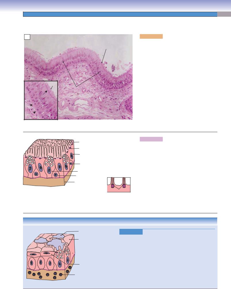
Pseudostratified Columnar Epithelium
A
Basal
bodies
Basement membrane
Cilia
Cilia
Pseudostratified
columnar
epithelium
37
Figure 3-9A. Pseudostratified ciliated columnar epithelium, trachea. H&E, 155; inset 247
The cells of pseudostratified ciliated columnar epithelium vary in shape and height, and their nuclei are staggered, giving the false impression of being arranged in two or three layers of cells. However, the basal aspect of each cell is in contact with the basement membrane. Most cells are tall and columnar, but there are also short basal cells, some of which are stem cells. The most widespread type of pseudostratified columnar epithelium is ciliated and is found lining the trachea and primary bronchi, the auditory tube, and part of the tympanic cavity. Nonciliated pseudostratified columnar epithelium is found throughout the epididymis and vas deferens in the male reproductive tract. Cilia on the apical surfaces of some cells are closely packed like bristles of a brush. The pink line indicated by the arrow (inset) is formed by the basal bodies, from which the cilia arise. The arrangement of the nuclei in pseudostratified columnar epithelium is more irregular than in stratified columnar epithelium.
B |
Cilia |
|
|
|
|
|
Basal body |
|
|
Ciliated columnar cell |
|
|
Basement membrane |
|
|
Goblet cell |
Cilia, basal body, |
|
Basal cell |
and microtubules |
D. Cui |
Connective |
|
|
tissue |
|
|
|
D. Cui |
|
|
Basal body |
Figure 3-9B. A representation of the pseudostratified ciliated columnar epithelium of the trachea.
Secretory goblet cells are interspersed among the ciliated columnar cells. Cilia are elongated, motile structures that are about 5 to 10 times longer than microvilli. The core of a cilium is composed of microtubules, which are inserted into basal bodies, electron-dense structures in the apical cytoplasm just below the cell membrane. The function of cilia is to aid in the transport of material along the surface of the cells, such as moving mucus and particulate matter out of the respiratory tract. Basal cells are short, located in the basal portion of the epithelium, and do not reach the lumen. The epithelium may appear to have more than one layer; in fact, all of its cells are in contact with the basement membrane.
CLINICAL CORRELATION
Mucus
C
Loss of cilia

 Remaining basal body

 Hyperplastic columnar cell
Squamous cell
Inflammatory cells in
the connective tissue
D. Cui
Figure 3-9C. Bronchitis.
Bronchitis is a disease marked by acute or chronic inflammation of the bronchial tubes (bronchi). The inflammation may be caused by infection (virus, bacteria) or by exposure to irritants (smoking or inhalation of chemical pollutants or dust.) Cigarette smoking is the leading cause of chronic bronchitis. The inflammatory process inhibits the characteristic activity of cilia, which is to trap and eliminate pollutants. Inflammation also increases the secretion of mucus. The inflamed area of the bronchial wall becomes swollen, and excess mucus may obstruct the airway. In chronic bronchitis, the surface epithelium may undergo hyperplasia and loss of cilia; the pseudostratified epithelium is often replaced by squamous epithelium. This process is called squamous metaplasia.

38 UNIT 2 ■ Basic Tissues
Cilia
Mucous secretory  granules
Basal bodies
Ciliated columnar cell
Goblet cell
Basal bodies
Figure 3-10. Respiratory epithelium, example of ciliated pseudostratified columnar epithelium. EM, 6,300; inset 11,500
This view of the respiratory epithelium includes only the apical half of the thickness of this pseudostratified columnar epithelium. Shown here are goblet cells and ciliated cells, the two most common of the approximately five cell types composing the respiratory epithelium. The goblet cells secrete mucus onto the surface of the epithelium. This mucus serves to trap airborne particles that have not been trapped by the nasal cavities. The ciliated cells convey the mucus with any captured material toward the larynx and the pharynx. Mucociliary escalator is the term sometimes used to refer to this system that protects the delicate respiratory structures of the lung from airborne microorganisms and foreign particulate matter.

CHAPTER 3 ■ Epithelium and Glands |
39 |
Three Apical Specializations of Epithelium
|
Cilia (motile) |
|
Microvilli (nonmotile) |
|
Stereocilia (nonmotile) |
|
A |
|
|
B |
|
C |
|
Cilia |
Basal |
Microvilli |
Terminal |
Stereocilia |
|
bodies |
web |
||||
|
|
|
Tight junction
Tight junction (zonula occludens)
Figure 3-11A. Cilia, basal body, and junctional complex. EM, 9,500; inset color photomicrograph 724
Cilia are 0.2 μm in diameter and 5 to 10 μm long, so they can be seen as individual structures with the light microscope. The core (axoneme) of each cilium is composed of microtubules and associated proteins, most notably the molecular motor dynein. The microtubules are arranged as nine peripheral doublets with two central singlets. Each cilium extends from a basal body just beneath the apical surface of the epithelial cell. Basal bodies also have microtubules as a major component. These form an orderly array of nine peripheral triplets with no central microtubules, an arrangement seen also in centrioles.
Figure 3-11B. Microvilli,terminalweb, and junctional complex. EM, 9,500; inset color photomicrograph 723
Microvilli of the intestinal epithelium are about 0.08 μm in diameter and 1 μm long, so they cannot be distinguished as individual structures with the light microscope, but the row of tightly packed microvilli can be seen as a brush border. The core of each microvillus contains a bundle of 6-nm actin filaments, which extend from the actin filaments that form the terminal web just beneath the apical surface of the cell.
Figure 3-11C. Stereocilia and junctional complex. EM, 9,500; inset color photomicrograph 565
Stereocilia are extremely long microvilli. Like ordinary microvilli, stereocilia are less than 0.1 μm in diameter, but they can attain lengths of 10 μm or more. Stereocilia are characteristic of the pseudostratified columnar epithelium of the ductus epididymis, which is the site of absorption of the large volumes of testicular fluid produced by the seminiferous tubules. The greatly expanded surface area afforded by the stereocilia probably contributes to this function.

40 |
UNIT 2 ■ |
Basic Tissues |
|
|
|
|
|
|
A |
|
Central |
Figure 3-12A. Cross section of cilia. |
|||
|
|
EM, 74,000 |
|
|
|
||
|
|
|
microtubule |
|
|
|
|
|
|
|
|
|
|
|
|
|
|
|
|
The structure of the axoneme (core) of a |
|||
|
Plasma |
|
Microtubules |
cilium can be readily appreciated from a |
|||
|
membrane |
|
|
high-magnification cross-sectional view. |
|||
|
|
|
|
The microtubules have an extremely |
|||
|
|
|
|
orderly, consistent arrangement |
into |
||
|
|
|
|
two separate, central microtubules sur- |
|||
|
|
|
|
rounded by nine sets of doublet micro- |
|||
|
Basal |
|
|
tubules. This is often termed the 9+2 |
|||
|
|
|
arrangement. |
|
|
|
|
|
body |
|
|
|
|
|
|
|
D. Cui |
|
|
|
|
|
|
|
B |
|
|
Figure 3-12B. |
Cross |
section |
of |
|
|
|
|
microvilli. EM, 74,000 |
|
|
|
|
|
|
Actin |
Many examples of columnar and |
|||
|
|
|
filaments |
||||
|
|
|
cuboidal epithelia comprise cells that |
||||
|
|
|
|
||||
|
Plasma |
|
|
bear microvilli, |
but absorptive |
cells |
|
|
membrane |
|
lining the small intestine provide the |
||||
|
|
|
|
||||
|
|
|
|
premier example of tightly packed |
|||
|
|
|
|
microvilli that provide a pronounced |
|||
|
|
|
|
increase in surface area of the plasma |
|||
|
|
|
|
membrane. Microvilli are less than half |
|||
|
Terminal web |
|
the diameter of cilia, and their cores |
||||
|
|
have a much simpler structure. In cross |
|||||
|
(actin filaments) |
|
section, the actin filaments that extend |
||||
|
|
|
|
||||
|
D. Cui |
|
|
from the terminal web into the microvilli |
|||
|
|
|
|
appear as a cluster of small dots. The |
|||
|
|
|
|
fuzzy coating on the membranes of the |
|||
|
|
|
|
microvilli is the glycocalyx. |
|
||
SYNOPSIS 3 - 1 Specialized Structures of the Epithelial Cell
■Apical surface (domain): Exposed to a luminal or external environment; site of primary function (absorption, protection, etc.).
■Microvilli, composed of actin microfilaments anchored to terminal web. Microvilli increase apical surface area to aid in absorption.
■Cilia, composed of microtubules, arise from basal bodies. Cilia aid in the transport of material across the surface of the epithelium.
■Stereocilia, unusually long microvilli that aid in absorption.
■Lateral surface (domain): Contains junctional complexes that connect cells to neighboring cells.
■Tight junctions (zonula occludens), specialized membrane proteins between the apical and the lateral surfaces of the cell. Surround the apical borders and serve as impermeable barriers.
■Adhering junctions (zonula adherens), beneath the tight junctions, form bandlike junctions, and link the cytoskeleton of one cell to neighboring cells. They provide mechanical stability of the cells.
■Desmosomes (macula adherens), spotlike junctions, which assist in cell-to-cell attachment.
■Gap junctions, communicating junctions that permit passage of ions and small molecules between neighboring cells.
■Basal surface (domain): Contains junctional complexes and basement membrane. Basolateral folds may also be present.
■Hemidesmosome, a junction (one half of a desmosome) connecting cells to the underlying basement membrane.
■Basement membrane, consists of basal lamina and reticular lamina, which provide an underlying sheet for epithelial cells.
■Basolateral folds, corrugations of the cell membrane in the lateral and basal regions of the cell, which increase cell surface area and are involved with ion and fluid transport.

CHAPTER 3 ■ Epithelium and Glands |
41 |
Stratified Squamous Epithelium (Keratinized)
A
Keratinized dead cells
Rete
Stratified ridge squamous
epithelium Epidermis (keratinized) of the skin
Dermal papilla |
|
|
Basement |
Dermis of the skin |
membrane |
|
|
(Connective tissue) |
Connective |
|
tissue |
Figure 3-13A. Stratified squamous epithelium, palm of the hand (thick skin). H&E, 78; inset 96
Thick skin (palms and soles) and thin skin (most other body surfaces) are covered by keratinized stratified squamous epithelium. Skin includes epidermis (stratified squamous epithelium) and dermis (connective tissue). The top layer of the keratinized stratified squamous epithelium consists of dead cells (corneocytes), which lack nuclei. This tough keratinized layer resists friction and is impermeable to water. The cells of outer layers of the epithelium are flattened, and the middle and most basal layers of cells are more polyhedral or cuboidal. Only the cells in the deepest layer are in contact with the basement membrane. Note that the interface between the epithelium and the underlying connective tissue is expanded by unique features, such as dermal papillae and rete ridges throughout most of the stratified squamous epithelium. The white dashed line indicates the depth of the epithelial layer (epidermis) and the boundaries of the dermal papilla and rete ridge.
B |
Keratinized dead cells |
(stratum corneum) |
|
|
Squamous |
|
cell |
|
Basal |
|
cuboidal |
|
cell |
|
Basement |
|
membrane |
D. Cui |
Connective tissue |
Figure 3-13B. Stratified squamous epithelium in thin skin. H&E, 207
Stratified squamous epithelium that covers thin skin is similar to that of thick skin, although its superficial keratinized layer (stratum corneum) is much thinner than in thick skin. Keratinized stratified squamous epithelium is composed of several layers of cells. The superficial layers are formed by dead cells whose nuclei and cytoplasm have been replaced with keratin. Under the keratinized layer is the squamous cell layer; these cells are flat. The intermediate layers contain cells that are polyhedral. The cells close to the basement membrane are cuboidal in shape and are called basal cells; they are stem cells that are continuously dividing and migrating from the basal layer toward the surface as they differentiate.
CLINICAL CORRELATION
|
Piled |
C |
parakeratotic |
scales |
Stratum |
Microabscesses |
corneum |
|
Stratum |
|
granulosum |
|
Stratum |
|
Spinosum |
Micropustules |
|
Inflammatory connective tissue
D. Cui |
Dilated capillary |
Figure 3-13C. Psoriasis.
Psoriasis is a common chronic inflammatory skin disease typically characterized by pinkto salmoncolored plaques with silver scales and sharp margins.
T lymphocyte–mediated immunologic reactions are believed to cause the clinical features. Symptoms and signs include itching, joint pain, nail pitting, and nail discoloration. Pathologic examinations reveal a thickened epidermis caused by increased epidermal cell turnover and extensive overlying parakeratotic scales. Neutrophils may migrate into the epidermis from dilated capillaries to form microabscesses (within the parakeratotic area of the stratum corneum layer of the epidermis) and micropustules (within the stratum granulosum and spinosum layers of the epidermis) as shown in this illustration.

42 UNIT 2 ■ Basic Tissues
Stratified Squamous Epithelium (Nonkeratinized)
A
Epithelium
|
|
|
sue |
|
|
e tis |
|
|
tiv |
|
|
nnec |
|
|
|
Co |
|
|
|
Stratified squamous epithelium (nonkeratinized)-keratinized)
Nucleated |
Epithelium |
squamous |
|
cells |
|
|
Connective |
|
tissue |
Figure 3-14A. Stratified squamous epithelium, esophagus. H&E, 78; inset 175
Stratified squamous epithelium (nonkeratinized) is usually wet on its surface and is found lining the mouth, oral pharynx, esophagus, true vocal cords, and vagina. Nonkeratinized stratified squamous epithelium is similar to keratinized squamous epithelium, but the flattened surface cells retain their nuclei, and there is no keratinization of these cells.
In some patients with long histories of gastroesophageal reflux and heartburn, the stratified squamous epithelium in the esophageal-stomach junction may be replaced by metaplastic columnar epithelium (see Fig. 3-14C). The dashed lines illustrate the depth of the epithelial layer.
B


 Squamous cell
Basement membrane
Basal cuboidal cell
 Connective tissue
D. Cui
Figure 3-14B. A representation of nonkeratinized stratified squamous epithelium.
This type of epithelium is formed by multiple layers of cells. The top surface layers are composed of flattened and nucleated live cells, which do not form keratin. Other general features of nonkeratinized stratified squamous epithelium are similar to keratinized squamous epithelium: The basal layer has cuboidal or low column-shaped cells in contact with a basement membrane, intermediate layer cells are polyhedral in shape, and nuclei become progressively flatter as the cells move toward the surface.
CLINICAL CORRELATION
C

 Metaplastic columnar cell
 Goblet cell


 Inflammatory cells
D. Cui 
Figure 3-14C. Barrett Syndrome (Barrett Esophagus). Barrett syndrome (Barrett esophagus) is a complication of chronic gastroesophageal reflux disease marked by metaplasia of the stratified squamous epithelium of the distal esophagus into a simple columnar epithelium as a response to prolonged reflux-induced injury. Patients with Barrett syndrome have a high risk of developing adenocarcinoma (cancer of the esophagus). This illustration shows the metaplastic columnar cells and goblet cells that have replaced the normal squamous epithelium and the inflammatory cells (mainly lymphocytes and plasma cells) infiltrating the connective tissue.

CHAPTER 3 ■ Epithelium and Glands |
43 |
Stratified Cuboidal Epithelium
A
Stratified cuboidal epithelium
Figure 3-15A. Stratified cuboidal epithelium, salivary gland. H&E, 175; inset 234
Stratified cuboidal epithelium lines the ducts of the salivary glands. This uncommon type of epithelium has a very limited distribution. It may be found forming the ducts of some large exocrine glands and sweat glands. It functions to form a conduit for the secretory products of the gland. This type of epithelium is composed usually of only two layers of cuboidal cells, with the basal layer of cells often appearing incomplete.
B

 Cuboidal cell
Basement membrane
 Cuboidal cell
Connective tissue
D. Cui
Figure 3-15B. A representation of stratified cuboidal epithelium in the duct of a salivary gland.
Stratified cuboidal epithelium usually has only two, occasionally three, layers of cuboidal cells. The top layer is composed of uniform cuboidal cells, whereas the basal cells sometimes appear to form an incomplete layer (Fig. 3-15A). Cells in stratified cuboidal epithelium often have smooth apical surfaces, and nuclei are centrally located.
CLINICAL CORRELATION
C


 Stone in lumen (calcium)
 Distorted cuboidal cell


 Inflammatory cells
D. Cui &L Lynch 


Figure 3-15C. Salivary Gland Swelling.
Salivary gland swelling with inflammation (sialadenitis) is a clinical condition that can result from blockage of a duct or ducts, so that saliva is not able to exit into the mouth. This causes the saliva to back up inside the duct, resulting in gland swelling. The patient will feel pain when chewing food. The most common cause of blockage is a salivary stone (calculus), which forms from salts contained in the saliva. A blocked duct and gland filled with stagnant saliva may become infected with bacteria. A typical symptom of a blocked salivary duct is swelling that worsens just before mealtime. Sometimes, a small stone may be ejected into the mouth without medical intervention. A dentist may be able to push the stone out by pressing on the side of the obstructed duct. Removal of a stone may require surgery or lithotripsy treatment by focused, high-intensity acoustic pulses.

44 UNIT 2 ■ Basic Tissues
Stratified Columnar Epithelium
A
Stratified columnar epithelium
Connective
tissue
Stratified columnar cells
Figure 3-16A. Stratified columnar epithelium, eyelid. H&E, 155; inset 295
Stratified columnar epithelium can be found lining the palpebral conjunctiva of the eyelid. The anterior surface of the eyelid is covered by keratinized stratified squamous epithelium (epidermis of thin skin); the posterior surface of the eyelid, which is in contact with the surface of the eyeball, is lined by stratified columnar epithelium as demonstrated here. The basal cells are cuboidal in shape, and the surface layer cells are low columnar in shape (only slightly taller than wide). The conjunctiva has a smooth surface that is kept moist and lubricated by tears and a mucinous substance in the normal condition. The arrowheads point to columnar cells of the surface layer of the epithelium (inset).
B
 Columnar cell
 Basement membrane
 Cuboidal cell

 Connective tissue
D. Cui
Figure 3-16B. A representation of the stratified columnar epithelium lining the conjunctiva of the eye.
Stratified columnar epithelium usually has two or three layers; the top layer is made up of columnar cells, and the basal layer normally consists of cuboidal cells. Stratified columnar epithelium is not a common type of epithelium and is found in only a few places in the body, for example, the larger ducts of some exocrine glands and the lining of the palpebral conjunctiva of the eyelid.
CLINICAL CORRELATION
C

 Granulated surface


 Inclusion body
|
Lymphocyte |
D. Cui &L Lynch |
Macrophage |
Figure 3-16C. Trachoma.
Trachoma is a chronic contagious conjunctivitis (eye disease) characterized by inflammatory granulation on the conjunctival epithelium surface caused by the bacteria Chlamydia trachomatis. This form of “pink eye” often presents with bilateral keratoconjunctivitis with symptoms of tearing, discharge, photophobia, pain, and swelling of the eyelids. It can cause eyelid deformities and turned-in eyelashes that scrape against the cornea. If left untreated, ulceration and infection of the cornea may occur. Trachoma can even cause loss of vision if scarring occurs on the central part of the cornea. Lymphocytes and macrophages invade underlying connective tissue as part of the inflammatory response. The epithelial hyperplasia and inclusion bodies in the epithelial cells are illustrated here.
CHAPTER 3 ■ Epithelium and Glands |
45 |
Transitional Epithelium (Stratified Epithelium)
A
Dome shaped surface cells
Connective |
Lumen |
tissue |
Transitional epithelium
Connective
tissue
Figure 3-17A. Transitional epithelium, urinary bladder. H&E, 155; inset 250
Transitional epithelium has the special characteristic of being able to change shape to accommodate a volume change in the organ it lines. In the relaxed state, transitional epithelium contains four to six cell layers, and each surface cell appears dome shaped, often containing two nuclei (these cells are “binucleate”). This picture illustrates transitional epithelium in a relaxed state (in most slides, this tissue is unstretched and the surface cells appear dome shaped). The lumen of the bladder appears as a white space. The transitional epithelium lining the urinary tract including the bladder, ureter, and major calyces of the kidney is also referred to as urothelium. The black dashed line indicates the thickness of the epithelium.
B
Dome-shaped cell
Basement membrane
Basal cuboidal cell
Connective tissue
D. Cui
Figure 3-17B. Transitional epithelium (relaxed).
The relaxed, normal transitional epithelium is composed of four to six layers of cells. The cells located basally are smaller, low columnar or cuboidal cells. By contrast, the cells located in the most superficial layer are larger and exhibit a rounded, dome shape that bulges into the lumen.
C
Flattened top cell
Basement membrane
Connective tissue
D. Cui
Figure 3-17C. Transitional epithelium (distended).
A presentation of transitional epithelium in the distended state is shown. These cells change shape according to the degree of distention of the bladder. When the transitional epithelium is stretched, the top dome-shaped cells become flattened squamous cells and the epithelium becomes thinner.
SYNOPSIS 3 - 2 Pathological Terms for Epithelial Tissue
■Metastasis: The spread of a malignant neoplasm from its site of origin to a remote site, usually through blood and lymphatic vessels (Fig. 3-2C).
■Dyslipidemia: A general term describing a disorder of lipoprotein metabolism causing abnormal amounts of lipids and lipoproteins in the blood; certain dyslipidemias constitute a major risk factor for the development of atherosclerosis, such as hypercholesterolemia (Fig. 3-3C).
■Osteomalacia: Abnormal bone mineralization producing weak, soft bones; may be caused by vitamin D deficiency or kidney disorders, including renal Fanconi syndrome (Fig. 3-6C).
■Metaplasia: The reversible process by which one mature cell type changes into another mature cell type, as in squamous metaplasia of respiratory or glandular epithelia (Figs. 3-9C and 3-14C).
■Microabscess: Collection of neutrophils and neutrophil debris within the parakeratotic scale in the skin disease psoriasis (Fig. 3-13C).
■Micropustule: Collection of neutrophils within the epidermis, abutting the parakeratotic scale in the skin disease psoriasis (Fig. 3-13C).
■Parakeratosis: Persistence of the nuclei of keratinocytes into the stratum corneum of the skin or mucous membranes; parakeratotic scales containing neutrophils are seen in the skin disease psoriasis (Fig. 3-13C).

46 UNIT 2 ■ Basic Tissues
TABLE 3 - 2 |
|
Epithelium |
|
|
|
|
Types of |
|
Number of |
Type of Cells in the |
Apical |
Main Locations |
Main Functions |
Epithelia |
|
Layers |
Epithelium |
Surface |
(Lining) |
|
Simple |
|
One |
Flattened, squamous |
Smooth |
Cornea, blood, and |
Fluid transport, |
|
||||||
squamous |
|
|
epithelial cells |
|
lymphatic vessels— |
lubrication, and |
epithelium |
|
|
|
|
endothelium; surface |
exchange |
|
|
|
|
|
of body cavities— |
|
|
|
|
|
|
mesothelium (pleural, |
|
|
|
|
|
|
pericardial, peritoneal); |
|
|
|
|
|
|
alveoli in the lung |
|
Simple |
|
One |
Cuboidal epithelial cells |
Smooth/short |
Kidney tubules, thyroid |
Absorption, |
|
||||||
cuboidal |
|
|
(height equal to width) |
microvilli; |
follicles; small ducts |
secretion, and |
epithelium |
|
|
|
long microvilli |
of exocrine glands and |
transportation |
|
|
|
|
depending on |
surface of ovary |
|
|
|
|
|
location |
|
|
Simple |
|
One |
Absorptive columnar cells |
Mostly |
Most of digestive |
Secretion, |
|
||||||
columnar |
|
|
and secretary cells, such as |
microvilli; |
tract and gallbladder; |
absorption, |
epithelium |
|
|
goblet cells |
cilia in some |
oviducts and ductuli |
protection, and |
|
|
|
|
locations |
efferentes |
transportation |
Pseudostratified |
|
One |
Ciliated columnar cells, |
Mostly cilia; |
Most of respiratory |
Secretion, |
|
||||||
columnar |
|
|
goblet cells, and short basal |
stereocilia |
tract; ductus deferens |
transportation, |
epithelium |
|
|
cells not reaching lumen; all |
in some |
and epididymis |
and absorption |
|
|
|
cells rest on the basement |
locations |
|
|
|
|
|
membrane |
|
|
|
Stratified |
|
Several |
Flattened surface cells, |
Keratinized |
Epidermis of the skin; |
Protection |
|
||||||
squamous |
|
|
polygonal cells in the |
or nonkerati- |
oral cavity, epiglottis, |
(barrier) |
epithelium |
|
|
middle layers, and cuboidal |
nized surface |
and esophagus; vagina |
|
|
|
|
cells in basal layer |
layer |
|
|
Stratified |
|
Two to three |
Cuboidal cells |
Mostly |
Large ducts of exocrine |
Transportation |
cuboidal |
|
|
|
smooth |
glands and ducts |
|
epithelium |
|
|
|
|
of sweat gland (not |
|
|
|
|
|
|
common type) |
|
Stratified |
|
Two to three |
Low columnar surface cells |
Smooth |
Large ducts of exocrine |
Transportation |
|
||||||
columnar |
|
|
and cuboidal basal cells |
|
glands; conjunctiva of |
and protection |
epithelium |
|
|
|
|
the eye (not common |
|
|
|
|
|
|
type) |
|
Transitional |
|
Four to six layers |
Dome-shaped surface cells |
Smooth |
Urinary tract |
Transportation |
|
||||||
epithelium |
|
(relaxed); two |
(relaxed), polygonal in the |
|
|
and protection |
|
|
to three layers |
middle layer, cuboidal cells |
|
|
(distensible |
|
|
(distended) |
in the basal layer |
|
|
property) |
|
|
|
|
|
|
|
SYNOPSIS 3 - 3 Functions of Epithelial Tissue
■Promotes gliding between two surfaces (mesothelium of pleural cavity [Fig. 3-2A,B]).
■Senses changes in blood pressure, oxygen tension, and blood flow and controls blood coagulation (endothelium of blood vessels [Fig. 3-3A,B]).
■Pumps the excess fluid out of the stroma and keeps the cornea clear (simple squamous epithelium in cornea).
■Mediates gas exchange (type 1 pneumocytes and simple squamous epithelium in the alveoli of the lung).
■Absorbs material from a lumen (simple cuboidal epithelium in kidney and simple columnar epithelium in small and large intestines [Figs. 3-6A,B and 3-7A,B]).
■Transports material along a surface (pseudostratified ciliated columnar epithelium in the respiratory tract [Fig. 3-9A,B]).
■Provides conduit for fluid (simple and stratified cuboidal and columnar epithelia forming ducts of some large exocrine glands [Fig. 3-15A,B]).
■Protects the body from abrasion and injury (stratified squamous epithelium in the skin and esophagus [Figs. 3-13A,B and 3-14A,B]).
■Becomes highly distensible when the bladder is filled with urine and the tissue is stretched (transitional epithelium in bladder [Fig. 3-17A–C]).
■Secretes mucus, hormones, and proteins (secretory epithelium, glands [Figs. 3-18 and 3-19]).