

- •Dedication
- •Preface
- •Acknowledgments
- •Figure Credits
- •Expert Consultants and Reviewers
- •Contents
- •Descriptive Terms for Normal Cells
- •Descriptive Terms for Abnormal Cells and Tissues
- •Epithelium
- •Glands
- •Introduction and Key Concepts for Connective Tissue
- •Cartilage
- •Bone
- •Introduction and Key Concepts for the Nervous System
- •Peripheral Blood Cells
- •Hemopoiesis
- •Introduction and Key Concepts for the Circulatory System
- •The Cardiovascular System
- •Introduction and Key Concepts for the Lymphoid System
- •Cells in the Lymphoid System
- •Introduction and Key Concepts for the Respiratory System
- •Conducting Portion
- •Respiratory Portion
- •Introduction and Key Concepts for the Urinary System
- •Introduction and Key Concepts for the Integumentary System
- •Oral Mucosa
- •Teeth
- •Introduction and Key Concepts for the Digestive Tract
- •Introduction and Key Concepts for the Endocrine System
- •Introduction and Key Concepts for the Male Reproductive System
- •Introduction and Key Concepts for the Female Reproductive System
- •Introduction and Key Concepts for the Eye
- •Introduction and Key Concepts for the Ear
- •Introduction
- •Preservation versus Fixation
- •Fixatives and Methods of Fixation
- •Sectioning and Mounting
- •Staining
- •Index

410 UNIT 3 ■ Organ Systems
Figure 21-11C |
Clinical Correlation: Otitis Media |
Figure 21-12A |
Clinical Correlation: Vestibular Schwannoma |
Figure 21-12B |
Clinical Correlation: CT and MRI of Inner Ear Structures |
Synopsis 21-1 |
Pathological and Clinical Terms for the Ear |
Introduction and Key Concepts for the Ear
The ear is a complex structure that serves two important sensory functions, hearing (through the auditory system) and balance (through the vestibular system). Sensory receptor organs that serve the two functions are supplied by two distinct branches of cranial nerve (CN) VIII, the acoustic branch and the vestibular branch. The ear can be divided into three general regions, the outer ear, middle ear, and inner ear (Fig. 21-1A).
(1) The outer ear consists of a pinna (auricle), an irregularly shaped structure with a core of cartilage covered on both sides by thin skin, and an external auditory meatus that conducts sound to the middle ear. (2) The middle ear includes the tympanic membrane, the tympanic cavity containing the ossicles, and the auditory tube (Fig. 21-1A,B). The tympanic membrane, the landmark between the outer ear and middle ear, covers the medial end of the external auditory meatus and converts sound waves in the air to mechanical vibrations. The tympanic cavity is an air-filled space that contains the ossicles, three tiny bones that conduct the mechanical vibrations of the tympanic membrane to the oval window of the cochlea. The tympanic cavity is connected to the nasopharynx by the auditory tube (eustachian tube), thereby allowing the air pressure on each side of the tympanic membrane to be equalized when the ambient air pressure changes (e.g., by changes in altitude). (3) The inner ear consists of structures contained within the bony labyrinth, a system of tunnels and cavities in the petrous portion of the temporal bone, the hardest bone in the body (Figs. 21-2 and 21-3A). The structures include the cochlear labyrinth or cochlea (Latin for “snail shell”), which subserves hearing. It contains a spiral, fluid-filled tunnel within which a membranous tube, the cochlear duct, is suspended (Fig. 21-3A,B). The sensory receptors that detect sound are located in a strip of specialized epithelium, the organ of Corti (spiral organ), in the cochlear duct. The vestibular labyrinth consists of a complex group of fluid-filled tunnels and cavities in the temporal bone that contain a group of interconnected membranous structures, the semicircular ducts, utricle, and saccule (Figs. 21-2 and 21-3A). The sensory receptors that are involved in balance are located in specialized regions of the semicircular ducts (rotation) and in the utricle and saccule (static head position and acceleration).
Auditory System
Sound is normally produced by compression and rarefaction waves in air, of various frequencies, that impinge upon the tympanic membrane where they are converted into mechanical vibrations in the ossicles. The mechanical vibrations, in turn, are transferred to the fluid of the vestibule at the oval window (Fig. 21-2). This fluid is perilymph, a sodium-rich fluid that is similar in composition to cerebrospinal fluid and extracellular fluid. The resulting vibrations, or pressure waves, in
the perilymph spread into the scala vestibuli of the cochlear labyrinth and act upon mechanoreceptors in the cochlear duct to produce the sensation of hearing (Fig. 21-6A). The cochlear duct is a membranous tube, triangular in cross section, that coils inside the spiral tunnel in the cochlea (Fig. 21-3A,B). It is suspended within the cochlear labyrinth so that it divides the labyrinth into two tunnels, the scala vestibuli (above) and the scala tympani (below), which are connected with each other by a small opening, the helicotrema (Figs. 21-4A,B and 21-6A). The cochlear duct encloses the scala media, a space containing endolymph, a potassium-rich fluid similar in composition to intracellular fluid. The scala media is bounded above by the vestibular membrane, below by the basilar membrane, and externally by the spiral ligament and stria vascularis (Fig. 21-5). The sensory receptors for hearing are specialized epithelial cells (hair cells) in the organ of Corti. The hair cells get their name from clumps of stereocilia that project from their apical surfaces and contact an overlying gelatinous structure, the tectorial membrane. The organ of Corti sits on the basilar membrane and extends its entire length, from the base of the cochlea to the apex. It contains a single row of inner hair cells and three or four rows of outer hair cells (Figs. 21-5 and 21-7A). In humans, there are about 3,500 inner hair cells and 12,000 outer hair cells, yet 95% of the afferent axons in the auditory nerve contact only inner hair cells. The primary function of the inner hair cells appears to be basic frequency and loudness discrimination, whereas the outer hair cells appear to be primarily concerned with the fine-tuning of frequency discrimination in the cochlea (Figs. 21-6C and 21-7A,B). When sound waves cause pressure waves to occur in the scala vestibuli and scala media, the basilar membrane vibrates up and down, and a shearing force is generated between the surface of the organ of Corti and the tectorial membrane. The shearing force bends the stereocilia of the hair cells, leading to the release of neurotransmitters by the hair cells and the initiation of action potentials in auditory nerve axons (Fig. 21-6A,B).
Vestibular System
The sense of balance is critical to our ability to walk, run, jump, or even just stand still with eyes closed. One important source of neural signals that aid in controlling such behaviors is the peripheral vestibular apparatus. This includes the vestibular labyrinth, consisting of the vestibule, a cavity within the temporal bone, and three semicircular canals, curved tunnels that connect with the vestibule (Fig. 21-2). One canal is in approximately the horizontal plane; the other two are in approximately the vertical plane and at right angles to each other. These cavities in the bone are filled with perilymph. Floating within the vestibule are two membranous saclike structures, the utricle and saccule. Within each of the semicircular canals is a membranous tube called a semicircular duct, which joins the utricle at each of its ends (Figs. 21-2 and 21-3A). The utricle, saccule, and semicircular ducts contain endolymph. The semicircular ducts detect

CHAPTER 21 ■ Ear |
411 |
rotational movements of the head. Each duct has an enlargement at one end where it joins the utricle. This swelling is called the ampulla and contains the sensory receptors that are stimulated by rotational movements (Fig. 21-9A). A short wall of connective tissue, the crista ampullaris, extends across part of each ampulla. Hair cells, similar to those of the organ of Corti, cover the upper surface of the crista. Their stereocilia and kinocilia are embedded in a gel-like structure, the cupula, which blocks the ampulla. When the head turns, the inertia of the endolymph in the semicircular ducts causes the fluid to push against the cupula and deflect the cilia of the hair cells, thereby initiating action potentials in axons in the vestibular nerve (Figs. 21-8B
and 21-9A,B). Vestibular hair cells are also clustered in a small region of the utricle, the macula utriculi (Figs. 21-8B and 21-10A,B). The stereocilia and kinocilia of these hair cells are embedded in a gelatinous structure, the otolithic membrane (Fig. 21-10A). Thousands of tiny calcium carbonate crystals, otoconia, are clustered on the surface of the otolithic membrane (Fig. 21-10A–C). These crystals are heavier than the surrounding endolymph. Gravity or linear acceleration, therefore, exerts a force on the otoconia, causing the underlying cilia to be deflected and consequently sending a neural signal to the central nervous system (CNS) related to head position or acceleration. The saccule contains a similar region, the macula sacculi.
412 |
UNIT 3 ■ |
Organ Systems |
|
|
|
A |
Outer |
Middle |
Inner |
|
|
ear |
ear |
ear |
Semicircular canals
Temporal bone
Vestibular nerve
Malleus |
Facial nerve |
Pinna
 Cochlear nerve
External |
auditory |
meatus |
|
||
|
|
Vestibule
Cochlea
Tympanic membrane |
|
|
||
|
Incus |
|
|
|
Cartilage |
Tympanic |
Stapes |
|
|
(in oval |
Round |
|||
|
cavity |
|||
|
window) |
|||
|
|
window |
||
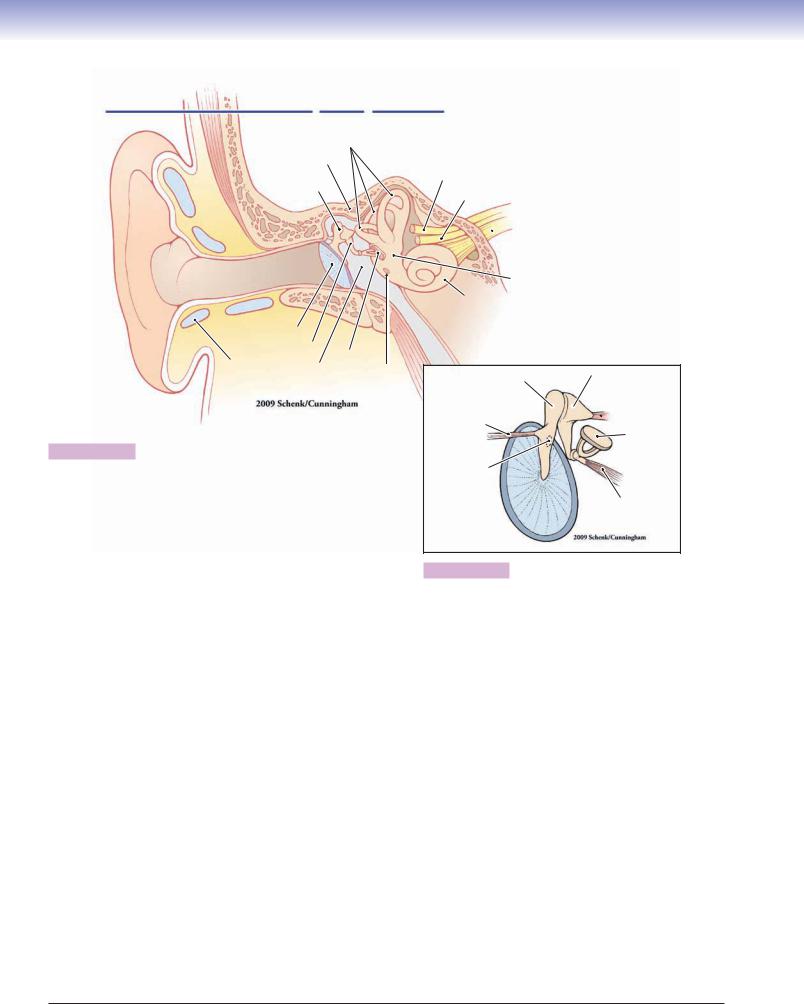
Figure 21-1A. Overview of the ear: Outer, middle, and inner ear.
The ear is divided into three regions: the outer ear, middle ear, and inner ear. The outer ear includes the pinna and the external auditory meatus. The pinna (or auricle) is an irregularly shaped structure of elastic cartilage covered by a layer of perichondrium (connective tissue) and thin skin. One important function of the pinna is to selectively filter higher frequency sound waves and, therefore, aid in spatial localization of sounds in the environment. The external auditory meatus is a tunnel that carries sound waves to the tympanic membrane, where the compressions and rarefactions of air are converted into mechanical vibrations. The outer third of the meatus is lined with a continuation of the cartilage of the pinna and a continuation of the perichondrium and thin skin that covers the pinna. In the inner two thirds of the meatus, the skin adheres directly to the periosteum of the temporal bone. The middle ear is an air-filled cavity (tympanic cavity) that is separated from the outer ear by the tympanic membrane. It contains three tiny bones, the ossicles, which are the smallest bones in the body. These ossicles transfer sound-induced movement of the tympanic membrane to the fluid contained in the cochlea, where the vibrations are transduced into nerve impulses. The middle ear is connected to the posterior region of the nasopharynx by the auditory tube (eustachian tube), which allows the equalization of air pressure on each side of the tympanic membrane. The inner ear contains the cochlea and the vestibular apparatus. The cochlea, the snail shell–shaped organ of hearing, includes a membranous tube lying within a fluid-filled tunnel in the temporal bone (see Fig. 21-2). Auditory hair cells in the cochlea are excited by vibratory movements of the fluid and generate action potentials in auditory nerve fibers. The vestibular apparatus is the body’s sensory organ for balance and consists of membranous structures contained within the three semicircular canals and the vestibule as well as some accessory structures (see Fig. 21-2). Vestibular hair cells within the semicircular ducts
(inside the semicircular canals) detect rotational movement of the head in three dimensions. Vestibular hair cells within the utricle and saccule (inside the vestibule) detect static head position and linear acceleration in the horizontal and vertical planes, respectively. The auditory and vestibular branches of CN VIII innervate these structures.
 Auditory tube
Malleus |
Incus |
Posterior incudal Anterior malleal 

 ligament
ligament
|
|
Stapes |
|
Insertion |
|
|
|
of tensor |
|
|
|
tympani |
Tympanic |
Stapedius |
|
muscle |
|||
membrane |
muscle |
||
|
B
Figure 21-1B. Middle ear structures.
The tympanic membrane (viewed here from its medial side) is a thin, semitransparent cone-shaped sheet of collagenous fibers and fibroblasts, covered on the outer side by a very thin layer of skin and on the inner side by the mucosa that lines the rest of the tympanic cavity. The first of the ossicles, the malleus (hammer), is attached to the upper half of the tympanic membrane, the incus (anvil) is attached to the malleus by a saddle-shaped synovial joint, and the stapes (stirrup) is attached to the incus by a ball- and-socket synovial joint. The footplate of the stapes is attached to the oval window of the vestibule. When sound waves cause the tympanic membrane to vibrate, the chain of ossicles rotates about the anterior malleal and posterior incudal ligaments, causing the footplate of the stapes to rock on the oval window and therefore producing waves of compression in the fluid that fills the bony labyrinth. This lever arrangement increases the pressure on the oval window approximately 20-fold compared to the air pressure on the tympanic membrane. The tensor tympani muscle contracts during very loud sounds, reducing the movement of the ossicles and the oval window.
CHAPTER 21 ■ |
Ear |
|
413 |
|
Endolymphatic sac |
|
|
Bone |
|
|
|
Semicircular |
|
Brain tissue |
|
canal |
|
||
|
Endo- |
|
|
|
lymphatic |
|
|
Semicircular |
duct |
|
|
ducts |
|
|
Subarachnoid space |
|
Ampulla |
|
|
|
|
Dura mater |
|
|
Crista |
|
|
|
|
|
|
|
|
|
Perilymphatic duct |
|
|
|
(cochlear duct) |
|
|
Vestibule |
|
|
Utricle |
Cochlea |
Scala vestibuli |
|
|
Maculae |
|
|
|
|
|
|
Saccule |
|
Cochlear duct |
|
|
(scala media inside) |
|
|
|
|
|
Oval window |
|
Scala tympani |
|
|
Vestibular |
||
|
|
|
|
Mastoid |
|
|
(Reissner) |
|
|
membrane |
|
cavities |
|
|
|
auditory |
|
Bone |
|
Round |
Scala vestibuli |
||
External |
|
window |
(perilymph) |
|
|
|
|
meatus |
Tympanic |
|
|
|
membrane |
|
|
Auditory |
Osseous |
|
(Eustachian) |
Scala tympani |
|
tube |
spiral |
|
|
lamina |
(perilymph) |
Scala media (endolymph)
 Organ of Corti
(spiral organ)
Basilar membrane
Cross section of cochlea
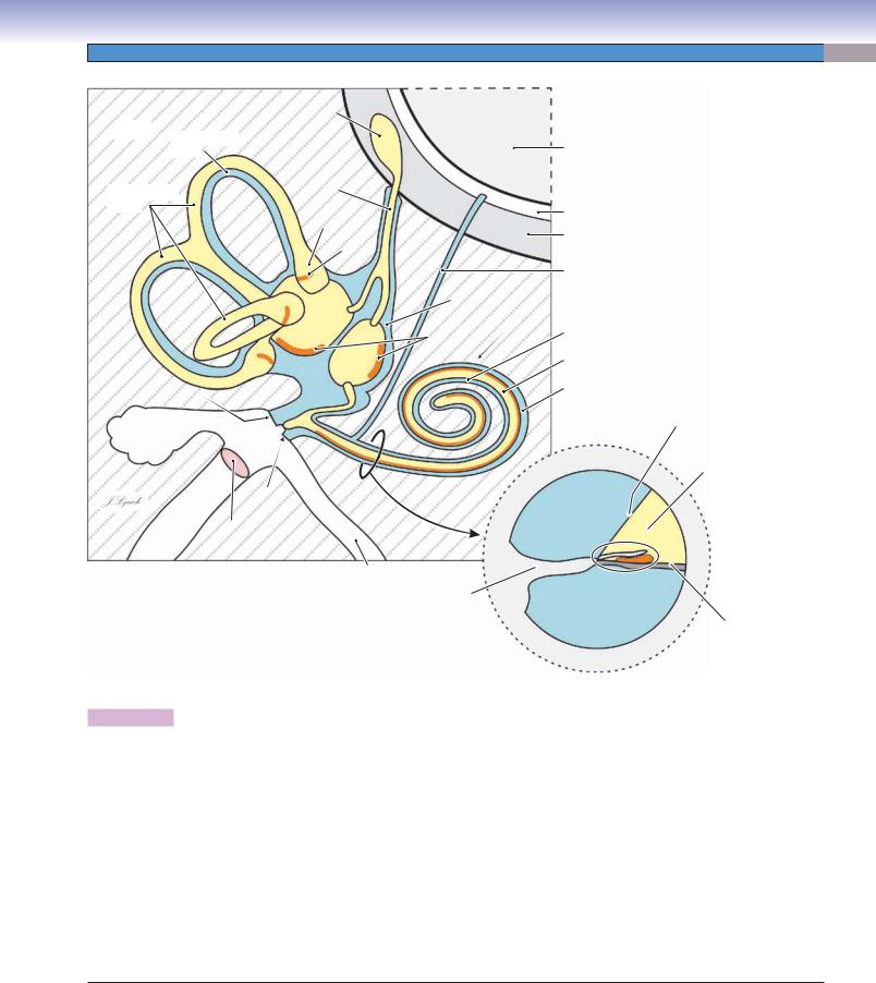
Figure 21-2. Major structures of the inner ear.
The bony labyrinth is a complex cavity in the petrous portion of the temporal bone, the hardest bone in the body. The cavity is lined with endosteum and contains perilymph (blue shading), a clear fluid with a composition very similar to that of cerebrospinal fluid. The bony labyrinth is divided into three portions, the vestibule, the cochlea (anteriorly), and the semicircular canals (posteriorly). The membranous labyrinth, consisting of the three semicircular ducts, the utricle, and the saccule, lies within the bony labyrinth. The walls of the membranous labyrinth are, in general, composed of a thin layer of fibrous connective tissue; a thin layer of more delicate, more vascularized connective tissue; and an internal lining of simple epithelium. Two saclike structures, the utricle and saccule, are found in the vestibule. A semicircular duct lies within each of the semicircular canals. A similar but more complicated structure, the cochlear duct, lies within the bony cochlea. These membranous structures are continuous with each other and contain endolymph (yellow shading), a fluid with a high concentration of K+ (potassium) ions that is unique in the body. Specialized regions within the membranous labyrinth (indicated by thick orange lines) contain receptor cells innervated by branches of the vestibulocochlear nerve (CN VIII). These include the cristae (singular, crista, from Latin for “crest”) in each of the ampullae (singular, ampulla, from Latin for “flask” or “bottle”) of the semicircular ducts, the maculae (singular, macula, from Latin for “spot”) of the utricle and saccule, and the organ of Corti (spiral organ) in the cochlear duct. The receptor cells in all these regions are specialized epithelial cells (hair cells) that transduce movements of the basilar membrane into nerve impulses (see Figs. 21-5C, 21-7, and 21-11A).
414 UNIT 3 ■ Organ Systems
A
Endolymphatic sac
Semicircular |
|
|
Vestibular nerve |
ducts |
|
|
|
|
Ampulla |
Vestibular |
(CN VIII) |
|
|
ganglia |
|
|
|
(of Scarpa) |
|
|
Fig. 21-9B |
Facial nerve |
|
|
|
|
|
|
|
|
(CN VII) |
|
Utricle |
|
|
|
Saccule |
|
Cochlear nerve |
|
|
|
(CN VIII) |
Fig. 21-8B |
|
|
|
Fig. 21-10B,C |
|
|
Fig. 21-4B |
Ductus reuniens |
|
|
|
|
|
Fig. 21-6 |
|
Position of |
|
|
|
oval window |
|
|
|
in osseous |
|
|
|
labyrinth |
Cochlear |
|
Apex |
|
duct |
|
|
|
|
of cochlea |
|
|
Base |
Fig. 21-3B |
|
|
|
||
|
of cochlea |
Fig. 21-4A |
|
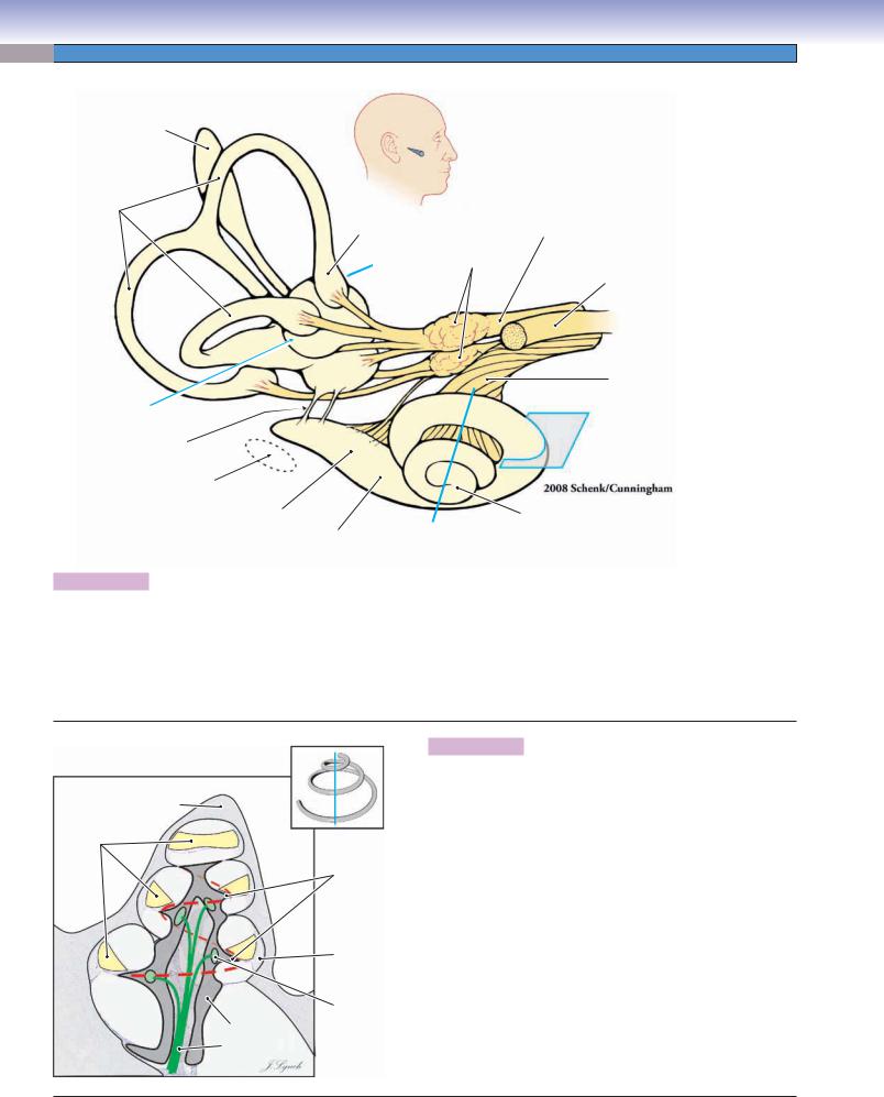
Figure 21-3A. Membranous labyrinth.
The endolymph-filled membranous labyrinth floats within the perilymph-filled osseous (bony) labyrinth (Fig. 21-2). This anatomically correct drawing shows the position of the cochlea and semicircular canals within the head. The labyrinth is viewed from the direction indicated by the pointer in the small inset of the head. The position of the nerves that innervate the cochlea, cristae of the ampullae, and maculae of the utricle and saccule are illustrated. The positions and planes of section of the photomicrographs that follow in this chapter are indicated by blue lines, arrows, or boxes. The dashed oval indicates the position of the oval window in the osseous labyrinth (Fig. 21-2).
B
Apex
of cochlea
Cochlear duct
Osseous spiral lamina
Spiral ligament
Bone |
Scala |
Spiral |
|
tympani |
ganglion |
||
|
|||
|
|
Modiolus |
|
Base |
|
Cochlear |
|
of cochlea |
nerve |
||
Figure 21-3B. Cochlear duct and modiolus.
The membranous cochlear duct lies within a spiral-shaped tunnel in the temporal bone. In this illustration, the cochlea has been cut in half along the plane indicated by the single blue line in Figure 21-3A and in the inset. The spiral of the cochlear duct makes about two and one-half turns from the base of the cochlea to its apex. The outer bony surface of the cochlea is indicated in light gray. A spiral, screw-shaped bony structure, the modiolus forms the central core of the cochlea and is indicated in darker gray. A spiral cavity within the modiolus contains the cell bodies of the spiral ganglion (green) and the proximal axons of the cochlear nerve
(CN VIII). The osseous (bony) spiral lamina curves around the modiolus like the threads of a screw (red dashed line and inset). The central edge of the cochlear duct is attached to the spiral lamina; the outer wall of the cochlear duct is attached to the spiral ligament. Histological sections of these structures are shown in Figures 21-4A and 21-4B.
CHAPTER 21 ■ Ear |
415 |
Auditory System
A |
|
Basal turn |
Middle turn |
Apical turn |
|
|
|||
Base |
|
Scala |
|
|
|
media |
Cochlear |
||
of cochlea |
|
|||
|
|
|
|
duct |
|
Scala |
Scala |
|
Modiolus |
|
tympani |
vestibuli |
|
|
|
|
|
||
Apex
of cochlea
CN VIII
Osseous spiral lamina
Spiral ganglion
Basilar |
|
Auditory |
membrane |
Tensor |
tube |
|
|
|
|
tympani |
Cartilage “C” |
|
muscle |
|
|
|
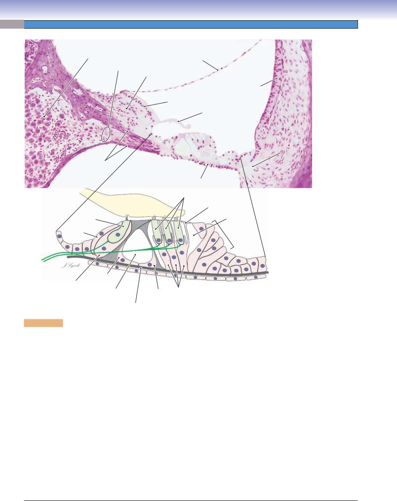
Figure 21-4A. Cross section of cochlea. H&E, 22
The cochlea consists of a spiral tunnel in the temporal bone and associated membranous structures within that tunnel. The tunnel makes two and three-quarter turns as it proceeds from the wide base of the cochlea to its apex. The cross section shows the cochlea in its approximate anatomical orientation (plane of section is indicated by the blue line through the cochlea in Fig. 21-3A). The tunnel is lined with endosteum and is larger at the base, becoming progressively narrower toward the apex. It is divided into two sections, the scala (“staircase”) vestibuli and the scala tympani. These two sections are separated by the membranous cochlear duct (Figs. 21-2 and 21-3A,B), which encloses the scala media. The cochlear duct contains the sensory receptors of the cochlea. The vestibule opens into the scala vestibuli, and sound waves are transmitted from the oval window to the sensory receptors by this route. The scala vestibuli is continuous with the scala tympani at the cochlear apex via a small opening, the helicotrema (see Fig. 21-6A). The scala tympani extends from the helicotrema to the round window of the tympanic cavity. The auditory tube connects the middle ear cavity with the nasopharynx to allow air pressure in the middle ear to equilibrate with that of the surrounding environment. The auditory tube runs in a groove in a C-shaped band of cartilage. The structures associated with the scalae vestibuli, tympani, and media (dashed rectangle) are shown at higher magnification in Figure 21-4B.
B
Scala vestibuli
Spiral ganglion
Scala media
Scala tympani
Vestibular (Reissner) membrane
Stria vascularis
Cochlear duct
Spiral limbus
Organ
of Corti
Spiral ligament
Basilar membrane
Osseous spiral lamina
Figure 21-4B. Scala media and organ of Corti. H&E, 84
It is traditional to represent the internal structures of the cochlea as if the basilar membrane is horizontal. This photomicrograph has, therefore, been rotated 90 degrees counterclockwise from its position in Figure 21-4A. The cochlear duct (dotted line) is a roughly triangular structure that lies between the scala vestibuli and the scala tympani. The fluidfilled space within the cochlear duct is the scala media (yellow shading). In this photomicrograph, the cochlear duct is bounded by the bony labyrinth and basilar membrane below, the stria vascularis on the right, and the vestibular membrane (Reissner membrane) above. The basilar membrane supports the organ of Corti, which is described more fully in following figures. The stria vascularis (see Fig. 21-5) is a specialized, thickened region of stratified epithelium. In contrast to most types of epithelium, it is highly vascularized by a dense meshwork of capillaries. The stria vascularis is instrumental in maintaining the high K+ concentration of the endolymph in the scala media. Lateral to the stria vascularis, the endosteum is much thickened and forms the spiral ligament, to which the outer edge of the basilar membrane connects. The vestibular membrane consists of two layers of squamous epithelial cells on either side of a basal lamina. The tectorial membrane (not labeled) normally rests on the hair cells of the organ of Corti (see the illustration in Fig. 21-5). However, it is often distorted or damaged during tissue processing.
416 UNIT 3 ■ Organ Systems
Spiral |
|
|
|
ganglion |
Auditory |
|
Vestibular |
|
|
(Reissner) |
|
|
nerve |
|
|
|
|
membrane |
|
|
fibers |
Spiral |
|
|
|
||
Bone |
|
limbus |
|
|
|
|
Stria |
|
|
|
vascularis |
|
|
|
Interdentate |
|
|
|
cells |
|
|
|
Tectorial |
|
|
|
membrane |
Spiral ligament
Bony spiral lamina
 Modiolus
Basilar membrane
|
Outer hair |
|
|
cells |
|
Tectorial membrane |
Reticular |
|
Inner hair |
lamina |
|
Outer |
||
cell |
tunnel |
|
Border |
Cells of |
|
cell |
||
Hensen |
||
|
Afferent nerve fibers
|
Inner pillar |
|
|
|
Inner phalangeal cell |
|
Outer phalangeal |
|
|
cell |
Inner tunnel |
Outer pillar |
Organ of Corti |
|
|
cells |
|||
|
of Corti |
cell |
||
|
|
|
||
Basilar membrane
Figure 21-5. Organ of Corti and associated structures. H&E, 189
The organ of Corti is a band of specialized epithelial cells that rest on the basilar membrane, a thin sheet of fibrous connective tissue that extends from the osseous (bony) spiral lamina to the spiral ligament. The surface of the basilar membrane in the scala tympani is covered by a thin layer of vascularized connective tissue and elongated mesothelial cells. The organ of Corti contains the auditory hair cells, the receptor cells for hearing, as well as several types of supporting cells. The hair cells have bundles of 50 to 100 stereocilia (hairs) protruding from their upper surfaces. The transduction of sound waves to nerve impulses is based upon changes in the polarization of the hair cell membrane that occur when their apical stereocilia are bent during the vibration of the basilar membrane by sound waves (see Fig. 21-6A,B). The hair cells are in synaptic contact with afferent and efferent nerve fibers of the auditory branch of CN VIII (efferent fibers not illustrated). Auditory hair cells are divided into two groups: inner hair cells and outer hair cells. In humans, there are about 3,500 inner hair cells arranged in a single row and about 12,000 outer hair cells arranged in three or sometimes four rows (see Fig. 21-7A). The hair cells are surrounded by a variety of supporting cells, including pillar cells, phalangeal cells, border cells, and cells of Hensen. The inner and outer hair cells are separated by inner and outer pillar cells. These cells have long, thin processes that include dense bundles of microtubules and extend from the basilar membrane to the upper surfaces of the hair cells. Pillar cells surround a triangular, fluid-filled space, the inner tunnel of Corti. The basal and lateral aspects of inner hair cells are surrounded by inner phalangeal cells. By contrast, outer phalangeal cells cup only the lower third of each outer hair cell, whereas the upper two thirds of each outer hair cell is surrounded by a fluid-filled space. The spaces between the upper surfaces of the outer hair cells are filled by processes of phalangeal cells. These processes form the reticular lamina. Tight junctions connect the phalangeal cell processes and the apical surfaces of the hair cells to form a barrier that separates the endolymph of the scala media from the cells of the organ of Corti. Columnar epithelial cells called border cells mark the medial extent of the organ of Corti; columnar epithelial cells called cells of Hensen mark its lateral extent. The tectorial membrane hangs over the organ of Corti and deflects the stereocilia of the hair cells when sound waves move the basilar membrane. The tectorial membrane is a gelatinous structure, containing fine filaments, which is secreted by columnar epithelial cells (interdentate cells) on the surface of the spiral limbus. It is commonly distorted during tissue processing; its normal position is illustrated.

CHAPTER 21 ■ Ear |
|
|
|
417 |
|
A |
|
|
|||
|
Figure 21-6A. |
Sound transduction. |
|||
Vestibule |
|
As shown in this diagram of a straightened cochlea, sound waves |
|||
|
Base |
are transmitted into the perilymph (blue) of the scala vestibuli by |
|||
|
Scala vestibuli |
movements of the footplate of the stapes on the oval window. |
|||
|
Basilar membrane |
The membranous round window provides pressure relief for the |
|||
|
sound waves within the closed chamber of the bony labyrinth. |
||||
Oval window |
Spiral lamina |
The basilar membrane (gray) is a thin sheet of fibrous connective |
|||
tissue that supports the organ of Corti (Fig. 21-5). The basilar |
|||||
|
|||||
|
|
||||
|
Apex |
membrane is narrower at the base of the cochlea (about 0.21 mm) |
|||
Round window |
|
than at the apex of the cochlea (about 0.36 mm). It is also stiffer |
|||
Scala tympani |
|
at the base of the cochlea than at the apex. These properties cause |
|||
|
the basilar membrane to vibrate preferentially (resonate) near the |
||||
Spiral ligament |
|
||||
|
base when stimulated at high frequencies (red arrows) and near |
||||
|
|
||||
Cochlea |
|
the apex (blue arrows) when stimulated at low frequencies. This |
|||
Helicotrema |
arrangement creates a tonotopic map along the organ of Corti and |
||||
(unrolled) |
|||||
|
is one of the ways in which the cochlea encodes sound waves of |
||||
|
|
||||
|
|
different frequencies into trains of nerve impulses that can be pro- |
|||
|
|
cessed by the nervous system to produce the sensation of pitch. |
|||
B
Tectorial membrane |
|
Scala media |
|
|
(endolymph) |
|
Original position |
|
Scala vestibuli |
Basilar membrane |
|
(perilymph) |
||
|
||
|
|
Figure 21-6B. Stereocilia displacement.
When a sound wave increases the pressure in the perilymph of the scala vestibuli, the pressure in the endolymph of the scala media increases simultaneously, because the vestibular membrane is very thin and delicate. This increased pressure moves the tectorial membrane and basilar membrane downward (large red arrow) and away from their original positions (indicated by the ghost outline). Because the centers of rotation of the tectorial membrane and basilar membrane are different, the downward movement of the two membranes induces a transverse displacement of the tips of the hair cells (small red arrows). This bending of the stereocilia opens K+ channels and causes a change in the membrane potential of the hair cells. The potential change (depolarization) induces the release of transmitter molecules and produces action potentials in the afferent nerve fibers of the cochlear nerve.
C |
Inner hair |
Outer hair |
|
cell |
cell |
|
|
Reticular |
|
Cuticular |
lamina |
|
|
|
|
plate |
|
|
|
Phalangeal |
|
|
cell process |
Phalangeal  cells
cells
Afferent axon 
Efferent axon
Figure 21-6C. Auditory hair cells.
There are several differences between the inner and outer hair cells. The stereocilia of inner hair cells are arranged in a straight line, whereas the stereocilia of the outer hair cells are arranged in a “V” or “W” pattern (see Fig. 21-7A,B). Inner hair cells are completely surrounded by inner phalangeal cells; only the lower third of outer hair cells is cupped by the cell bodies of outer phalangeal cells. About 95% of the sensory nerve fibers in the auditory nerve contact inner hair cells. A single afferent axon typically contacts only one inner hair cell, and each inner hair cell has synaptic contact with at least 10 afferent axons (green). By contrast, a single afferent axon may branch and contact as many as 10 outer hair cells. In addition, there are efferent nerve fibers (orange) that originate in auditory centers in the brainstem and make synaptic contacts on hair cells or on afferent nerve endings. These efferent fibers play a role in tuning the excitability of the hair cells. The majority of the efferent endings are on outer hair cells.
418 |
UNIT 3 ■ |
Organ Systems |
|
|
|
|
|
|
|
|
A |
|
Stereocilia |
Tip links |
|
|
|
||
Inner
hair cells
B
Heads of inner pillar cells
Outer hair cells
Reticular |
Microvilli |
|
lamina |
Reticular |
Process of |
|
||
|
lamina |
phalangeal cell |
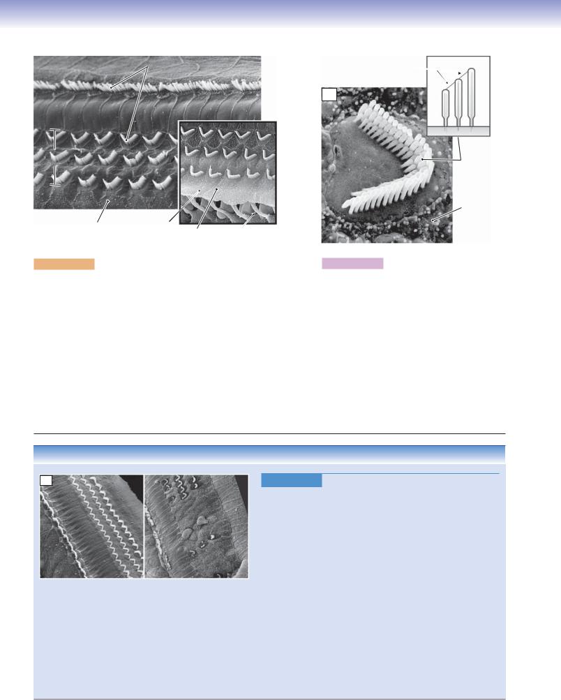
Figure 21-7A. Inner and outer hair cells. SEM, 1,300
Stereocilia
 Hair cell
Phalangeal cell process
 Microvillus
Figure 21-7B. Stereocilia of an outer hair cell.
SEM, 5,000
The arrangement of inner and outer hair cells is illustrated in these scanning electron micrographs. The row of stereocilia of the inner hair cells is separated from the outer hair cells by the heads of the inner pillar cells. The “V” or “W” pattern of the stereocilia of the outer hair cells is clearly seen. Outer hair cells are particularly important for frequency discrimination. They possess the unique property of being able to actively change their physical length in response to changing electrical fields. This property contributes to the frequency selectivity of hair cell responses. The inset shows the thin processes of the outer phalangeal cells that flatten out to form the reticular lamina. This lamina serves to isolate the endolymph in the scala media from the perilymph in the scala tympani. The upper surfaces of the hair cells are smooth, whereas the surrounding processes of phalangeal cells are covered with microvilli.
The stereocilia of outer hair cells are typically arranged in three rows, with the tallest row on the outside of the “V.” Each cilium is narrower at its base than in its body (inset). The cilia are connected at their tips by fine filaments (tip links), which play a critical role in changing the ionic permeability of the cell membrane when the stereocilia are bent. The permeability change initiates a sequence of events that results in the release of transmitter molecules, leading to action potentials in the afferent nerve fibers. Outer hair cells vary in length along the basilar membrane, with the shortest at the basal end of the cochlea and the longest at the apical end.
CLINICAL CORRELATION
C
OHCs |
OHCs |
IHCs
|
IHCs |
Normal organ of Corti: |
Inner and outer hair cells |
Inner hair cells (IHCs) and |
damaged by prolonged noise |
outer hair cells (OHCs) |
|
Figure 21-7C. Sensorineural Hearing Loss. SEM, 376 Extended exposure to loud sounds can impair hearing. These scanning electron micrographs compare a normal mammalian organ of Corti (left) with one that was subjected to high-intensity sound for several days (right). Outer hair cells are more subject to damage than inner hair cells. Durations of loud noise as short as a few minutes can produce detectable damage to stereocilia; longer duration exposure (as illustrated here) causes death of hair cells. Damaged hair cells are not replaced in mammals, although they are replaced in some birds and reptiles. In mammals, the holes left by dying hair cells are filled by the processes of phalangeal cells in order to maintain the barrier between the endolymph and perilymph. Hair cells are normally lost with advancing age (presbyacusis). Hair-cell damage can also be produced by prolonged exposure to high dosages of aminoglycoside antibiotics (e.g., streptomycin, neomycin), some diuretics (e.g., furosemide), and some chemotherapy agents. When only outer hair cells are damaged, there is an overall loss of sensitivity and a profound loss of frequency discrimination (e.g., ability to understand speech). A loss of both inner and outer hair cells leads to complete deafness, which cannot be ameliorated by hearing aids.

CHAPTER 21 ■ Ear |
419 |
Vestibular System
A
Figure 21-8A. Sensory receptors.
|
Temporal |
Anterior |
bone |
semicircular |
|
duct |
|
Posterior |
|
Utricle |
|
|
|
semicircular |
|
|
duct |
|
|
|
|
Saccule |
Horizontal |
|
|
semicircular |
|
Macula |
duct |
|
|
|
of saccule |
|
|
|
|
Crista |
Ampulla |
Vestibule |
|
||
ampullaris |
Macula |
|
|
of utricle |
|
The vestibular apparatus of the inner ear contains sensory receptors that detect rotation of the head in space, linear acceleration, and the static position of the head. The sensory receptors are hair cells that are similar in many, but not all, respects to the hair cells of the auditory system (see Fig. 21-11A,B). Rotational movements of the head are detected by hair cells located in the crista ampullaris of the anterior, posterior, and horizontal semicircular ducts (located within their respective semicircular canals). Horizontal acceleration is detected by hair cells in the macula of the utricle; vertical acceleration is detected by hair cells in the macula of the saccule. Static head position is detected by combining signals from the maculae of the utricle and saccule. The dashed rectangle indicates the approximate position of the photomicrograph in Figure 21-8B.
B
Temporal bone
Perilymph
Position of |
|
Endolymph |
cupula |
Ampulla |
Utricle
Crista
 ampullaris
 Fig. 21-9B
Macula utriculi
Fig. 21-10B
Vestibule
Figure 21-8B. Crista ampullaris and macula utriculi. H&E, 60
A low-power photomicrograph that includes the ampulla of a semicircular canal, the utricle, and a portion of the vestibule is shown. The semicircular canal within the temporal bone is filled with perilymph. The membranous labyrinth, filled with endolymph, floats within this bony canal (Fig. 21-2). The sensory receptors of both the vestibular system and the auditory system are in contact with the endolymph. The crista ampullaris contains vestibular hair cells and the sensory receptors of the vestibular system and is described in detail in Figure 21-9A,B. A gelatinous structure, the cupula, surrounds the crista ampullaris and forms a wall across the ampulla (Fig. 21-9A). The cupula is normally lost during tissue processing. Movement of the endolymph during head rotation deflects the cupula and, thereby, bends the cilia of the hair cells. The large fluid-filled utricle contains the macula utriculi, a sense organ that measures linear acceleration and static position of the head. The macula utriculi contains vestibular hair cells with cilia that are embedded in a gelatinous structure, the otolithic membrane. This membrane is covered by tiny crystals (otoconia), which have a higher specific gravity than the surrounding endolymph and, consequently, are influenced by gravity and acceleration. The macula is described in further detail in Figure 21-10A–C.

420 UNIT 3 ■ Organ Systems
Figure 21-9A. Ampulla of the semicircular canal.
Ampulla
Cupula
Hair cells
Crista ampullaris
A
Each of the three semicircular ducts has an enlargement called the ampulla near one of the points at which the duct joins the utricle. There is a ridge, the crista ampullaris, on the floor of each ampulla. Partially surrounding the ridge and extending to the ceiling of the ampulla is a wall, the cupula, which completely blocks the duct. The cupula consists of a firm gel of proteins and polysaccharides. This structure is normally dissolved during the tissue preparation process and only remnants are typically seen in histological sections. When the head rotates, the endolymph within the semicircular ducts moves (red arrows) and exerts pressure on the cristae and their respective cupulae, causing them to deflect slightly. This deflection bends the hair cells in the cristae (Fig. 21-11B) and modulates the frequency of action potentials that are going to the brainstem vestibular centers, thereby producing the sensation of motion.
Remnant |
Support |
cells |
|
of cupula |
Cupula |
Connective |
|
tissue |
Basal |
|
|
|
lamina |
Dark |
Hair cells |
Hair cells |
|
cells |
|
|
Planum |
|
semilunatum |
B
Figure 21-9B. Crista ampullaris. H&E, 166
The crista ampullaris (also known as the ampullary crest) is a projection of connective tissue covered with epithelium within the ampulla. The epithelium consists of hair cells, support cells, and dark cells. The cilia of the hair cells are embedded in the gelatinous material of the cupula. The hair cells are cradled by supporting cells that rest on the basal lamina of the epithelium. There are two distinct types of hair cells in the cristae, termed type I and type II hair cells. These will be described in greater detail in Figure 21-11A,B. The planum semilunatum is a region of endothelium composed of a single layer of cells called “dark cells,” because they stain more intensely than other epithelial cells in the internal ear. Dark cells display cytological characteristics of cells with high metabolic activity and are believed to be important in controlling the ionic composition of the endolymph. They are found in several other locations within the labyrinthine ducts, including the stria vascularis.
CLINICAL CORRELATION
Normal membranous |
Dilated membranous labyrinth |
labyrinth |
in Ménière’s disease |
|
(endolymphatic hydrops) |
Figure 21-9C. Ménière Disease.
Ménière disease is a disorder of the labyrinth of the inner ear, characterized by intermittent episodes of hearing loss, tinnitus, aural pressure, and vertigo. Its causes are uncertain but may include autoimmune disorders, viral infections, genetic predisposition, allergies, and head trauma. Disorders of secretory cells in the membranous labyrinth and endolymphatic sac may produce ionic imbalance between endolymph and
Cperilymph, resulting in endolymphatic hydrops (swelling of the membranous labyrinth) and producing many of the above symptoms. Diagnosis is based on history, clinical symptoms, audiometry, and vestibular testing. Postmortem histopathologic findings may include perisaccular fibrosis, atrophy of the endolymphatic sac, and other membranous changes. Treatments include reduction of caffeine and salt intake, diuretics, antinausea medications, glucocorticoid therapy, intratympanic gentamicin injection, surgical labyrinthectomy, and vestibular nerve section.

CHAPTER 21 ■ Ear |
|
|
421 |
Otoconia |
|
||
Figure 21-10A. |
Macula of the utricle. |
||
A
Otolithic
 membrane
Support cell
|
|
Type I |
Afferent |
Type II |
hair cell |
|
||
nerve axon |
hair cell |
|
The utricle and saccule are similar in structure. The walls consist of an outer fibrous layer, an intermediate layer of vascularized connective tissue, and an inner epithelial lining. In both the utricle and the saccule, there is a region of specialized epithelium termed the macula (see Fig. 21-2), which is 2 to 3 mm in diameter. The macula contains two types of sensory hair cells, classified as type I and type II (see Fig. 21-11A). The sensory epithelium of the macula is overlaid by a gelatinous structure, called the otolithic membrane, which is similar in makeup to the cupula of the ampulla. The stereocilia and kinocilia of the hair cells are embedded in the membrane. Hundreds of tiny crystals, otoconia, are attached to the surface of the otolithic membranes. These crystals have a higher specific gravity than the surrounding endolymph and are consequently affected by gravity or linear acceleration. Changes in head position or acceleration, therefore, cause the otoconia-weighted otolithic membrane to deflect the cilia of the hair cells and, thus, trigger changes in the frequency of nerve impulses generated by the hair cells.
B
Otolithic Otoconia membrane
Hair cells and support cells
Connective tissue
Axons of utricular nerve
Figure 21-10B. Macula of the utricle with otoconia. H&E,
260
This photomicrograph shows a cross section of the macular region of the wall of the utricle (an enlargement of the area indicated by the dashed rectangle in Fig. 21-8B). The otoconia, which stain darkly in this H&E stain, lie on the surface of the otolithic membrane; the otolithic membrane itself is almost transparent. The support cells provide mechanical support to the hair cells and also secrete the substance of the otolithic membrane.
Dizziness is one of the most common reasons adults seek medical care. The term has two general meanings: (1) A feeling of light-headedness or “about to faint” or (2) a feeling that the individual is spinning or that the room is spinning. This latter feeling is properly called vertigo. Twenty percent of all complaints of dizziness involve problems related to the otoconia of the utricle and saccule (see Fig. 21-10C).
C
Figure 21-10C. Otoconia from the macula of the utricle.
SEM, scale bar = 10 μm
Otoconia are crystals of almost pure calcium carbonate that form in the region of the macular hair cells. The otoconia lie on the surface of the otolithic membrane, attached by a proteinaceous substance that is not well understood.
The number of otoconia present decreases with age, contributing to the balance difficulties often experienced by older individuals. In addition, crystals or small groups of crystals sometimes become detached from the otolithic membrane and drift into a semicircular canal or even become attached to the hair cells in a canal. This dislocation can disrupt the normal neural signals from the labyrinth and produce a type of severe vertigo termed benign paroxysmal positional vertigo. Treatment that involves sequences of different head positions and movements can usually improve or eliminate the symptoms. The exact maneuver depends upon which semicircular canal is involved.

422 |
UNIT 3 ■ |
Organ Systems |
|
|
|
Kinocilium |
Stereocilia |
|
|
|
Figure 21-11A. |
Vestibular hair cells. |
||
Basal body
Calyx nerve  ending
|
|
Efferent |
|
|
|
nerve |
Afferent |
|
Afferent |
fibers |
|
|
|
nerve |
|
|
nerve |
|
fiber |
A |
fiber |
Type I hair cell |
Type II hair cell |
|
|||
Type I hair cells are pyriform (pear shaped) and have basally located nuclei. They are almost completely surrounded by a single, chalice-shaped synaptic terminal (calyx) of a large afferent nerve fiber. Each type I hair cell is innervated by a single nerve axon, and each axon branches to innervate only a few hair cells. Type I hair cells receive few efferent endings, which contact the afferent nerve endings rather than the hair cell itself. Type II cells are more cylindrical in shape, with more centrally located nuclei. These cells are contacted by multiple small boutonlike synaptic endings associated with both afferent and efferent nerve fibers. Type II cells receive axon terminals from multiple nerve fibers, each of which branches to contact many type II hair cells. Both types of vestibular hair cells have a single kinocilium (with a typical basal body and a ring of nine double microtubules) on one side of the apical surface. A group of 40 to 100 stereocilia of various lengths are arranged in a hexagonal array next to the kinocilium.
|
A |
B |
C |
Figure 21-11B. |
Excitation and inhibition in hair cells. |
|
|
|
|
Vestibular hair cells continuously release a small amount of |
|
|
|
|
|
neurotransmitter at the afferent terminal synapses, producing a |
|
|
|
|
|
moderate frequency of action potentials in afferent nerve fibers in |
|
|
|
|
|
the resting state (B). When movement of endolymph causes the |
|
|
|
|
|
cupula or otolithic membrane to deflect the hair cell stereocilia |
|
|
|
|
|
toward the kinocilium, the amount of neurotransmitter released |
|
|
|
|
|
goes up and the frequency of action potential discharge increases |
|
|
|
|
|
(A). When the stereocilia are deflected away from the kinocilium, |
|
|
|
|
|
less neurotransmitter is released and the frequency of the action |
|
|
|
|
|
potentials decreases (C). Hair cells in different regions of the |
|
|
|
|
|
peripheral vestibular apparatus have their kinocilia and stereo- |
|
|
|
|
|
cilia oriented in different directions. The CNS integrates informa- |
|
B |
Action potentials |
|
|
tion from the various hair cells to form a central representation of |
|
|
|
the position of the head in space, the direction of any movement |
|||
|
|
|
|
of the head, and the rate of change (acceleration) of any move- |
|
|
|
|
|
ment. |
|
|
|
|
|
|
|
|
|
|
|
|
|
CLINICAL CORRELATION
C
Inflamed and bulging |
Silicone ventilation |
tympanic membrane, |
tube in place |
viewed through operating |
|
microscope |
|
Figure 21-11C. Otitis Media.
Otitis media is a bacterial or viral infection involving the middle ear. The mucosal lining of the middle ear produces serous exudate and pus when inflamed. In children (younger than age 3 years), the auditory tube is not fully developed and drainage of infection is problematic. The consequent buildup of fluid in the middle ear may cause severe pain (otalgia) and a temporary conductive hearing loss. The tympanic membrane becomes erythematous and bulges outward from the fluid pressure (left). (Because of the limited field of view of the operating microscope, only the anterior half of the membrane is visible.) Most cases resolve spontaneously, although antibiotics are commonly prescribed to speed recovery. In cases of severe and repeated infections, a ventilation tube may be inserted into an incision in the tympanic membrane to relieve pressure in the middle ear (right). These tubes usually stay in place for about 1 year before they spontaneously extrude. Very rarely, middle ear infections may spread locally and produce mastoiditis or labyrinthitis.

CHAPTER 21 ■ Ear |
423 |
CLINICAL CORRELATIONS
Schwannoma |
Basilar artery |
 Pons
Pons
Vertebral artery
Medulla 
Cerebellum
A
Ventral (anterior) surface of the brain
Figure 21-12A. Vestibular Schwannoma.
Vestibular schwannoma (sometimes called acoustic schwannoma or, incorrectly, acoustic neuroma) is a Schwann cell–derived benign tumor, usually arising from the vestibular branch of CN VIII during the fifth or sixth decade of life. Risk factors include prolonged exposure to loud noise, childhood exposure to low-dose radiation, and possible links to parathyroid adenoma. Symptoms include hearing loss, headache, vertigo, tinnitus, and facial pain. The tumor is usually unilateral. It appears as a well-circumscribed, encapsulated mass. The tumor attaches to the nerve but can usually be separated from the nerve. Histologically, schwannomas arise from perineural elements of Schwann cells. Areas of alternately dense and sparse cellularity, called Antoni A and Antoni B regions, are characteristic of the tumor. Treatment options include surgical removal of the tumor, stereotactic radiosurgery, stereotactic radiotherapy, and proton beam therapy.
Internal |
Temporal |
|
|
auditory |
Internal auditory |
||
lobe |
|||
meatus |
meatus |
||
|
|||
Ossicles |
|
|
Vestibule and semicircular canal
M |
Mastoid |
Horizontal |
Cerebellum |
cavity |
semicircular canal |
|
|
|
|
and vestibule |
|
B
CT scan of temporal |
Axial MRI scan |
|
bone in horizontal plane |
||
|
Figure 21-12B. CT and MRI of Inner Ear Structures.
Recent developments in imaging resolution have made it possible to distinguish many of the anatomical features of the inner ear in human subjects. Computed tomography (CT) imaging of the temporal bones is helpful for evaluating bony anatomy, such as the middle ear ossicles and bony labyrinthine structures. CT is useful in cases of trauma to look for fractures and dislocations, infection and inflammatory processes to evaluate for bone erosion, and congenital abnormalities to explain hearing dysfunction. Magnetic resonance imaging (MRI) is most often performed to evaluate a patient with sensorineural hearing loss. It is used to exclude a vestibular schwannoma and, occasionally, to evaluate for infection. The brainstem, internal auditory canals, cranial nerves, and membranous labyrinthine structures (perilymph-filled cochlea, vestibule, and semicircular canals) are well evaluated on MRI. In the CT scan (left), bone is light and perilymph dark; these relations are reversed in the T2-weighted MRI (right).
SYNOPSIS 21 - 1 Pathological and Clinical Terms for the Ear
■Sensorineural hearing loss: Hearing loss that involves the loss of hair cells or neurons; accounts for about 90% of all hearing loss. Can occur after soundor drug-induced damage to hair cells, disease-induced damage to neurons in the auditory system, and loss of hair cells with advancing age (Fig. 21-7C).
■Hydrops: Excessive accumulation of clear, watery fluid in a tissue or cavity; endolymphatic hydrops refers to an accumulation of endolymph within the membranous labyrinth (Fig. 21-9C).
■Tinnitus: Abnormal noise in the ear (e.g., ringing, whistling, hissing, roaring, chirping), ranging from “mild” to “extremely annoying”; inner ear trauma produced by loud noise is the leading cause; also occurs in Ménière disease. Advancing age is frequently accompanied by gradual loss of hair cells, producing sensorineural hearing impairment and tinnitus (Figs. 21-9C and 21-12A).
■Vertigo: The illusory sensation of spinning or tilting most frequently caused by inner ear disturbances. Can last for minutes, days, or weeks and can be incapacitating; often accompanied by severe nausea (Figs. 21-9C, 21-10C, and 21-12A).
■Conductive hearing loss: Decrease in sound conduction to the inner ear. Possible causes include buildup of fluid pressure in the middle ear because of infection, blockage of external auditory meatus by wax, and disorders or traumatic damage of the ossicles (Fig. 21-11C).
■Erythema (erythematous): Redness of a tissue as a result of inflammation (Fig. 21-11C).
■Otalgia: Earache (Fig. 21-11C).
■Benign: Description of a tumor that is nonmalignant, that is, does not invade surrounding tissues and does not metastasize to other locations in the body (Fig. 21-12A).