

CHAPTER 1 ■ An Illustrated Glossary of Histological and Pathological Terms |
5 |
Descriptive Terms for Abnormal Cells and Tissues
Acute Inflammation. H&E, 388
Acute inflammation is an immediate response by the immune system due to tissue injury from many causes including infection, necrosis, and trauma. It results in a local increase in blood flow, tissue edema due to increased vascular permeability, and an increased number of acute inflammatory cells (chiefly polymorphonuclear leukocytes or neutrophils). A confluent collection of neutrophils is an abscess.
Image: Acute appendicitis showing an infiltration of neutrophils within layers of smooth muscle in the appendiceal wall is shown.
Apoptosis. H&E, 155
Apoptosis is the process of cell death initiated by either physiologic or pathologic causes. Pathologic apoptosis may be seen in malignant neoplasms, cells damaged by radiation or chemicals, tissues infected by viruses, and immunologic damage as seen in graft-versus-host disease.
Image: Colonic mucosa demonstrating marked apoptosis of the crypt epithelial cells is shown in a patient with graft-versus-host disease following a bone marrow transplant.
Atrophy. H&E, 99
Pathologic atrophy refers to a decrease in cell size as a result of various factors including denervation, decreased use, aging, decreased blood supply and nutrients, and pressure.
Image: Skeletal muscle showing denervation atrophy is pictured here. Note the larger, normal myocytes in the right portion of the image, and the smaller, atrophic myocytes in the left portion of the image. In this case, damage to a motor neuron or axon caused atrophy of a group of muscle fibers it served.
Calcification. H&E, 199
Tissue calcification is abnormal and is broadly divided into metastatic calcification and dystrophic calcification. Metastatic calcification occurs in normal, healthy tissues in patients who are hypercalcemic due to vitamin D intoxication, renal failure, or increased parathyroid hormone or in patients with bone destruction. Dystrophic calcification occurs in dying or necrotic tissues in patients with normal serum calcium.
Image: Dystrophic calcification in an intraductal papilloma of the breast is shown. Tissues that assume a papillary morphology tend to develop calcifications at the tips of degenerating papillae. Other examples include papillary thyroid carcinoma and serous papillary neoplasms of the ovaries. Round, lamellated calcifications are called psammoma bodies.
Chronic Inflammation. H&E, 199
Chronic inflammation is an ongoing inflammatory process, typically weeks to months in duration. It may be seen in infectious processes, like viral hepatitis, autoimmune disease, and toxic exposures. Acute and chronic inflammations commonly coexist, as in active chronic gastritis due to infection of the gastric mucosa by the bacteria Helicobacter pylori. The inflammatory cells participating in chronic inflammation include lymphocytes, plasma cells, mast cells, and eosinophils.
Image: This stomach biopsy shows chronic gastritis; note the plasma cells within the lamina propria. No neutrophils are seen, which would indicate a concomitant active, acute inflammatory process.

6UNIT 1 ■ Basic Principles of Cell Structure and Function
Cellular Accumulations
Alpha 1-Antitrypsin. PASD (periodic acid-Schiff with diastase digestion), 173 Alpha 1-antitrypsin deficiency is an autosomal recessive disorder characterized by low serum concentrations of the enzyme alpha 1-antitrypsin, which inhibits or inactivates proteases and elastases. Alpha 1-antitrypsin deficiency may cause neonatal hepatitis with later cirrhosis and pulmonary panacinar emphysema. Image: A liver biopsy in a patient with alpha 1-antitrypsin deficiency shows accumulated alpha 1-antitrypsin as hyaline globules in this PAS stain. The globules remain after the tissue has been treated with diastase (diastase resistant).
Amyloid. H&E, 173
Amyloid is an abnormal protein caused by many pathological conditions, the most common of which include AL amyloid, caused by light chains secreted by plasma cells in plasma cell myeloma or monoclonal B-cell neoplasms; AA amyloid, seen in chronic inflammatory conditions; and Ab amyloid in Alzheimer disease. Many other types of amyloids exist.
Image: A lymph node containing AL amyloid in a patient with small lymphocytic lymphoma is shown. In H&E-stained preparations, amyloid is amorphous and eosinophilic. Amyloid stained with Congo red shows a characteristic green birefringence when viewed with polarized light.
Fatty Change. H&E, 173
Fatty change, or steatosis, is the abnormal intracellular accumulation of lipid. Although it can occur in many organs, it is most commonly seen in the liver because of a variety of causes including alcohol abuse, hepatitis C, genetic predisposition, medications, toxins, and diabetes mellitus.
Image: This liver biopsy shows marked steatosis with intracellular lipid vacuoles.
Lewy Body. Immunohistochemistry for alpha-synuclein, 431
A Lewy body is an intracytoplasmic oval with a halo, formed in neuromelanincontaining neurons in patients with idiopathic Parkinson disease.
Image: In this immunoperoxidase preparation, brown pigment indicates the presence of alpha synuclein (arrow), the main protein in the inclusion.
Neurofibrillary Tangles. Silver stain, 173
In patients with Alzheimer disease, microtubule-associated protein tau and abnormally phosphorylated neurofilaments form neurofibrillary tangles within neurons.
Image: This silver-stained slide shows a twisted, black helix (arrows) in the cytoplasm of a neuron from a patient with Alzheimer disease.

CHAPTER 1 ■ An Illustrated Glossary of Histological and Pathological Terms |
7 |
Granulomatous Inflammation. H&E, 99
Granulomatous inflammation is a type of chronic inflammation characterized by localized aggregates of macrophages called epithelioid histiocytes. Collectively, these collections of histiocytes are termed granulomas. Granulomatous inflammation is characteristic of certain bacterial infections, particularly Mycobacterium tuberculosis, fungal infections with organisms like Histoplasma capsulatum, and many other disease processes. Granulomatous inflammation may contain areas of necrosis, as in mycobacterial or fungal infections (caseous necrosis), or may be noncaseating as in the granulomatous disease sarcoidosis.
Image: This lymph node biopsy contains abundant noncaseating granulomas in a patient with sarcoidosis.
Hyperplasia. H&E, 99
Hyperplasia represents the increase in the number, not size, of cells within an organ or a tissue. Contrast this to hypertrophy below in which the cell size, not number, increases. Hyperplasia and hypertrophy both may result in a larger organ or tissue. Hyperplasia can be the result of hormonal stimulation, as seen in hyperplasia of the endometrial cells of the uterus in response to estrogen stimulation.
Image: This is an endometrial biopsy showing hyperplasia of the glandular endometrium. The glands are increased in number and are abnormally close together. Endometrial hyperplasia is a risk factor for the development of adenocarcinoma of the endometrium.
Hypertrophy. H&E, 497
Hypertrophy is a compensatory mechanism by which the size of cells increases because of various stimuli, resulting in the increase in the size of the corresponding organ. Cardiac myocytes hypertrophy in response to increased workload because of hypertension or valvular dysfunction. In systemic hypertension, as the cardiac myocytes enlarge, the heart itself enlarges, producing left ventricular hypertrophy with a thickened muscular wall.
Image: This image of a hypertrophied cardiac myocyte in a patient with hypertension shows an enlarged, hyperchromatic (deeply staining) nucleus, referred to as a “boxcar” nucleus.
Hydropic Change. H&E, 155
Hydropic change is an early reversible cell injury characterized by cellular swelling due to perturbations in cellular membrane ion-pump function. Image: This kidney biopsy shows swollen tubular epithelial cells with cytoplasmic clearing due to edema.
Karyorrhexis. H&E, 747
Karyorrhexis refers to a pattern of nuclear change seen in irreversibly damaged cells, similar to pyknosis (below), in which the nucleus breaks apart and fragments.
Image: This image shows a nucleus undergoing karyorrhexis (arrow) in a malignant neoplasm.

8UNIT 1 ■ Basic Principles of Cell Structure and Function
Metaplasia. H&E, 199
Metaplasia is the reversible change of one mature cell type to another as a result of an environmental stimulus. Examples of metaplasia include squamous metaplasia of the endocervical glandular epithelium of the cervix, squamous metaplasia of ciliated respiratory epithelium in smokers, and intestinal metaplasia of gastric mucosa as a result of chronic gastritis.
Image: A section of stomach shows chronic gastritis with intestinal metaplasia. Note the presence of goblet cells (arrow) not normally present in the gastric mucosa.
Monomorphism. H&E, 155
Monomorphism is a term describing cell populations that show little difference in size and shape of the cell itself, the nucleus, or both. Benign neoplasms and well-differentiated malignant neoplasms may be monomorphic.
Image: This image shows a monomorphic population of cells in a duodenal gastrinoma. The cells are relatively uniform in size and shape, as are the nuclei.
Multinucleation. H&E, 155
Most normal cells contain a single nucleus, with the exception of the osteoclast, which is multinucleated. Multinucleated cells can be seen in a variety of conditions including reactive bone conditions, malignant bone and soft tissue tumors, granulomatous inflammation, and foreign body giant cell reactions.
Images: The first image (left) shows a giant cell (arrow) in a granuloma of sarcoidosis. The second image (right) shows an extensive foreign body giant cell reaction (dashed line) to suture material (arrow).
Pleomorphism. H&E, 199
Pleomorphism is a term describing cell populations that show differences in the size and shape of the cell itself, the nucleus, or both. It is typically used to describe neoplasms. In general, poorly differentiated malignant neoplasms may have a pleomorphic appearance.
Image: This image shows extreme cellular and nuclear pleomorphism in this case of malignant fibrous histiocytoma, pleomorphic type. Compare this image to that of the monomorphic gastrinoma above.
Pyknosis. H&E, 747
Pyknosis is a morphologic change in the nucleus of an irreversibly damaged cell characterized by condensation and increased basophilia.
Image: This image shows pyknosis (arrow) in a dying cell in a malignant neoplasm.

CHAPTER 1 ■ An Illustrated Glossary of Histological and Pathological Terms |
9 |
Scar. H&E, 16
A scar, or cicatrix, is the result of a complex healing process involving an initial inflammatory response followed by the formation of new blood vessels, tissue remodeling, and wound contraction. Abnormal healing processes include keloid formation and hypertrophic scars.
Image: This image shows a dermal scar characterized by horizontal collagenous bands and an absence of skin adnexa–like hair follicles.
Necrosis
Necrosis represents the death of living cells due to irreversible cell injury. Depending on the tissue involved, necrosis will assume one of several morphologic patterns associated with the processes involved in cell death.
H&E, 97
Caseous necrosis is a form of necrosis characterized by obliteration of the underlying tissue architecture and the formation of amorphous granular necrotic debris, which grossly appears “cheesy,” hence the name caseous. Caseous necrosis is highly characteristic of infection with M. tuberculosis and certain fungi and is a type of granulomatous inflammation.
Image: This image shows a lymph node biopsy with granulomatous inflammation and necrosis (to the right of the dashed line) in a patient with H. capsulatum infection.
Coagulative Necrosis. H&E, 97
Coagulative necrosis is a form of necrosis characterized by preservation of cellular outlines. Examples include necrosis of cardiac myocytes in myocardial infarction and renal necrosis.
Image: This image shows global coagulative necrosis in a transplanted kidney due to compromised perfusion after the transplant. Note the preserved architecture with the glomerulus in the center surrounded by renal tubules. Note also the pale eosinophilic staining with lack of nuclear staining.
Fat Necrosis. H&E, 193
Fat necrosis is a specific type of necrosis seen in fatty, or adipose, tissue. Damage to adipocytes causes release of lipids and cell death followed by aggregates of foamy macrophages containing the released lipids. Fat necrosis is seen in damage to fatty tissue by trauma as well as enzymatic digestion as seen in acute pancreatitis.
Image: This image shows fat necrosis in subcutaneous adipose tissue after previous surgery. Note the abundant foamy macrophages containing lipid droplets (arrow).
Liquefactive Necrosis. H&E, 48
Liquefactive necrosis may be seen after bacterial infections or infarcts involving the central nervous system.
Image: This image shows liquefactive necrosis in an infarcted area of the brain. Note the intact white matter in the lower portion of the image and the granular liquefactive necrosis in the upper portion of the image.

10 UNIT 1 ■ Basic Principles of Cell Structure and Function
Pigments
Pigments are colored substances found within tissue macrophages or parenchymal cells. Pigments may be endogenous, those produced by the body, or exogenous, those originating outside of the body. Melanin, lipofuscin, and hemosiderin are the most common endogenous pigments. The most common exogenous pigment is carbon.
Anthracosis. H&E, 155
Anthracosis is an exogenous pigment composed of carbonaceous material from smoking and air pollution. Inhaled carbon is taken up by alveolar macrophages and transported to lymph nodes. Anthracotic tissues are black in gross appearance.
Image: This lymph node from the hilar region of the lung shows abundant macrophages containing black carbonaceous material.
Melanin. H&E, 388
Melanin pigment, a product of melanocytes, can normally be seen in the basal keratinocytes of the skin. In some chronic inflammatory skin conditions, melanin is released into the dermis and taken up by dermal macrophages, or melanophages.
Image: Black-brown melanin pigment is present within the papillary dermal macrophages.
Lipofuscin. H&E, 388
Also known as lipochrome, lipofuscin is a yellow-brown pigment related to tissue aging. Lipofuscin is insoluble and composed of phospholipids and lipids as a result of lipid peroxidation. It is commonly seen in the liver and heart.
Image: This hypertrophic cardiac myocyte from an older individual contains lipofuscin granules adjacent to the nucleus.
Hemosiderin. H&E, 155 (left); Prussian blue, 155 (right) Hemosiderin is the tissue storage form of iron, which appears as granular, coarse, golden-brown pigment. Hemosiderin is formed from the breakdown of red blood cells and is taken up into tissue macrophages. It may be seen in tissues in which remote bleeding has occurred or in any condition in which excess iron is present. Images: The first image (left) shows abundant hemosiderin-laden macrophages in soft tissue where past bleeding has occurred. The second image (right) is a Prussian blue preparation of the same tissue, showing the deep blue staining of hemosiderin.
Ulcer. H&E, 25
An ulcer represents the discontinuity of an epithelial surface, which may involve skin or mucous membranes. Ulcers may be caused by infectious processes, chemical exposures, prolonged pressure, or vascular compromise. They typically form crater-shaped lesions with a superficial fibrinopurulent layer and an underlying vascular and fibroblastic proliferation called granulation tissue.
Image: This is an image of a gastric ulcer. Note the intact mucosa transitioning to the ulcer with a fibrinopurulent surface. The formation of gastric ulcers is closely associated with infection with H. pylori. Gastric ulcers may be benign or malignant, representing gastric adenocarcinoma.

|
|
|
2 |
Cell Structure and |
|
Function |
||
|
|
|
|
|
|
|
|
|
|
|
|
|
|
|
Introduction and Key Concepts for Cell Components
Synopsis 2-1 |
Functions of Major Cell Components |
Figure 2-1 |
Major Structures of the Cell |
Cell Ultrastructure
Figure 2-2 |
Membranes Define the Major Components |
|
and Compartments of the Cell |
Figure 2-3 |
The Nucleus and Its Components |
Figure 2-4 |
Cytoplasmic Organelles |
Figure 2-5A |
Cytoplasmic Organelles: Rough Endoplasmic |
|
Reticulum and the Golgi Complex |
Figure 2-5B |
Cytoplasmic Organelles: Smooth Endoplasmic |
|
Reticulum |
Figure 2-6A |
Cell Surface and Cytoskeleton, Intestinal |
|
Absorptive Cells |
Figure 2-6B |
Cytoskeleton, the Dendrite of a Neuron |
Cell Structure
Figure 2-7A |
Cell Components in Light Microscopy |
Figure 2-7B |
Wide Range of Cell Sizes, Spinal Ganglion |
|
Sensory Neuron |
Cell Structure Correlates with Function
Figure 2-8A,B Protein-Secreting Cells, Plasma Cells
Figure 2-9A,B Exocrine Protein–Secreting Cells, Pancreatic
Acinar Cells
Figure 2-10A,B Steroid Hormone–Secreting Cells, Adrenal
Cortex
Figure 2-11A,B Ion-Pumping Cells, Renal Tubule
11

12 UNIT 1 ■ Basic Principles of Cell Structure and Function
Introduction and Key Concepts for |
both in the turnover of intrinsic cellular components and in the |
||
Cell Components |
breakdown of the material ingested by endocytosis. |
||
Other types of membrane-delimited vesicles: Substances other |
|||
|
|
than the secretory products of secretory granules and the |
|
The cell is the basic structural and functional unit of the body, |
|||
hydrolytic enzymes of lysosomes may be sequestered within |
|||
and variations in cell structure account for the remarkable diver- |
|||
vesicles. Examples are proteases within proteasomes, unfinished |
|||
sity in the morphology and function of the body’s basic tissues |
|||
polypeptides in transfer vesicles, and oxidases and catalase in |
|||
and organs. The cell’s repertoire of constituents is limited, but |
|||
peroxisomes. |
|||
the possible combinations of quantities and arrangements of |
|||
Smooth endoplasmic reticulum (SER): This is a membrane- |
|||
these constituents are numerous. |
|||
delimited labyrinth, usually in the form of tubules rather than |
|||
|
|
||
Nucleus (Fig. 2-1) |
flattened cisternae. Its multiple functions include synthesis of |
||
membrane phospholipids, degradation of some hormones and |
|||
Nuclear envelope: This is a double-membrane structure consist- |
|||
toxic substances (liver), synthesis of steroid hormones, and |
|||
ing of a perinuclear cistern sandwiched between the inner and |
sequestration of calcium stores (striated muscle). |
||
the outer nuclear membranes. The nuclear envelope protects the |
Mitochondrion: This is a double-membrane organelle (like the |
||
genetic material and separates it from the diverse molecules and |
nucleus) that generates adenosine triphosphate (ATP) to fuel |
||
structures of the cytoplasm. The nuclear envelope is continuous |
energy-requiring activities of the cell. It also functions in some spe- |
||
with the rough endoplasmic reticulum. |
cialized synthetic pathways such as that for steroid hormones. |
||
Nuclear pore: These gaps in the nuclear envelope are bridged by |
|
||
a pore complex of numerous proteins. This structure controls |
Cell Surface (Fig. 2-1) |
||
the traffic of molecules and particles between the nucleus and |
|||
Cilia: These are cylindrical extensions about 200 nm thick |
|||
the cytoplasm. |
|||
Heterochromatin: This tightly coiled chromatin is inaccessible |
and 5 to 10 μm long. They are usually motile, with a whipping |
||
for transcription, and it appears as basophilic clumps in speci- |
motion that consists of a power stroke and a recovery stroke. |
||
mens prepared for light microscopy. |
An axoneme of microtubules in a nine-doublet, two-singlet |
||
Euchromatin: This is chromatin in a more extended form, |
arrangement provides the stiffness and movement of cilia. |
||
accessible to transcription. In the light microscope, it appears |
Microvilli: These are cylindrical, nonmotile extensions, typi- |
||
as less densely stained, basophilic regions of the nucleus. |
cally about 80 nm in diameter and 1 μm long. In some cells, |
||
Nucleolus: One or more of these spherical, basophilic structures |
they are several micrometers long, in which case, they are called |
||
develop in the nucleus if the cell is generating ribosomes. |
stereocilia. Actin filaments form the cores of microvilli. |
||
|
|
Zonula occludens: Also called tight junctions, these are sites |
|
Cytoplasm (Fig. 2-1) |
of fusion between plasmalemmas of adjacent epithelial cells |
||
that separate the lumen from the underlying connective tissue. |
|||
Ribosome: This is an egg-shaped particle (~20 30 nm) that |
Tight junctions prevent materials from passing from one com- |
||
attaches to a messenger RNA (mRNA) molecule and generates |
partment to another by diffusing through the space between |
||
a polypeptide according to the code provided by the nucleotide |
adjacent epithelial cells. |
||
sequence of the mRNA strand. |
Zonula adherens: These are sites of mechanical adhesion |
||
Polyribosome: This is formed by the alignment of several ribo- |
between adjacent epithelial cells. They are generally associated |
||
somes along the length of an mRNA molecule. When an mRNA |
with and parallel to tight junctions. Actin filaments of the ter- |
||
for a cytosolic or mitochondrial protein is being translated, the |
minal web are anchored by proteins at the cytoplasmic faces of |
||
polyribosome is not associated with endoplasmic reticulum, but |
the membranes of the two cells. |
||
rather it is suspended in the cytosol as a free polyribosome. |
Macula adherens: Also called desmosomes, these, like zonula |
||
Rough endoplasmic reticulum (RER): This typically consists of |
adherens, are sites of mechanical adhesion between cells, but |
||
flattened membrane-delimited cisternae that are studded with |
they are configured as spots rather than bands. Intermediate |
||
ribosomes. As polypeptides are synthesized by the ribosomes, |
filaments are anchored to proteins at the cytoplasmic surfaces |
||
they are sequestered by insertion into the cisternae. Small shut- |
of the membranes of the two cells. |
||
tle vesicles (transport vesicles) move the newly synthesized poly- |
Gap junctions: Also called nexus junctions, these are sites of |
||
peptides from the RER to the Golgi complex. |
close (~2 nm) apposition between plasmalemmas of adjacent |
||
Golgi complex: This consists of a stack of flattened membra- |
cells. The small space between the membranes is bridged by |
||
nous saccules (cisternae). The Golgi complex receives poly- |
connexons, each of which is a cylindrical complex of proteins |
||
peptides from the RER, modifies them (e.g., by sulfation, |
forming a pore. Gap junctions allow free flow of small ions |
||
glycosylation), and sorts and packages them according to their |
between cells, so that the cells are electrically coupled. |
||
destination as constituents of lysosomes, secretory granules |
Basolateral folds: Infoldings of the plasmalemma at the basal |
||
(e.g., pancreatic acinar cell), or cytoplasmic granules (e.g., |
surface of an epithelial cell provide greatly increased surface area |
||
neutrophilic leukocyte). |
in support of extensive traffic of substances between the cytosol |
||
Secretory vesicle: Also called secretory granules, these are |
and the underlying interstitial compartment. Such folds are par- |
||
membrane-delimited packages of secretory products that can |
ticularly prominent in epithelial cells that function in removing |
||
be either stored in the cytoplasm or immediately secreted by |
water and ions from a lumen. Lateral folds can have an additional |
||
exocytosis of the vesicle at the cell surface. |
function of contributing mechanical strength to a layer of cells by |
||
Lysosome: This is a membrane-delimited vesicle characterized |
interlacing with complementary folds of an adjacent cell. |
||
by its low pH and the content of a variety of hydrolases that can |
Basal lamina: This is an extracellular layer at the interface |
||
digest proteins, lipids, and polysaccharides. Lysosomes function |
of a cell and the adjacent connective tissue. Basal laminae are |
||

CHAPTER 2 ■ Cell Structure and Function |
13 |
characteristic of the basal surfaces of epithelia, but they are not restricted to epithelia. Examples of other cells with basal laminae are muscle cells and the Schwann cells that enshroud the axons of peripheral nerves.
Cytoskeleton (Fig. 2-1)
Actin filaments: These are thin (6 nm) filaments that form a feltwork (cortex) beneath the plasmalemma of many cells. They form a skeletal core of microvilli, and they also function in the movement of motile cells. In contractile cells, actin filaments interact with myosin (thick) filaments.
Intermediate filaments: These are relatively stable 10-nm to 12-nm filaments that provide structural support for the cell.
Different specific proteins that form these filaments are characteristic of particular groups of cell types.
Microtubules: These are hollow tubes about 25 nm in diameter. In conjunction with other proteins, particularly kinesin and dynein, they serve as tracks for movement of materials within the cytoplasm. Microtubules also form centrioles and the basal bodies and axonemes (cores) of cilia.
Centrosome: This is composed of a pair of centrioles embedded in amorphous material. The two centrioles are oriented at right angles to each other, and each is composed of nine triplet sets of microtubules. The centrosome functions in organizing the array of microtubules in the cell’s cytoplasm and in developing the spindle apparatus during cell division.
SYNOPSIS 2 - 1 Functions of Major Cell Components
Nucleus
■Nucleus: Synthesizes all types of RNA; replicates its DNA.
■Heterochromatin: Condenses inactive DNA.
■Euchromatin: Renders DNA accessible to transcription.
■Nucleolus: Produces ribosomal RNA; assembles ribosome particles.
■Nuclear envelope: Segregates DNA from cytoplasmic constituents.
■Nuclear pore: Controls access of molecules that move between the nucleus and the cytoplasm.
Cell Surface
■Microvillus: Increases the area of plasmalemma at a free (apical) surface of an epithelial cell.
■Basal or basolateral fold: Increases the area of plasmalemma at the basolateral surface of an epithelial cell.
■Cilium: Moves the material along the apical surface of an epithelial cell.
■Basal lamina: Contributes to the boundary between a cell and its surrounding interstitium.
Cytoskeleton
■Actin filament: Contributes to contraction, cell motility, and cell stiffness.
■Myosin filament: Interacts with actin to produce contraction.
■Intermediate filament: Contributes to structural (mechanical) strength of a cell.
■Microtubule: Provides tracks for intracellular movement of molecules and particles; generates movement of cilia and movement of chromosomes during cell division.
Cytoplasm
■Mitochondria: Generate ATP and contribute to synthesis of some molecules.
■Ribosome: Translates mRNA to polypeptides.
■Rough endoplasmic reticulum (RER): Synthesizes proteins to be confined by or associated with membranes.
■Smooth endoplasmic reticulum (SER): Contributes to lipid metabolism, drug detoxification, and calcium regulation.
■Golgi complex: Modifies, packages, and traffics proteins.
■Lysosome: Degrades extraneous material.
■Vesicles: Segregate molecules from cytosol.

14 UNIT 1 ■ Basic Principles of Cell Structure and Function
|
Cilium |
Actin |
|
Secretory |
|
filaments |
Microvillus |
vesicles |
|
|
|
|
|
Zonula |
|
|
|
|
occludens |
|
|
|
|
Zonula |
|
|
|
|
adherens |
Microtubules |
|
|
|
Macula |
|
|
|
adherens |
|
|
|
Golgi |
|
|
|
apparatus |
|
|
|
|
|
|
|
Intermediate |
|
|
|
|
filaments |
|
|
|
|
Gap |
|
|
|
|
junction |
|
Centrosome |
|
|
|
|
Nuclear pore |
|
|
|
|
Heterochromatin |
|
|
|
|
Euchromatin |
|
|
Rough |
|
|
|
|
endoplasmic |
Lysosome |
Nucleolus |
|
|
reticulum |
|
|
|
||
|
|
|
|
|
|
Nuclear lamina |
|
|
|
|
Nuclear envelope |
|
|
|
|
|
|
Ribosome |
|
|
|
|
|
Free |
|
|
|
|
polyribosome |
|
|
|
|
Mitochondrion |
Smooth |
|
|
|
|
endoplasmic |
|
|
|
|
reticulum |
|
|
|
|
J. Naftel |
Hemidesmosome |
Basal lamina |
Basolateral fold |
|
Figure 2-1. Major structures of the cell.
CHAPTER 2 ■ Cell Structure and Function |
15 |
Cell Ultrastructure
Rough  endoplasmic reticulum
Nuclear envelope
Smooth endoplasmic reticulum
Secondary lysosome

 Mitochondria
Plasmalemma
Figure 2-2. Membranes define the major components and compartments of the cell. EM, 19,000
All of the ultrastructural features of cells can be viewed with electron microscopy under optimum conditions of specimen preparation, orientation, and magnification. The unit membrane that delimits the cell and many of its major components is seen as a thin, electron-dense line in transmission electron micrographs. Segments of membrane that are oriented vertically to the plane of section are most sharply defined, whereas membrane segments that are oriented at or nearly parallel to the plane of section cannot be readily discerned. Identification of the major organelles relies, to a great extent, on the characteristic arrangements of the membranes that define them. Two major structures, the nucleus and the mitochondria, are bounded by double membranes. Identification of mitochondria is aided by the infoldings of the inner mitochondrial membrane that form shelflike or tubular cristae, extending through the interior of the organelle. The inner nuclear membrane typically has heterochromatin apposed to it, and the outer nuclear membrane is often studded with ribosomes. The membrane that forms the RER encloses a cisternal space in the shape of either disks or tubules, and identification is confirmed by the presence of polyribosomes on the outer surface of the membrane. SER typically has a branching tubular form, so that, in sections, patches of circular, ovoid, and Y-shaped profiles of membrane are seen.

16 UNIT 1 ■ Basic Principles of Cell Structure and Function
J. Naftel |
Cisterna of nuclear envelope
Outer nuclear membrane
Inner nuclear membrane
Nucleolus
Rough endoplasmic reticulum
Euchromatin
Nuclear pore
(vertical section)
Heterochromatin |
J. Naftel |
Nuclear pores
(face-on view)
Figure 2-3. The nucleus and its components. EM, 43,000; inset 42,000
The outer and inner nuclear membranes and the perinuclear cisternae are readily identified in electron micrographs if there is adequate magnification and a favorable plane of section. In some cells, the outer nuclear membrane is studded with ribosomes, and the perinuclear cisternae are continuous with the cisternae of RER. In a cell that is actively synthesizing proteins, the nuclear envelope has numerous nuclear pores that can be identified in electron micrographs as interruptions in the double-membrane arrangement of the nuclear envelope. Some face-on views of nuclear pores can be seen in the inset. It can be seen that the pores are not simply openings, but rather each has a diaphragm. Chromatin that is highly condensed, or heterochromatin, is much more electron dense than chromatin that is accessible to transcription, or euchromatin. Clumps of heterochromatin tend to be located adjacent to the inner nuclear membrane, with gaps that correspond to sites of nuclear pores. Nucleoli appear similar to heterochromatin but can usually be distinguished by a more complex substructure of granular, fibrous, and nucleolar organizer components.

CHAPTER 2 ■ Cell Structure and Function |
17 |
Double membrane of  mitochondrion
Cristae of mitochondrion
Golgi complex
Cisternae of rough endoplasmic reticulum
Lysosome
Figure 2-4. Cytoplasmic organelles. EM, 66,000
Mitochondria vary both in size and shape and in the appearance of their cristae, yet they are generally the easiest organelles to identify in transmission electron micrographs. The striking features are the double membrane and the cristae, which are inwardly directed extensions of the inner mitochondrial membrane. The RER typically consists of flattened, membrane-delimited sacs. The distinguishing feature of RER is the presence of ribosomes (polyribosomes) attached to the outer surface of the membrane. SER also consists of membrane-delimited spaces, but these are usually in the form of a labyrinth of branching tubules. The surface of the membrane of SER is smooth, with no attached ribosomes. Golgi complexes are composed of stacks of flattened, membranedelimited sacs along with associated vesicles. Shapes of the sacs can vary, but often they are bowl shaped, so that there is a convex face (the forming face) and a concave face (the maturing face).
18 UNIT 1 ■ Basic Principles of Cell Structure and Function
A |
|
|
|
Figure 2-5A. |
Cytoplasmic organ- |
|
|
|
|
|
elles: Rough |
endoplasmic |
reticulum |
|
|
|
|
and the Golgi complex. EM, 49,000 |
||
|
|
|
|
This view includes only a small area |
||
|
|
|
Golgi |
at the edge of the nucleus of a cell |
||
|
|
|
complex |
that is actively synthesizing proteins |
||
|
|
|
|
for secretion. Both euchromatin and |
||
|
|
|
|
heterochromatin can be seen, but the |
||
|
|
|
|
nucleolus, although present in the |
||
|
|
|
Condensing |
cell, is not in view here. The nuclear |
||
|
|
|
(secretory) |
pore is the gateway for materials |
||
|
|
|
vesicle |
leaving the nucleus, for example, |
||
|
|
|
|
mRNA, tRNA (transfer RNA), and |
||
|
|
|
Outer |
preribosomal |
particles. |
Entering |
|
|
|
the nucleus through nuclear pores |
|||
|
|
|
nuclear |
|||
|
|
|
membrane |
are histones and other proteins of |
||
|
|
|
|
chromatin, DNA polymerases and |
||
|
|
|
Cisterna of |
RNA polymerases, and |
ribosomal |
|
|
|
|
rough |
proteins. The outer nuclear mem- |
||
|
|
|
endoplasmic |
brane is studded with ribosomes, an |
||
|
|
|
reticulum |
indication that the nuclear envelope |
||
|
|
|
|
|||
|
|
|
|
is continuous with the RER, which |
||
|
|
|
Nuclear |
is abundant in this cell. A part of the |
||
|
|
|
||||
|
|
|
Golgi complex is identifiable as a |
|||
|
|
|
pore |
|||
stack of flattened membranous sacs with smooth surfaces. The small vesicles associated with the Golgi complex include transport vesicles that convey polypeptides from the RER.
B
Plasmalemma
Smooth endoplasmic reticulum
Intermediate filaments
Nucleus
 Mitochondrion
Figure 2-5B. Cytoplasmic organelles: Smooth endoplasmic reticulum. EM, 33,000
Smooth endoplasmic reticulum is considerably less conspicuous in appearance than RER with its broad, flattened cisternae and arrays of attached ribosomes. The usual configuration of SER is a labyrinth of branching tubules with swellings. Thus, it presents in sections as profiles of smooth-surfaced circular or oval membranes, much the same as slices through spherical vesicles. The true distinctive structure of the SER is revealed by the occasional profiles that have a Y-shaped or branching lumen, as can be seen in this image. The cytoplasm in this view also contains some mitochondria and numerous intermediate filaments coursing through the cytosol. Some of the filaments near the plasmalemma of the cell are more likely actin filaments, which are concentrated in the cortex (outer layer) of many cells.

CHAPTER 2 ■ Cell Structure and Function |
|
|
19 |
|
|
|
|
||
A |
|
Figure 2-6A. |
Cell surface and cyto- |
|
|
|
skeleton, intestinal absorptive cells. EM, |
||
|
|
|||
|
|
73,000 |
|
|
Microvilli with core of actin filaments
Zonula occludens
Zonula adherens
Terminal web (actin filaments)
Intermediate filaments
Macula adherens
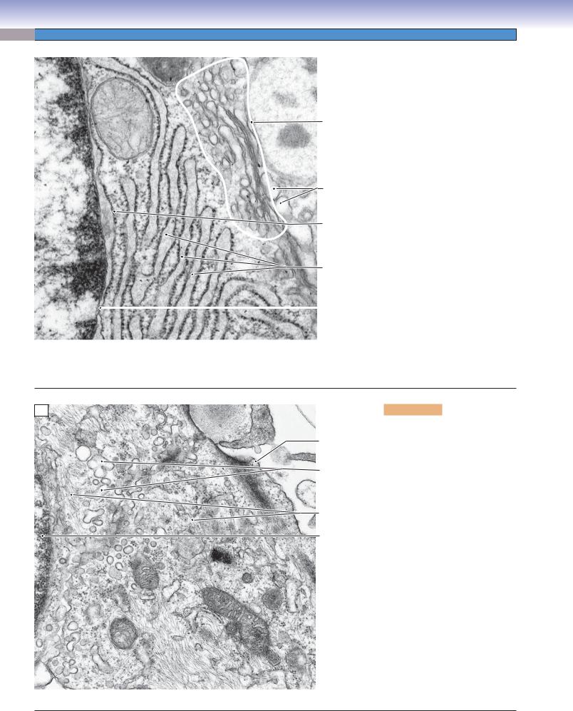
This image is restricted to a very small part of the surfaces of two absorptive cells (enterocytes) in the wall of the small intestine. The plane of section is tangential to the plane of the surface, so that the right side of the image shows numerous microvilli that project into the lumen and function to greatly increase the surface area exposed to the contents of the intestine. The electron-dense dots in the cores of the microvilli are actin filaments, which, in this case, provide for stiffness rather than motility. These actin filaments extend from the microvilli into the cytoplasm as part of the terminal web of actin filaments near the cell surface. Components of a junctional complex provide a seal (zonula occludens) and adhesion (zonula adherens and macula adherens) between neighboring enterocytes. Beneath the terminal web of actin filaments are thicker filaments, intermediate filaments, which provide mechanical strength to the cell. Some of the intermediate filaments are anchored in the macula adherens.
B
Microtubules
Intermediate  filaments
/neurofilaments)
Mitochondria
Synaptic vesicles
Figure 2-6B. Cytoskeleton, the dendrite of a neuron. EM, 20,000
The central structure in this view is a dendrite of a neuron. It is a process extending from the cell body of the cell. Dendrites and axons, the other type of neural process, require cytoskeletal elements both for mechanical support and for conveying essential molecules, particles, and organelles over distances that can be quite extensive. The structural support is provided mainly by the intermediate filaments, which are termed neurofilaments in neurons because of their specialized molecular structure. Intermediate filaments appear in electron micrographs as electron-dense single lines when they course within the plane of the section or as electrondense dots when their orientation is vertical to the plane of the section. The movement of molecules and particles along the lengths of dendrites and axons requires microtubules as tracks and motor molecules (dyneins and kinesins) to transport the cargo structures and molecules along the microtubules. Because of their tubular structure, microtubules appear as a closely paired set of parallel lines when they course within the plane of the section or as circles when their orientation is vertical to the plane of the section.

20 UNIT 1 ■ Basic Principles of Cell Structure and Function
Cell Structure
A
Inactive nucleus  (heterochromatin)
Secretory granules (contents extracted)
Secretory granules (contents eosinophilic)
 Nucleolus
 Euchromatin
Heterochromatin
Bilobed nucleus
Basophilic cytoplasm (RER)
Granules of
eosinophilic Hof (Golgi)  leukocyte
Figure 2-7A. Cell components in light microscopy. H&E, 1,075
Only the larger cell components can be distinguished individually by light microscopy. Examples of such readily identifiable components include the nucleus, nucleoli, blocks of heterochromatin and euchromatin, larger secretory vesicles, and larger secondary lysosomes. Because of its limited resolution of about 0.2 μm, light microscopy cannot distinguish smaller cellular components, such as ribosomes, centrioles, cytoskeletal elements, and most primary lysosomes and mitochondria, although the presence of some of these structures can be inferred by their influence on staining reactions when they are present in abundance. For example, ribosomes, whether free or associated with endoplasmic reticulum, impart basophilic staining to a region of cytoplasm where they are concentrated. By contrast, mitochondria impart acidophilic (eosinophilic) staining to regions of cytoplasm where they are concentrated. In cells with a large Golgi complex, its presence can sometimes be distinguished as an unstained region adjacent to the nucleus.
B
Shrinkage artifact
Cytoplasm of neuronal cell body with basophilic patches of RER
Euchromatin in nucleus
 of neuronal cell body
Nucleolus
Nucleus of satellite cell
Figure 2-7B. Wide range of cell sizes, spinal ganglion sensory neuron. H&E, 780
Human cells have a wide range of sizes and shapes. Some of the largest cell types, such as oocytes and megakaryocytes, can be as much as 100 μm in diameter. Similar diameters can be seen in cell bodies of neurons with long axons such as the sensory neurons in this illustration. The enormity of the cell body in the center of the image can be appreciated if its size is compared with that of the satellite cells that surround it. The nucleolus in the nucleus of the neuron is equal in size to the entire nucleus of the satellite cell. The large volume of cytoplasm that surrounds the nucleus of the neuron contains large expanses of RER and Golgi complexes. As large as the cell body is, it amounts to less than 1% of the total cell volume because the axon may be over a meter in length. Cell types with the smallest volumes include spermatozoa, lymphocytes (spherical cells with diameters as little as 5 μm), and endothelial and alveolar cells
(extremely thin cells that allow exchange of gases and other materials between compartments).

CHAPTER 2 ■ Cell Structure and Function |
21 |
Cell Structure Correlates with Function
Nuclear envelope
Euchromatin
Nucleolus
Secretory vesicle
Heterochromatin
Rough endoplasmic reticulum
A
Figure 2-8A. Protein-secreting cells, plasma cell. EM, 17,000
Plasma cells function to synthesize and secrete immunoglobulin, a glycoprotein. Once these cells have differentiated from stimulated B lymphocytes, they secrete the antibodies as fast as they are generated for 1 to 2 weeks before they die. The major structures required for this process are RER, the Golgi complex, and secretory vesicles. The RER is the site of synthesis and sequestering of the polypeptides of the antibody. Posttranslational modification and packaging occur in the Golgi complex, and the secretory vesicles convey the product to the cell surface. Because plasma cells do not store the immunoglobulin, few, if any, secretory vesicles are seen in the cytoplasm. Rather, the cytoplasm is packed with RER, and there is a large Golgi complex (not visible in this section) located adjacent to the nucleus. The nucleus has one or more well-developed nucleoli, but there is a considerable amount of heterochromatin, considering that this is an active protein-secreting cell. The explanation may be that only one protein, an antibody molecule, is secreted, and the cell is terminally differentiated, so it will never divide. The immunoglobulin is secreted into the surrounding interstitial compartment, from which it can enter circulation through walls of small blood or lymph vessels. Even though the cell is not sharply polarized, the nucleus tends to occupy an eccentric position, with the Golgi complex near the center of the cell.
Basophilic |
Nucleus |
Figure 2-8B. |
Light microscopic appearance of plasma cells. H&E, 1,200 |
|
|
||
cytoplasm (RER) |
|
|
|
The appearance of plasma cells in light microscopy is consistent with the ultrastructure of these antibody-secreting cells. The nucleus has a mixture of heterochromatin and euchromatin in an arrangement that is variously described Nucleolus as a “clock face” or checkerboard pattern. A large nucleolus or two occupy the center of the nucleus. The cytoplasm is basophilic as a result of the ribonucleic acid associated with the extensive RER. Depending upon the orientation of the cell in the section, a pale area, called the hof, can be seen adjacent to the nucleus.
This unstained area is the location of the Golgi complex.
B
Site of Golgi complex (hof)
22 UNIT 1 ■ Basic Principles of Cell Structure and Function
A
Lumen
Secretory granules
Golgi complex
Euchromatin in nucleus
Nucleolus
Rough endoplasmic reticulum
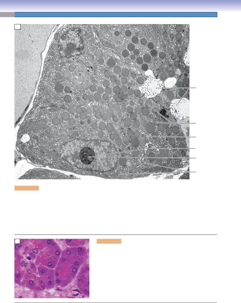
Figure 2-9A. Exocrine protein–secreting cells, pancreatic acinar cell. EM, 7,000
There are many examples of glandular epithelial cells that function to synthesize and secrete proteins and glycoproteins into a lumen. All cells have in common the equipment needed for this function, and all cells are distinctly polarized, with a typical arrangement of cellular constituents. The necessary organelles are the RER, Golgi complex, and secretory vesicles (often called secretory granules). The RER functions to synthesize polypeptides, sequester them, and initiate posttranslational modifications such as glycosylation. The polypeptides are then shunted by small transport vesicles to the Golgi complex for further modification and packaging into secretory vesicles, which convey the products to the part of the cell’s plasmalemma that borders a lumen or free surface. The nucleus of an exocrine protein–secreting cell can vary in shape and position, but it will typically have nucleoli evident and a substantial proportion of its chromatin in the euchromatin (transcription-capable) form.
B Nucleolus
Secretory granules
Lumen
Basophilic |
Site of Golgi |
|
cytoplasm |
||
|
Figure 2-9B. Light microscopic appearance of an exocrine protein– secreting cell. H&E, 750
Light microscopic views of exocrine secretory cells in sections are consistent with the structures that can be discerned in electron micrographs. The nucleus usually exhibits nucleoli and a considerable amount of euchromatin. The cytoplasm at the basal end of the cell is basophilic owing to the concentration of RER at this location. The position of the Golgi apparatus may be evident as a pale, unstained area of cytoplasm at the apical-facing edge of the nucleus. The sizes and staining characteristics of the secretory granules vary according to the type of cell and secretory product. In this example, the secretory granules are acidophilic (eosinophilic).
CHAPTER 2 ■ Cell Structure and Function |
23 |
A
Golgi complex
Nuclear envelope
Smooth endoplasmic reticulum
Nucleolus
Secondary lysosome
Rough endoplasmic reticulum
Mitochondrion with tubular cristae
Lipid droplet
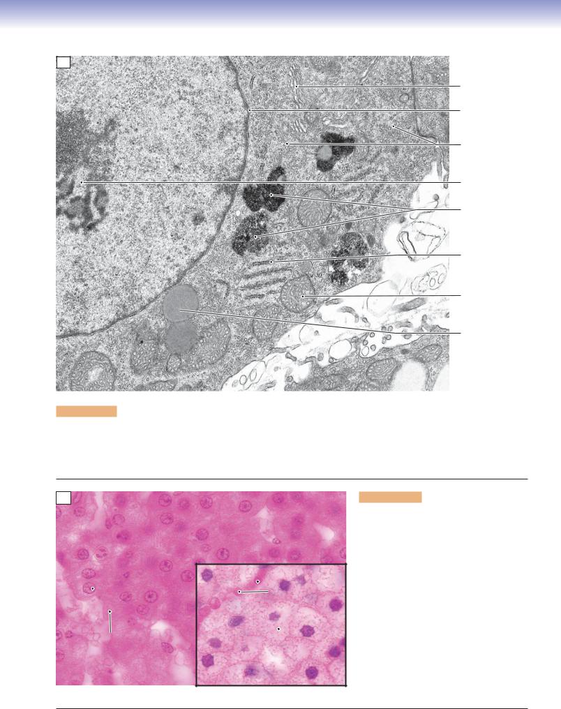
Figure 2-10A. Steroid hormone–secreting cells, adrenal cortex. EM, 21,000
Adrenal cortical cells, testicular interstitial cells, and ovarian follicular cells are examples of cell types that function to synthesize and secrete steroid hormones. Each of these cells have in common the equipment needed for synthesis of steroid hormones. The necessary components include lipid droplets containing cholesterol esters as substrates, SER, and mitochondria that function together in the synthesis of hormones. The mitochondria characteristically have cristae that are tubular rather than shelflike.
B
Nucleolus
 in nucleus
Eosinophilic
cytoplasm
 Red cells in capillary

 Cytoplasm filled with lipid droplets
Figure 2-10B. Light microscopic appearance of steroid hormone–secreting cells. H&E, 800 main panel and inset
Light microscopic views of steroid hormone– synthesizing cells reveal only a hint of the distinctive set of structures that characterize the cytoplasm of these cells. The eosinophilia (acidophilia) of the cytoplasm is largely attributable to the abundance of mitochondria. Steroid hormone–secreting cells vary in the amount of cholesterol stored in the form of lipid droplets, which appear in most preparations as empty vacuoles because their contents are extracted during specimen preparation. The cells in the main panel are in the zona reticularis and have few droplets, similar to the cell in the electron micrograph (Fig. 2-10A). The cells in the inset are from the zona fasciculata and have numerous lipid droplets.

24 UNIT 1 ■ Basic Principles of Cell Structure and Function
A
Nucleus
Mitochondria
Basolateral folds of renal tubule epithelial cell
Basal lamina of renal tubule cell
Interstitial compartment
Red blood cell in capillary
Figure 2-11A. Ion-pumping cells, renal tubule. EM, 9,060
Examples of epithelial cells that function primarily to move ions from a lumen to the interstitial compartment form the walls of some renal tubules and of some ducts of salivary glands. Perhaps surprisingly, the molecular pumps are located not in the apical cell membrane but rather in the plasmalemma of the basal and lateral surfaces of the cells. These ATP-requiring pumps transfer sodium ions from the cytosol of the cell into the interstitial compartment. Sodium ions are pulled from the lumen at the apex of the cell by the gradient created by the basolateral pumps. The surface area of the membrane containing the pumps is greatly increased by numerous, deep basolateral folds. The energy required to pump ions against a concentration gradient is supplied by numerous mitochondria packed into the cytoplasm of the basolateral folds.
B |
Renal medulla |
|
|
Proximal tubule cells |
Collecting duct |
||
cells with channels, |
|||
with basolateral folds, |
|||
but without ion pumps |
|||
ion pumps, and abundant |
|||
|
|||
mitochondria |
|
||
Striated duct
Striations
Figure 2-11B. Light microscopic appearance of ion-pumping cells. H&E, 770 main panel; 710 inset
Light microscopic views of cells that pump ions from one compartment to another are consistent with the ultrastructural features of these cells. The cytoplasm is distinctly acidophilic (eosinophilic in an H&E-stained specimen) owing to the abundance of mitochondria, particularly in the basal portions of the cells. In paraffin sections such as the main panel view of the renal medulla, the basolateral folds are usually difficult to discern. The inset shows a plastic-embedded, 1-μm thick section of a duct from a submandibular salivary gland. Evidence of the basolateral folds can be seen here as vertical stripes in the basal cytoplasm of these cells, which function to retrieve ions from the secretion (saliva). This appearance is the basis of the term striated duct for this structure.
UNIT 2 ■ Basic Tissues
3 |
Epithelium and |
|
Glands |
||
|
|
|
|
|
|
|
|
|
Epithelium
Introduction and Key Concepts for Epithelium
Figure 3-1A |
Overview of Types of Simple Epithelia |
Figure 3-1B |
Overview of Types of Stratified Epithelia |
Table 3-1 |
Classification of Epithelium |
Simple Squamous Epithelium |
|
Figure 3-2A,B |
Simple Squamous Epithelium, Oviduct |
Figure 3-2C |
Clinical Correlation: Mesothelioma |
Figure 3-3A,B |
Simple Squamous Epithelium, Blood Vessels |
Figure 3-3C |
Clinical Correlation: Atherosclerosis |
Figure 3-4 |
Simple Squamous Epithelium in the Medulla of the Kidney |
Simple Cuboidal Epithelium |
|
Figure 3-5A,B |
Simple Cuboidal Epithelium, Thyroid |
Figure 3-5C |
Clinical Correlation: Hyperthyroidism |
Figure 3-6A,B |
Simple Cuboidal Epithelium, Kidney Tubules |
Figure 3-6C |
Clinical Correlation: Renal Fanconi Syndrome |
Simple Columnar Epithelium
Figure 3-7A,B Simple Columnar Epithelium, Small Intestine
Figure 3-7C Clinical Correlation: Celiac (Coeliac) Disease
Figure 3-8A,B Components of the Junctional Complex
Pseudostratified Columnar Epithelium
Figure 3-9A,B |
Pseudostratified Columnar Epithelium, Trachea |
Figure 3-9C |
Clinical Correlation: Bronchitis |
Figure 3-10 |
Pseudostratified Columnar Epithelium (Respiratory Epithelium) |
Three Apical Specializations of Epithelium |
|
Figure 3-11A |
Cilia |
Figure 3-11B |
Microvilli |
25

26 UNIT 2 ■ Basic Tissues
Figure 3-11C |
Stereocilia |
Figure 3-12A |
Cross Section of Cilia |
Figure 3-12B |
Cross Section of Microvilli |
Synopsis 3-1 |
Specialized Structures of the Epithelial Cell |
Stratified Squamous Epithelium (Keratinized) |
|
Figure 3-13A |
Stratified Squamous Epithelium, Thick Skin |
Figure 3-13B |
Stratified Squamous Epithelium, Thin Skin |
Figure 3-13C |
Clinical Correlation: Psoriasis |
Stratified Squamous Epithelium (Nonkeratinized) |
|
Figure 3-14A,B |
Stratified Squamous Epithelium, Esophagus |
Figure 3-14C |
Clinical Correlation: Barrett Syndrome (Barrett Esophagus) |
Stratified Cuboidal Epithelium |
|
Figure 3-15A,B |
Stratified Cuboidal Epithelium, Salivary Gland |
Figure 3-15C |
Clinical Correlation: Salivary Gland Swelling |
Stratified Columnar Epithelium |
|
Figure 3-16A,B |
Stratified Columnar Epithelium, Eyelid |
Figure 3-16C |
Clinical Correlation: Trachoma |
Transitional Epithelium (Stratified Epithelium) |
|
Figure 3-17A |
Transitional Epithelium, Urinary Bladder |
Figure 3-17B |
Transitional Epithelium, Relaxed |
Figure 3-17C |
Transitional Epithelium, Distended |
Synopsis 3-2 |
Pathological Terms for Epithelial Tissue |
Table 3-2 |
Epithelium |
Synopsis 3-3 |
Functions of Epithelial Tissue |
Glands (Exocrine Glands)
Introduction and Key Concepts for Glands
Exocrine Glands Classified by Product
Figure 3-18A |
Parotid Gland |
Figure 3-18B |
Duodenum |
Figure 3-18C |
Sublingual Gland |
Figure 3-18D |
Skin (Scalp) |
Figure 3-19A |
Serous Acinus from the Parotid Gland |
Figure 3-19B |
Mucous Acinus from the Submandibular Gland |
Synopsis 3-4 |
Glands Classified by Product |
Exocrine Glands Classified by Morphology |
|
Figure 3-20A,B |
Unicellular Glands |
Figure 3-21A,B |
Simple Tubular Glands |
Figure 3-22A,B |
Simple Branched Tubular Glands |
Figure 3-23A,B |
Simple Coiled Tubular Glands |
Figure 3-24A,B |
Simple Acinar Glands |
Figure 3-25A,B |
Simple Branched Acinar Glands |
Figure 3-26A,B |
Compound Tubular Glands |
Figure 3-27A,B |
Compound Acinar Glands |
Figure 3-28A,B |
Compound Tubuloacinar Glands |
Table 3-3 |
Glands Classified by Morphology |