


Contents
Preface vii
Acknowledgments ix
Figure Credits x
Expert Consultants and Reviewers xi
Unit 1 ■ BASIC PRINCIPLES OF CELL STRUCTURE AND FUNCTION 1
1 ■ An Illustrated Glossary of Histological and Pathological
Terms 1
2 ■ Cell Structure and Function 11
Unit 2 ■ BASIC TISSUES 25
3 ■ Epithelium and Glands 25
4 ■ Connective Tissue 56
5 ■ Cartilage and Bone 79
6 ■ Muscle 98
7 ■ Nervous Tissue 115
8 ■ Blood and Hemopoiesis 134
Unit 3 ■ ORGAN SYSTEMS 155
9 ■ |
Circulatory System |
155 |
10 ■ |
Lymphoid System |
178 |
11 ■ |
Respiratory System |
199 |
12 ■ |
Urinary System 220 |
|
13 ■ |
Integumentary System 241 |
|
14 ■ |
Oral Cavity 256 |
|
15 ■ |
Digestive Tract 279 |
|
xiii

xiv Contents
16 ■ Digestive Glands and Associated Organs 304
17 ■ Endocrine System 323
18 ■ Male Reproductive System 344
19 ■ Female Reproductive System 369
20 ■ Eye 388
21 ■ Ear 409
Appendix ■ General Principles of Tissue Preparation and Staining 425
Index 431

UNIT 1 ■ Basic Principles of Cell Structure and Function
An Illustrated
1 Glossary of
Histological and
Pathological Terms
Descriptive Terms for Normal Cells
Cell Shape
Cytoplasm Features
Nucleus Features
Cell Size
Descriptive Terms for Abnormal Cells and Tissues
Acute Inflammation
Apoptosis
Atrophy
Calcification
Chronic Inflammation
Cellular Accumulations
Granulomatous Inflammation
Hyperplasia
Hypertrophy
Hydropic Change
Karyorrhexis
Metaplasia
Monomorphism
Multinucleation
Pleomorphism
Pyknosis
Scar
Necrosis
Pigments
Ulcer
1
2UNIT 1 ■ Basic Principles of Cell Structure and Function
Descriptive Terms for Normal Cells
Cell Shape
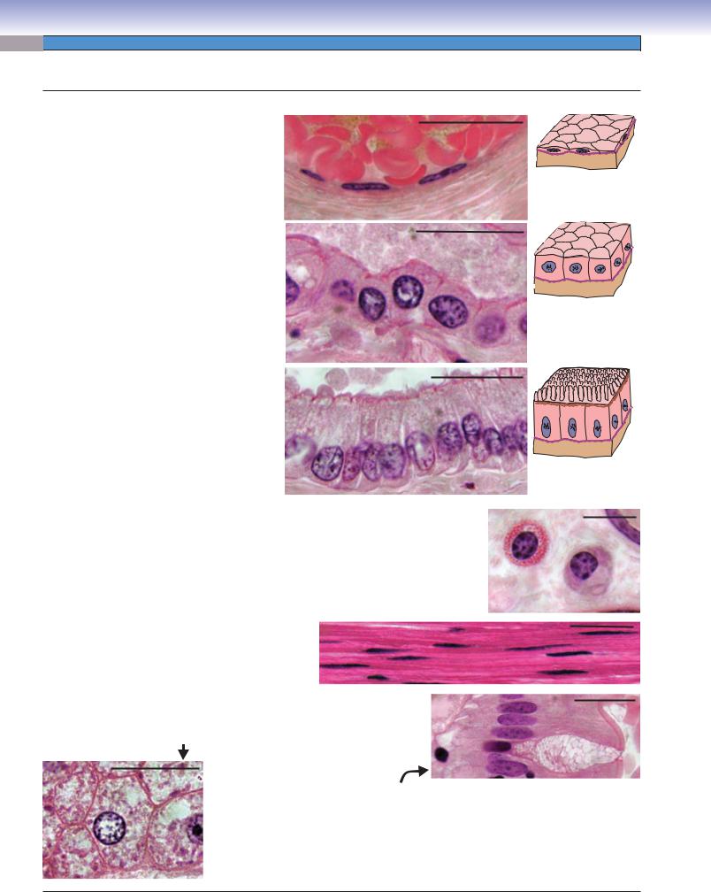
Squamous: |
This is an epithelial cell that has |
|
a flattened, |
“squamous” shape. Usually, the |
20 μm |
squamous cells we see in sections are cut edgewise
so that they appear very thin, and about all we see
D. Cui
is the flattened nucleus. The example here shows nuclei of three endothelial cells lining a vein full of erythrocytes.
Cuboidal: This is an epithelial cell with equal |
|
height and width. The example here is a layer of |
20 μm |
cuboidal epithelial cells lining a small duct in the |
|
pancreas. |
|
Columnar: This is an epithelial cell with the height distinctly greater than the width. The example here is a layer of columnar cells lining a large duct in the pancreas.
D. Cui
20 μm
D. Cui
Spherical or ovoid: These terms describe ball-shaped or egg-shaped cells, respectively. The examples here are two spherical cells, a mast cell on the left and a plasma cell on the right. These residents of connective tissue can assume other shapes depending on pressures from neighboring structures.
Fusiform: These cells are elongated and tapering at the two ends. The examples here are smooth muscle cells packed together in parallel. The elongated nucleus of each cell is easy to see, but the boundaries of the long spindleshaped cells are difficult to distinguish.
10 μm
20 μm
Polyhedral: Multiple flattened surfaces give the appearance of a
pentagon, hexagon, and so on. The hepatocyte shown displays 20 μm six sides.
20 μm
Polarized: These cells have a distinct orientation with one end of the cell being different from the other. Each columnar cell in the above example of intestinal epithelium has an apical end on the right that is different than the basal end on the left. The apical ends are exposed to the lumen. Most of the cells are absorptive cells that have a brush border and a dense row of microvilli extending from their apical surfaces. The cell that appears to have vacuoles filling the apical two thirds of its cytoplasm is a goblet cell, a mucus-secreting cell. What appear to be vacuoles are actually secretory vesicles that lost their contents during processing of the tissue.

CHAPTER 1 ■ An Illustrated Glossary of Histological and Pathological Terms |
3 |
Cytoplasm Features
Basophilia versus acidophilia (eosinophilia): In a
hematoxylin and eosin (H&E)–stained section, 20 μm basophilic components stain blue-purple; aci-
dophilic components stain pink-red. The arrow indicates the basal part of the cytoplasm of a cell of a salivary gland duct. The acidophilic (pink) staining results from a concentration of mitochondria in this part of the cell. The arrowhead indicates the cytoplasm of a plasma cell. The basophilic (bluish) staining results from a concentration of rough endoplasmic reticulum.
Granules: These vesicles stain because they retained at least
some of their contents during tissue preparation. The arrow 20 μm indicates small acidophilic granules filling the cytoplasm of
an eosinophil. The arrowhead indicates large acidophilic secretory granules in the apical cytoplasm of a Paneth cell. Note that the basal cytoplasm surrounding the nucleus of the Paneth cell stains basophilic, indicating large amounts of rough endoplasmic reticulum in this region of the cell.
Vacuolated cytoplasm: This type of cytoplasm contains what appear to  be empty holes. Usually, these represent either lipid droplets or vesicles 20 μm
whose contents were washed out during processing of the tissue. What appear to be empty spaces in the cells from the adrenal cortex shown here were actually occupied, in the living state, by droplets of lipid (cholesterol) that were extracted during tissue preparation.
Abundant versus scant: This pair of terms describes a substantial amount (volume) of cytoplasm (cell 1) or a slight amount of cyto-
plasm (cell 2) surrounding the nucleus. These are inexact terms but 1 2 are sometimes useful in describing a cell.
20 μm

4UNIT 1 ■ Basic Principles of Cell Structure and Function
Nucleus Features
Large versus small: This is another |
|
|
|||
imprecise set of terms, but useful if it’s |
1 |
|
|||
possible to make comparisons with nuclei |
2 |
||||
of other cells in the field of view. Label 1 |
|||||
indicates a relatively large nucleus; labels |
|
3 |
|||
3, 4, and 6 indicate small nuclei. |
|
|
|||
Euchromatic |
versus |
heterochromatic: |
|
||
|
4 |
||||
Nuclei of most cells show us a mixture of |
|
||||
euchromatin |
(dispersed, |
lightly |
stained, |
|
|
|
|
||||
accessible to transcription) and hetero- |
|
|
|||
chromatin (condensed, |
darkly |
stained, |
|
|
|
inactive). However, there is a wide variabil- |
|
5 |
|||
ity in the relative proportions of the two |
|
||||
forms. Nucleus 1 has mostly euchroma- |
|
|
|||
tin. Nucleus 6 is highly heterochromatic. |
|
|
|||
Nuclei 3 and 4 have mixes of heterochro- |
|
|
|||
matin and euchromatin. Notice that the |
|
6 |
|||
size of a nucleus is roughly proportional |
|
||||
to its content of euchromatin. |
|
|
|
||
Nucleoli prominent: Presence of an obvi- |
|
|
|||
ous nucleolus (nucleus 1) or nucleoli |
|
|
|||
indicates that the cell is actively synthe- |
|
20 μm |
|||
sizing ribosomes and, therefore, proteins. |
|
||||
Typically, nucleoli are present in a nucleus
that has most of its chromatin in the form of euchromatin, but there are exceptions (for example, some malignant tumor cells).
Mitotic nucleus: When a cell divides, the nuclear envelope dissolves, and the chromosomes are dark, condensed particles. Label 2 |
||
indicates the chromosomes of a metaphase cell. Label 5 indicates two clusters of chromosomes in an anaphase cell. |
|
|
Simple versus segmented: A simple nucleus appears as a single structure that can have a variety |
10 μm |
|
of shapes (round, oval, indented, fusiform, irregular). All the nuclei in this photomicrograph are |
||
|
||
simple nuclei. A segmented nucleus, which is typical of some types of white blood cells, appears in |
|
|
sections as two or more distinct parts (lobes), as seen in the cell in the center of this small panel. |
|
|
Inferences from nucleus appearance: The appearance of the nucleus provides some clues about the state of the cell’s activity. Cells that are active in protein synthesis will usually have fairly large nuclei, prominent nucleoli, and a preponderance of euchromatin. Examples of such cells are rapidly dividing cells (which are busy duplicating cell constituents to be divided among daughter cells), cells that secrete proteins, and cells that have a very large area of membrane and a large volume of cytoplasm to maintain (e.g., neurons).
Cell Size
Large versus small: Although many cell types have diameters falling in the range of 10 to 20 μm, the total spectrum of cell sizes is much broader. There are cells much smaller (e.g., inactive lymphocyte at about 6 μm) or much larger (e.g., megakaryocyte at about 100 μm). If cell volume and surface area are considered things can get pretty tricky, because many cells are not compact spheres or simple geometric shapes but have other shapes that can be extremely complex.