

- •Dedication
- •Preface
- •Acknowledgments
- •Figure Credits
- •Expert Consultants and Reviewers
- •Contents
- •Descriptive Terms for Normal Cells
- •Descriptive Terms for Abnormal Cells and Tissues
- •Epithelium
- •Glands
- •Introduction and Key Concepts for Connective Tissue
- •Cartilage
- •Bone
- •Introduction and Key Concepts for the Nervous System
- •Peripheral Blood Cells
- •Hemopoiesis
- •Introduction and Key Concepts for the Circulatory System
- •The Cardiovascular System
- •Introduction and Key Concepts for the Lymphoid System
- •Cells in the Lymphoid System
- •Introduction and Key Concepts for the Respiratory System
- •Conducting Portion
- •Respiratory Portion
- •Introduction and Key Concepts for the Urinary System
- •Introduction and Key Concepts for the Integumentary System
- •Oral Mucosa
- •Teeth
- •Introduction and Key Concepts for the Digestive Tract
- •Introduction and Key Concepts for the Endocrine System
- •Introduction and Key Concepts for the Male Reproductive System
- •Introduction and Key Concepts for the Female Reproductive System
- •Introduction and Key Concepts for the Eye
- •Introduction and Key Concepts for the Ear
- •Introduction
- •Preservation versus Fixation
- •Fixatives and Methods of Fixation
- •Sectioning and Mounting
- •Staining
- •Index

CHAPTER 20 ■ Eye |
|
389 |
Tunica Vasculosa (Tunica Media) |
||
Figure 20-9A |
Overview of the Iris and Nearby Structures |
|
Figure 20-9B |
Iris |
|
Figure 20-9C |
Constrictor Pupillae Muscle and its Function |
|
Figure 20-9D |
Dilator Pupillae Muscle and its Function |
|
Figure 20-10A |
Anterior Surface of the Lens |
|
Figure 20-10B |
Posterior Surface of the Iris |
|
Figure 20-11A |
Overview of the Ciliary Body and Nearby Structures |
|
Figure 20-11B |
Ciliary Processes and the Ciliary Muscle |
|
Figure 20-11C |
Ciliary Body, Transverse and Posterior Views |
|
Figure 20-12A |
Posterior Portion of the Ciliary Body |
|
Figure 20-12B |
Ora Serrata |
|
Figure 20-12C |
Clinical Correlation: Outflow of Aqueous Humor and Glaucoma |
|
Retina (Tunica Interna) |
|
|
Figure 20-13A |
Overview of the Retina; Fovea |
|
Figure 20-13B |
Macular Region of the Retina and the Retinal Layers |
|
Figure 20-13C |
Peripheral Region of the Retina and Distribution of the Rods and Cones |
|
Figure 20-14A,B |
A Representation of Rods and Codes |
|
Table 20-1 |
Comparison of Rods and Cones |
|
Figure 20-15A–C |
Retinal Layers |
|
Synopsis 20-1 |
Retinal Layers |
|
Figure 20-16A |
Clinical Correlation: Age-Related Macular Degeneration |
|
Figure 20-16B |
Clinical Correlation: Retinal Detachment |
|
Optic Nerve |
|
|
Figure 20-17A |
Optic Nerve and Optic Disk |
|
Figure 20-17B |
Clinical Correlation: Papilledema |
|
Synopsis 20-2 |
Clinical Terms for the Eye |
|
Introduction and Key Concepts for the Eye
The eye is the organ of vision, perhaps the most important of the sensory modalities. The eye converts light into nerve impulses in a way that allows the brain to be aware of the individual’s visual surroundings. In our study, we divide the structure of the eye into three general categories: those structures which protect the eye (eyelids); those structures which help form a visual image of what the individual is looking at (cornea, lens, sclera, and associated structures); and those structures which convert the visual image into nerve impulses and conduct the impulses to the brain (retina and optic nerve), where they are analyzed to produce the sensation of vision.
The basic structure of the eye is that of a hollow sphere with optical elements on the anterior surface that focus an inverted image of the surroundings onto the inside of the posterior wall. The primary structural element of the sphere is the tunica fibrosa, or tunica externa, which consists of the sclera and the cornea. Lining the inside of the tunica externa is the tunica vasculosa, consisting of the choroid, the ciliary body, and the iris. The eye is filled with a transparent liquid
(aqueous humor) and a transparent gel (vitreous body). In the anterior portion of the eye is the lens, which is flexible and can adjust the focus of the image depending on the distance from the eye to the object being viewed. The image is focused on the retina, a layer of neurons and neural receptors that line the internal surface of the posterior two thirds of the eye. The axons of the ganglion cell neurons in the retina leave the eye and form the optic nerve.
The Eyelids
The eyelids protect the eyes from injury by foreign objects and also maintain a thin film of moisture on the surface of the cornea that prevents the cornea from drying out and becoming opaque. Each eyelid consists of an outer layer of skin; a middle layer of muscle, glands, and connective tissues (tarsal plate); and an inner layer of conjunctival tissue (palpebral conjunctiva). There are several types of glands in the eyelid that aid in keeping the cornea moist, including meibomian glands, glands of Zeis, glands of Moll, and accessory lacrimal glands (Figs. 20-3 and 20-4).
Several muscles are associated with the eyelids. These include (1) the orbicularis oculi muscle, a circular sheet of striated muscle that is innervated by the facial nerve (cranial nerve

390 UNIT 3 ■ Organ Systems
[CN] VII) and functions to close the eyelids; (2) the levator palpebrae superioris muscle, a thin flat striated muscle that originates in the orbit, passes forward, and inserts into the upper eyelid. It is innervated by the oculomotor nerve (CN III) and is responsible for opening the eyelid and holding it open; and (3) the superior tarsal muscle (Müller muscle), a bundle of smooth muscle that arises from the interstitia of the levator muscle and inserts on the upper end of the tarsal plate and superior conjunctiva of the eyelid. The superior tarsal muscle is innervated by sympathetic nerve fibers from the superior cervical ganglion and helps to raise the upper eyelid.
Tunica Fibrosa (Tunica Externa)
The outermost structures of the eye are the cornea and sclera (Fig. 20-1).
THE CORNEA is a transparent tissue that covers the anterior sixth of the eye (Figs. 20-5 and 20-6). The cornea contains no blood vessels and aids in focusing the visual image onto the retina. It consists of five layers. The thickest layer, the stroma, comprises 90% of the thickness of the cornea and consists of collagen fibers and fibroblasts embedded in an extracellular matrix (Fig. 20-6B). The anterior surface of the cornea is covered by a thin layer of pavement epithelium (stratified squamous epithelium) resting on the Bowman membrane. The posterior surface of the cornea is covered by a layer of corneal endothelium (simple squamous epithelium) that is only one cell thick and rests on the Descemet membrane (Figs. 20-5A and 20-6B).
THE SCLERA is a tough, thin structure consisting of dense, irregular, opaque connective tissue that comprises the posterior five sixths of the outer surface of the eyeball (Figs. 20-1, 20-11A, and 20-12A). The cornea and sclera are continuous with each other at the limbus (Fig. 20-11A). The extraocular muscles, which move the eyes in their orbits, insert in the sclera. The conjunctival tissue, which covers the inner surfaces of the eyelids, also attaches to the sclera.
Refractive Media of the Eye
THE LENS is a transparent, flexible, biconvex structure that is suspended from the ciliary processes by zonular fibers. The curvature of the lens can be changed by contraction or relaxation of the ciliary muscles (under control of parasympathetic nerve fibers of the oculomotor nerve) so that the image of nearby or distant objects can be focused on the retina. The lens has three components: the lens capsule, the subcapsular epithelium, and the lens fibers (Figs. 20-7 and 20-8). The lens capsule is a transparent basement membrane that surrounds the entire lens. Immediately beneath it, on only the anterior surface of the lens, is a single layer of squamous cells, the subcapsular epithelium (Fig. 20-10A). In the region of the equator of the lens, proliferating epithelial cells become elongated, are displaced toward the center of the lens, and lose their nuclei. They are then called lens fibers and comprise the major bulk of the lens (Fig. 20-10A).
THE AQUEOUS HUMOR is a thin, watery, transparent fluid that is produced continuously by the ciliary body and fills the anterior chamber. It exits the anterior chamber in the region of the angle of the anterior chamber. It is produced by the
ciliary processes, which are rich in capillaries (Figs. 20-11C and 20-12C).
THE VITREOUS BODY is a transparent gelatinous substance that fills the eye between the posterior surface of the lens and the retina (Fig. 20-1). Its composition is predominantly water with small amounts of collagen and hyaluronic acid. The surface of the vitreous body is covered by a layer of condensed vitreous fibers called hyaloid membrane. It is in contact with the posterior lens capsule, the zonular fibers, the posterior portion of the ciliary epithelium (pars plana), the retina, and the optic nerve head (Fig. 20-1). The vitreous body is important in maintaining the transparency and shape of the eye.
Tunica Vasculosa (Tunica Media)
The tunica vasculosa (sometimes called the uveal tract) lies just internal to the tunica externa and consists of the iris (anteriorly), the ciliary body, and the choroid (posteriorly).
THE IRIS is a thin diaphragm of tissue in the anterior chamber, composed of a highly vascularized, loose connective tissue stroma, two groups of contractile elements, the anterior iridal border, and the posterior iridal border. The posterior iridal border contains two layers of pigmented epithelium, the anterior iridal epithelium (anterior pigmented epithelium) and the posterior iridial epithelium (posterior pigmented epithelium). Two groups of muscle fibers regulate the diameter of the pupil, the circular hole in the center of the iris, and adjust the amount of light entering the eye (Fig. 20-9). The circular constrictor pupillae muscle (smooth muscle fibers) reduces the size of the pupil under the influence of parasympathetic nerve fibers; the radial fibers of the dilator pupillae muscle (myoepithelial cells) act to increase the size of the pupil under the influence of sympathetic nerve fibers.
THE CILIARY BODY lies interior to the anterior margin of the sclera, between the choroid and the iris. It is composed of two concentric rings of tissue, the pars plicata and the pars plana, and includes epithelial tissue, a stroma of connective tissue, and smooth muscle fibers (Figs. 20-9A and 20-11). The muscles of the ciliary body control the curvature of the lens and, therefore, function to focus the visual image on the retina. The epithelium of the ciliary body has two layers, a pigmented layer and a nonpigmented layer. The latter secretes the aqueous humor, which fills the anterior chamber of the eye and leaves the anterior chamber in the region of the anterior chamber angle (Fig. 20-12C).
THE CHOROID is a highly vascularized tissue containing some collagen fibers that is loosely attached to the overlying sclera (Fig. 20-13A). The inner surface of the choroid adheres tightly to the pigment epithelium layer of the retina. The innermost layer of the choroid is the choriocapillaris, which supplies oxygen and nutrients to the outer layers of the retina. Bruch membrane delineates the junction between the choriocapillaris and the retinal pigment epithelium (Fig. 20-13B).
Retina (Tunica Interna)
The retina consists of a thin sheet of neurons that covers the inner surface of the posterior two thirds of the eye and a layer of cuboidal epithelial cells that sit on the choroid and contain

CHAPTER 20 ■ Eye |
391 |
melanin (retinal pigment epithelium). In general, the retina can be divided into an optic (neural) retina and a non-optic (nonneural) retina. The neural retina, often referred to as simply “retina,” contains neural elements and has the visual functions described below. The nonneural retina has no neural elements and no visual function. It is the anterior continuation of the pigmented layer, which covers the surface of the ciliary body and the posterior surface of the iris.
Unlike the rest of the eye, the retina develops as a part of the central nervous system (CNS). It contains five types of neural elements: photoreceptor cells (rods and cones), bipolar cells, horizontal cells, amacrine cells, and ganglion cells (Fig. 20-15). The retina can be divided into 10 layers, some of which contain the nuclei of cells and others of which contain cell processes and synapses (Figs. 20-13 and 20-15).
The retina is not homogeneous throughout its extent. Over most of the retina, the focused light of the visual image must pass through all of the neural layers, as well as small blood vessels, before reaching the photoreceptors. This degrades the image to a certain extent. However, in the fovea centralis, a small
region near the posterior pole of the eye, the superficial layers are displaced to the side, and light strikes the photoreceptors directly (Figs. 20-1 and 20-13A). The visual image in this region is perceived with the greatest detail. The area immediately surrounding the fovea centralis is the macula lutea (Fig. 20-13B). This is the thickest region of the retina and contains a high concentration of both cones (for color vision) and rods (for low-light vision [Fig. 20-14]). More peripherally, the retina becomes thinner. There are fewer ganglion cells, fewer cones, and a relatively higher proportion of rods (Fig. 20-13C). These changes cause visual acuity to be reduced but sensitivity to low light levels to increase.
Optic Nerve
The ganglion cells are the output cells of the retina (Fig. 20-15C). The axons of ganglion cells travel in the nerve fiber layer to the optic disk, where the nerve fibers exit the eye and form the optic nerve (Fig. 20-17A). There are no photoreceptors in the optic disk; this absence produces a small blind spot in the visual field. The appearance of the optic disk when viewed through an ophthalmoscope is an important diagnostic aid.

392 UNIT 3 ■ Organ Systems
|
|
Extraocular muscles |
|
|
|
|
Sclera |
|
|
|
|
Choroid |
|
|
|
|
Retina |
|
|
|
|
Ora serrata |
|
|
|
|
|
Ciliary muscle |
Ciliary body |
|
|
|
Ciliary processes |
|
|
|
|
|
|
|
|
|
Zonular fibers |
|
|
Macula |
Vitreous body |
|
|
|
Lens |
Pupil |
|
|
|
Fovea |
|
||
|
|
|
||
|
|
Aqueous |
|
|
Cerebrospinal |
|
humor |
Cornea |
|
|
Optic disk |
Iris |
|
|
fluid |
|
|
||
|
(Optic nerve papilla) |
|
|
|
|
|
Anterior chamber |
||
Central retinal |
|
|
||
|
|
Posterior chamber |
||
artery and vein |
|
|
||
Optic nerve |
|
Sclera |
|
|
|
|
Choroid |
|
|
Retina
D. Cui
Figure 20-1. Overview of the eye.
The yellow dashed line indicates light coming in and projecting on the fovea.
Structures of the Eye
I.Tunica Fibrosa (Tunica Externa)
A.Cornea
1.Pavement epithelium (anterior corneal epithelium)
2.Bowman membrane
3.Corneal stroma (corneal substantia propria)
4.Descemet membrane
5.Endothelium (posterior corneal epithelium)
B.Sclera
1.Episclera (blood vessels and adipose tissue)
2.Sclera proper
II.Tunica Vasculosa (Tunica Media)
A.Choroid
1.Bruch membrane
2.Choriocapillaris
3.Choroid propria
B.Iris
1.Pigment epithelium
2.Stroma
3.Constrictor pupillae muscle
4.Dilator pupillae muscle
C.Ciliary body
1.Ciliary processes
2.Ciliary epithelium
3.Ciliary muscle
III.Refractive Media of the Eye
A.Lens (biconvex, flexible, transparent structure)
1.Lens capsule
2.Subcapsular epithelium
3.Lens fibers
4.Zonular fibers
B.Aqueous humor (transparent fluid occupying the space between the lens and cornea)
C.Vitreous body (refractive gel filling the interior of the globe posterior to the lens)
IV. Retina (Tunica Interna)
A.Fovea
B.Macula
C.Peripheral retina V. Optic nerve

CHAPTER 20 ■ Eye |
393 |
|
Fig. 20-11A,B,C |
|
Fig. 20-13B |
Fig. 20-9B,C,D |
|
Fig. 20-7C |
||
Fig. 20-13A |
||
Fig. 20-5A,B |
||
|
Fig. 20-6A,B |
Fig. 20-7B
Fig. 20-10A
Fig. 20-13C |
Fig. 20-7D |
Fig. 20-8A
Fig. 20-17A,B
Fig. 20-12A
Fig. 20-12B
D. Cui
Figure 20-2. Orientation of detailed eye illustrations.
Structures of the Eye with Figure Numbers
Eyelid |
Ciliary Body |
Figure 20-3A |
Figure 20-11A |
Figure 20-3B |
Figure 20-11B |
Figure 20-3C |
Figure 20-11C |
Figure 20-4A |
Figure 20-12A |
Figure 20-4B |
Figure 20-12B |
Figure 20-4C |
Figure 20-12C |
Cornea |
Retina |
|
Figure 20-5A |
Figure 20-13A |
|
Figure 20-5B |
Figure 20-13B |
|
Figure 20-5C |
Figure 20-13C |
|
Figure 20-6A |
Figure 20-14A |
|
Figure 20-6B |
Figure 20-14B |
|
Lens |
Figure 20-15A |
|
Figure 20-15B |
||
Figure 20-7A |
||
Figure 20-15C |
||
Figure 20-7B |
||
Figure 20-16A |
||
Figure 20-7C |
||
Figure 20-16B |
||
Figure 20-7D |
||
|
||
Figure 20-8A |
Optic Nerve |
|
Figure 20-8B |
Figure 20-17A |
|
Figure 20-8C |
Figure 20-17B |
|
Figure 20-10A |
|
|
Iris |
|
|
Figure 20-9A |
|
|
Figure 20-9B |
|
|
Figure 20-9C |
|
|
Figure 20-9D |
|
|
Figure 20-10B |
|
|
|
|

394 UNIT 3 ■ Organ Systems
Eyelid
Tendon of the |
|
|
levator |
Superior |
|
palpebrae |
tarsal muscle |
|
muscle |
|
|
|
Meibomian |
|
|
(Tarsal) |
|
|
glands |
|
Orbicularis |
|
|
oculi muscle |
Tarsal |
|
|
||
Skin |
Plate |
|
Eyelash |
Palpebral |
|
conjunctiva |
||
follicles |
||
|
||
A |
Lid margin |
Figure 20-3A. Overview of the upper eyelid. H&E, 7.6
A low-power photomicrograph of the upper eyelid is shown. The eyelids contain an outer layer of skin; a middle layer of muscles, glands, and tarsal plate; and an inner layer of conjunctival tissue
(palpebral conjunctiva). Eyelids cover and protect the eye from the environment, injury, and intense light. They also maintain a smooth corneal surface by spreading a film of lacrimal fluid (tears) evenly over the cornea to moisten the eye. The skin of the eyelids is thin, loose, and delicate and, therefore, may permit extreme swelling. The internal lid is covered by a palpebral conjunctiva, a layer of stratified low columnar epithelium. It is continuous with the bulbar conjunctiva where it covers the sclera of the eyeball. The tarsal plate is a dense fibroelastic tissue, which provides flexible support. The tarsal glands (meibomian glands) are embedded in it. The eyelashes are located in the anterior margins of the eyelids.
Levator |
D. Cui |
palpebrae |
|
superioris |
|
muscle and |
|
its tendon |
|
Orbicularis |
|
oculi |
|
muscle |
|
Sweat |
|
gland |
|
Skin |
|
Gland |
|
of Zeis |
|
B |
Eyelash Gland |
|
of Moll |
Accessory lacrimal
 gland (of Krause)

 Superior
  tarsal muscle

 Accessory lacrimal


 gland (of Wolfring)
Meibomian (tarsal) gland
Tarsal plate
Palpebral conjunctiva
Figure 20-3B. A representation of the upper eyelid, glands, and muscles that control eyelid movements.
Several types of glands in the eyelids include (1) meibomian (tarsal) glands, sebaceous glands that produce a lipid-rich substance; (2) glands of Zeis, modified sebaceous glands associated with the follicles of the eyelashes; (3) glands of Moll, modified sweat glands, associated with eyelash follicles; and (4) accessory lacrimal glands, serous glands that contribute to tears. The muscles associated with upper eyelids are the (1) orbicularis oculi muscle, a circular sheet of striated muscle which functions to close the eyelids; (2) levator palpebrae superioris muscle, a thin, flat, striated muscle that arises from the apex of the orbit and inserts into the posterior surface of the orbicularis oculi muscle and the skin of the upper eyelid, and which opens the eyelid; and (3) superior tarsal muscle (Müller muscle), smooth muscle that arises from the interstitia of the levator muscle and inserts on the upper end of the tarsal plate and superior conjunctiva of the lid. It joins the levator palpebrae superioris muscle in raising the upper eyelid. (For innervation, see text of Fig. 20-4C.)
CLINICAL CORRELATION
D. Cui
Lymphocyte


 Lipid-laden macrophage
Gland |
Nodule |
|
|
of Zeis |
|
|
Obstructed meibomian gland |
C |
|
Figure 20-3C. Chalazion.
Chalazion is a chronic eyelid inflammatory lesion that results from the obstruction of the ducts of either the Zeis or meibomian glands, or both. Trapped sebaceous secretions leak into the surrounding tissue and cause a granulomatous inflammation. This is frequently associated with blepharitis and occasionally becomes secondarily infected. Early symptoms and signs include eyelid swelling and erythema. In time, it changes into a firm nodule within the eyelid or the tarsal plate. Histologic examination reveals granulation tissue characterized by focal aggregation of epithelium-like (epithelioid) cells, lymphocytes, giant cells of Langerhans, and yellow lipid-laden macrophages. Treatment options include warm compresses applied to the outer lid until acute symptoms disappear, topical antibiotics, and surgical incision if the lesion is large and disturbs vision.
CHAPTER 20 ■ Eye |
395 |
A |
|
Meibomian |
|
|
(tarsal) |
|
|
gland |
|
|
Tarsal |
|
|
plate |
|
Gland of Moll |
|
|
Eyelash |
|
|
follicles |
|
|
Gland |
Palpebral |
Eyelashes |
of Zeis |
|
|
conjuctiva |
Figure 20-4A. Upper eyelid (lower part), glands of the eyelid. H&E, 68
Eyelids (palpebrae) consist of upper and lower eyelids. The structural components of the upper eyelid are similar to those of the lower eyelid, although the upper eyelid is more mobile. Eyelashes and their follicles are visible at the margin of the eyelid. The tarsal glands, also called meibomian glands, are large sebaceous glands embedded in the tarsal plate. The glands associated with eyelashes are (1) glands of Moll and (2) glands of Zeis. The glands of Moll are modified sweat glands near the base of the eyelash. They have unbranched tubules, which begin in a simple spiral rather than coiling in a glomerular shape, as do ordinary sweat glands (see Fig. 20-3B). The glands of Zeis are small, modified sebaceous glands that are sometimes called ciliary glands. They are close to the eyelash follicles and empty their secretions into the follicles.
B
Orbicularis oculi muscle
Hair
follicles Skin
Skin 
Meibomian |
|
(tarsal) |
|
gland |
|
oculi |
Palpebral |
conjuctiva |
|
Orbicularis |
Duct of |
muscle |
tarsal |
|
|
|
plate |
Figure 20-4B. Upper eyelid (middle part). H&E, 68
The outer layer of the eyelid is covered by thin skin (see Fig. 3-13 and Chapter 13 “Integumentary system”), a keratinized stratified squamous epithelium, over a loose elastic connective tissue layer. The skin contains hair follicles, which are much smaller than eyelash follicles (found only in the lid margin; see Fig. 20-3A). Orbicularis oculi muscle fibers are located beneath the skin. The inner surface of the lid is a layer of palpebral conjunctiva, covered by stratified low columnar epithelium, which is in contact with the eyeball. The tarsal (meibomian) glands, embedded in the tarsal plate, lie between the orbicularis muscles and palpebral conjunctiva. Each gland has a single duct that opens at the lid margin. Their lipid secretion creates a surface on the tear film that prevents lacrimal fluids (tears) from evaporating from the surface of the eyeball. This secretion also lubricates the cornea and edges of the eyelids.
C |
Orbicularis |
Tendon of the |
Superior |
|
oculi muscle |
levator |
|
(skeletal muscle) |
palpebrae |
tarsal |
|
|
|
muscle |
muscle |
Meibomian (tarsal) gland
Orbicularis oculi muscle
Figure 20-4C. Upper eyelid (upper part), muscular control of upper eyelid movement. H&E, 68; inset 272
Three types of muscles control upper eyelid movement.
(1) The orbicularis oculi muscle is a sheet of striated muscle that is oriented in a circle around the eye. It is innervated by the facial nerve (CN VII) and is responsible for closing the eyelids. (2) The levator palpebrae superioris muscle is a band of striated muscle that originates in the orbit, passes forward, and inserts into the upper eyelid. This muscle is innervated by the oculomotor nerve (CN III) and functions to open the eyelid and hold it up. The levator palpebrae muscle is not visible here, but its tendon is clearly seen. (3) The superior tarsal muscle, also called the Müller muscle, is a smooth muscle that inserts on the superior tarsal plate. It is innervated by sympathetic nerve fibers from the superior cervical ganglion and works with the levator palpebrae superior muscle to raise the upper eyelid. Damage to the levator palpebrae superior muscle, the superior tarsal muscle, or their innervation can cause ptosis (drooping of the eyelid).

396 UNIT 3 ■ Organ Systems
Tunica Fibrosa (Tunica Externa)
A |
|
Pavement epithelium |
Pavement |
|
|
|
(anterior corneal epithelium) |
epithelium |
|
Anterior |
||||
|
|
|||
Bowman membrane
Bowman membrane
Stroma
Descemet Descemet membrane membrane
|
Corneal |
Corneal |
Posterior |
endothelium |
endothelium |
|
|
Figure 20-5A. Cornea. H&E, 77.5; insets 173
The cornea is a transparent and avascular tissue, which is composed of five layers: three cellular layers (epithelial layers and stroma) and two noncellular layers (Bowman membrane and Descemet membrane). These include (1) pavement epithelium (anterior corneal epithelium); (2) the Bowman membrane (basement membrane of the pavement epithelium); (3) stroma, consisting of fibroblasts (also called keratocytes in the cornea) and alternating lamellae of collagen fibers; (4) the Descemet membrane
(basement membrane of the posterior corneal epithelium (endothelium); and (5) the corneal endothelium
(posterior corneal epithelium). The cornea has a rich nerve supply from CN V. The superficial corneal layer contains numerous sensory nerve fibers, and irritation can cause severe eye pain. CN V also carries the afferent limb of the corneal reflex.
B
D. Cui
Pavement epithelium (cellular layer)
Bowman membrane (noncellular layer)
Stroma (cellular layer)
Descemet membrane 

 (noncellular layer)
 Endothelium
(cellular layer)
Figure 20-5B. A representation of the cornea.
Pavement epithelium is formed by stratified squamous epithelium consisting of four to six layers and is about 50 mm in thickness; it has a high capacity for regeneration. The transparency of the cornea is due to its avascularity and due to its state of relative dehydration. Corneal endothelium is a single layer of squamous cells. This layer functions in the regulation of water in the stroma. The primary task of corneal endothelium is to pump the excess fluid out of the stroma and, therefore, is critical for keeping the cornea clear. If this endothelium is damaged, the result may be corneal swelling due to fluid retention within the stroma and loss of its transparency.
CLINICAL CORRELATION
C
Cornea flap
Laser beam |
Exposed Bowman membrane |
D. Cui |
Figure 20-5C. LASEK.
Laser refractive surgery reshapes the cornea in order to focus images more accurately onto the retina. The most common procedures include laser in-situ keratomileusis (LASIK), and laser-assisted epithelial keratomileusis (LASEK). Laser techniques have been continuously modified to reduce complications and enhance surgical outcomes. Unlike with LASIK, the most recent LASEK surgery saves the epithelium by using an alcohol solution to weaken epithelial cell adhesion so the epithelial layer can be lifted as a flap. After the epithelial flap is moved out of the way, excimer laser energy is applied through the Bowman membrane layer and into the upper stroma to reshape the cornea. The epithelial flap is then returned to its original position. The advantages of LASEK are a reduction of postoperative discomfort, a decreased risk of infection, and an increase in the overall thickness of the untouched area of the cornea.

CHAPTER 20 ■ Eye |
397 |
Corneal
stroma
Collagen
fibers
Bowman membrane 
Basal epithelial cells 
A
 Nuclei of the
epithelialit li l cellsll
Posterior (Fig. 20-6B) Anterior (Fig. 20-6A)
Figure 20-6A. Pavement epithelium (anterior epithelium). EM, ×6,000
The anterior corneal epithelium is a stratified squamous epithelium that covers the external surface of the cornea. The lack of keratinization and the uniform thickness of the epithelium contribute to the essential transparency of the cornea. The epithelial cells have a fairly fast turnover rate of about one week. The surface cells are not perfectly smooth, but rather have small microvilli (only a few are preserved here) that serve to anchor the vitally important tear film. Not shown in this field are free nociceptive nerve endings that penetrate the epithelium and provide the afferent limb of the corneal blink reflex. Bowman membrane is the commonly used term for the thick basement membrane at the interface between the anterior corneal epithelium and the corneal stroma.
Corneal stroma
Keratocytes
(fibroblasts)
Nuclei of endothelial cells
Descemet
membrane Collagen
  fibers
B
Figure 20-6B. Corneal endothelium (posterior corneal epithelium).
EM, 4,500
The posterior corneal epithelium is sometimes called the corneal endothelium. It is a simple squamous epithelium that faces the aqueous humor of the anterior chamber. Descemet membrane is the commonly used term for the sharply defined basement membrane of the posterior epithelium. Descemet membrane is unusual in that it consists largely of an orderly network of type VIII collagen, a relatively rare collagen type. The epithelial cells are joined by tight junctions, and they control the movement of water, ions, and metabolites between the stroma and the aqueous humor, the source of nutrition for the corneal stroma. Orderly layers of type I collagen fibrils form the bulk of the corneal stroma. The extremely flattened fibroblasts that produce and maintain the stroma are called keratocytes.

398 UNIT 3 ■ Organ Systems
Refractive Media of the Eye
A
Cornea
|
Posterior chamber |
Iris |
|
Anterior chamber |
Fig. 20-7B |
Lens cortex |
|
Lens |
|
nucleus |
Fig. 20-7D |
|
|
|
Ciliary |
Lens |
|
|
body |
|
|
|
|
|
Fig. 20-7C |
||
Figure 20-7A. Overview of the lens. H&E, 11
The lens is a biconvex, avascular, flexible, and colorless transparent structure. Anterior to the lens is aqueous humor and posterior to it is the vitreous body. The lens is composed of the lens capsule, subcapsular epithelium, and lens fibers. The peripheral region of each lens fiber contains a nucleus and organelles, which form the bulk of the lens called the lens cortex. In the central part of the lens, the fibers lose their nuclei and organelles; this region is filled with crystalline proteins and is called the lens nucleus. The lens is held in place by zonular fibers (position indicated by dashed lines; see Figs. 20-8A and 20-11C). The anterior chamber is the space between the cornea and the iris. The posterior chamber is a narrow space between the iris and the posterior zonular fibers of the lens. These chambers are filled with aqueous humor and communicate through the pupil.
Figure 20-7B. The anterior region of the lens. H&E, 272; inset
680
B |
Lens capsule |
Anterior |
|
|
|
|
|
Subcapsular epithelium |
|
|
(anterior lens epithelium) |
Anterior |
|
|
lens epithelium |
|
|
Lens |
cortex |
|
|
|
|
The anterior lens is covered by a thickened lens capsule (see Figs. 20-8B and 20-10A), a transparent basement membrane that envelops the entire lens. Beneath it is a single layer of flat, squamous cells (subcapsular epithelium).
Figure 20-7C. The posterior region of the lens. H&E, 272; inset 680
The posterior lens is covered by a thin lens capsule (see Fig. 20-8B). No subcapsular epithelium lies beneath the capsule in this region. The posterior lens surface is in contact with the vitreous body, a transparent gel, which contains water, collagen, and hyaluronic acid and fills the interior of the globe posterior to the lens.
Figure 20-7D. The equatorial region of the lens. H&E, 272; inset 544
The cells of the subcapsular epithelium in the equatorial region of the lens are increased in height, and most cells are cuboidal in shape. The sizes of the lens fibers and their nuclei are increased in this region. The equatorial surface of the capsule is connected to the zonular fibers, which hold the lens in place (Fig. 20-8A).
C |
|
|
|
D |
|
|
|
|
|
|
|
Subcapsular |
Lens |
|
|
|
|
|
epithelium |
|
|
|
|
|
|
(cuboidal cells) |
|
|
Lens cortex |
|
|
|
Nuclei of |
capsule |
|
|
|
|
|
|
|
|
|
|
|
|
peripheral |
|
|
|
|
|
|
lens fibers |
|
|
|
Posterior lens |
|
|
|
|
|
|
suface |
|
|
|
|
|
Posterior lens surface |
|
|
|
Lens fibers |
|
|
|
|
|
|
|
|
Posterior |
|
|
|
Subcapsular |
||
|
|
|
epithelium |
|||
|
|
|
|
|
|
|

CHAPTER 20 ■ Eye |
399 |
A
Iris
Zonular
 fibers
Ciliary Ciliary muscle processes
Ciliary body
Figure 20-8A. Lens and zonular fibers. H&E, 34
The zonular fibers are also called the zonules of Zinn. The combination of all the zonular fibers is referred to as the suspensory ligament of the eye. These fibers form a connection between the ciliary body and the equatorial (lateral) region of the lens (see Fig. 20-11C,D). One end of each zonular fiber is attached to a ciliary process, and the other end of the fiber is embedded in the capsule of the lens. The capsule is thicker on the anterior and lateral surfaces than on the posterior surface. The zonular fibers have an important function during accommodation, which is to adjust the tension of the lens (thereby changing the curvature of the lens) to enable an object to be focused on the retina. Some ciliary muscle fibers are arranged in a circle at the base of the iris. Contraction of these fibers decreases the diameter of the circle and, therefore, decreases the tension on the zonular fibers so the lens becomes more round (the curvature increases). Relaxation of the ciliary muscle increases tension on the zonular fibers and the lens becomes flattened (curvature decreases).
B
Figure 20-8B. A representation of the lens and its function.
Anterior
Zonular |
|
|
|
fibers (relaxed) |
|
Anterior lens capsule |
|
|
|
||
Lens curvature |
|
Subcapsular |
|
increases |
|
epithelium |
|
Lens |
|
|
|
cortex |
Lens nucleus |
|
|
D. Cui |
|
Nucleus of |
|
Lens curvature |
|
lens fibers |
|
decreases |
|
Lens |
|
|
Posterior |
||
Zonular |
fibers |
||
lens capsule |
|||
|
|||
fibers (stretched) |
Posterior |
|
|
|
|
The lens is transparent and is composed of a lens capsule, subcapsular epithelium, and lens fibers. The function of the lens is to focus light on the retina. When focusing on a distant object, the ciliary muscle is relaxed, tension on the zonular fibers increases, and the anteroposterior thickness of the lens decreases. To focus on a near object, the ciliary muscle contracts to release the tension on the zonular fibers and the thickness of the lens increases. Ciliary muscle contraction also pulls the choroid forward to aid in focusing objects on the retina. (The actions of the ciliary muscle are also discussed in Figs. 20-8A and 20-11B.) The ability of the lens to accommodate decreases with age (presbyopia). The most common disorder associated with the lens is the cataract.
CLINICAL CORRELATION
Abnormal anterior lens cortex tissue
C |
Degenerating |
|
protein |
Wrinkled capsule |
|
Normal anterior lens cortex tissue
D. Cui |
|
|
Necrosis |
Disrupted |
|
lens fibers |
||
|
Figure 20-8C. Cataract. H&E, 51
Cataract is a condition of opacity in the lens of the eye. Crystallin protein in the lens degenerates, becoming insoluble and opaque. In cataractous lenses, lens fibers are edematous and sometimes necrotic. These changes alter the normal continuity of the lens fibers. The capsule may become wrinkled. The inset photomicrograph shows a cluster of altered protein in the anterior lens cortex. Cataract is usually an age-related disorder that causes partial or total blindness if left untreated. Risk factors include age, smoking, alcohol use, sunlight exposure, diabetes mellitus, and systemic corticosteroid use. Cataracts are usually bilateral and progress slowly. The visual acuity decrease is directly related to the density of the cataract. Types of cataracts include senile cataract, congenital cataract, traumatic cataract, toxic cataract, and cataract associated with systemic disease. The senile cataract is the most common form. Medical intervention consists of removing the opacified lens from the eye and implanting an artificial intraocular lens.

400 UNIT 3 ■ Organ Systems
Tunica Vasculosa (Tunica Media)
A
|
Cornea |
|
Posterior chamber |
||||
Iris |
|
|
Anterior chamber |
|
|
|
Ciliary |
|
|
|
|
|
|
||
|
|
|
|
Fig. 20-9C,D |
body |
||
|
|
|
|
|
|||
|
Fig. |
|
|
|
|
|
|
|
|
|
|
|
|
|
|
|
|
|
|
|
|
|
|
|
20-9B |
|
|
|
|
||
Lens
Position of zonular fibers
B
Anterior |
Anterior iridial border |
|
iridial |
Iris stroma |
|
epithelium |
||
(stroma iridis) |
||
|
Dilator pupillae muscle
Constrictor pupillae
Posterior muscle iridial
epithelium Lens
Figure 20-9A. Overview of the iris and nearby structures. H&E,
11
A low-magnification photomicrograph of the anterior part of the eye is shown. The cornea, iris, lens, ciliary body, and their anatomical relationships are reviewed here. The iris arises from the anterior part of the ciliary body and separates the anterior and posterior chambers. The iris covers part of the anterior surface of the lens and forms the pupil, which regulates the amount of light that enters the eye.
Figure 20-9B. Iris. H&E, 136
The anterior iridial border (anterior surface of the iris) consists of a discontinuous layer of fibroblasts and melanocytes. Beneath it is a thick layer of loose connective tissue, called the iris stroma (stroma iridis), which contains some fibers, cells (fibroblasts and melanocytes), and blood vessels. The posterior surface of the iris is covered by two layers of heavily pigmented epithelial cells, which completely block light coming into the eye (except the light through the pupil). These are the anterior iridial epithelium (anterior pigment epithelium) and the posterior iridial epithelium (posterior pigment epithelium). (See also Fig. 20-8D.)
Figure 20-9C. Constrictor pupillae muscle and its function.
H&E, 272; inset 628
The pigment epithelium of the iris is partially in contact with the lens capsule. There is a thick layer of concentrically oriented smooth muscle in the stroma of the iris, which is called the constrictor pupillae muscle (sphincter muscle). It is innervated by postganglionic parasympathetic fibers from the ciliary ganglion and serves to decrease the pupillary size when the eye is exposed to strong light.
Figure 20-9D. Dilator pupillae muscle and its function. H&E,
473; inset 680
The dilator pupillae muscle of the iris is composed of radially arranged myoid processes of myoepithelial cells of the anterior pigmented epithelium, located more peripherally in the iris than the constrictor pupillae muscle (also see Fig. 20-8B). It is innervated by postganglionic sympathetic fibers from the superior cervical ganglion and serves to increase pupillary size when the light is dim.
C
Anterior iridial border
D
Dilator pupillae muscle
Lens |
capsule |
|
Iris stroma |
Anterior |
Melanocytes |
|
||
|
iridial |
|
|
epithelium |
|
Constrictor pupillae |
|
|
muscle |
Constrictor |
|
|
pupillae muscle |
|
|
Posterior |
|
Lens |
iridial |
|
epithelium |
Dilator |
|
|
|
pupillae |
|
|
muscle |

CHAPTER 20 ■ Eye |
401 |
|
|
Figure 20-10A. |
Anterior surface |
|
A |
|
|||
Lens |
of the lens. EM, 3,600 |
|||
|
||||
|
A simple epithelium, the lens epi- |
|||
|
epithelium |
thelium, covers the anterior surface |
||
|
|
|||
|
|
of the lens. The heights of the cells |
||
|
Lens fiber filled with |
vary, ranging from squamous near |
||
|
protein crystallin |
the center of the anterior surface to |
||
|
Nuclei of the |
columnar at the edge (equator) of |
||
|
the lens. Because the lens develops |
|||
|
lens epithelial cells |
from a ball of epithelial cells (lens |
||
|
|
|||
|
|
vesicle) that invaginate from the |
||
|
|
surface ectoderm (lens placode), |
||
|
|
the basement membrane of the epi- |
||
|
|
thelium covers the surface of the |
||
|
|
lens as the lens capsule. The part |
||
|
|
of the lens capsule at the equator |
||
|
|
of the lens serves as the insertion |
||
|
|
for zonule fibers of the lens suspen- |
||
|
|
sory ligament. Behind the lens epi- |
||
|
|
thelium is an orderly array of lens |
||
|
|
fibers. Each lens fiber is a highly |
||
|
Lens |
elongated, crystallin-filled remnant |
||
|
of an epithelial cell that extends the |
|||
|
cortex |
|||
|
Lens |
full thickness of the lens. The lens |
||
|
capsule |
fibers in this view are seen in cross |
||
|
|
section. |
||
 B
B
AE PE
 Nucleusl of
stromal cell
Anterior |
|
piridialgment |
|
epithelial cellssl |
Nucleus |
|
JunctionJunctionboorderborder Myoid
processes
Posterior |
piridialgment |
epithelial cells |
Melanin
granules
Figure 20-10B. Posterior surface of the iris. EM, 4,600
The posterior surface of the iris is covered by double epithelium derived from the inner and outer layers of the lip of the original optic cup. The cells of the posterior iridial epithelium are tall and densely packed with melanin granules. The cells of the anterior iridial epithelium are more complicated in shape. Part of the cytoplasm of these cells contains melanin granules, similarly to the posterior epithelial cells; however, these cells also extend contractile processes into the adjacent iridial stroma. These myoid processes are filled with actin filaments, and, because of their radial orientation, the diameter of the pupil is increased when they contract. Therefore, the myoepithelial cells of the anterior iridial epithelium collectively constitute the pupillary dilator. At the border between the posterior and anterior iridial epithelia ([PE and AE, respectively] inset), the apices of the epithelial cells contact each other.

402 UNIT 3 ■ Organ Systems
Posterior |
Sclera |
Anterior |
|
Figure 20-11A. |
Overview of the ciliary body and nearby |
|
|
|
|
|
|
|
|
|
structures. H&E, 19 |
||
|
|
|
|
||
|
Limbus |
|
|
(corneoscleral |
|
|
junction) |
|
|
Cornea |
|
Ciliary Ciliary muscle |
|
|
body |
Ciliary |
|
Ciliary processes |
||
muscle |
||
|
||
Zonular fibers |
|
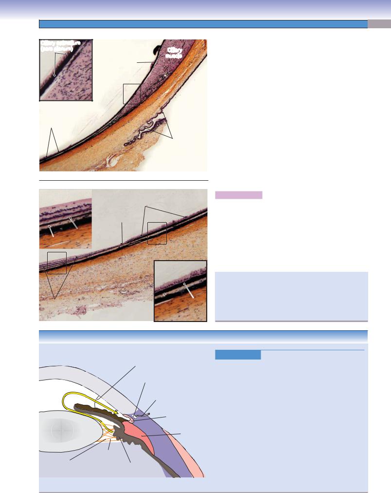
The ciliary body is located internally to the anterior margin of the sclera. The transition between the cornea and sclera is the limbus (corneoscleral junction). This is an important landmark for eye surgery procedures. The surface of the anterior portion of the ciliary body (ciliary process) has zonular fibers attached to it and is in contact with the aqueous humor. The surface of the posterior portion of the ciliary body is in contact with the vitreous body.
Figure 20-11B. Ciliary processes and the ciliary muscle.
H&E, 87; inset 348
Anterior chamber angle |
Posterior |
|
|
(iridocorneal angle) |
Anterior |
||
|
chamber |
||
|
|
chamber |
|
|
|
|
|
Lens |
Iris |
|
A |
|
|
|
|
Ciliary
processes Ciliary muscle
Ciliary processes have loose connective tissue cores and are covered by two layers of epithelium: (1) a nonpigmented layer and (2) a pigmented layer. The apical surfaces of the two epithelial layers face each other. Their basal surfaces each rest on a basement membrane, one bordering the ciliary stroma and the other bordering the aqueous humor. The cells are firmly connected by junctional complexes. The ciliary muscle contains three smooth muscle fiber groups: (1) longitudinal muscle fibers, which stretch the choroid to alter the opening of the anterior chamber angle for drainage of aqueous humor; (2) radial muscle fibers, which increase tension on the zonular fibers and cause the lens to flatten, allowing the eyes to focus for distant vision; and (3) circular muscle fibers, which relax the tension on the zonular fibers and cause the lens to become more convex to accommodate for near vision. Ciliary muscles are innervated by parasympathetic nerve fibers of the oculomotor nerve.
Zonular fibers
Pigmented
epithelium
 Unpigmented
epithelium
Figure 20-11C. Ciliary body, transverse and posterior views.
H&E, 34; inset 62
The ciliary body consists of (1) the ciliary ring (pars plana), the region that contains a ring of smooth muscle (ciliary muscle) surrounded by loose connective tissue and covered by the ciliary epithelium and (2) the ciliary processes (pars plicata), fingerlike structures which contain many fenestrated capillaries that
Bproduce aqueous humor. Aqueous humor flows from the posterior chamber through the pupil to the anterior chamber, then passes into the trabecular meshwork and finally into the canal of Schlemm (see Fig. 20-12C).
Posterior
Sclera
Ciliary
epithelium Ciliary muscle
Trabecular meshwork
Trabecular |
|
meshwork Canal of |
Ciliary |
Schlemm |
processes |
Canal of
Schlemm
|
angle |
Anterior |
|
chamber |
|
Anterior
Zonular fibers
Bulbar
conjunctiva
Ciliary process (pars plicata)
Lens


 Ora serrata
 Retina
|
D. Cui |
|
Ciliary ring |
|
(pars plana) |
|
Zonular fiber |
C |
Ciliary process |
|
CHAPTER 20 ■ Eye
Ciliary epithelium (pars planum)
Ciliary epithelium (pars planum)
Ciliary epithelium
Sclera
403
|
|
|
Figure 20-12A. |
Posterior portion of the ciliary body. |
|
|
A |
|
|||
Ciliary |
|
H&E, 34; inset 102 |
|||
|
|
||||
muscle |
|
|
|
|
|
|
|
|
The ciliary body lies posterior to the root of the iris, anterior |
||
|
|
|
to the ora serrata, and interior to the sclera (see Fig. 20-1). |
||
|
|
|
The ciliary body is triangular in shape. The anterior portion is |
||
|
|
|
thick and the posterior portion gradually becomes thinner and |
||
|
|
|
ends at the ora serrata (Fig. 20-12B). The two cell layers of the |
||
|
|
|
ciliary epithelium cover the entire surface of the ciliary body. |
||
|
|
|
|
||
|
|
|
Figure 20-12B. |
Ora serrata. H&E, 34; inset 102 |
|
|
|
|
The ora serrata is a denticulate border (junction) between |
||
|
|
|
the ciliary body and the retina; this is an important anatomic |
||
Bulbar |
|
|
landmark for the ophthalmologist. The extended ciliary epi- |
||
conjunctiva |
|
|
thelium from the ciliary body is shown on the right side of |
||
|
|
|
the picture. The anterior portion of the retina is shown on |
||
|
|
|
the left side of the picture. The pigmented ciliary epithelium |
||
|
|
|
and its basement membrane are continuous with the retinal |
||
|
|
|
pigmented epithelium and the Bruch membrane. |
||
Retina |
|
|
B |
|
|
Ciliary epithelium |
|
|
|
Ora serrata |
|
|
Retinal |
|
|
Bruch |
pigmented |
|
|
membrane epithelium |
|
|
|
Sclera
Retina
Ciliary epithelium
 Pigmented Basement ciliary membrane epithelium
Figure 20-12C. Outflow of aqueous humor.
Aqueous humor is produced in the posterior chamber by the epithelium that lines the ciliary processes of the ciliary body (see Fig. 20-11B,C). It flows through the pupil from the posterior chamber to the anterior chamber (yellow arrow), where it is absorbed into the trabecular meshwork (Fig. 20-11B). The aqueous humor then diffuses through connective tissue and epithelial tissue into the canal of Schlemm. Aqueous veins connect the canal of Schlemm to episcleral veins, where the aqueous humor is absorbed into the body’s venous circulation.
Interference with the normal flow of aqueous humor leads to increased intraocular pressure, and a serious medical condition, glaucoma, may result. The most likely sites of blockage of the flow of aqueous humor are at the trabecular meshwork and the endothelial lining of the canal of Schlemm rather than the venous collector system.
CLINICAL CORRELATION
C
|
|
Anterior chamber angle |
|
Cornea |
|
|
|
Anterior |
|
Canal of Schlemm |
|
|
|
||
chamber |
|
Aqueous vein |
|
|
|
||
|
|
Trabecular |
|
|
|
meshwork |
|
|
|
Ciliary |
|
|
|
muscle |
|
|
Posterior |
Sclera |
|
Zonular fibers |
chamber |
Ciliary process |
|
Vitreous body |
|||
J.Lynch |
|
Figure 20-12C. Glaucoma.
Glaucoma is a group of eye diseases that produce elevated intraocularpressure,usuallybecauseofobstructionoftheaqueous humor outflow (see Figs. 19-11B and 19-12C). Glaucoma results in damage to the optic nerve and is a major cause of blindness. (1) Open-angle glaucoma is the most common type. Fragments from normal cell degeneration become deposited within the trabecular meshwork and endothelial lining of the canal of Schlemm and reduce the absorption of aqueous fluid. Intraocular pressure rises slowly over a long period of time. Peripheral vision may be reduced before the patient is aware of the loss. As the pressure rises, the optic disk becomes cupped.
(2) Acute angle-closure glaucoma is also common. Occlusion of the anterior chamber angle occurs when the peripheral iris obstructs aqueous outflow. Intraocular pressure can rise quickly and reach very high levels. Patients may present with severe headache and eye pain, malaise, nausea, and vomiting. Immediate medical intervention is necessary to prevent vision loss.

404 UNIT 3 ■ Organ Systems
Retina (Tunica Interna)
A
Macula
lutea
Fovea centralis
Choroid
Substantia propria
Sclera
Episclera
Figure 20-13A. Overview of the retina; fovea. H&E, 17
The retina is a multilayered sheet of neural tissue covering the inner aspect of the posterior two thirds of the eyeball (Figs. 20-13B,C and 20-15A–C). There are regional variations in its structure: the macular (central) region is generally thicker than the peripheral region, and cone receptors predominate in the central retina, whereas rods are more numerous in the periphery. The fovea centralis is a small depression in the central retina caused by the displacement of the superficial layers, thereby allowing incoming light more direct access to the photoreceptors in this area. The photoreceptors here consist entirely of miniature cones, and there are no blood vessels in this region. These characteristics permit maximum visual acuity to be achieved in the fovea. The macula lutea is the region immediately surrounding the fovea centralis. Macula lutea means “yellow spot”; this region appears yellow in the living retina when viewed with an ophthalmoscope.
B
Bruch Pigment membrane epithelial
cells
Choriocapillaris
Inner limiting membrane (10)
Nerve fiber layer (9)
 Ganglion cell layer (8)
 Inner plexiform layer (7)
 Inner nuclear layer (6)
 Outer plexiform layer (5)
 Outer nuclear layer (4)
 Outer limiting layer (3)
 Photoreceptor layer (2)
 Pigment epithelium layer (1)
Choroid
Figure 20-13B. Macular region of the retina and the retinal layers. H&E, 184; inset 368
The macular region of the retina is relatively thick, and its ganglion cell layer contains many layers of nuclei. However, both the macular and peripheral regions of the retina have the same histologic layers: (1) the pigment epithelium layer, a layer of cuboidal cells that contain melanin granules; (2) the photoreceptor layer, external segments of rods and cones; (3) the outer limiting layer, a junction complex between the Müller cells and photoreceptor cells; (4) the outer nuclear layer, containing nuclei of the photoreceptor cells; (5) the outer plexiform layer, containing processes of photoreceptor cells, bipolar cells, and horizontal cells; (6) the inner nuclear layer, containing nuclei of bipolar cells, horizontal cells, amacrine cells, and Müller cells;
(7) the inner plexiform layer, containing processes of the cells in the adjacent layers; (8) the ganglion cell layer, containing nuclei of the ganglion cells; (9) the nerve fiber layer, containing axons of the ganglion cells; and (10) the inner limiting membrane, which is the basement membrane of the Müller cells.
C
Nuclei of rods
Nuclei of cones
|
Inner limiting membrane |
|
Nerve fiber layer |
|
Ganglion cell layer |
|
Inner plexiform layer |
|
Inner nuclear layer |
|
Outer plexiform layer |
|
Outer nuclear layer |
|
Outer limiting layer |
|
Photoreceptor layer |
|
Pigment epithelium |
Choroid |
layer |
Figure 20-13C. Peripheral region of the retina and distribution of the rods and cones. H&E, 184; inset 694
The peripheral part of the retina is thinner than the macular region and its ganglion cell layer becomes a single layer of nuclei. The rod photoreceptors are more numerous in the peripheral retina. The nuclei of rod cells are small and round and spread throughout the depth of the outer nuclear layer. The cone photoreceptors are present in both the center and periphery of the retina but are most highly concentrated in the fovea and macula. The nuclei of cone cells are large and ovoid in shape and often located at the base of the outer nuclear layer. Rod and cone cells are both photoreceptor neurons. Rods are specialized for motion detection and vision in dim light. Cones are specialized for fine visual acuity and color vision. Note the difference in thickness between the macular and peripheral regions of the retina and the relatively low number of ganglion cells in the peripheral retina.
CHAPTER 20 ■ Eye |
|
|
405 |
A |
|
B |
|
Synaptic |
Synaptic vesicles |
|
Synaptic |
region |
|
region |
|
|
|
||
|
Inner rod fiber |
|
|
|
|
Inner cone fiber |
|
Nuclear |
Nucleus |
|
|
region |
|
|
|
|
|
|
|
|
|
|
Nuclear |
|
|
Nucleus |
region |
|
Outer rod fiber |
|
|
|
|
|
|
|
|
Outer cone fiber |
|
Inner |
Mitochondrion |
|
|
segment |
|
|
|
|
Mitochondrion |
|
|
|
|
Inner segment |
|
|
|
|
|
|
Basal body |
Basal body |
|
|
Modified cilium |
|
|
|
|
|
|
|
|
|
Modified cilium |
Outer |
Disk |
|
|
|
|
|
|
segment |
|
|
|
|
Plasma membrane |
|
Outer segment |
|
D. Cui |
|
D. Cui |
Figure 20-14A and B. A representation of rods and cones.
Rods and cones are photoreceptor cells in the retina. They are similar in structure and both have (1) a synaptic region (2) a nuclear region (3) inner segments, and (4) outer segments. There are numerous synaptic vesicles in the synaptic regions, where the photoreceptors synapse with the dendrites of bipolar cells. The nuclear regions of rods and cones lie in the outer nuclear layer. The inner and outer segments form the photoreceptor layer of the retina. The inner segments contain mitochondria, rough endoplasmic reticulum, Golgi apparati, and other organelles that support the synthesis of proteins. The outer segments are in contact with the apical region of the pigmented epithelium cells. Outer segments of rods are composed of a series of superimposed disks, which have individual membranes, are stacked on each other, and are enclosed within the plasma membrane. Outer segments of cones have disks that are formed by invaginations of the plasma membrane. The membranes of the disks are continuous with the plasma membrane of the cell. The inner region is much wider than the outer region, which gives a cone appearance. Other differences between rods and cones are listed in Table 20-1.
TABLE 20 - 1 Comparison of Rods and Cones
Types of |
Synaptic |
Nuclear |
Inner Segment |
Outer Segment |
Distribution in |
Main |
Photoreceptor |
Region |
Region |
|
|
the Retina |
Function |
Cells |
|
|
|
|
|
|
|
|
|
|
|
|
|
Rods |
Spherule |
Small, |
Fewer |
Cylindrical in shape; disk |
Numerous in the |
Vision in |
|
synapses with |
round |
mitochondria than |
membranes are separate |
peripheral retina; |
dim light; |
|
dendrites of |
nucleus |
cones; synthesized |
and are not connected |
none in fovea |
motion |
|
one bipolar |
|
proteins passed |
to the outside plasma |
|
detection |
|
neuron |
|
only to newly |
membrane; disks contain |
|
|
|
|
|
forming disks |
rhodopsin protein |
|
|
Cones |
Pedicle |
Large, |
More |
Cone shaped; disk |
Highly |
Color |
|
synapses with |
ovoid |
mitochondria than |
membranes are connected |
concentrated in |
vision; |
|
dendrites of |
nucleus |
rods; synthesized |
to the plasma membrane |
the fovea and |
perception |
|
several bipolar |
|
proteins passed |
and form invaginated |
macula lutea; |
of fine detail |
|
neurons |
|
to entire outer |
membranes; disks contain |
less numerous in |
|
|
|
|
segment |
iodopsin protein |
periphery |
|
|
|
|
|
|
|
|

406 UNIT 3 ■ Organ Systems
A
 Inner limiting membrane (10)
 Nerve fiber layer (9)
 Ganglion cell layer (8)
 Inner plexiform layer (7)
 Inner nuclear layer (6)
 Outer plexiform layer (5)
 Outer nuclear layer (4)
 Outer limiting layer (3)
 Photoreceptor layer (2)
Pigment epithelium
 layer (1)
 Bruch membrane

 B
B
Ganglion cell
Cone
Rod
10 μm
Figure 20-15A. Retinal layers. H&E, 463
There are 10 histologic layers in the retina; see Figure 20-13B and Synopsis 20-1 for details of the components in each layer.
Figure 20-15B. Scanning electron micrograph of retinal layers.
Freeze-fracture preparation, 1,200
This freeze-fracture preparation of the peripheral retina shows the threedimensional shapes of the various cells in the retina. This section from the peripheral retina has fewer ganglion cells than the macular retina illustrated at left. Compare Figure 20-13B and C.
C
Light
|
|
Inner limiting |
|
|
membrane |
Müller cell |
|
Nerve fiber layer |
|
|
|
Ganglion |
|
Ganglion cell layer |
cell |
|
|
Amacrine |
|
Inner plexiform layer |
cell |
|
|
Müller cell |
|
Inner nuclear layer |
Bipolar cell |
|
|
|
|
|
Horizontal |
|
Outer plexiform layer |
cell |
|
|
|
|
|
Rod cell |
T. Yang |
Outer nuclear layer |
Cone cell |
|
Outer limiting layer |
Pigment cell |
|
|
|
Photoreceptor layer |
|
|
|
|
|
Neural |
Pigment |
|
outflow |
epithelium layer |
Figure 20-15C. A representation of the functional retinal layers.
Light passes through all retinal layers to activate the photoreceptor cells (rods and cones), and excess light is absorbed by pigmented epithelial cells. The photoreceptors transduce the light into electrochemical signals, which pass to the conducting neurons (bipolar cells, then ganglion cells). Horizontal and amacrine cells are association neurons that have long dendrites. The dendrites of the horizontal cells synapse with photoreceptors and contribute to the elaborate neuronal connections in the outer plexiform layer. The dendrites of amacrine cells are in contact with bipolar and ganglion cells and modulate signals in the inner plexiform layer. The ganglion cells collect all visual information and send it along their axons in the optic nerve. Müller cells are large supporting neuroglial cells and extend from the inner limiting membrane to the outer limiting membrane. Their processes surround all the neuronal elements of the retina.

CHAPTER 20 ■ Eye |
407 |
SYNOPSIS 20 - 1 Retinal Layers
(1)Pigment epithelium layer, a layer of cuboidal cells which are rich in melanin granules. This layer is important in absorbing excess light and in esterifying vitamin A.
(2)Photoreceptor (rods and cones) layer, consists of inner and outer segments of the photoreceptor cells.
(3)Outer limiting layer, a plexus of junction complexes that joins the membranes of the photoreceptor cells and Müller cells (retinal glial cells).
(4)Outer nuclear layer, contains nuclei of the photoreceptor cells (rods and cones).
(5)Outer plexiform layer, consists of axodendritic synapses between the axons of the photoreceptor cells and dendrites of the bipolar and horizontal cells.
(6)Inner nuclear layer, contains nuclei of bipolar cells, horizontal cells, amacrine cells, and Müller cells.
(7)Inner plexiform layer, composed of axodendritic synapses between the bipolar cell axons, ganglion cell dendrites, and processes of the amacrine cells.
(8)Ganglion cell layer, contains nuclei of the ganglion cells.
(9)Nerve fiber layer, contains axons of the ganglion cells.
(10) Inner limiting membrane, the basement membrane of the Müller cells.
The ganglion cells and their axons are part of the central nervous system. They cannot regenerate following severe damage. Embryologically, the pigmented epithelium layer and the neural layers of the retina originate from different layers of the optic vesicle. Therefore, retinal detachment may occur between the pigmented epithelium layer and the rest of the neural retina.
CLINICAL CORRELATIONS
A
J. Lynch |
|
|
Bruch membrane |
Abnormal blood |
Choroid |
|
vessels, leaking fluid |
|
Figure 20-16A. Age-Related Macular Degeneration.
Age-related macular degeneration (ARMD) is a degenerative eye disease affecting the central portion of the retina. Symptoms include blurred vision, distortion of straight lines, and worsening of color vision. There are two forms: exudative (wet) and nonexudative (dry). Wet ARMD is characterized by the growth of new vessels from the choroidal circulation and serous fluid leakage from the new vessels. This causes detachment of the retina and macula, producing a rapid and irreversible loss of central vision. Treatment options include laser photocoagulation, surgery, and drugs. Dry ARMD is characterized by subretinal drusen deposits (focal eosinophilic material arising from the Bruch membrane and lying between the pigment epithelium and the Bruch membrane), and atrophy and degeneration of the outer retina, including the retinal pigment epithelium, Bruch membrane, and choriocapillaris. The causes of ARMD are not well understood, but risk factors include age, smoking, family history, hypertension, and ethnicity (higher in non-Hispanic Caucasians).
B
Photoreceptor Detached layer
retina
Vitreous fluidfilled space
Retinal
 hole
hole
Pigment epithelium
 Vitreous fluid
D. Cui
Figure 20-16B. Retinal Detachment. H&E, 62
Retinal detachment is an eye condition in which the sensory retina separates from the underlying retinal pigment epithelium and choroid (see red arrows). It is categorized into three major types:
(1) rhegmatogenous retinal detachment ([RRD] illustrated) is the most common; it occurs when vitreous fluid leaks into the subretinal space through a break in the retina; (2) exudative or serous detachment occurs in association with inflammation or a tumor;
(3) traction detachment occurs when scar tissue on the retina’s surface contracts and causes the retina to separate from the retinal pigment epithelium. Retinal detachment is a medical emergency that can lead to permanent vision loss. Time is critical for surgical reattachment of the retina, which will usually preserve vision. Symptoms include flashes, floaters, and visual field distortion or loss. Treatment options include laser photocoagulation, cryoretinopexy, vitrectomy, and silicone oil repair.

408 UNIT 3 ■ Organ Systems
Optic Nerve
A
Sclera
Neuroglial cells
Retina nerve fibers
Optic |
disk |
|
(optic |
|
|
|
papilla) |
|
Lam |
|
in |
|
a |
cribrosa |
|
Optic nerve (myelinated)
Retinal nerve fibers (nonmyelinated)
Subarachnoid space
(filled with CSF)
Pia mater
Figure 20-17A. Optic nerve and optic disk. H&E, 22; inset, 83
The optic nerve is a nerve fiber trunk formed by the convergence of retinal ganglion cell axons at the posterior pole of the eye. From there, they leave the globe on their way to the brain. Each optic nerve contains about one million myelinated axons and even more neuroglial cells. The surface of the optic nerve is covered by pia mater, which is continuous with that on the surface of the brain. The optic disk (optic nerve papilla) is the small circular site in the retina where the retinal nerve fiber layer (nonmyelinated nerve fibers) continues into the optic nerve. The nonmyelinated nerve fibers begin to acquire myelin at the level of the lamina cribrosa (thin dotted line), a perforated, sievelike region of the sclera through which optic nerve fibers and blood vessels pass. The myelinated segments of ganglion cell axons, therefore, form the optic nerve. The neuroglial cells include oligodendrocytes, which produce myelin for axons in the CNS, and astrocytes, which perform several nutritive and supportive functions.
CLINICAL CORRELATION
Normal |
Optic |
CSF |
disk |
B
Elevated
CSF pressure
 Swollen and elevated optic disk
|
Engorged and |
D. Cui |
tortuous central vein |
Figure 20-17B. Papilledema.
Papilledema is a noninflammatory swelling of the optic disk. It is produced by increased intracranial pressure that is transmitted along the optic nerve sheath as elevated cerebrospinal fluid pressure, and it is, therefore, a sign of a potentially life-threatening condition. The increased pressure disrupts blood circulation within the optic nerve, causing leakage of water, protein, and other contents into the extracellular spaces of the optic disk. The most noticeable symptoms of increased intracranial pressure are headache and vomiting. Clinical findings include a swollen and elevated optic disk (papilledema), engorged and tortuous retinal veins, and focal hemorrhages. Cerebral tumors, subdural hematoma, malignant hypertension, and hydrocephalus are the most common causes of increased intracranial pressure and papilledema. Because papilledema is a sign of many systemic intracranial and spinal diseases, the correct diagnosis of the underlying disease is essential for proper treatment.
SYNOPSIS 20 - 2 Clinical Terms for the Eye
■Blepharitis: Inflammation of the eyelids and especially of their margins (Fig. 20-3C).
■Ptosis: Drooping or falling down, usually of the upper eyelid, because of muscle weakness or paralysis (Fig. 20-4C).
■Open angle (glaucoma): Raised intraocular pressure in which obstruction to aqueous flow occurs at the ultrastructural level, within the walls of the tiniest interstices of the trabecular meshwork (Fig. 20-12C).
■Closed angle (glaucoma): The flow of aqueous fluid is blocked by the root of the iris. This can be a generalized closure (hyperopic eyes) or in multiple focal adhesions caused by inflammation (peripheral anterior synechiae [Fig. 20-12C]).
■Cupping of the optic disk: Glaucoma destroys retinal axons as they traverse the optic disk in the walls of a central, cupshaped conduit. As the walls disappear, the cup enlarges (Fig. 20-12C).
■Dry macular degeneration: Scattered drusen damage the pigment epithelium of the macula, gradually decreasing visual acuity (Fig. 20-16A).
■Wet macular degeneration: Abnormal capillaries break through the retinal pigment epithelium and leak underneath the retina. When close to the fovea, they can decrease visual acuity (Fig. 20-16A).
■Limbus: An anatomic transition zone between cornea and sclera (corneoscleral junction), which is an important landmark for eye surgery procedures (Fig. 20-11A).
■Ora serrata: A denticulate border (junction) between the ciliary body and the retina; this is an important anatomic landmark
for the ophthalmologist (Fig. 20-12B).

21 Ear
Introduction and Key Concepts for the Ear
Figure 21-1A |
Overview of the Ear: Outer, Middle, and Inner Ear |
Figure 21-1B |
Middle Ear Structures |
Figure 21-2 |
Major Structures of the Inner Ear |
Figure 21-3A |
Membranous Labyrinth |
Figure 21-3B |
Cochlear Duct and Modiolus |
Auditory System |
|
Figure 21-4A |
Cross section of Cochlea |
Figure 21-4B |
Scala Media and Organ of Corti |
Figure 21-5 |
Organ of Corti and Associated Structures |
Figure 21-6A |
Sound Transduction |
Figure 21-6B |
Stereocilia Displacement |
Figure 21-6C |
Auditory Hair Cells |
Figure 21-7A |
Inner and Outer Hair Cells |
Figure 21-7B |
Stereocilia of an Outer Hair Cell |
Figure 21-7C |
Clinical Correlation: Sensorineural Hearing Loss |
Vestibular System |
|
Figure 21-8A |
Sensory Receptors |
Figure 21-8B |
Crista Ampullaris and Macula Utriculi |
Figure 21-9A |
Ampulla of the Semicircular Canal |
Figure 21-9B |
Crista Ampullaris |
Figure 21-9C |
Clinical Correlation: Ménière Disease |
Figure 21-10A |
Macula of the Utricle |
Figure 21-10B |
Macula of the Utricle with Otoconia |
Figure 21-10C |
Otoconia from the Macula of the Utricle |
Figure 21-11A |
Vestibular Hair Cells |
Figure 21-11B |
Excitation and Inhibition in Hair Cells |
409