

узи сосудов шеи
.pdfRadioGraphics
RG f Volume 25 ● Number 6 |
|
|
Tahmasebpour et al 1571 |
||||
|
|
|
|
|
|
|
|
|
Table 1 |
|
|
|
|
|
|
|
Criteria for Diagnosis of ICA Stenosis with Gray-Scale and Doppler US |
|
|
|
|||
|
|
|
|
|
|
||
|
|
Primary Parameters |
Additional Parameters* |
||||
|
|
|
|
|
|
|
|
|
|
ICA PSV |
Degree of |
ICA/CCA |
ICA EDV |
||
|
Degree of Stenosis (%) |
(cm/sec) |
Plaque† (%) |
PSV Ratio |
(cm/sec) |
|
|
|
Normal |
125 |
None |
2.0 |
40 |
||
|
50 |
125 |
50 |
2.0 |
40 |
||
|
50–69 |
125–230 |
50 |
2.0–4.0 |
40–100 |
||
|
70 but less than near |
230 |
50 |
4.0 |
100 |
||
|
occlusion |
|
|
|
|
|
|
|
Near occlusion |
High, low, or undetectable |
Visible |
Variable |
Variable |
||
|
Total occlusion |
Undetectable |
Visible, no detectable |
NA |
NA |
||
|
|
|
lumen |
|
|
|
|
|
|
|
|
|
|
|
|
Source.—Reference 21.
*EDV end-diastolic velocity, NA not applicable.
†Estimated value based on the diameter reduction at gray-scale and color Doppler imaging.
Figure 16. Severe stenosis (70% to near occlusion) of the ICA. Duplex US image of the left ICA shows a high PSV (366 cm/ sec), a significant amount of visible plaque, the presence of aliasing despite a high color scale setting (114 cm/sec), color flow turbulence immediately distal to the stenotic segment, broadening of the PW Doppler spectrum, and a high end-diastolic velocity (182 cm/sec).
dure of choice for diagnosis of dissection because detection of the intimal flap has not been reliable and because the intimal tear may be too high in the ICA to be assessed with carotid US. Findings that might be encountered at carotid US include the following: visualization of the flap at grayscale imaging, reversed flow in the true channel and antegrade flow in the false channel at color Doppler imaging, and a damped high resistive flow pattern proximal to the dissection at PW Doppler spectral imaging. Such findings may require alternative imaging modalities, such as MR or CT angiography, to assist in diagnosis.
Normal flow in the CCA is usually greater than 45 cm/sec. High flow ( 135 cm/sec) in both CCAs may be due to high cardiac output in hypertensive patients or young athletes. Low flow
( 45 cm/sec) in both CCAs is likely to be secondary to poor cardiac output from cardiomyopathies, valvular heart disease, or extensive myocardial infarction. Arrhythmias can be a real problem. The PSV value will be low if measured after a premature ventricular contraction, and it will be high after a compensatory pause. Measurements of the ICA and CCA velocities after differing rhythms can alter the ICA/CCA PSV ratio. The PSV should be measured after a regular beat. If this is not possible, the result will be limited.
An abnormal mid-systolic deceleration in the PW Doppler waveform of the right CCA and ICA may be due to a partial or complete right subclavian steal (22). Severe stenosis in the innominate
RadioGraphics
1572 November-December 2005 |
RG f Volume 25 ● Number 6 |
Figure 17. Severe stenosis of the innominate artery. PW Doppler spectral image of the right CCA shows a tardus-parvus waveform, which is suggestive of a severe stenosis proximal to the point of sampling. A severe stenosis of the innominate artery was subsequently demonstrated at angiography. EDV end-diastolic velocity.
artery may manifest as a tardus-parvus waveform (a prolonged systolic acceleration time with low PSV) in the right CCA and ICA (Fig 17). This waveform generally indicates a severe stenosis proximal to the point of sampling. Imaging with an alternative modality may be recommended to determine the exact location of the stenosis.
Contralateral carotid disease or vertebrobasilar disease may alter the overall flow dynamics (7). The absolute velocity in an individual vessel must be correlated with the overall cardiovascular status of the patient and the caliber of the vessel.
Near Occlusion
and Total ICA Occlusion
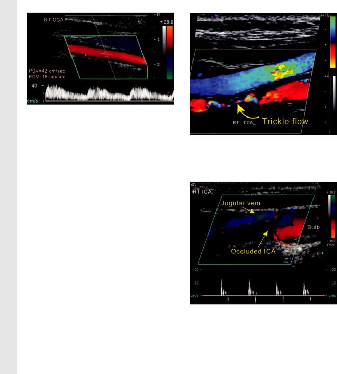
The distinction of near occlusion versus total occlusion is clinically extremely important. Patients with near occlusion may be surgical candidates, while patients with total occlusion are not. The number of false-positive interpretations due to lack of flow detection can be reduced by attention to the technical detail but cannot be eliminated. Alternative imaging modalities such as catheter angiography or multisection CT angiography may be helpful to distinguish between total and near occlusions (23,24). The hallmark of a near occlusion is the “string sign” or “trickle flow” at color Doppler imaging (Fig 18). Power Doppler US may also be helpful with older equipment in searching for trickle flow; we have found no diagnostic advantage for power Doppler imaging over color Doppler imaging at our institution. Recommendations for optimal color and PW Doppler imaging parameters to enhance the detection of
Figure 18. Trickle flow in the ICA. Color Doppler image shows a narrow patent channel (the string sign) in the right ICA. This finding is suggestive of near occlusion of the ICA.
Figure 19. Thud flow. Color Doppler image of the right ICA and carotid bulb shows no flow in the ICA lumen and reversed flow in the bulb at the point of occlusion. The red and blue arrows indicate the direction of the reversed flow at the point of obstruction (thud flow). The PW Doppler spectrum also demonstrates thud flow, which manifests as damped systolic flow and reversed flow in early diastole.
trickle flow in near occlusions are provided in Table 2.
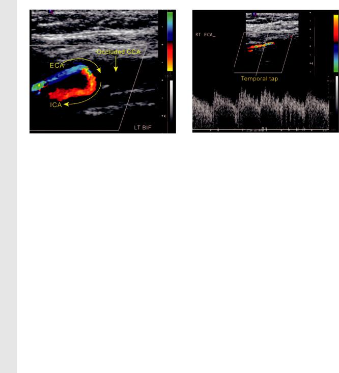
When a single patent vessel beyond the carotid bifurcation is identified, the operator must confidently determine if the vessel represents the ICA or the ECA. The single most reliable parameter for differentiating the ECA from the ICA is the presence of ECA branches in the neck. The temporal tap maneuver involves tapping on the superficial temporal artery and looking for reflected flow in the ECA. This has not proved reliable, as
RG f Volume 25 ● Number 6 |
Tahmasebpour et al 1573 |
RadioGraphics
Figure 20. Internalization of the ECA. Color Dopp- |
Figure 21. PW Doppler spectrum in internalization |
ler image of the left carotid bifurcation shows no flow |
of the ECA. PW Doppler spectral image shows a re- |
in the distal CCA. The ICA and ECA are both patent, |
versed low resistive flow pattern with delayed systolic |
but flow in the ECA is reversed to supply antegrade |
acceleration (tardus wave) in the ECA. The patient had |
flow in the ICA above the level of the occluded CCA. |
an occluded CCA. In addition, reflections from the |
The curved arrows indicate the direction of blood flow |
temporal tap maneuver are demonstrated as ripples in |
from the ECA to the ICA. |
the Doppler spectrum. |
|
Table 2 |
|
|
|
Optimal Color and PW Doppler Imaging Parameters for Enhancing |
||
|
Detection of Trickle Flow in Near Occlusion of the ICA |
||
|
|
|
|
|
Parameter |
Recommended Setting |
|
|
|
|
|
|
Transducer frequency |
7 MHz |
|
|
Color box |
Steer to the center or straight position |
|
|
Sample volume box |
Steer to the center or straight position |
|
|
Focal zone |
At the level of the diseased segment |
|
|
Color velocity scale |
Decrease to 15 cm/sec |
|
|
PW Doppler scale |
Decrease to 15 cm/sec |
|
|
Color Doppler gain |
Increase to the point of visible background noise |
|
|
PW Doppler gain |
Increase to the point of visible background noise |
|
|
Wall filter |
Decrease to low |
|
|
Color threshold |
Increase to 80% |
|
|
Sample volume gate |
Increase to 2.5 mm |
|
|
|
|
|
the reflected flow from tapping on the temporal |
locities may be low, which may necessitate chang- |
artery can also be detected in the ICA and CCA |
ing the velocity settings in order to detect the flow |
(25). |
(Fig 21). |
In a total ICA occlusion, there is a characteris- |
ECA Stenosis |
tic “to-and-fro” flow pattern at the point of occlu- |
|
sion known as “thud flow” at color Doppler and |
The ECA is an important collateral pathway in |
PW Doppler imaging (Fig 19). Other findings are |
patients with ipsilateral ICA occlusion and recur- |
direct visualization of a thrombus at gray-scale |
rent symptoms; this may influence the surgical |
imaging, absent flow at color Doppler imaging, |
decisions involving revascularization of the ste- |
and damped resistive flow in the CCA at PW |
notic ECA (26). However, an isolated ECA ste- |
Doppler imaging (externalization of the CCA). |
nosis may not be clinically significant, with no |
In near or total occlusion of the CCA, reversal |
need to change patient care. |
of flow direction in the ECA via collateral vessel |
|
recruitment to a patent ICA may occur (internal- |
|
ization of the ECA) (Fig 20). In this setting, ve- |
|
1574 November-December 2005 |
RG f Volume 25 ● Number 6 |
RadioGraphics
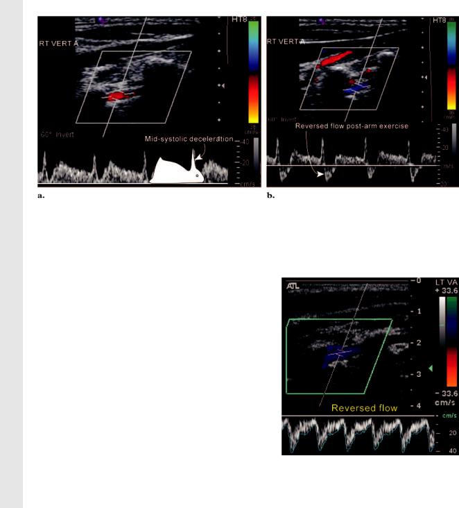
Figure 22. Occult and partial subclavian steal. (a) PW Doppler spectral image of the right vertebral artery shows midsystolic deceleration with antegrade late-systolic velocities (occult steal). (b) PW Doppler spectral image obtained after the patient exercised the right arm (by opening and closing the hand for 2 minutes). The Doppler spectrum shows midsystolic deceleration with retrograde late-systolic velocities. The subclavian artery “steals” blood from the vertebral artery to supply the ischemic arm.
Vertebral Artery and Subclavian Steal
Carotid US can show patency, direction of blood flow, and, to some extent, relative size of the left versus right vertebral arteries. Carotid US is not accurate for identification of a focal stenosis in the vertebral artery. Congenital and acquired occlusions or near occlusions can all appear alike.
Identification of the vertebral artery is achieved
by locating the CCA in a sagittal view and sweep- |
|
|
ing the transducer laterally to the transverse pro- |
|
|
cesses of the cervical spine, where the vertebral |
|
|
artery can be demonstrated with color Doppler |
|
|
imaging. Since the vertebral artery is located deep |
|
|
in the neck, increasing the color Doppler gain |
|
|
may aid visualization. PW Doppler spectral analy- |
|
|
sis of the vertebral artery provides necessary infor- |
Figure 23. Complete subclavian steal. PW |
|
mation to demonstrate the presence of a subcla- |
||
Doppler spectral image of the left vertebral artery |
||
vian steal. On the basis of the hemodynamic |
shows completely reversed flow. |
|
changes in the vertebral artery, there are three |
|
|
types of subclavian steals. |
|
|
In occult steal (minimal hemodynamic |
Partial subclavian steal corresponds to moder- |
|
changes), PW Doppler imaging may show ante- |
ate hemodynamic changes. The PW Doppler |
|
grade flow with midsystolic deceleration, which |
spectrum shows partially reversed flow. The PW |
|
may temporarily convert to a more abnormal |
Doppler spectrum in occult and partial subcla- |
|
waveform (with reversed late-systolic flow) in re- |
vian steal may resemble the profile image of a rab- |
|
sponse to reactive hyperemia in the ipsilateral arm |
bit (the “bunny rabbit” sign) (27). |
|
after arm exercise (27) (Fig 22). |
In complete (full) subclavian steal, flow in the |
|
|
vertebral artery is completely reversed (Fig 23). |
|
|
This may be associated with ischemic symptoms |
|
|
in the ipsilateral arm. |

RadioGraphics
RG f Volume 25 ● Number 6 |
Tahmasebpour et al 1575 |
Conclusions
Carotid US offers a noninvasive evaluation of the extracranial neck portions of the carotid and vertebral arteries for atherosclerotic disease. Standardized technical parameters, scanning methods, Doppler analysis, and interpretation enhance the accuracy and reproducibility of the results.
Acknowledgment: The authors greatly thank Andrea J. Phillips, BSc, MBBS, MRCP, FRCR, for her expert assistance in editing the manuscript.
References
1.Kochanek KD, Smith BL. Deaths: preliminary data for 2002. Natl Vital Stat Rep 2004;52(13): 1– 47.
2.Grant EG, Barr LL, Borgstede J, et al. ACR guideline for the performance of an ultrasound examination of the extracranial cerebrovascular system. Reston, Va: American College of Radiology, 2002; 577–580.
3.Zagzebski JA. Doppler instrumentation. In: Rowland J, Potts L, eds. Essentials of ultrasound physics. St Louis, Mo: Mosby, 1996.
4.Tahmasebpour HR, Cooperberg PL, Segan-Hoff- man J, et al. Velocity quantifications in the carotid ultrasound with Doppler angle set at 44 vs 60 degree (abstr). In: Radiological Society of North America scientific assembly and annual meeting program. Oak Brook, Ill: Radiological Society of North America, 2003; 297.
5.Bluth EI, Stavros AT, Marich KW, et al. Carotid duplex sonography: a multicenter recommendation for standardized imaging and Doppler criteria. RadioGraphics 1988;8:487–506.
6.Nelson TR, Pretorius DH. The Doppler signal: where does it come from and what does it mean? AJR Am J Roentgenol 1988;151:439 – 447.
7.Beckett WW, Davis PC, Hoffman JC. Pitfalls in duplex carotid evaluation contralateral to significant stenosis/occlusion. AJNR Am J Neuroradiol 1990;11:1049 –1053.
8.Kennedy J, Quan H, Ghali WA, et al. Importance of the imaging modality in decision making about carotid endarterectomy. Neurology 2004;62(6):
901–904.
9.Nguyen-Huynh MN, Lev MH, Rordorf G. Spontaneous recanalization of internal carotid artery occlusion. Stroke 2003;34(4):1032–1034.
10.O’Leary DH, Polak JF. Intima-media thickness: a tool for atherosclerosis imaging and event prediction. Am J Cardiol 2002;90(10C):18L–21L.
11.Baldassarre D, Amato M, Bondioli A, et al. Carotid artery intima-media thickness measured by ultrasonography in normal clinical practice correlates well with atherosclerosis risk factors. Stroke 2000;31:2426 –2430.
12.Ebrahim S, Papacosta O, Whincup P. Carotid plaque, intima-media thickness, cardiovascular risk factors, and prevalent cardiovascular disease in men and women. Stroke 1999;30:841– 850.
13.Kofoed SC, Gronholdt ML, Wilhjelm JE, et al. Real-time spatial compound imaging improves reproducibility in the evaluation of atherosclerotic
carotid plaques. Ultrasound Med Biol 2001; 27(10):1311–1317.
14.Homma S, Hirose N, Ishida H, et al. Carotid plaque and intima-media thickness assessed by B-mode ultrasonography in subjects ranging from young adults to centenarians. Stroke 2001;32: 830 – 835.
15.Lal BK, Hobson RW 2nd, Pappas PJ, et al. Pixel distribution analysis of B-mode ultrasound scan images predicts histologic features of atherosclerotic carotid plaques. J Vasc Surg 2002;35(6):
1210 –1217.
16.Sabetai MM, Tegos TJ, Nicolaides AN, et al. Hemispheric symptoms and carotid plaque echomorphology. J Vasc Surg 2000;31(1):39 – 49.
17.Polak JF, Shemanski L, O’Leary DH, et al. Hypoechoic plaque at US of the carotid artery: an independent risk factor for incident stroke in adults aged 65 years or older. Radiology 1998;208(3): 649 – 654. [Published correction appears in Radiology 1998;209(1):288 –289.]
18.Fu¨ rst H, Hart WH, Jansen I, et al. Color flow Doppler sonography in the identification of ulcerative plaques in patients with high-grade carotid artery stenosis. AJNR Am J Neuroradiol 1992;13: 1581–1587.
19.Weskott HP. B-flow: a new method for detecting blood flow. Ultraschall Med 2000;21(2):59 – 65.
20.Tahmasebpour HR, Fix C, Cooperberg PL, et al. Clinical application of twinkle artifact in ultrasonography (abstr). In: Radiological Society of North America scientific assembly and annual meeting program. Oak Brook, Ill: Radiological Society of North America, 2004; 734.
21.Grant EG, Benson CB, Moneta GL, et al. Carotid artery stenosis: gray-scale and Doppler US diagno- sis—Society of Radiologists in Ultrasound Consensus Conference. Ultrasound Q 2003;19(4): 190 –198.
22.Saden SE, Grant EG, Carroll BA, et al. Innominate artery occlusive disease: sonographic findings (abstr). In: Radiological Society of North America scientific assembly and annual meeting program. Oak Brook, Ill: Radiological Society of North America, 2003; 296.
23.Chen CJ, Lee TH, Tseng YC, et al. Multi-slice CT angiography in diagnosing total versus near occlusions of the internal carotid artery: comparison with catheter angiography. Stroke 2004;35(1): 83– 85.
24.Paciaroni M, Caso V, Cardaioli G, et al. Is ultrasound examination sufficient in the evaluation of patients with internal carotid artery severe stenosis or occlusion? Cerebrovasc Dis 2003;15(3):173–176.
25.Kliewer MA, Freed KS, Hertzberg BS. Temporal artery tap: usefulness and limitations in carotid sonography. Radiology 1996;201(2):481– 484.
26.Boontje AH. External carotid artery revascularization: indications, operative techniques and results. J Cardiovasc Surg (Torino) 1992;33(3):315–318.
27.Kliewer MA, Hertzberg BS, Kim DH, et al. Vertebral artery Doppler waveform changes indicating subclavian steal physiology. AJR Am J Roentgenol 2000;174(3):815– 819.