


C H A P T E R 8
PANCREAS
Cases 8.1–8.27 Inflammatory Diseases
Acute Pancreatitis
Chronic Pancreatitis
Cases 8.28–8.70 Masses
Solid
Cystic
Cases 8.71–8.78 |
Miscellaneous |
This page intentionally left blank

8. PANCREAS 657
CASE 8.1
Findings
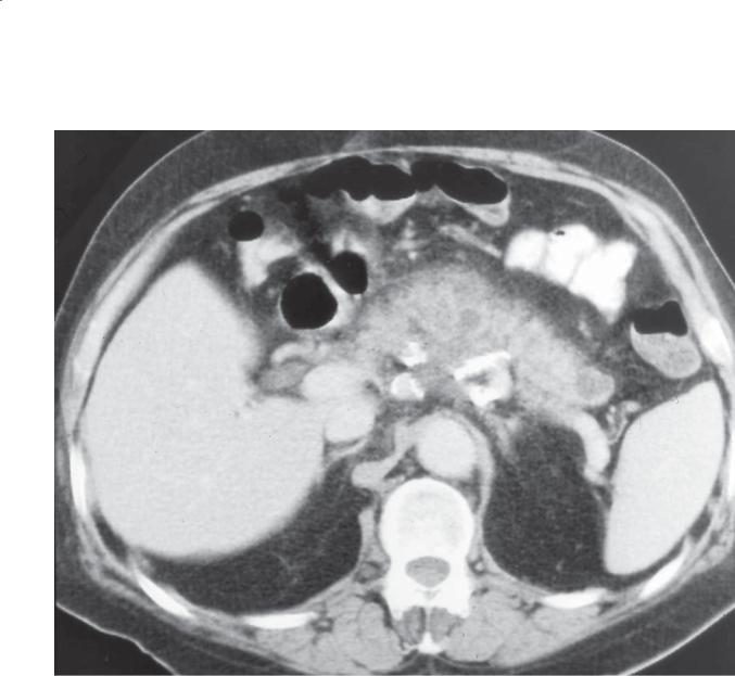
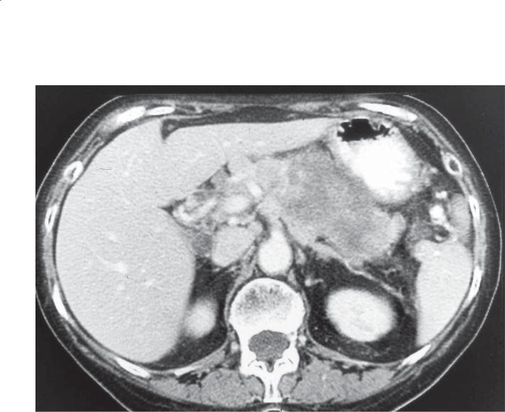
Contrast-enhanced CT. Peripancreatic stranding and an effusion are present about the tail of the pancreas. The pancreas is homogeneously perfused.
Di erential Diagnosis
Acute pancreatitis
Diagnosis
Acute pancreatitis
Discussion
Acute pancreatitis usually is caused by either alcoholism or biliary tract disease (gallstones). In approximately one-fourth of patients, the cause of pancreatitis is never established. Pancreas divisum (cases 8.72–8.74) has also been implicated as a cause of acute pancreatitis due to functional obstruction of the pancreatic duct at the accessory papilla of Santorini. Other, less common causes include drugs, hypercalcemia, hyperlipidemia, viral infections,
endotoxins, trauma, and others. Symptoms include abdominal pain, nausea, vomiting, and distention. Serum amylase and lipase levels are increased in about 80% of patients with acute pancreatitis. The absolute values of the enzymes do not necessarily correlate with the severity of disease.
Th e CT appearance of acute pancreatitis usually takes 2 forms: diffuse glandular enlargement or a normal-sized gland with a peripancreatic effusion (as in this case). Focal pancreatitis (case 8.4) is less common. The gland can be normal-appearing in mild pancreatitis. The staging system reported by Balthazar and colleagues can be helpful to estimate the severity of disease. Grade A refers to a normalappearing pancreas. Progressively increasing grades of severe pancreatitis are B) focal or diffuse enlargement, C) intrinsic pancreatic abnormality with inflammatory changes in the peripancreatic tissues, D) a fluid collection or phlegmon, and E) 2 or more large phlegmonous collections or peripancreatic gas.
Disease type: Inflammatory Diseases
658 MAYO CLINIC GASTROINTESTINAL IMAGING REVIEW
CASE 8.2
Findings
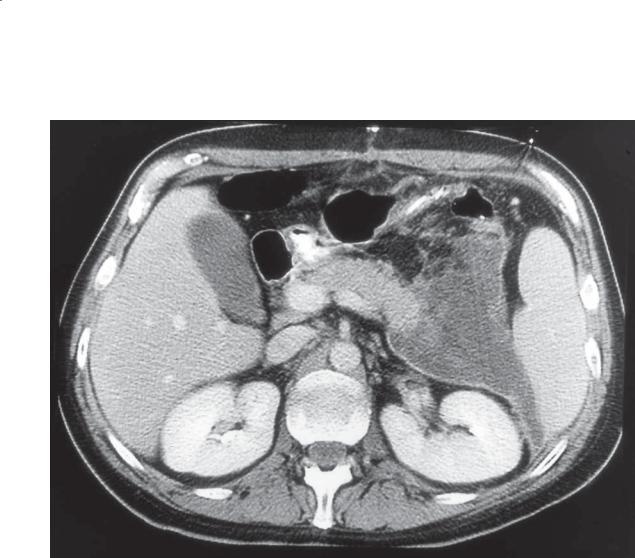
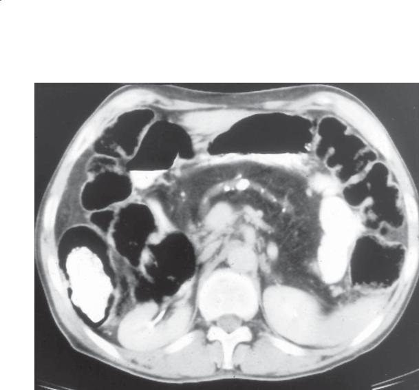
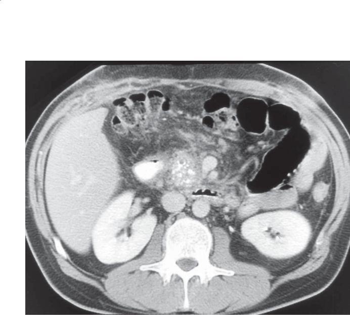
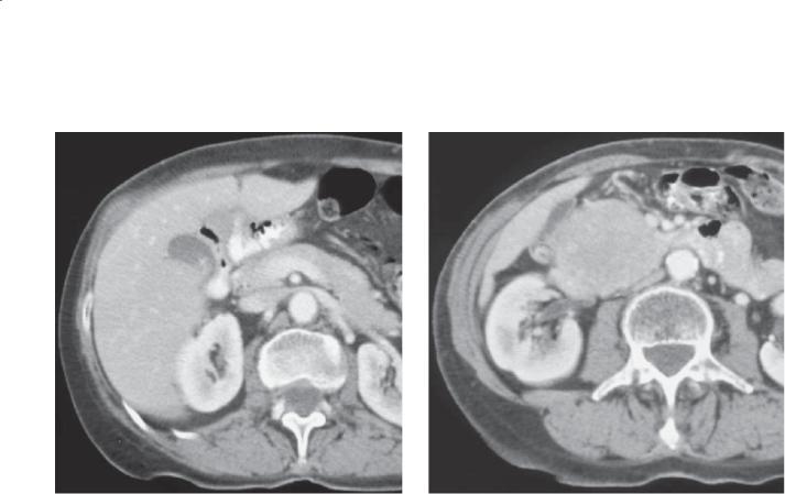
Contrast-enhanced CT. The pancreas is diffusely enlarged. There are several intrapancreatic fluid collections. A small peripancreatic effusion is present.
Di erential Diagnosis
Acute pancreatitis
Diagnosis
Acute pancreatitis
Discussion
Th is case shows findings of severe acute pancreatitis with the presence of 2 or more intrapancreatic fluid collections (grade E, Balthazar criteria). Patients with severe acute pancreatitis usually have a protracted clinical illness and have higher incidences of pancreatic abscess and dying of complications of their illness.
Disease type: Inflammatory Diseases
CASE 8.3
Findings
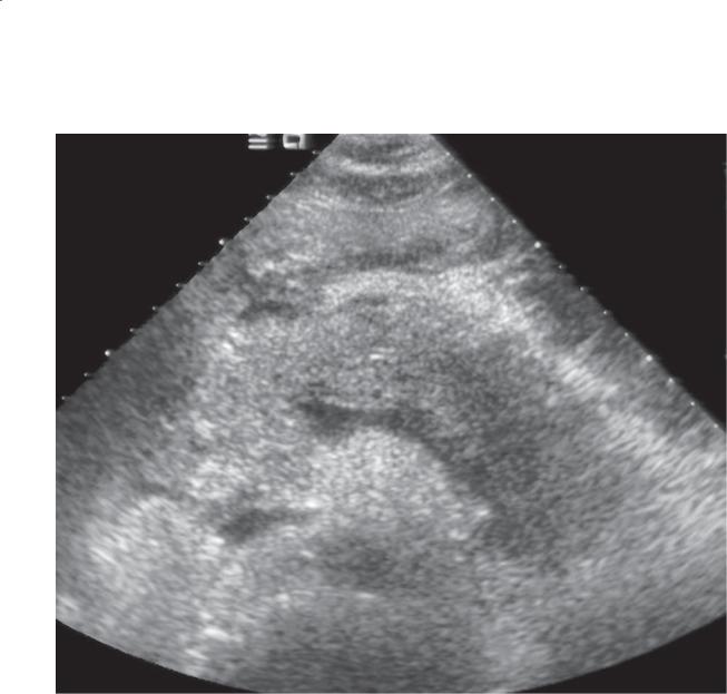
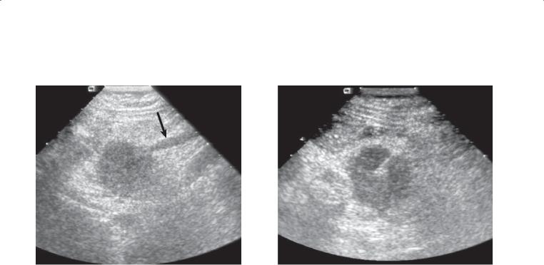
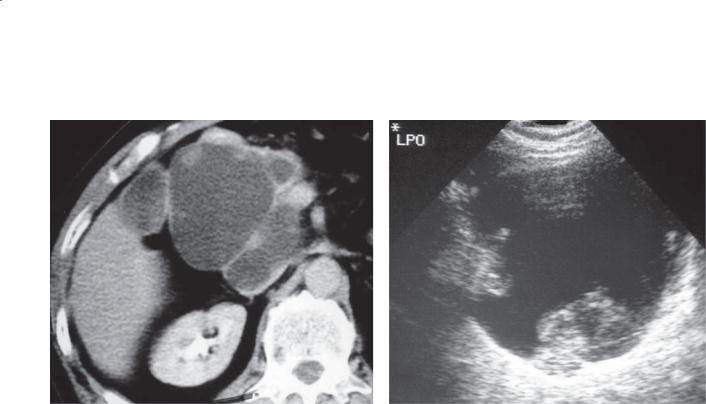
Sonogram. The pancreas is diffusely enlarged and has decreased echogenicity with a small amount of peripancreatic fluid.
Di erential Diagnosis
Acute pancreatitis
Diagnosis
Acute pancreatitis
8. PANCREAS 659
Discussion
Findings of diffuse acute pancreatitis include enlargement of the gland, hypoechogenicity relative to the liver, and peripancreatic fluid. These sonographic findings usually are seen only in moderate or severe acute pancreatitis. The pancreas often has a normal sonographic appearance in mild cases of pancreatitis. One of the major roles of sonography in patients with acute pancreatitis is to assess for stone disease in the biliary system as a possible cause for the pancreatitis. Differentiation between necrotic and nonnecrotic pancreatitis is not possible with sonography.
Disease type: Inflammatory Diseases
660 MAYO CLINIC GASTROINTESTINAL IMAGING REVIEW
CASE 8.4
A B
Findings
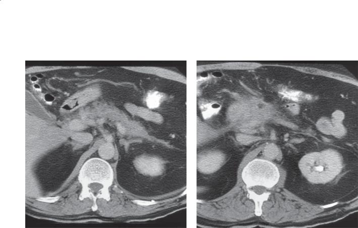
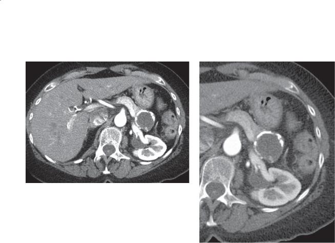
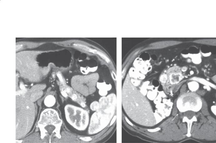
Contrast-enhanced CT. A and B. There is focal enlargement of the pancreatic head. The body and tail of the pancreas are of normal size. There is blurring of the tissue planes about the mass in the head of the gland. Soft tissue thickening and stranding are present in the left anterior pararenal space.
Di erential Diagnosis
1.Focal pancreatitis
2.Ductal adenocarcinoma
3.Islet cell tumor
Diagnosis
Focal pancreatitis
Discussion
Focal pancreatitis occurs in less than 20% of patients with acute pancreatitis. Usually the pancreatic head is involved. Often the disease is mild and due to choledocholithiasis. As in this case, other common findings of acute pancreatitis are present, including loss of discrete planes between the mass and the retroperitoneal fat and peripancreatic soft tissue stranding.
Disease type: Inflammatory Diseases
CASE 8.5
Findings
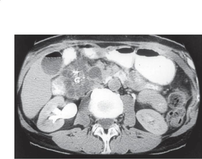
Contrast-enhanced CT. A large fluid collection is present in the region of the pancreatic tail. There is no perfusion of the pancreatic tail.
Di erential Diagnosis
Acute pancreatitis with pancreatic necrosis
Diagnosis
Acute pancreatitis with pancreatic necrosis
8. PANCREAS 661
Discussion
Pancreatic necrosis should be excluded in each patient with acute pancreatitis. The absence of perfusion of a segment(s) of the pancreas is diagnostic for pancreatic parenchymal necrosis. This is the most severe stage
of pancreatic inflammation and is associated with the highest morbidity and mortality rates. Common complications associated with pancreatic necrosis
include infection, pseudoaneurysm formation, splenic and portal vein thrombosis, biliary obstruction, and many systemic problems.
Disease type: Inflammatory Diseases
662 MAYO CLINIC GASTROINTESTINAL IMAGING REVIEW
CASE 8.6
A B
C
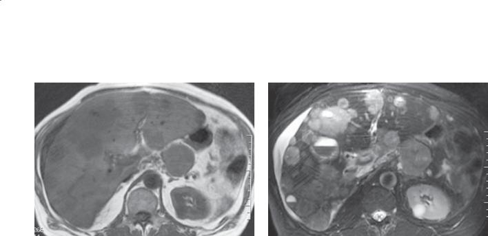
Findings
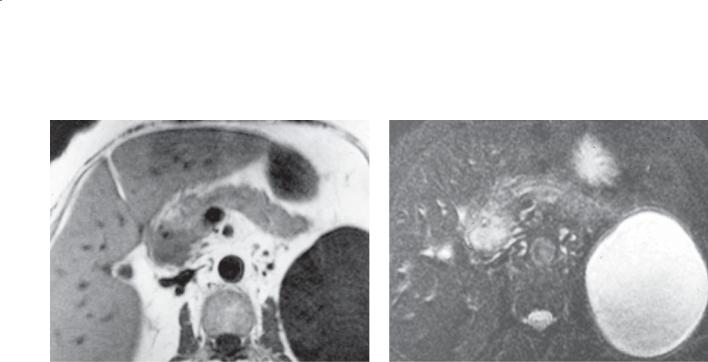
A.T1-weighted MRI. Hypointense changes are seen within the lesser sac. The pancreas is not identified.
B.T2-weighted MRI. Hyperintense changes are seen within the lesser sac. There is a soft tissue component in the region of the pancreatic tail.
C.Contrast-enhanced MRI. Only a small amount of enhancing pancreatic tissue is visible within the tail region. Nonenhancing fluid occupies the lesser sac.
Di erential Diagnosis
Acute pancreatitis with pancreatic necrosis
Diagnosis
Acute pancreatitis with pancreatic necrosis
Discussion
Contrast-enhanced MRI is very sensitive to soft tissue enhancement. The body of the pancreas is at greatest risk of necrosis because the head and tail have more collateral vessels from the gastroduodenal arcade and splenic hilar vessels. Necrosis is associated with considerable morbidity and mortality and is also susceptible to secondary infection or abscess.
Disease type: Inflammatory Diseases
CASE 8.7
Findings
Contrast-enhanced CT. A large fluid collection is present in the region of the body and tail of the pancreas. The fluid collection is well demarcated and contains fat. The pancreas is normal in size and enhancement (tail seen on more cephalad images).
Di erential Diagnosis
Acute pancreatitis with a pseudocyst
Diagnosis
Acute pancreatitis with a pseudocyst
8. PANCREAS 663
Discussion
Fluid collections can be identified in about half of all patients with acute pancreatitis. Most fluid
collections are absorbed within 2 to 3 weeks. These are referred to as peripancreatic fluid collections. Fluid collections that last more than 6 weeks are referred
to as pseudocysts and usually have a thick fibrous wall. These collections usually indicate a persistent communication with the pancreatic duct. They tend to remain the same size but can enlarge over time. Pseudocysts can compress adjacent organs and cause obstruction or pain. Because of the potential for complications (pancreatic peritonitis, abscess,
pseudoaneurysm formation and rupture), pseudocysts more than 4 cm in diameter are usually drained, even if asymptomatic.
Disease type: Inflammatory Diseases

664 MAYO CLINIC GASTROINTESTINAL IMAGING REVIEW
CASE 8.8
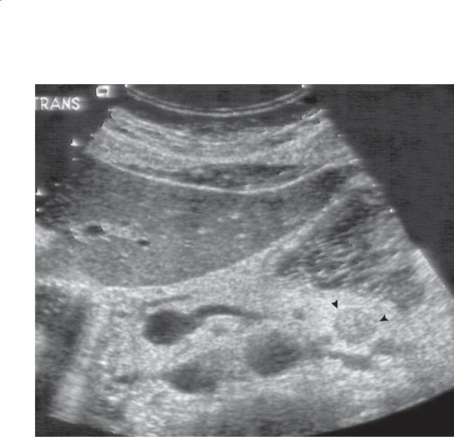
Findings
Transverse sonogram. The pancreas is diffusely enlarged and has decreased echogenicity (arrowheads). A large, mixed echogenicity fluid collection (arrows) is located anterior to the pancreas, which contains echogenic debris.
Di erential Diagnosis
Acute pancreatitis (with associated complicated peripancreatic fluid collection)
Diagnosis
Acute pancreatitis (with infected peripancreatic fluid collection)
Discussion
Sonography is often a valuable tool to determine the nature of a peripancreatic fluid collection or pseudocyst. Complicated peripancreatic fluid collections or pseudocysts containing hemorrhagic or infected fluid often exhibit homogeneous fluid attenuation on CT. Sonography easily shows the
complexity of this peripancreatic fluid collection in the setting of acute pancreatitis. On sonography-guided aspiration in this case, infected fluid was found.
Disease type: Inflammatory Diseases
CASE 8.9
Findings
Contrast-enhanced CT. A large fluid collection is present in the lesser sac. An irregular collection of contrast enhancement is present within the fluid collection.
Di erential Diagnosis
1.Pancreatic pseudocyst
2.Pancreatic fluid collection with pseudoaneurysm
Diagnosis
Pancreatic fluid collection with pseudoaneurysm
8. PANCREAS 665
Discussion
Pancreatitis can lead to weakening of the walls of the vessels that come into contact with the pancreatic enzymes produced during an episode of acute pancreatitis. As the wall weakens, a pseudoaneurysm can develop. Ruptured pseudoaneurysms bleed into the retroperitoneum, bowel lumen, or peritoneal space. This is usually a sudden and catastrophic event with a high mortality rate. Recognition of
the pseudoaneurysm and surgical or endovascular repair or embolization are critical to preventing lifethreatening hemorrhage.
Disease type: Inflammatory Diseases
666 MAYO CLINIC GASTROINTESTINAL IMAGING REVIEW
CASE 8.10
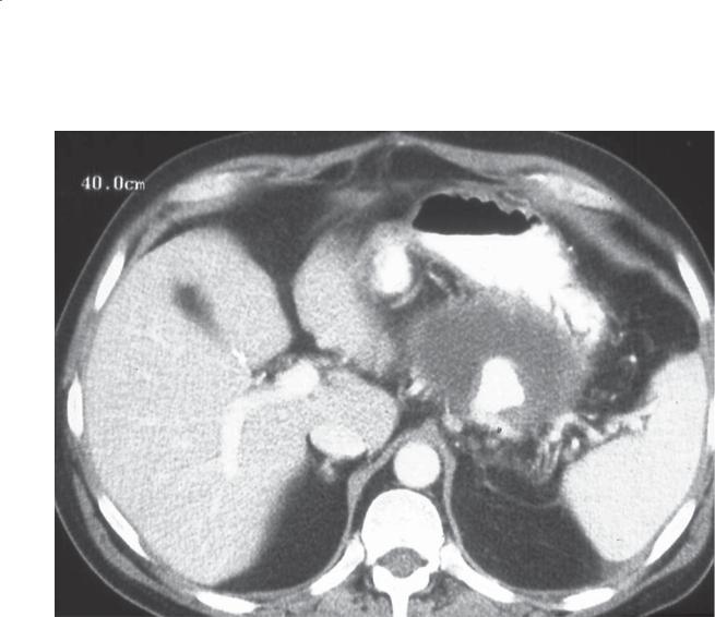
Findings
Unenhanced CT. The pancreas is enlarged and contains multiple regions of high attenuation. Peripancreatic, mesenteric, and abdominal fluid also is present.
Di erential Diagnosis
Hemorrhagic pancreatitis
Diagnosis
Hemorrhagic pancreatitis
Discussion
Pancreatic hemorrhage is an uncommon finding in patients with pancreatitis. The inflammatory changes can cause erosion of the pancreatic vessels, pseudoaneurysm formation, and eventually
retroperitoneal hemorrhage. High-attenuation fluid on unenhanced CT is key to the diagnosis of pancreatic hemorrhage. If patients are unstable from blood loss, emergency angiography with embolization of the bleeding vessel may be required. Usually, pancreatic necrosis also is present.
Disease type: Inflammatory Diseases
8. PANCREAS 667
CASE 8.11
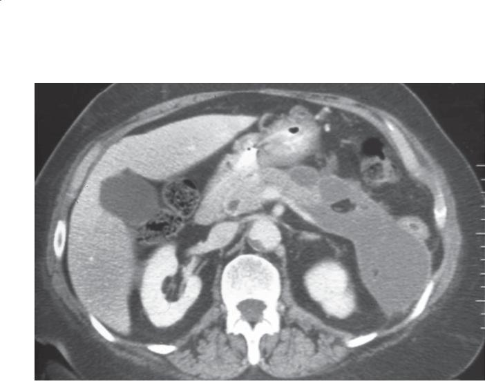
Findings
Contrast-enhanced CT. Fluid and gas are present within the lesser sac.
Di erential Diagnosis
Pancreatic abscess
Diagnosis
Pancreatic abscess
Discussion
Pancreatic abscess usually develops as a complication of pancreatic necrosis. Pancreatic pseudocysts also can
become infected. These can develop from a few days to several weeks after the onset of acute pancreatitis.
Systemic signs of infection and sepsis usually are present, although occult presentations can occur. Most abscesses present only as an abnormal fluid collection. Poorly defined fluid collections that are present 2 to 4 weeks after the onset of pancreatitis are highly suspicious for an abscess. In suspicious cases, percutaneous aspiration of the fluid for microbiologic assessment is required. About 20% of abscesses contain gas (as in this case), a feature making the diagnosis much more likely. Recognition of an abscess is important, because untreated it has a high mortality rate.
Disease type: Inflammatory Diseases
668 MAYO CLINIC GASTROINTESTINAL IMAGING REVIEW
CASE 8.12
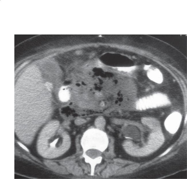
Findings
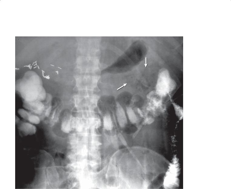
Single-contrast barium enema (postevacuation view). Separation of the stomach and colon with an abnormal gas collection in the region of the lesser sac (arrows). The left transverse and upper descending colon is narrowed. Multiple surgical clips are in the right upper quadrant.
Di erential Diagnosis
Pancreatic abscess with inflammatory narrowing of the colon (colon cutoff sign)
Diagnosis
Pancreatic abscess with inflammatory narrowing of the colon (colon cutoff sign)
Discussion
Pancreatic abscess containing gas can be recognized on abdominal radiographs. Gas located between the colon and the stomach is usually extraluminal and within the lesser sac. A pancreatic abscess without gas is usually not visible on an abdominal radiograph. Pancreatic inflammatory fluid preferentially occupies the left anterior pararenal space. Inflammatory fluid in this location often bathes the colon and causes colonic spasm. Persistent spasm causes partial colonic obstruction with gaseous dilatation of the colon proximal to the splenic flexure. This constellation of events is radiographically referred to as the “colon cutoff sign.” It may be the only clue to the presence of pancreatitis on an abdominal radiograph.
Disease type: Inflammatory Diseases
CASE 8.13
Findings
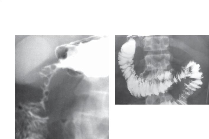
CASE 8.13. UGI. Marked narrowing of the proximal duodenum. The mucosa through this narrowed region is intact.
CASE 8.14. UGI. Widening of the duodenal C loop and thickening of the folds of the proximal small bowel.
Di erential Diagnosis
1.Acute pancreatitis
2.Pancreatic mass
3.Pancreatic abscess
Diagnosis
Acute pancreatitis
8. PANCREAS 669
CASE 8.14
Discussion
Acute pancreatitis can cause various upper gastrointestinal abnormalities. Enlargement of the duodenal C loop is most common and can be due to pancreatic enlargement or peripancreatic fluid
collections. Narrowing of the duodenal lumen usually is due to severe spasm and can result in gastric outlet obstruction. Occasionally, the inflammation can be long-lasting and result in a duodenal stricture. If the pancreatic effusion is large enough, the inflammatory changes can extend beyond the lesser sac and involve the small bowel mesentery. Thickening of the folds in the proximal small bowel often is associated with extension of the peripancreatic effusion into the small bowel mesentery and with pancreatic ascites.
Disease type: Inflammatory Diseases
670 MAYO CLINIC GASTROINTESTINAL IMAGING REVIEW
CASE 8.15
Findings |
Discussion |
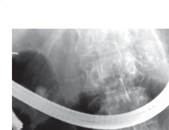
Abdominal radiograph. Multiple calcifications in |
Chronic pancreatitis is characterized by irreversible |
the upper abdomen are located in the region of the |
morphologic and functional pancreatic damage. |
pancreas. |
Most cases are due to alcoholism. Hyperlipidemia, |
|
hyperparathyroidism, trauma, and pancreas divisum |
Di erential Diagnosis |
have been associated with this condition. Pain, weight |
Chronic pancreatitis |
loss, diabetes, and malabsorption are found clinically. |
|
The calcifications in chronic pancreas are ductal |
Diagnosis |
in location, usually small and irregular in shape. |
Chronic pancreatitis |
The presence of pancreatic ductal calcifications is |
|
pathognomonic of chronic pancreatitis. |
Disease type: Inflammatory Diseases
CASE 8.16
Findings
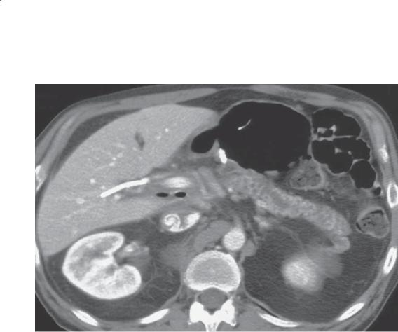
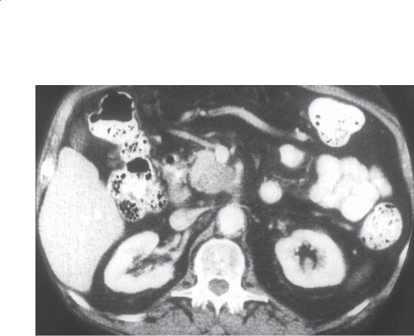
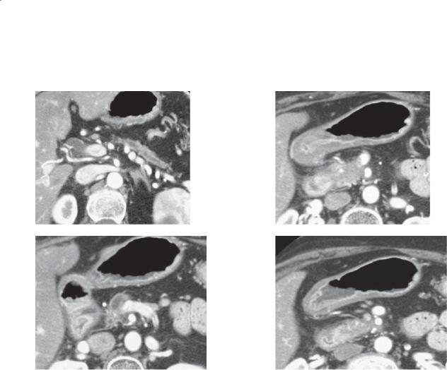
Contrast-enhanced CT. The pancreas is atrophic. Main and side branch ductal dilatation is present. A biliary stent and cystgastrostomy tube are present.
Di erential Diagnosis
1.Chronic pancreatitis
2.Intraductal pancreatic mucinous neoplasm
3.Pancreatic ductal obstruction—cause undetermined
Diagnosis
Chronic pancreatitis
8. PANCREAS 671
Discussion
Th e CT diagnosis of chronic pancreatitis depends on assessment of the pancreatic parenchyma and pancreatic duct and the presence of calculi. The pancreatic parenchyma can vary from enlarged to normal to atrophic. An atrophic gland is a helpful finding when present. Pancreatic ductal dilatation and dilatation of its side branches are characteristic of chronic pancreatitis. Ductal dilatation should be continuous to the ampulla. It is important to see ductal dilatation throughout the gland to exclude an obstructing neoplasm. Approximately half of patients with chronic pancreatitis have ductal calcifications. Calculi are the most reliable finding for the disease. It may be difficult to differentiate intraductal pancreatic mucinous neoplasm (IPMN) from ductal changes
in chronic pancreatitis. Generally, ductal dilatation is the predominant finding with IPMN, whereas parenchymal atrophy dominates with chronic pancreatitis. In equivocal cases, ERCP or endoscopic ultrasonography is recommended.
Disease type: Inflammatory Diseases

672 MAYO CLINIC GASTROINTESTINAL IMAGING REVIEW
CASE 8.17
A B
Findings
Contrast-enhanced CT. A and B. The biliary and main pancreatic ducts are dilated. There are coarse calcifications within the pancreatic duct. A benign left renal cyst is present.
Di erential Diagnosis
1.Chronic pancreatitis (with benign stricture)
2.Chronic pancreatitis (with ductal adenocarcinoma)
Diagnosis
Chronic pancreatitis (with associated benign biliary duct stricture)
Discussion
Pancreatic ductal calcifications are the most helpful finding to suggest the diagnosis of chronic pancreatitis. The calculi are often small and irregular in shape. They can be coarse in appearance (as in this case). Those associated with familial pancreatitis and cystic fibrosis are usually the largest stones. Dilatation of the biliary tree raises the possibility of a mass in the region
of the pancreatic head or distal common bile duct. A careful search for a mass should be performed. Biliary strictures can occur from changes of chronic
pancreatitis (as in this case). Pancreatic cancers develop in 2% to 3% of patients with chronic pancreatitis; therefore, a change in the imaging findings or clinical symptoms should prompt a careful search for tumor.
Disease type: Inflammatory Diseases
CASE 8.18
Findings
Contrast-enhanced CT. There is fatty replacement of the pancreas. Several calculi are visible within the main pancreatic duct.
Di erential Diagnosis
1.Chronic pancreatitis
2.Cystic fibrosis
Diagnosis
Chronic pancreatitis with fatty replacement of the pancreas
8. PANCREAS 673
Discussion
Fatty replacement of the pancreas most often is found in patients with chronic pancreatitis, obesity, diabetes, or cystic fibrosis. It also can occur sporadically without definite cause. Pancreatic insufficiency may or may not accompany fatty replacement. Pancreatic insufficiency usually indicates loss of more than 90% of functioning pancreatic tissue. Diabetes and malabsorption are
the main symptoms of pancreatic insufficiency. This patient had chronic pancreatitis. Pancreatic ductal calcifications led to ductal obstruction and secondary pancreatic parenchymal fatty replacement.
Disease type: Inflammatory Diseases
674 MAYO CLINIC GASTROINTESTINAL IMAGING REVIEW
CASE 8.19 |
CASE 8.20 |
Disease type: Inflammatory Diseases

Findings
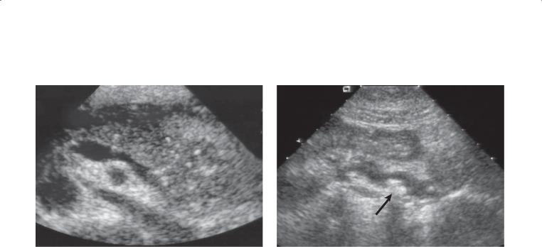
CASE 8.19. Sonogram. The pancreatic gland is generous in size with decreased echogenicity and multiple echogenic foci in the visualized body and tail of the pancreas.
CASE 8.20. Sonogram. The main pancreatic duct is substantially dilated and contains a large posteriorly shadowing stone (arrow). There is diffuse atrophy of the visualized pancreatic gland.
Di erential Diagnosis
Chronic pancreatitis
Diagnosis
Chronic pancreatitis
8. PANCREAS 675
Discussion
Each of these two cases has a classic sonographic appearance of chronic pancreatitis. Case 8.19 has multiple tiny echogenic foci (calculi) scattered in the visualized pancreatic parenchyma. Shadowing may or may not be visible depending on the size of the calcifications. These tiny calcifications are in an intraductal location, but side branch ducts are generally not visible at sonography. The size and echotexture of the pancreas can vary in chronic
pancreatitis. Although nonspecific, the enlargement and relative hypoechogenicity of the visualized pancreas in case 8.19 favor some degree of acute inflammation. The pancreas tends to be atrophic in cases of chronic pancreatitis without associated acute inflammation. When calcifications migrate
from side branches into the main pancreatic duct, or when calcifications form primarily in the main duct, an appearance such as that in case 8.20 may be seen.
Chronic main duct obstruction by stones often results in an irregular or beaded dilatation of the duct and associated atrophy of the pancreatic gland.
Disease type: Inflammatory Diseases
676 MAYO CLINIC GASTROINTESTINAL IMAGING REVIEW
CASE 8.21
CASE 8.22A
A
Disease type: Inflammatory Diseases
CASE 8.22B
B
Findings
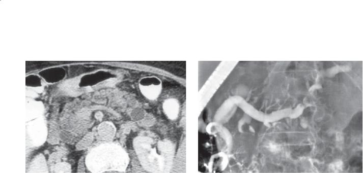
CASE 8.21. ERCP. The main pancreatic duct is irregularly dilated and contains several filling defects in the region of the pancreatic head.
CASE 8.22. A. ERCP. The pancreatic duct in the head of the pancreas is irregular in contour. Side branch ducts are variable in shape—dilated, irregular in contour, and beaded. Within the body and tail of the pancreas, the duct is dilated and irregular in contour.
B. Contrast-enhanced CT. The main pancreatic duct is mildly dilated and is irregular and beaded in contour.
Di erential Diagnosis
Chronic pancreatitis
Diagnosis
Chronic pancreatitis
8. PANCREAS 677
Discussion
ERCP and MRCP are the most helpful techniques to make the diagnosis of chronic pancreatitis. Typical changes include ductal dilatation, ductal contour irregularity, and clubbing and stenosis of the side branches. Filling defects within the ducts are usually calculi. Advanced changes of chronic pancreatitis lead to a chain-of-lakes or beaded appearance of the side branches.
Disease type: Inflammatory Diseases
678 MAYO CLINIC GASTROINTESTINAL IMAGING REVIEW
CASE 8.23
A
B
C
Findings
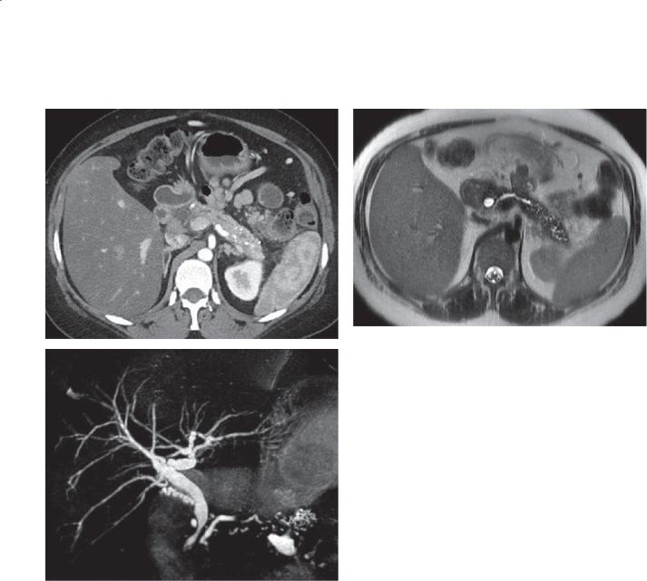
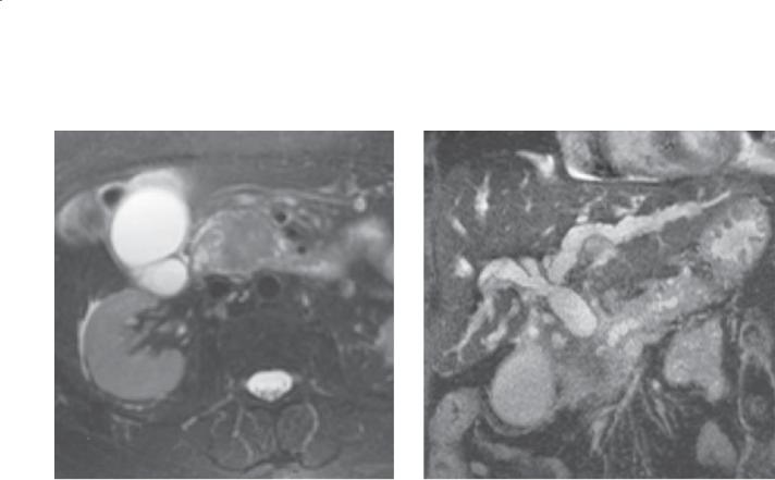
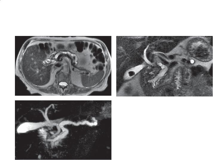
A.Contrast-enhanced CT. The pancreatic duct is segmentally dilated and there are pancreatic parenchymal calcifications.
B.T2-weighted MRI. The main pancreatic duct is segmentally dilated and there are multiple dilated pancreatic ductal side branches.
C.MRCP, coronal. The main and side branch pancreatic ducts are segmentally dilated. The distal common
bile duct is moderately dilated, and narrowing of the intrapancreatic portion of the bile duct is visible.
Di erential Diagnosis
Chronic pancreatitis
Diagnosis
Chronic pancreatitis
Discussion
Th e presence of pancreatic parenchymal calcifications is pathognomonic of chronic pancreatitis. Because
of uneven fibrosis during the healing process after resolution of the pancreatic inflammation, ductal strictures and upstream dilatation will develop. Usually, multiple bouts of pancreatitis are required (with irreversible pancreatic parenchymal damage) before chronic pancreatitis is apparent at imaging. The common bile duct can be affected in the same manner because its distal end courses through the pancreas. MRI (MRCP) is sensitive for visualization of fluid-filled structures such as the pancreatic duct and its side branch ducts.
Disease type: Inflammatory Diseases
CASE 8.24
Findings
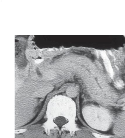
Contrast-enhanced CT. Multiple calcifications are present within the pancreatic head. There is mild soft tissue stranding within the peripancreatic tissues.
Di erential Diagnosis
1.Chronic pancreatitis
2.Familial pancreatitis
Diagnosis
Familial pancreatitis
8. PANCREAS 679
Discussion
Familial pancreatitis is a term used to encompass 2 types of disease: 1) a familial occurrence of pancreatitis in kindreds with hyperlipidemia,
hyperparathyroidism, cystic fibrosis, and cholelithiasis and 2) an autosomal dominant inherited syndrome referred to as hereditary pancreatitis. Patients with the latter type have an early age at onset of pancreatitis (often during childhood), pain, disability, and common complications including pancreatic insufficiency, pseudocyst formation, and an increased incidence of pancreatic carcinoma. Calcifications usually are large within dilated pancreatic ducts.
Disease type: Inflammatory Diseases
680 MAYO CLINIC GASTROINTESTINAL IMAGING REVIEW
CASE 8.25
A B
Findings
Contrast-enhanced CT. A. Peripherally calcified, waterattenuation mass is in the region of the pancreatic
tail. B. Same image, bone windows. The peripheral calcification is better seen within the cyst wall.
Di erential Diagnosis
1.Splenic artery aneurysm
2.Splenic artery pseudoaneurysm
3.Chronic pseudocyst
Diagnosis
Chronic pseudocyst
Discussion
Pseudocysts develop during the acute phase of pancreatitis as a result of persistent communication between the pancreatic ductal system and the peripancreatic space(s). As the inflammation subsides, the pressure in the main pancreatic ductal system is reduced, and normal flow through the pancreatic duct to the duodenum resumes. Under these circumstances, peripancreatic fluid collections are reabsorbed. If a pancreatic stricture develops, the pressure differences within the pancreatic ductal system may favor continued excretion of fluid into a peripancreatic collection or pseudocyst. If this abnormal communication persists, a chronic pseudocyst will develop. Chronic inflammation within the cyst wall can lead to calcification (as in this case).
Aneurysms or pseudoaneurysms will have at least some vascular enhancement or attachment to the artery (visible on other adjacent images or reformatted views—not shown). Pseudoaneuryms
should be suspected in patients with a history of recent pancreatitis.
Disease type: Inflammatory Diseases
8. PANCREAS 681
CASE 8.26
A B
C
Findings
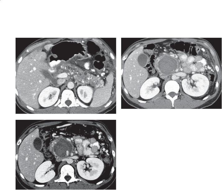
Contrast-enhanced CT. A. The pancreas appears mildly enlarged and contains multiple calcifications and dilated ducts. B. A moderate mixed-attenuation mass is in the region of the pancreatic head. C. The mixed attenuation mass contains a small enhancing aneurysm.
Di erential Diagnosis
1.Chronic pancreatitis with pseudoaneurysm formation
2.Chronic pancreatitis with enhancing neoplasm
Diagnosis
Chronic pancreatitis with pseudoaneurysm formation
Discussion
Th e presence of pancreatic calcifications and dilated ducts is consistent with the diagnosis of chronic pancreatitis. A mixed-attenuation mass could be due to hemorrhage within an aneurysm or a tumor. The presence of a small aneurysm within the mass is typical of a pseudoaneurysm. Tumor is less likely because the mass does not enhance except for the small aneurysm. Chronic pancreatitis is usually due to multiple bouts of acute pancreatitis. Adjacent inflammation and enzymatic action can weaken nearby vessels, leading to pseudoaneurysm formation. These pseudoaneurysms need to be treated either surgically or with the placement of vascular stents and coils in order to prevent rupture of the aneurysm and death.
Disease type: Inflammatory Diseases

682 MAYO CLINIC GASTROINTESTINAL IMAGING REVIEW
CASE 8.27
A B
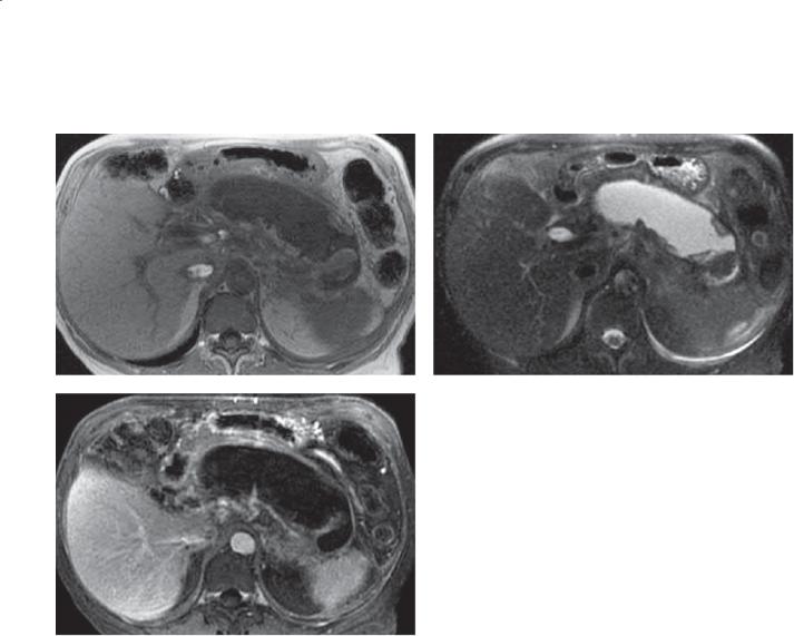
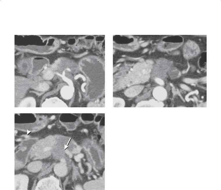
Findings
A.Contrast-enhanced CT. The pancreas appears enlarged. There is a hypoattenuating rim around the pancreas that includes a focus of fat.
B.Contrast-enhanced MRI. The pancreas is engulfed in a sausage-shaped region of moderate signal intensity.
Di erential Diagnosis
1.Acute pancreatitis
2.Autoimmune pancreatitis
3.Adenocarcinoma of the pancreas
Diagnosis
Autoimmune pancreatitis
Discussion
Autoimmune pancreatitis is a type of chronic pancreatitis, characterized by lymphocytic infiltration of the pancreas with fibrosis. The disease is twice as common among men than women and usually affects older persons (mean age, 60 years). Most patients present with painless jaundice or abdominal pain; 17% of patients have inflammatory bowel disease. Increased levels of immunoglobulin G4 are common. The disease can be systemic and affect other organs besides the pancreas, including lung, biliary system, renal tubules, and salivary glands. Associated conditions include Sjögren syndrome, primary sclerosing cholangitis, and systemic lupus erythematosus. Oral corticosteroid therapy usually is effective.
Disease type: Inflammatory Diseases

|
8. PANCREAS 683 |
|
|
|
|
|
TABLE 8.1 |
|
Inflammatory Diseases of the Pancreas |
CASE |
|
|
|
|
Acute diffuse pancreatitis |
Diffuse enlargement or peripancreatic effusion. Soft |
8.1–8.3 |
|
tissue stranding |
|
|
|
|
Acute focal pancreatitis |
Segmental mass, fluid, and soft tissue stranding |
8.4 |
|
|
|
Necrotizing pancreatitis |
Nonenhancement of a segment after intravenous conrast. |
8.5 and 8.6 |
|
Hemorrhage in some cases |
|
|
|
|
Complications of acute pancreatitis |
Complications include pseudocysts, abscesses, |
8.7–8.14 |
|
pseudoaneurysms, splenic or portal vein thrombosis, |
|
|
hemorrhage |
|
|
|
|
Chronic pancreatitis |
Ductal calcifications. Ductal dilatation. |
8.15–8.23, 8.26 |
|
Parenchymal atrophy |
|
|
|
|
Familial pancreatitis |
Positive family history. Early onset. Ductal dilatation |
8.24 |
|
with coarse calcifications |
|
|
|
|
Chronic pseudocyst |
Well-defined water attenuation mass adjacent or within |
8.25 |
|
pancreas—usually indicates pancreatic ductal stricture. |
|
|
Cyst wall calcification possible |
|
|
|
|
Autoimmune pancreatitis |
Older persons. Associated autoimmune disorders. |
8.27 |
|
Mild symptoms |
|
|
|
|
Disease type: Inflammatory Diseases
684 MAYO CLINIC GASTROINTESTINAL IMAGING REVIEW
CASE 8.28
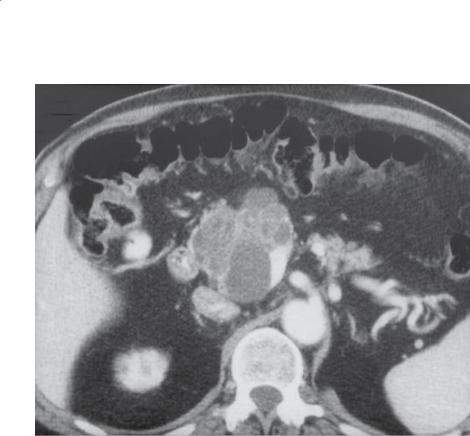
Findings
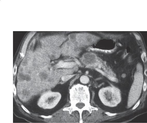
Contrast-enhanced CT. A large hypoattenuating mass is present in the body of the pancreas. The pancreatic duct in the region of the tail is dilated. Multiple hepatic metastases are present.
Di erential Diagnosis
Ductal adenocarcinoma
Diagnosis
Ductal adenocarcinoma
Discussion
Pancreatic ductal adenocarcinoma is the fifth most common cause of death from cancer in the United States (less common than lung, colon, breast, and prostate). Despite imaging advances, there has been no improvement in the mortality from this disease. This is likely due to these tumors being discovered after they have already metastasized and to their rapid growth characteristics. Radiologists have an important role in the detection of this tumor (to explain symptoms) and in the staging of disease extent.
Most ductal cancers are hypoattenuating, but small tumors may be detectable only if they alter the texture of the pancreatic parenchyma or obstruct the main pancreatic duct. Staging is based on assessment of local disease extent, vascular encasement, lymphatic or peritoneal spread, and hepatic metastases. In this case, the typical features of a nonresectable ductal cancer are present: a large mass obstructing the pancreatic duct, and hepatic metastases.
Disease type: Masses
CASE 8.29
A
Findings
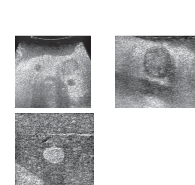
Sonogram. A. Transverse view of the pancreas shows a large hypoechoic mass in the head of the pancreas with marked dilatation of the main pancreatic duct to the level of this mass (arrow). B. A biopsy needle is visualized within the pancreatic mass.
Di erential Diagnosis
Ductal adenocarcinoma
Diagnosis
Ductal adenocarcinoma
8. PANCREAS 685
B
Discussion
Th e most common sonographic appearance of pancreatic carcinoma is a poorly defined hypoechoic mass with associated pancreatic duct dilatation. The biliary ducts also are often dilated if the tumor is in the head of the pancreas. This associated ductal dilatation is less common in other solid hypoechoic pancreatic masses, which include islet cell tumors, metastases, lymphoma, or focal pancreatitis. Although Doppler ultrasonography can be useful for determining vascular involvement of the tumor, contrast-enhanced CT or MRI is considered a better staging tool. When clinically indicated, percutaneous ultrasonographyguided biopsy often is used to obtain a tissue diagnosis. Advantages of sonography-guided biopsy over CT-guided biopsy include the ability to displace overlying bowel loops with transducer compression and real-time visualization of peripancreatic vessels during the biopsy.
Disease type: Masses
686 MAYO CLINIC GASTROINTESTINAL IMAGING REVIEW
CASE 8.30
A B
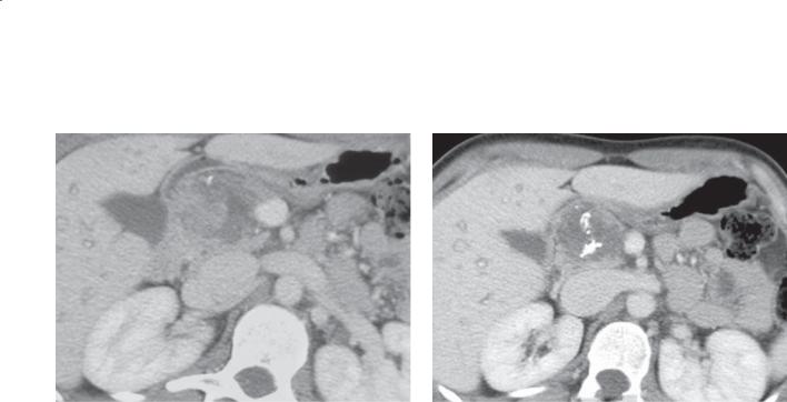
Findings
Contrast-enhanced CT. A. Pancreatic phase of contrast enhancement. A hypoattenuating region is present within the head of the pancreas (arrows). B. Portal venous phase of contrast enhancement. The pancreatic head is nearly isoattenuating. The duodenum is seen lateral to the pancreatic head.
Di erential Diagnosis
1.Ductal adenocarcinoma
2.Focal pancreatitis
Diagnosis
Ductal adenocarcinoma
Discussion
Multidetector scanners allow rapid acquisition of image data during multiple different phases of contrast enhancement. Hypovascular masses (such as ductal adenocarcinoma) usually are optimally identified during the pancreatic phase of contrast enhancement. Tissue contrast between the enhancing pancreatic parenchyma and a hypoattenuating mass is usually maximal. In most patients, this occurs approximately 45 seconds after initiation of contrast material administration. Portal venous phase imaging is useful for the detection of hepatic metastases and regional lymphadenopathy. In patients with a suspected pancreatic carcinoma, dual-phase imaging (pancreatic and portal venous phases) is recommended.
Disease type: Masses
CASE 8.31
A
Findings
A.T1-weighted MRI. A hypointense mass is present within the head of the pancreas.
B.T2-weighted MRI. The mass is hyperintense. The tumor does not extend beyond the pancreas. A large benign cyst is present in the upper pole of the left kidney.
Di erential Diagnosis
1.Ductal adenocarcinoma
2.Islet cell tumor
3.Pancreatic metastases
4.Focal pancreatitis
Diagnosis
Ductal adenocarcinoma
8. PANCREAS 687
B
Discussion
Either CT or MRI is used as the primary imaging method for assessment of the pancreas. Pancreatic cancers at MRI are hypointense on T1-weighted images and hyperintense on T2-weighted images. Small tumors are best depicted on T1-weighted images with use of fat saturation. With this technique, the pancreas appears hyperintense compared with the hypointense mass. Dynamic imaging after intravenous administration of gadolinium also can be used to identify a hypoenhancing mass. Findings at dynamic imaging mimic those found at CT. Diffusion imaging can be helpful to evaluate equivocal findings on other pulse sequences or to identify a small lesion that is otherwise not well seen.
Disease type: Masses
688 MAYO CLINIC GASTROINTESTINAL IMAGING REVIEW
CASE 8.32
A B
Findings
A.Contrast-enhanced MRI. A solid, hypointense mass is located within the pancreatic head.
B.MRCP. Both the biliary system and the pancreatic duct have dilatation to the level of the pancreatic head. A hypointense mass is present in the pancreatic head.
Di erential Diagnosis
1.Ductal adenocarcinoma
2.Focal pancreatitis
Diagnosis
Ductal adenocarcinoma (double duct sign)
Discussion
Secondary findings can be helpful for the detection of small pancreatic tumors. Secondary findings include ductal dilatation proximal to the tumor (this includes dilatation of both the biliary and the pancreatic ducts if the tumor is located in the pancreatic head—as in this case), vascular encasement, hepatic metastases, regional lymphadenopathy, peritoneal carcinomatosis, and ascites.
Disease type: Masses
CASE 8.33
Findings
Contrast-enhanced CT. The uncinate process of the pancreas is enlarged, rounded, and hypoattenuating. Soft tissue extends from the uncinate process to the superior mesenteric artery and obliterates the fat plane about the artery.
Di erential Diagnosis
Ductal adenocarcinoma
Diagnosis
Ductal adenocarcinoma
8. PANCREAS 689
Discussion
Th e uncinate process is normally triangular, located posterior to the superior mesenteric vein. It is normally isoattenuating with the remainder of the pancreas. Rounding of the uncinate process should prompt consideration of a mass. The additional findings of a change in attenuation compared with the remainder
of the gland and extension of the process to encase the superior mesenteric artery are typical of a carcinoma.
Disease type: Masses
690 MAYO CLINIC GASTROINTESTINAL IMAGING REVIEW
CASE 8.34
A C
B D
Findings
Contrast-enhanced CT. A through D. The pancreatic duct in the region of the tail of the pancreas is dilated, and the pancreatic parenchyma is atrophic. The duct ends in the region of the pancreatic head. The texture of the slightly hypoattenuating obstructing mass is homogeneous soft tissue. Soft tissue extends and encases the superior mesenteric artery and deforms the superior mesenteric vein.
Di erential Diagnosis
Ductal adenocarcinoma
Diagnosis
Ductal adenocarcinoma
Discussion
Not all pancreatic cancers are hypoattenuating at CT. Some are isoattenuating with the pancreatic
parenchyma. Detection can be difficult if the mass is small and does not alter the contour of the normal pancreas. Identification of the site of the obstructed pancreatic duct is a key finding in localizing the mass. In some cases, the mass alters the normal pancreatic parenchymal texture, often appearing more solid and homogeneous than the normal gland. Atrophy of the parenchyma upstream from the mass usually indicates chronicity of the obstruction.
Another clue to the presence of tumor in this case is tumor extension into the fat about the superior mesenteric artery and vein. Normally there is always a cuff of fat around the artery. The superior mesenteric vein (SMV) is normally in contact with the neck of the pancreas without a surrounding fatty cuff. The SMV is normally either round or elliptical. In this case, the vein is deformed by the adjacent mass, indicating venous invasion.
Disease type: Masses
CASE 8.35
Findings
Contrast-enhanced CT, reformatted curved plane image. The pancreatic duct is dilated to the level of a hypoattenuating mass. There is atrophy of the
pancreatic parenchyma about the dilated duct. Tumor is seen to extend about the common bile duct with bile duct dilatation above the mass. An enlarged lymph node is present adjacent to the pancreas.
Di erential Diagnosis
Pancreatic ductal cancer
Diagnosis
Pancreatic adenocarcinoma
8. PANCREAS 691
Discussion
Curved reformatted views in the plane of the pancreatic duct can be helpful to visualize small pancreatic tumors, especially those that do not deform the normal pancreatic contour. Staging for pancreatic cancer includes assessment for local (vascular invasion, gastric, biliary) and distal (lymph nodes, liver, peritoneum) spread.
Disease type: Masses
692 MAYO CLINIC GASTROINTESTINAL IMAGING REVIEW
CASE 8.36
Findings
Contrast-enhanced CT. There is a large hypoattenuating mass in the body of the pancreas. The mass extends posteriorly and encases the superior mesenteric artery.
Di erential Diagnosis
Ductal adenocarcinoma
Diagnosis
Ductal adenocarcinoma, nonresectable
Discussion
A key growth characteristic of ductal cancers is the proclivity to encase vascular structures and invade perineural structures. Vascular encasement is a major cause of nonresectability. Perineural invasion is the major reason that patients experience back pain with these tumors. In this case, the tumor is not resectable because of encasement of the superior mesenteric artery.
Nonresectability occurs when any of the following exist:
Arterial invasion: usually the superior mesenteric artery or celiac axis
Venous invasion: limited involvement of the superior mesenteric vein (SMV) or junction of the SMV and portal vein usually is considered
resectable and reconstructable. More extensive venous involvement may not be resectable (depending on local surgical views), except in selected specialty centers
Regional lymphadenopathy with metastatic tumor: Today, sampling of enlarged nodes with endoscopic ultrasonography-performed biopsy is common preoperatively
Distant metastases: most common to liver, peritoneum, and lungs
Disease type: Masses
CASE 8.37
A
Findings
Contrast-enhanced CT. A. There is a large hypoattenuating mass in the region of the pancreatic head. The superior mesenteric vein is occluded. The superior mesenteric artery is encased, and there is mass effect on the medial aspect of the duodenum. B. Multiple hepatic metastases and periportal lymphadenopathy are visible.
Di erential Diagnosis
Nonresectable ductal adenocarcinoma
Diagnosis
Nonresectable ductal adenocarcinoma
8. PANCREAS 693
B
Discussion
Nonresectability usually is determined on the basis of vascular invasion by the tumor (superior mesenteric artery encasement, extensive involvement or occlusion of the superior mesenteric vein, or splenic vein occlusion), regional lymphadenopathy, and hepatic metastases or peritoneal metastases. In this case, vascular, lymphatic, and hepatic criteria are met, and thus the tumor is nonresectable. Radiographic staging is highly beneficial, because patients with advanced disease can be spared unnecessary operation.
Disease type: Masses

694 MAYO CLINIC GASTROINTESTINAL IMAGING REVIEW
CASE 8.38
Findings
Contrast-enhanced CT. A large hypoattenuating region is present in the body and tail of the pancreas. The splenic vein is narrowed and encased by soft tissue.
Di erential Diagnosis
1.Ductal adenocarcinoma
2.Focal pancreatitis
Diagnosis
Ductal adenocarcinoma
Discussion
Pancreatic tumors may not deform the contour of the gland (as in this case). Detection usually depends on a difference in enhancement between the tumor and the pancreatic parenchyma. Texture differences
between the normal parenchyma that may contain fatty marbling and a solid tumor also may be a clue to the presence of a mass.
Encasement of the splenic artery and vein without involvement of the celiac axis or superior mesenteric artery usually requires a distal pancreatectomy and splenectomy. In such cases, multiple upper abdominal collaterals often are visible.
Disease type: Masses
8. PANCREAS 695
CASE 8.39
A B
C
Findings
Contrast-enhanced CT. A and B. Pancreatic phase. An infiltrating low-attenuation mass is present in the body of the pancreas and encases the celiac axis and superior mesenteric artery. C. Portal venous phase. The lowattenuation pancreatic mass has occluded the splenic vein (arrow), and multiple associated collateral vessels are seen in the upper abdomen (arrowhead).
Di erential Diagnosis
Ductal adenocarcinoma
Diagnosis
Ductal adenocarcinoma (nonresectable)
Discussion
Pancreatic ductal adenocarcinomas arising in the body or tail of the gland often result in splenic vein occlusion. Multiple collateral vessels in the upper abdomen in a patient with pancreatic adenocarcinoma are highly suggestive of tumor-related venous occlusion and should prompt a careful evaluation of the portal, splenic, and mesenteric veins. Dual-phase pancreatic imaging clearly shows the arterial encasement and venous occlusion in this case, which renders this pancreatic adenocarcinoma nonresectable.
Disease type: Masses
696 MAYO CLINIC GASTROINTESTINAL IMAGING REVIEW
CASE 8.40
A B
Findings
Contrast-enhanced CT. Biphasic arterial (A) and portal venous (B) phases of enhancement. During the arterial phase of contrast enhancement, there is a small hypervascular mass within the pancreatic tail. The mass is isoattenuating with the remainder of the pancreas during the portal venous phase.
Di erential Diagnosis
1.Islet cell tumor
2.Hypervascular metastasis
Diagnosis
Islet cell tumor, insulinoma
Discussion
Islet cell tumors of the pancreas are rare pancreatic neoplasms. It is important to try to differentiate them from ductal tumors because their prognosis and treatment are different. Islet cell tumors are slowgrowing neoplasms; more than 60% of patients are alive 10 years after the initial diagnosis. This contrasts with ductal tumors, with which there are very few 5-year survivors.
Islet cell tumors usually can be differentiated from ductal tumors by their vascularity, propensity to calcify, and their lack of vascular encasement. Islet cell tumors usually are hypervascular neoplasms and enhance briskly during the early phases of contrast enhancement. Because these tumors are slow-growing, calcification
can be found in many of them. Calcification is highly unusual among ductal tumors. Malignant islet cell tumors metastasize to regional lymph nodes and the liver, as is the case with ductal tumors, but vascular encasement and neural invasion are rare.
Clinically, islet cell tumors can be classified as hyperfunctioning or nonhyperfunctioning. The hyperfunctioning tumors are most commonly insulinomas and gastrinomas. Other unusual hyperfunctioning tumors include glucagonomas, somatostatinomas, and others producing a mixture of hormones. These tumors usually are recognized because of the clinical syndrome associated with hormone overproduction. Imaging is used to localize the tumor that is known to be present. Nonhyperfunctioning islet cell tumors are not associated with a clinical syndrome and usually are discovered incidentally or as a result of nonspecific clinical symptoms. Because of the lack of symptoms prompting early investigation, these tumors often are larger and many have metastasized at the time of diagnosis.
Th e patient described here had an insulinoma, the commonest hyperfunctioning tumor. These tumors are benign, solitary, and less than 2 cm in diameter in 90% of patients. This case illustrates the importance of scanning during the arterial phase of contrast enhancement to detect these hypervascular tumors at CT. Sonography, especially intraoperative sonography, is useful for the detection of these tumors. Dynamic enhanced MRI can also be used for diagnosis.
Disease type: Masses
CASE 8.41
Findings
Sonogram. Transverse view of the pancreas with water in the stomach (arrowheads) shows a single, welldefined, hypoechoic mass in the tail of the pancreas.
Di erential Diagnosis
1.Islet cell tumor
2.Other hypoechoic pancreatic mass
Diagnosis
Islet cell tumor, insulinoma
8. PANCREAS 697
Discussion
Th is case shows the importance of having the patient drink water before a sonographic examination for pancreatic islet cell tumor. The insulinoma in this case is easily visualized in the tail of the pancreas deep to the fluid-filled stomach. Visualization of the distal body and tail of the pancreas is often impossible if the stomach is gas-filled. Insulinomas most often are hypoechoic compared with the pancreas.
Disease type: Masses
698 MAYO CLINIC GASTROINTESTINAL IMAGING REVIEW
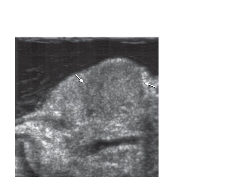
CASE 8.42
Findings
Intraoperative sonogram. A well-defined hypoechoic mass (arrows) is present in the head of the pancreas anterior to the main pancreatic duct.
Di erential Diagnosis
1.Islet cell tumor
2.Ductal adenocarcinoma
Diagnosis
Islet cell tumor, insulinoma
Discussion
Intraoperative sonography is very sensitive for detecting insulinomas. Studies have shown a near
100% sensitivity for detection of insulinomas with the combination of intraoperative ultrasonography and palpation of the gland. In addition to detection of pancreatic tumors, intraoperative ultrasonography is a valuable tool for evaluating the relationship of the tumor to the pancreatic duct. When an adequate margin of normal pancreatic tissue is present between the mass and the pancreatic duct, an enucleation procedure is performed. Without an adequate margin, a partial pancreatectomy is performed. Intraoperative ultrasonography also is used commonly after tumor
enucleation to assess for damage to the pancreatic duct. A normal dilated pancreatic duct after intravenous administration of secretin is highly suggestive of an intact duct.
Disease type: Masses

CASE 8.43
A
Findings
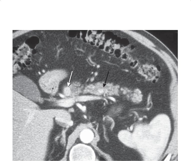
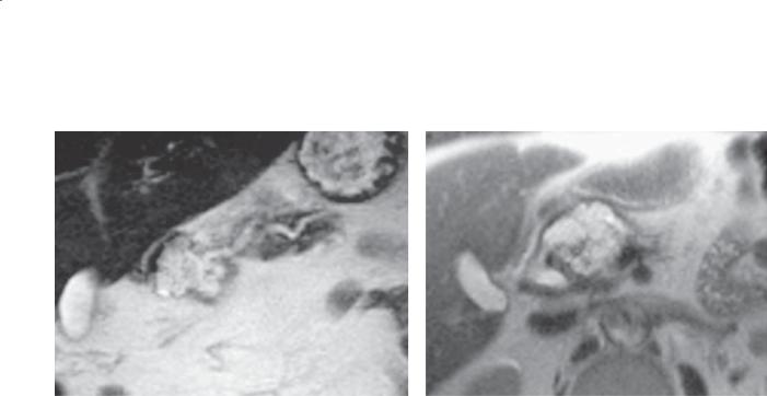
Contrast-enhanced CT. A. Enhancing nodule is in the region of the duodenal bulb. B. Tiny additional
enhancing nodule is within the duodenal bulb (arrow).
Di erential Diagnosis
1.Extrapancreatic islet cell tumor
2.Metastases
3.Brunner gland hyperplasia
4.Ectopic pancreatic rest
5.Gastrointestinal stromal tumor
Diagnosis
Extrapancreatic islet cell tumor (gastrinoma in this patient)
8. PANCREAS 699
B
Discussion
Gastrinomas arise outside the pancreas in about half of all patients and can be multiple (as in this case). These tumors usually are confined to a region known as the gastrinoma triangle—defined by the corners at the ampulla of Vater, junction of the cystic duct and common bile duct, and the junction of the neck and body of the pancreas. The tumors vary in size from 1 to 20 mm in diameter. Some gastrinomas arise within lymph nodes in the gastrinoma triangle.
About half of all gastrinomas arise in patients with multiple endocrine neoplasia type 1. These tumors tend to be multiple and small. They are less aggressive than those occurring sporadically.
Disease type: Masses
700 MAYO CLINIC GASTROINTESTINAL IMAGING REVIEW
CASE 8.44
A B
C D
Disease type: Masses

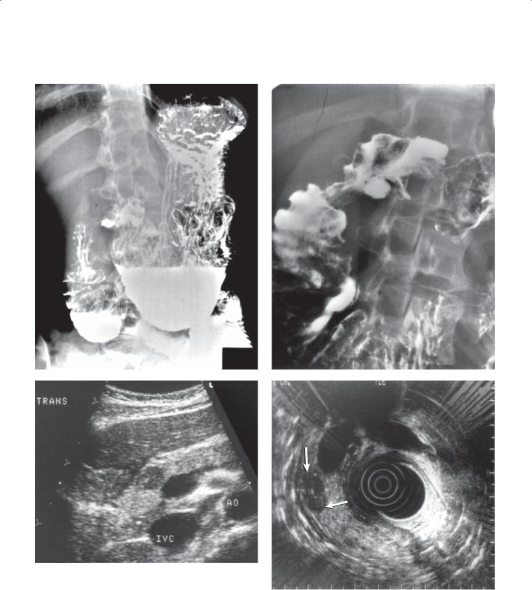
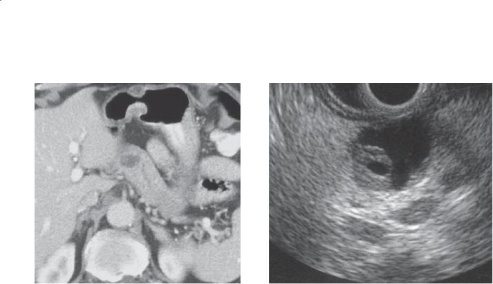
Findings
Double-contrast UGI. A and B. There is an excess amount of fluid in the stomach, which contains multiple thickened folds. There is an ulcer in the duodenal bulb. The duodenum is dilated and contains excess fluid.
C. Transverse sonogram of the pancreas. There is a 1-cm hypoechoic mass within the neck of the pancreas. AO, aorta; IVC, inferior vena cava.
D. Endoscopic sonogram. A 1-cm hypoechoic mass (arrows) in the neck of the pancreas is confirmed.
Di erential Diagnosis
Islet cell tumor, gastrinoma
Diagnosis
Islet cell tumor, gastrinoma
8. PANCREAS 701
Discussion
Gastrinomas are responsible for the Zollinger-Ellison syndrome. Affected patients have overproduction
of gastrin and excess stomach acid with subsequent thickening of the folds of the stomach, gastritis, duodenitis, peptic ulcer disease, and esophagitis. Although ulcerations can be found anywhere in the stomach or duodenum, the duodenal bulb is the commonest location. Nearly three-fourths of gastrinomas are malignant. These tumors can be multiple, and many occur in an extrapancreatic location (usually within the gastric antrum or proximal duodenum). Most are less than 2 cm in
diameter. Because of their small size, multiplicity, and extrapancreatic location, they are difficult to detect in their entirety with standard cross-sectional imaging tools. These tumors usually have a high concentration of somatostatin receptors, and the majority can be detected with octreotide scintigraphy.
Disease type: Masses
702 MAYO CLINIC GASTROINTESTINAL IMAGING REVIEW
CASE 8.45
A B
C
Findings
Intraoperative sonogram. A. Three well-defined hypoechoic masses are present in the body and tail of the pancreas. B. This coned-down view of one of these pancreatic lesions with a higher-frequency transducer more clearly shows the tumor anterior to the splenic vein. C. A well-defined hyperechoic mass is present in the liver with mild associated posterior acoustic shadowing.
Di erential Diagnosis
Metastatic islet cell neoplasm
Diagnosis
Metastatic islet cell tumors, metastatic gastrinoma
Discussion
Intraoperative ultrasonography is extremely sensitive for the evaluation of islet cell tumors. Islet cell tumors usually present as small, well-defined hypoechoic lesions. Unlike the primary tumors, metastatic islet cell lesions tend to be echogenic. The echogenic liver lesion in this case was metastatic gastrinoma. Echogenic hepatic lesions in the setting of islet cell neoplasm should be considered metastases until proved otherwise. The posterior acoustic shadowing behind the echogenic hepatic lesion (in this case) should
be another clue that this does not likely represent a cavernous hemangioma.
Disease type: Masses
CASE 8.46
Findings
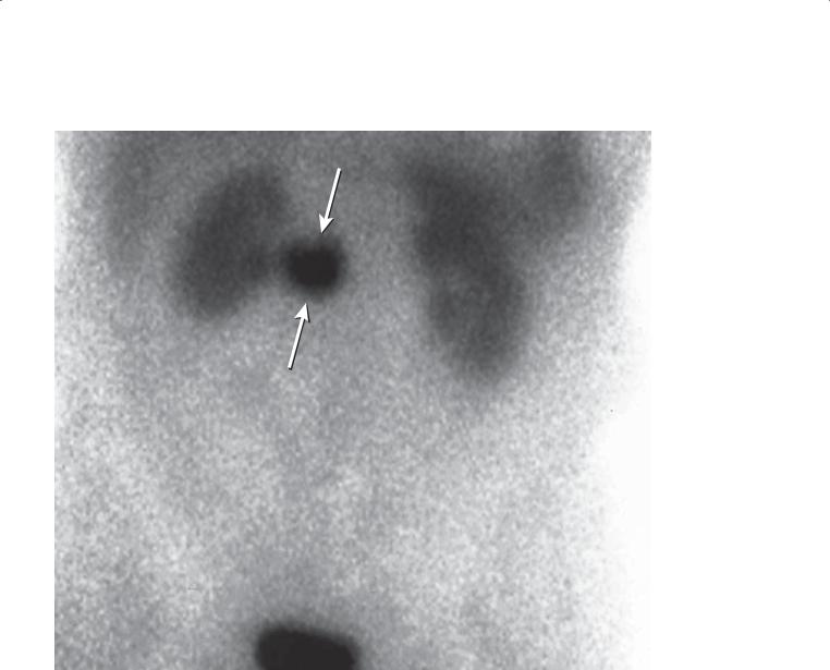
Indium-111-diethylenetriamine pentaacetic acidoctreotide scintigram. Abnormal uptake in the region of the pancreatic head (arrows).
Di erential Diagnosis
Islet cell tumor
Diagnosis
Islet cell tumor: gastrinoma
8. PANCREAS 703
Discussion
Octreotide scintigraphy is helpful for the diagnosis of gastrinomas because of the high concentration of somatostatin receptors within these tumors. This is a highly useful test to localize the gastrinoma in patients with Zollinger-Ellison syndrome. Other neuroendocrine tumors also have a high affinity for octreotide.
Disease type: Masses
704 MAYO CLINIC GASTROINTESTINAL IMAGING REVIEW
CASE 8.47
Findings
Contrast-enhanced CT. There are 2 small hyperenhancing masses within the body of the pancreas (arrows).
Di erential Diagnosis
Multiple endocrine neoplasia type 1 with multiple islet cell tumors
Diagnosis
Multiple endocrine neoplasia type 1 with multiple islet cell tumors
Discussion
Patients with multiple endocrine neoplasia type 1 have an inherited disorder of tumors arising in the pancreas (multiple islet cell tumors), parathyroid gland, and pituitary gland. Islet tumors behave differently from those arising sporadically. They are usually small, multiple, and biologically less aggressive. The small size of the tumors makes complete detection very difficult, even with intraoperative sonography.
Disease type: Masses

8. PANCREAS 705
|
TABLE 8.2 |
Hyperfunctioning Islet Cell Tumors |
|
|
|
TYPE |
KEY CLINICAL SYMPTOMS |
|
|
Insulinoma |
Hypoglycemia: sweating, trembling, palpitations, nervousness |
|
|
Gastrinoma |
Abdominal pain, diarrhea, vomiting, hematemesis, melena, weight loss |
|
|
Glucagonoma |
Necrolytic erythema migrans, diarrhea, diabetes, glossitis |
|
|
Vipoma |
WDHA syndrome: watery diarrhea, hypokalemia, achlorhydria |
|
|
Somatostatinoma |
Diarrhea, steatorrhea, weight loss |
|
|
Disease type: Masses
706 MAYO CLINIC GASTROINTESTINAL IMAGING REVIEW
CASE 8.48
A B
Findings
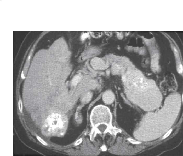
Contrast-enhanced CT. A and B. A hyperenhancing mass is in the body of the pancreas. An enlarged peripancreatic lymph node is present posterior to the pancreatic body, and two larger, but similar-appearing masses, are present in the liver.
Di erential Diagnosis
1.Islet cell tumor with metastases
2.Metastases to the pancreas and liver (primary unknown)
Diagnosis
Islet cell tumor with metastases—nonhyperfunctioning
Discussion
Nonhyperfunctioning islet cell tumors are not associated with a clinical syndrome and often present with nonspecific abdominal complaints. Usually
a large mass is detected in the pancreas. It can be distinguished from ductal cancer because of its hyperenhancement, lack of vascular encasement, and propensity to calcify. Treatment usually is symptomatic. Large tumors that do not respond to
symptomatic therapy may be treated with embolization as a nonoperative approach to debulking these
large masses.
Disease type: Masses
CASE 8.49
Findings
Contrast-enhanced CT (portal venous phase). There is a large mass in the body and tail of the pancreas. The mass contains calcification and enhances similarly to the spleen. A calcified mass also is present in the right lobe of the liver.
Di erential Diagnosis
Nonhyperfunctioning calcified islet cell carcinoma
Diagnosis
Nonhyperfunctioning calcified islet cell carcinoma
8. PANCREAS 707
Discussion
Th is case exemplifies the typical nonhyperfunctioning pancreatic islet cell tumor. Its large size and calcification without vascular encasement are characteristic of the tumor. Liver metastases also can calcify, indicating the slow growth of these lesions. The calcification usually is coarse and dystrophic in appearance.
Disease type: Masses
708 MAYO CLINIC GASTROINTESTINAL IMAGING REVIEW
CASE 8.50
A B
Findings
A.T1-weighted abdominal MRI. A 4-cm mass is present in the body of the pancreas. There are multiple hepatic metastases.
B.T2-weighted abdominal MRI. Multiple hyperintense masses correspond to those seen on the T1-weighted image. The masses in the liver are of variable intensity. Some contain a fluid-fluid layer. Ascites.
Di erential Diagnosis
1.Islet cell carcinoma with hepatic metastases
2.Neuroendocrine metastases to liver and pancreas
Diagnosis
Islet cell carcinoma with hepatic metastases
Discussion
Th is case is a typical example of a malignant nonhyperfunctioning islet cell tumor at MRI. MRI is useful to follow patients with this tumor because in many cases oral and intravenous contrast material are not necessary for assessing the size and progression of the lesions. In addition, because these hepatic lesions can become isoattenuating at CT, MRI is an optimal technique for completely evaluating the full extent
of the tumors. The fluid-fluid layers in the hepatic metastases are commonly seen in hypervascular metastases, especially neuroendocrine metastases (islet cell, carcinoid, pheochromocytoma).
Disease type: Masses
CASE 8.51
A
Findings
A.Contrast-enhanced CT. A cystic mass is present in the body of the pancreas. The pancreatic duct proximal to the mass is mildly dilated.
B.Endoscopic sonogram. The mass is predominantly cystic but contains a mural nodule.
Di erential Diagnosis
1.Ductal adenocarcinoma
2.Cystic metastases to the pancreas
3.Cystic islet cell tumor
4.Mucinous cystic neoplasm of the pancreas
Diagnosis
Cystic islet cell tumor
8. PANCREAS 709
B
Discussion
Islet cell tumors usually are solid lesions. Large tumors often undergo central necrosis and calcification but retain a rim of hyperenhancing tissue. Rarely, islet cell tumors appear cystic (water density) without evidence of hyperenhancement (as in this case). The sonogram reveals the complex (containing a septation and nodule) cystic nature of the mass. According to standard CT criteria, this most likely would have been
a ductal adenocarcinoma or cystic mucinous neoplasm. If the patient had a clinical syndrome associated with an islet cell tumor, then the radiologist should be suspicious that this is a cystic islet cell variant. The sonographic appearance of this mass is most consistent with a mucinous cystic neoplasm.
Disease type: Masses

710 MAYO CLINIC GASTROINTESTINAL IMAGING REVIEW
TABLE 8.3
Features of Ductal Adenocarcinoma and Islet Cell Tumors
FEATURE |
DUCTAL |
ISLET CELL |
Size |
≤4 cm |
0.5 mm–10 cm |
|
|
|
Vascularity |
Hypovascular |
Hypervascular |
|
|
|
Vascular encasement |
Common |
Rare |
|
|
|
Calcification |
Rare |
Common |
|
|
|
Ductal dilatation |
Common |
Possible (mass effect) |
|
|
|
Disease type: Masses
CASE 8.52
A
Findings
Contrast-enhanced CT. A and B. A mixed solid and cystic mass is present in the head of the pancreas. The inferior portion of this mass is predominantly cystic and contains coarse calcifications centrally.
Di erential Diagnosis
1.Mucinous cystadenocarcinoma
2.Serous cystadenoma, atypical
3.Solid and papillary epithelial neoplasm
4.Islet cell tumor
Diagnosis
Solid and papillary epithelial neoplasm
8. PANCREAS 711
B
Discussion
Solid and papillary epithelial neoplasms are rare tumors found almost exclusively in young women. These tumors can be predominantly cystic or solid or a combination of both. Intratumoral calcification is common, as in this case. The tumor usually is curable with surgical resection.
Disease type: Masses
712 MAYO CLINIC GASTROINTESTINAL IMAGING REVIEW
CASE 8.53
Findings
Contrast-enhanced CT. A large, well-circumscribed, low-attenuation mass is present in the head of the pancreas. A rim of solid soft tissue is present near its periphery.
Di erential Diagnosis
1.Mucinous cystadenocarcinoma
2.Serous cystadenoma, atypical
3.Solid and papillary epithelial neoplasm
4.Metastasis
Diagnosis
Solid and papillary epithelial neoplasm
Discussion
Th is rare tumor can resemble other, more common pancreatic tumors. The key to recognizing this tumor is a consistent history. It is most commonly found in young (average age, 24 years) females, often those of African descent. The tumor usually is curable with resection. At CT, it can be predominantly cystic or solid or a mixture. Intratumoral calcification occurs often. The cystic regions often are due to hemorrhage within the mass. At MRI, regions of high signal can be seen on the T1-weighted image.
Disease type: Masses
CASE 8.54
Findings
Contrast-enhanced CT. A large mixed-attenuation mass is present in the body of the pancreas. Enlarged lymph nodes are present about the celiac axis.
Di erential Diagnosis
1.Ductal adenocarcinoma
2.Islet cell carcinoma
3.Acinar cell carcinoma
Diagnosis
Acinar cell carcinoma
8. PANCREAS 713
Discussion
Acinar cell carcinoma is a rare pancreatic tumor. It usually is identified at an advanced stage in older men. No specific symptoms are associated with this tumor, but occasionally patients present with metastatic
fat necrosis due to the systemic release of lipase by the tumor. It usually presents as a larger tumor than ductal cancers and may be more encapsulated (less desmoplastic response compared with ductal cancer).
Disease type: Masses
714 MAYO CLINIC GASTROINTESTINAL IMAGING REVIEW
CASE 8.55
A B
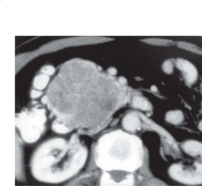
Findings
Contrast-enhanced CT. A and B. A 6-cm mass is present in the head and uncinate process of the pancreas. The mass is well circumscribed with mild homogeneous enhancement and no associated vascular encasement or pancreatic or biliary duct dilatation.
Di erential Diagnosis
1.Atypical ductal adenocarcinoma
2.Atypical islet cell tumor
3.Lymphoma
4.Acinar cell carcinoma
5.Metastasis
Diagnosis
Primary pancreatic lymphoma
Discussion
Non-Hodgkin lymphoma of the pancreas is rare and constitutes less than 1% of all pancreatic tumors. The pancreas is more commonly involved as a component of widespread lymphatic disease rather than as the primary location of tumor.
Several features of the mass in this case are extremely atypical for the more common solid pancreatic tumors, including ductal adenocarcinoma and islet cell tumor. The well-defined nature of the tumor, lack of encasement of adjacent vascular structures, and lack of pancreatic duct and biliary duct obstruction should be strong evidence that
this is unlikely to be a ductal adenocarcinoma. Nonhyperfunctioning islet cell tumors (case 8.48) can be large but usually have heterogeneous hyperenhancement. Cystic components and tumoral calcification are common. In contrast, pancreatic lymphomas usually present as large, mildly enhancing homogeneous masses. Lymphadenopathy or splenomegaly (not seen in this case) also favors the diagnosis of pancreatic lymphoma. Biopsy usually
is necessary to make the diagnosis. Pancreatic non-Hodgkin lymphoma generally is treated with chemotherapy with or without radiation.
Disease type: Masses
CASE 8.56
A
Findings
Contrast-enhanced CT (pancreatic phase of enhancement). A and B. Multiple hyperenhancing masses are present throughout the pancreas. The patient has had a right nephrectomy.
Di erential Diagnosis
1.Pancreatic metastases
2.Multiple islet cell tumors
Diagnosis
Pancreatic metastases, renal adenocarcinoma primary
8. PANCREAS 715
B
Discussion
Th e pancreas is an uncommon location for metastases, with the exception of renal adenocarcinoma and melanoma, which have a propensity to spread to this organ. Other tumors such as lung cancer and various soft tissue sarcomas also can spread to the pancreas.
Disease type: Masses
716 MAYO CLINIC GASTROINTESTINAL IMAGING REVIEW
CASE 8.57
Findings |
Discussion |
|
Sonogram. Transverse view of the pancreas shows 4 |
Pancreatic metastases are rare and found in only |
|
hypoechoic masses. |
3% of autopsies in patients with proven malignancy. |
|
|
|
Melanoma (as in this case) and metastatic renal |
Di erential Diagnosis |
adenocarcinoma have a propensity to involve the |
|
1. |
Islet cell tumors |
pancreas. However, other evidence of metastatic |
2. |
Metastases |
disease usually is present in addition to the pancreatic |
3. |
Lymphoma |
metastases. Pancreatic non-Hodgkin lymphoma also |
|
|
can present as multiple hypoechoic pancreatic masses. |
Diagnosis |
Concomitant intra-abdominal lymphadenopathy often |
|
Pancreatic metastases, melanoma primary |
is present. |
|
Disease type: Masses

|
8. PANCREAS 717 |
|
|
|
|
|
TABLE 8.4 |
|
Solid Masses of the Pancreas |
|
CASE |
|
|
|
Ductal adenocarcinoma |
Hypoattenuating mass. Ductal obstruction. |
8.28–8.39 |
|
Vascular encasement |
|
|
|
|
Islet cell tumor |
Hyperenhancing mass. Calcification. No vascular |
8.40–8.51 |
|
encasement. Clinical syndrome helpful |
|
|
|
|
Solid and papillary epithelial neoplasm |
Solid and cystic mass. Calcification common. |
8.52 and 8.53 |
|
Young women |
|
|
|
|
Acinar cell carcinoma |
Hypoenhancing mass. Usually larger than ductal |
8.54 |
|
adenocarcinoma. Older men. Fat necrosis |
|
|
|
|
Lymphoma |
Large, well-defined, homogeneous, mildly enhancing |
8.55 |
|
mass. Bulky lymphadenopathy or splenomegaly |
|
|
|
|
Metastases |
Renal carcinoma most common, hypervascular |
8.56 and 8.57 |
|
|
|
Focal pancreatitis |
Isoattenuating or hypoattenuating mass. Peripancreatic |
8.4 |
|
inflammatory changes. Clinical correlation important |
|
|
|
|
Disease type: Masses
718 MAYO CLINIC GASTROINTESTINAL IMAGING REVIEW
CASE 8.58
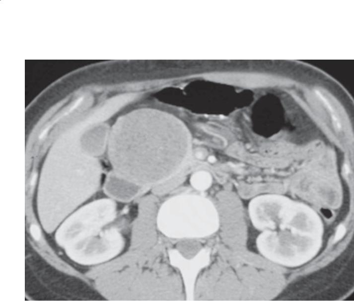
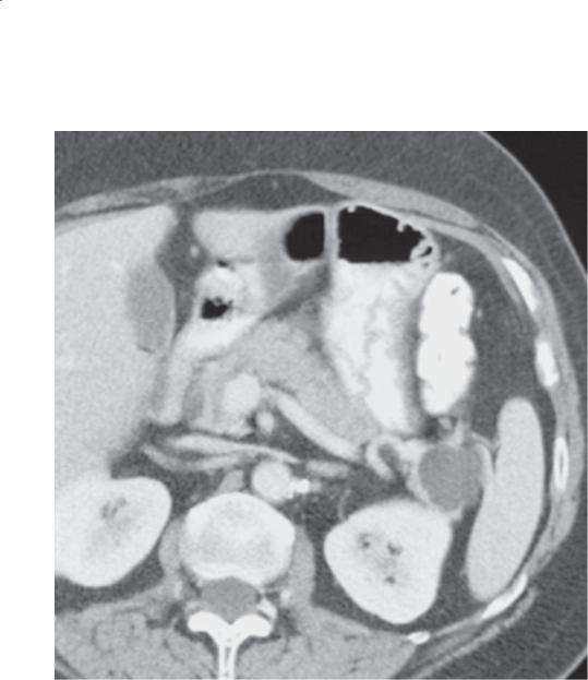
Findings
Contrast-enhanced CT. A large mass is present within the pancreatic head. The mass contains multiple small cysts and septal calcifications.
Di erential Diagnosis
Serous cystadenoma
Diagnosis
Serous cystadenoma
Discussion
Cystic masses of the pancreas are classified as either serous cystadenomas or mucinous cystic neoplasms. With few exceptions, serous cystadenomas are benign tumors that do not require resection unless symptomatic. Mucinous cystic neoplasms are considered premalignant (mucinous cystadenoma) or malignant (mucinous cystadenocarcinoma).
Differentiation of these tumors can be important because the treatment is potentially different (serous tumors can be observed; mucinous tumors are removed or closely followed). Differentiation of these tumors is not always possible, but some general rules can be helpful. Serous tumors have a honeycombed appearance, containing many (≥6) small (<2 cm) cysts. Occasionally, larger cysts are present in a serous tumor, but the majority of the cysts should be small. Mucinous tumors isolated from the main pancreatic duct usually have a few (<6) large (≥2 cm) cysts. Both types of tumors can
contain calcification(s). Septal calcification is typical of a serous tumor. A central scar is sometimes found with a serous tumor.
In this patient, the typical features of a serous tumor are present. The mass contains multiple small cysts. A few of the cysts are large, but the majority are less than 2 cm. Septal calcification is present.
Disease type: Masses
CASE 8.59
Findings
Contrast-enhanced CT. There is a large cystic mass in the pancreatic head. The mass contains both small and large cysts. A central region appears solid.
Di erential Diagnosis
1.Serous cystadenoma
2.Mucinous cystic neoplasm
3.Cystic—indeterminate neoplasm
Diagnosis
Cystic—indeterminate neoplasm (serous cystadenoma pathologically)
8. PANCREAS 719
Discussion
Some cystic neoplasms appear with features of both serous and mucinous subtypes. In this case the tumor contains both small and large cysts. These types
of tumors probably are best classified as a cystic— indeterminate mass. Further evaluation with biopsy can be helpful. Core biopsy of the septations can be diagnostic. Other centers analyze the cyst aspirate for levels of cancer antigen (CA) 19-9. The level of serum CA 19-9 is not increased in benign mucinous
lesions; however, it is increased in both malignant and inflammatory conditions (pancreatic pseudocyst). Our experience has shown that the majority of these indeterminate lesions are serous cystadenomas.
Disease type: Masses
720 MAYO CLINIC GASTROINTESTINAL IMAGING REVIEW
CASE 8.60
Findings
Contrast-enhanced CT. There is a low-attenuation mass in the pancreatic head. The mass is well circumscribed. A few cysts are identifiable within the mass.
Di erential Diagnosis
1.Serous cystadenoma
2.Mucinous cystic neoplasm
3.Lymphoma
Diagnosis
Serous cystadenoma
Discussion
Serous cystadenomas can contain cysts that are so small they approach or are below the spatial
resolution of the CT scanner. The mass may appear to be predominantly solid. In this case, a few cysts are
definitely detectable. The low attenuation of the mass is a clue to its cystic nature. The innumerable enhancing septations make the mass appear pseudosolid. These lesions also can appear solid at sonography because
of the many acoustic interfaces present within the mass. MRI is useful for proving the cystic nature of the mass. These lesions will be of very high intensity on T2-weighted images.
Disease type: Masses
CASE 8.61
A
Findings
Pancreatic MRI without intravenous contrast enhancement. A and B. There is a 4-cm mass in the pancreatic head with pancreatic ductal dilatation proximal to the mass. The mass is of very high signal intensity on T2-weighted images (A and B). The contour is lobulated (A), and the mass appears septated (B).
Di erential Diagnosis
Serous cystadenoma
Diagnosis
Serous cystadenoma
8. PANCREAS 721
B
Discussion
Pancreatic masses that are indeterminate at CT and sonography can sometimes be better characterized at MRI. Serous tumors, because of their cystic nature, are of high signal intensity on T2-weighted images (and of low signal on T1-weighted images). These tumors are well circumscribed. If the many septations that divide the tumor into a honeycomb appearance or a central scar can be identified (as in this case), a serous cystadenoma is suggested.
Disease type: Masses
722 MAYO CLINIC GASTROINTESTINAL IMAGING REVIEW
CASE 8.62
Findings
Contrast-enhanced CT. A 4-cm cystic mass bulges within the tail of the pancreas. The mass is slightly lobulated but appears unilocular without septations.
Di erential Diagnosis
1.Mucinous cystic neoplasm
2.Pseudocyst
Diagnosis
Mucinous cystadenoma
Discussion
Mucinous tumors isolated from the pancreatic duct usually have a few cysts (<6) that are 2 cm or more in diameter. This mass resembles a pseudocyst. Historical context is important, because patients with a recent episode of pancreatitis probably should have their mass followed, because it most likely represents a pseudocyst. Patients without a history of pancreatitis most likely have a mucinous tumor. Findings of thick septations and mural nodules are worrisome for a mucinous cystadenocarcinoma.
Disease type: Masses
CASE 8.63
A
Findings
A.Contrast-enhanced CT. There is a large cystic mass in the pancreatic head. The mass contains a few loculations and multiple enhancing nodules.
B.Transabdominal sonogram. A large cystic mass contains multiple mural nodules.
Di erential Diagnosis
1.Mucinous cystic neoplasm
2.Metastasis
Diagnosis
Mucinous cystadenocarcinoma
8. PANCREAS 723
B
Discussion
Th is case illustrates typical features of a mucinous tumor with a few large cystic spaces. The mural nodules are a worrisome finding for malignancy.
Disease type: Masses
724 MAYO CLINIC GASTROINTESTINAL IMAGING REVIEW
CASE 8.64
A B
C
Findings
A.T2-weighted MRI. The main pancreatic duct in the region of the body and tail of the pancreas is diffusely dilated. Pancreatic parenchymal atrophy is also present.
B.T2-weighted MRI, coronal. The pancreatic duct in the head of the pancreas is diffusely dilated, without evidence of an obstructing lesion.
C.MRCP. There is diffuse dilatation of the main pancreatic duct.
Di erential Diagnosis
1.Main duct, intraductal pancreatic mucinous neoplasm
2.Chronic pancreatitis
Diagnosis
Main duct, intraductal pancreatic mucinous neoplasm
Discussion
Mucinous tumors develop in isolation from the pancreatic duct or they arise within the main pancreatic duct or its side branches. Intraductal tumors are slow-growing tumors and can be multicentric.
As a well-differentiated neoplasm, they usually produce mucin that dilates the involved ducts. The small villous tumors may be detectable as frondlike enhancing filling defects within the dilated pancreatic duct. Chronic obstruction can lead to pancreatic parenchymal atrophy. Rarely, the mucin calcifies. The pathognomonic finding is the presence of mucin emanating from the papilla of Vater at ERCP.
Neoplastic obstruction of the duct usually is associated with a mass. Ductal dilatation in chronic pancreatitis is usually less severe and often associated with multiple ductal calcifications.
Disease type: Masses
CASE 8.65
A
Findings
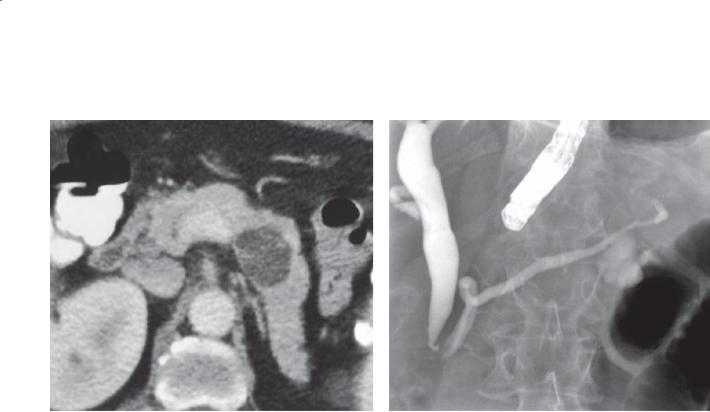
A.Contrast-enhanced CT. There are several cystic masses within the pancreas.
B.ERCP. The main and side branches of the pancreatic duct are dilated. There is a filling defect within the main pancreatic duct in the pancreatic tail.
Di erential Diagnosis
1.Intraductal pancreatic mucinous neoplasm
2.Chronic pancreatitis
Diagnosis
Main duct, intraductal pancreatic mucinous neoplasm
8. PANCREAS 725
B
Discussion
ERCP remains the standard for the diagnosis of intraductal pancreatic mucinous neoplasm. Pancreatic ductal dilatation with filling defect(s) is the main radiographic finding. Mucin flowing from the papilla of Vater is pathognomonic at ERCP. It is usually impossible to differentiate the ductal filling defects
as mucin or tumor. At CT, the intraductal mucin can mimic solid tissue attenuation (as in this case). This case illustrates the occasional difficulty in recognizing the continuously dilated pancreatic duct. In patients with equivocal CT findings, ERCP or MRCP is recommended.
Disease type: Masses
726 MAYO CLINIC GASTROINTESTINAL IMAGING REVIEW
CASE 8.66
Findings
Contrast-enhanced CT. There is a multicystic mass arising from the body of the pancreas. Mild pancreatic ductal dilatation is present proximal to the mass.
Di erential Diagnosis
1.Serous cystadenoma
2.Intraductal pancreatic mucinous neoplasm, side branch variant
Diagnosis
Intraductal pancreatic mucinous neoplasm, side branch variant
Discussion
Mucinous neoplasms arising in the pancreatic side branch ducts are increasingly common on all crosssectional imaging studies. In many cases, they have a unilocular or multilocular appearance similar to serous or mucinous cystic tumors. MRCP is very useful to make a definitive diagnosis and to follow these lesions. Identification of a dilated side branch duct, with or without a filling defect, is typical of intraductal pancreatic mucinous neoplasm arising in a side branch. Ductal dilatation due to mass effect, or as a consequence of excess mucin production, can occur. In the past, authors have referred to these side branch duct neoplasms as “ductectatic” mucinous cystadenomas.
Disease type: Masses
CASE 8.67
A
Findings
A.Contrast-enhanced CT. There is a 2.5-cm cystic mass within the body of the pancreas. The mass contains enhancing tissue.
B.ERCP. A cyst fills with contrast material from a pancreatic duct side branch within the pancreatic body.
Di erential Diagnosis
1.Intraductal pancreatic mucinous neoplasm arising from a pancreatic duct side branch
2.Pseudocyst
Diagnosis
Intraductal pancreatic mucinous neoplasm arising from a pancreatic duct side branch
8. PANCREAS 727
B
Discussion
At CT or MRCP, it is often very difficult to determine definitively whether the cystic mass communicates with the pancreatic ductal system. ERCP is the best technique for this question. All mucinous cystic neoplasms have similar histologic features and biologic behavior.
Management of these lesions is usually based on size. Side branch tumors 3 cm or larger are often referred for surgical resection. Smaller lesions have the lowest risk of malignant transformation and are followed periodically to assess for growth.
Disease type: Masses
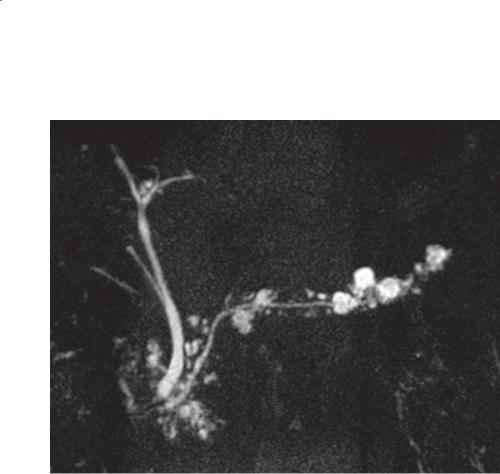
728 MAYO CLINIC GASTROINTESTINAL IMAGING REVIEW
CASE 8.68
Findings
T2-weighted MRI (MRCP), coronal. Multiple variablesized cystic lesions are seen within the pancreas; main pancreatic duct is of normal caliber.
Di erential Diagnosis
1.Side branch, intraductal papillary mucinous cystic neoplasms
2.Chronic pancreatitis
Diagnosis
Side branch, intraductal papillary mucinous cystic neoplasms
Discussion
Intraductal papillary mucinous cystic neoplasms are a subtype of mucinous cystic pancreatic neoplasms. These papillary tumors arise in the pancreatic side branch ducts and can be multiple (as in this case). They are relatively common among older persons and are believed to be premalignant or malignant tumors. Differentiation of benign from malignant is based on size and the presence of nodules within the lesions. The larger the lesion, the higher the likelihood that it will contain a malignancy. At our institution, we recommend operative resection if a cyst is 3 cm or larger in diameter or if it contains a soft tissue
enhancing nodule. Individual cysts less than than 3 cm are usually followed with interval imaging at MRI.
In chronic pancreatitis, the main pancreatic duct is irregular in caliber, and the side branches are mildly dilated and irregular. The side branch ducts in this case are too bulbous and cystic, and the main duct is smooth and of normal caliber.
Disease type: Masses

|
|
|
|
8. PANCREAS 729 |
|
|
|
|
|
|
|
|
|
TABLE 8.5 |
|
|
|
Mucinous Cystic Neoplasms: Subtypes |
|
|
|
|
|
|
|
|
|
|
|
LOCATION RELATIVE TO PANCREATIC DUCT |
|
APPEARANCE |
|
|
|
|
|
|
|||
Isolated from pancreatic duct |
|
Few large cysts; mural nodules and thick septations in |
|||
|
|
|
cystadenocarcionoma |
||
|
|
|
|||
Arising within main pancreatic duct (main duct |
|
Dilated main pancreatic duct, enhancing filling defects |
|||
intraductal pancreatic mucinous neoplasm) |
|
in pancreatic duct, mucin from papilla of Vater |
|||
|
|
||||
Arising within pancreatic duct side branches (side |
Dilated side branches can resemble mucinous or serous |
||||
branch intraductal pancreatic mucinous neoplasm) |
subtype, with or without main duct dilatation |
||||
|
|
||||
|
Features of Serous and Mucinous Cystic Pancreatic Tumors |
||||
|
|
|
|
|
|
|
FEATURE |
SEROUS CYSTADENOMA |
MUCINOUS CYSTADENOMA |
||
|
|
|
|
|
|
|
No. of cysts |
≥6 |
|
<6 |
|
|
|
|
|
|
|
|
Size of majority of cysts, cm |
<2 |
|
≥2 |
|
|
|
|
|
|
|
|
Calcification |
Septal |
|
Dystrophic |
|
|
|
|
|
|
|
|
Metastases |
No |
|
Possible |
|
|
|
|
|
|
|
|
Mural nodules |
No |
|
Cystadenocarcinoma |
|
|
|
|
|
|
|
|
Central scar |
Yes |
|
No |
|
|
|
|
|
|
|
Disease type: Masses
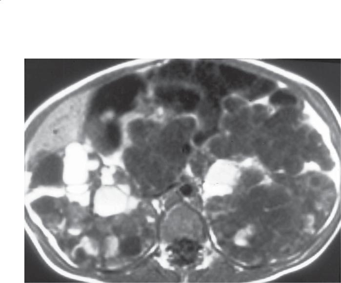
730 MAYO CLINIC GASTROINTESTINAL IMAGING REVIEW
CASE 8.69
Findings
T1-weighted MRI. Diffuse cystic changes throughout both kidneys and the pancreas. Many of the cysts in the kidneys are of high signal intensity.
Di erential Diagnosis
1.Polycystic renal disease
2.von Hippel-Lindau disease
Diagnosis
Polycystic renal disease
Discussion
Adult polycystic kidney disease is an autosomal dominant disease affecting more than 600,000 Americans and an estimated 12.5 million people worldwide. The disease causes multiple cysts on each kidney which enlarge and multiply over time. Kidneys progressively increase in size and, ultimately, renal failure occurs.
One-third of patients have cysts within their liver, and a smaller percentage have pancreatic cysts. Cysts developing in the pancreas and liver do not usually impair organ function.
Disease type: Masses
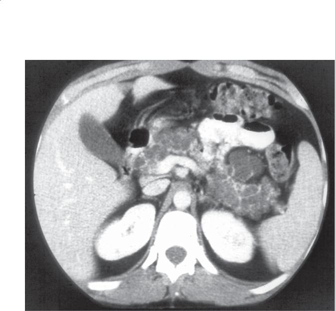
8. PANCREAS 731
CASE 8.70
Findings
Contrast-enhanced CT. The pancreas is replaced with innumerable tiny cysts.
Di erential Diagnosis
1.Polycystic kidney disease
2.von Hippel-Lindau disease
Diagnosis
von Hippel-Lindau disease
Discussion
Von Hippel-Lindau disease is an autosomal dominant multisystem inherited disorder. The commonest manifestations include retinal, cerebellar, spinal,
and medullary hemangioblastomas, renal cysts and carcinoma, pancreatic cysts, pheochromocytoma, and papillary cystadenoma of the epididymis. Patients also have an increased incidence of serous cystadenomas and islet cell tumors of the pancreas.
Th e onset of the disease occurs in early adulthood; retinal cancers develop at an average age of 25 years, cerebellar cancers at 30 years, and renal cancers at 37 years. In families with a high incidence of pheochromocytomas, adrenal tumors usually develop initially. Death usually occurs from hemangioblastomas or renal adenocarcinomas.
Th e patient in this case has the typical findings of diffuse pancreatic cystosis found in von Hippel-Lindau disease.
Disease type: Masses

732 MAYO CLINIC GASTROINTESTINAL IMAGING REVIEW
TABLE 8.6
Cystic Masses of the Pancreas |
|
CASE |
|
|
|
Serous cystadenoma |
≥6 cysts. <2 cm for majority of cysts. Honeycombed, central |
8.58–8.61 |
|
scar. Septal calcifications |
|
|
|
|
Mucinous cystadenoma or |
<6 cysts total. Majority ≥2 cm in diameter. Mural nodules |
8.62 and 8.63 |
cystadenocarcinoma |
and thick septations favor cystadenocarcinoma |
|
|
|
|
Intraductal pancreatic mucinous |
May arise in main duct or side branch ducts |
8.64–8.68 |
neoplasm |
|
|
|
|
|
Solid and papillary epithelial |
Can be predominantly cystic or solid or a combination. |
8.52 and 8.53 |
neoplasm (SPEN) |
Calcification common. Young African women |
|
|
|
|
Cystic islet cell tumor |
Unusual presentation, resembles mucinous tumor. Usually |
8.51 |
|
retains rim of hyperenhancing tissue. Clinical syndrome |
|
|
helpful |
|
|
|
|
Polycystic renal disease |
Disease obvious with renal cysts. Pancreatic cysts in 20% |
8.69 |
|
|
|
von Hippel-Lindau disease |
Pancreatic cystosis, islet cell, serous cystadenoma. Renal |
8.70 |
|
adenocarcinoma. Pheochromocytoma. Retinal or cerebellar |
|
|
hemangioblastomas |
|
|
|
|
Pseudocyst |
Fluid collection >6 weeks after acute pancreatitis. Persistent |
8.7 |
|
communication with pancreatic duct. Fibrous wall |
|
|
|
|
Abscess |
Poorly defined fluid collection that may contain gas |
8.11 |
|
|
|
Disease type: Masses

CASE 8.71
Findings
Contrast-enhanced CT. Multiple linear defects are visible through the body of the pancreas. Peripancreatic fluid is present.
Di erential Diagnosis
1.Pancreatic laceration or fracture
2.Pancreatitis with zones of necrosis
Diagnosis
Pancreatic laceration or fracture
8. PANCREAS 733
Discussion
Pancreatic trauma is usually due to gunshot wounds, direct blows to the abdomen, or high-speed automobile accidents. The mortality from these injuries approaches 20% and is nearly always associated with other abdominal injuries. Symptoms can be minor, but typically the symptoms mimic acute pancreatitis.
CT is usually used to make the diagnosis. Imaging features may mimic acute pancreatitis with diffuse or focal gland enlargement and soft tissue stranding and thickening. Occasionally, a hyperdense mass due to a hematoma is detected near the laceration. A lowattenuation defect within the gland is good evidence
for a laceration. Lacerations most often occur between the neck and body of the gland. These changes can
be subtle, and good CT technique with intravenous contrast material and thin collimation is important for optimal detection.
Disease type: Miscellaneous
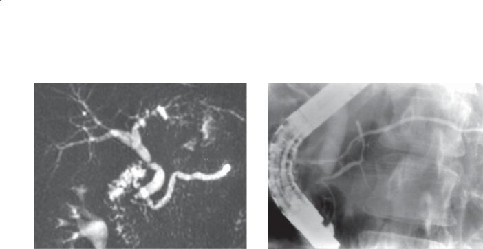
734 MAYO CLINIC GASTROINTESTINAL IMAGING REVIEW
CASE 8.72 |
CASE 8.73 |
Findings
CASE 8.72. MRCP. The pancreatic duct is uniformly dilated and empties via the accessory papilla into the duodenum.
CASE 8.73. ERCP. Both the dorsal and the ventral pancreatic ducts have been filled. There is no communication between these ducts.
Di erential Diagnosis
Pancreas divisum
Diagnosis
Pancreas divisum
Discussion
Pancreas divisum is a congenital anomaly of the pancreas in which the embryologic dorsal and ventral portions of the pancreas fail to fuse completely. As a result, the dorsal pancreatic anlage (drained by the duct of Santorini) does not communicate with the ventral pancreas (drained by the duct of Wirsung). The duct of Santorini empties via the accessory papillae (as in this case). The duct of Wirsung empties into the major papilla. Some authorities believe that patients with pancreas divisum have a higher incidence of pancreatitis because the main pancreatic duct does not empty normally.
Disease type: Miscellaneous
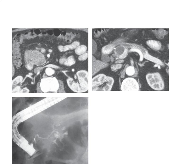
8. PANCREAS 735
CASE 8.74
A B
C
Findings
Contrast-enhanced CT. A. Th e head of the pancreas is generous in size but of normal homogeneous attenuation. B. Th e pancreatic duct in the body of the pancreas is dilated to the level of a well-defined fluid collection. C. ERCP. Only the duct of the ventral
pancreas is opacified with injection of the major papilla.
Di erential Diagnosis
1.Pancreas divisum with an obstructing pseudocyst
2.Pancreas divisum with a mucinous cystic neoplasm
Diagnosis
Pancreas divisum with an obstructing pseudocyst
Discussion
Pancreas divisum is believed to predispose to acute pancreatitis because the body and tail of the pancreas must drain by the smaller accessory papilla. The relative obstruction predisposes to pancreatitis. In this patient, a well-defined pseudocyst (the sequela of prior acute pancreatitis) is present in the body of the pancreas. The mass effect from the pseudocyst causes dilatation of the duct in the body and tail of the pancreas. ERCP is diagnostic of pancreas divisum with opacification of the small, rapidly arborizing ventral pancreatic duct. Although a mucinous cystic neoplasm could have this
appearance, it is much less likely with pancreas divisum.
Disease type: Miscellaneous
736 MAYO CLINIC GASTROINTESTINAL IMAGING REVIEW
CASE 8.75
CASE 8.76
Disease type: Miscellaneous

Findings
CASE 8.75. Contrast-enhanced CT. The duodenum is surrounded by a rim of tissue that contains multiple calcifications.
CASE 8.76. Contrast-enhanced CT. The pancreas encircles the normal duodenum.
Di erential Diagnosis
Annular pancreas
Diagnosis
CASE 8.75. Annular pancreas and chronic pancreatitis
CASE 8.76. Annular pancreas
Discussion
Annular pancreas is a congenital anomaly. Two theories exist about the abnormal embryologic development of the pancreas. One theory postulates that the ventral pancreas fails to atrophy, thereby encircling the
8. PANCREAS 737
duodenum. The other theory states that the ventral pancreas becomes adherent to the duodenum and is stretched around it as it rotates into normal position. About half of patients present with this abnormality as neonates, whereas the other half present during
adulthood. Symptoms are due to duodenal obstruction. Adults and older children remain asymptomatic
until the duodenal lumen is critically narrowed from either peptic ulcer disease (25%-50%) or pancreatitis (15%-30%). A gastrojejunostomy usually is performed to relieve the obstruction.
ERCP is usually the definitive test, showing the ventral pancreatic duct encircling the duodenum with a normal duct in the body and tail of the gland. In this case, the enhancing pancreatic tissue can be seen to encircle the duodenum (the low-attenuation region within the pancreas). The duodenal nature of the low-attenuation region was confirmed by tracing it on consecutive slices.
Disease type: Miscellaneous
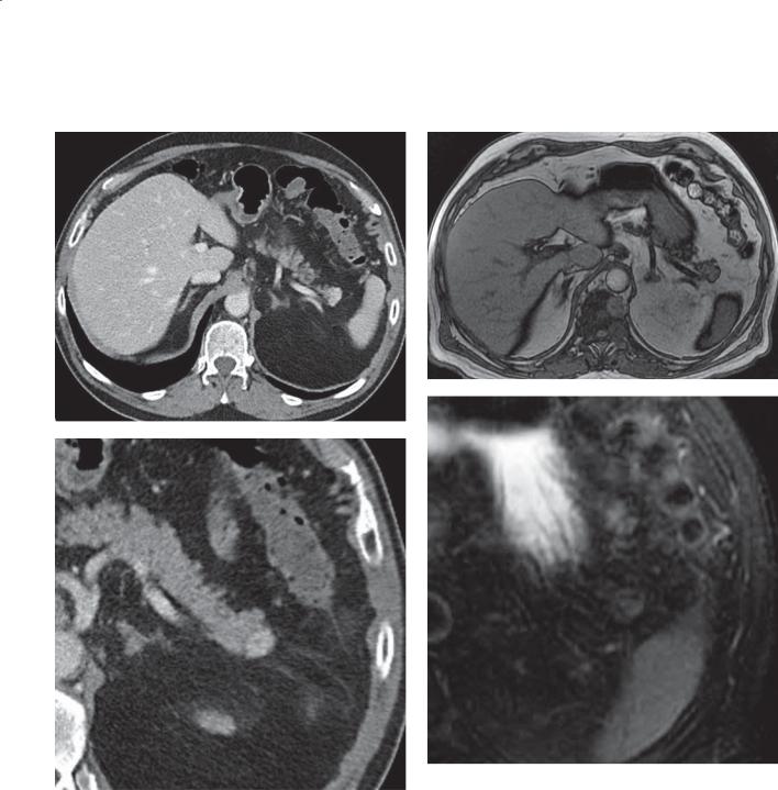
738 MAYO CLINIC GASTROINTESTINAL IMAGING REVIEW
CASE 8.77
A C
D
B
Disease type: Miscellaneous
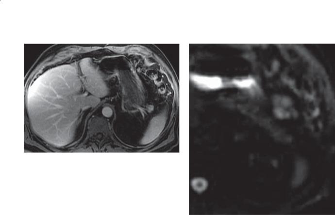
E
Findings
Contrast-enhanced CT. A. A hyperenhancing mass is in the tail of the pancreas.
B.Magnified contrast-enhanced CT. Hyperenhancing pancreatic tail mass is visible.
C.T1-weighted MRI. The above-described mass is difficult to discriminate from the pancreatic tail. It is of soft tissue intensity.
D.T2-weighted MRI. The mass is of higher intensity than the pancreatic tail and of the same intensity as the spleen.
E.Contrast-enhanced MRI. The mass enhances with intravenous gadolinium and is of the same intensity as the liver and spleen.
F.Diffusion-weighted MRI. There is no abnormal signal in the region of the mass.
Di erential Diagnosis
1.Intrapancreatic splenule
2.Islet cell tumor
3.Pancreatic metastasis
8. PANCREAS 739
F
Diagnosis
Intrapancreatic splenule
Discussion
Accessory spleens are common findings at crosssectional imaging. Approximately 20% are located in or near the pancreatic tail. These are congenital variants and range in size from a few millimeters to several centimeters. Radiologists must highly suspect these
on the basis of their location. The key to making the diagnosis is that the suspected abnormality follows the same imaging findings and enhancement pattern as the normal spleen. If the diagnosis is uncertain, technetium Tc 99m sulfur colloid or technetium Tc 99m heat-denatured red blood cell scintigraphy can be diagnostic.
Disease type: Miscellaneous
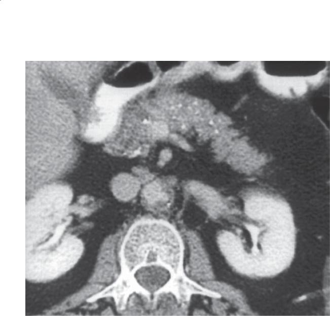
740 MAYO CLINIC GASTROINTESTINAL IMAGING REVIEW
CASE 8.78
Disease type: Miscellaneous

|
|
8. PANCREAS 741 |
Findings |
Discussion |
|
Contrast-enhanced CT. Multiple tiny calcifications are |
Sarcoidosis involving the abdomen is an unusual |
|
present throughout the pancreas. |
finding. Abnormalities in the abdomen may or |
|
|
|
may not be associated with changes in the chest |
Di erential Diagnosis |
typical of sarcoidosis. Abdominal findings include |
|
1. |
Histoplasmosis |
hepatosplenomegaly, lymphadenopathy, and |
2. |
Sarcoidosis |
calcifications in the liver, spleen, and pancreas. |
3. |
Chronic pancreatitis |
Other granulomatous diseases could present in |
|
|
a similar fashion. These findings are unusual for |
Diagnosis |
chronic pancreatitis because the calcifications are |
|
Sarcoidosis |
not intraductal and are not associated with ductal |
|
|
|
dilatation. |
Disease type: Miscellaneous

742 MAYO CLINIC GASTROINTESTINAL IMAGING REVIEW
TABLE 8.7
Miscellaneous Pancreatic Conditions |
CASE |
|
|
|
|
Pancreatic laceration or fracture |
Resembles acute pancreatitis with history of |
8.71 |
|
abdominal trauma. Linear lucency at fracture site |
|
|
|
|
Pancreas divisum |
Rapid arborization of the ventral pancreatic duct |
8.72–8.74 |
|
with injection of the major papilla. Dorsal duct |
|
|
fills from minor papilla. Perhaps predisposes to |
|
|
pancreatitis |
|
|
|
|
Annular pancreas |
Pancreatic tissue or duct encircles the |
3.41 and 3.42, 8.75 and 8.76 |
|
duodenum |
|
|
|
|
Intrapancreatic splenule |
Usually in pancreatic tail; follows signal and |
8.77 |
|
enhancement of spleen |
|
|
|
|
Sarcoidosis |
Scattered nonductal calcifications |
8.78 |
|
|
|
Disease type: Miscellaneous

8. PANCREAS 743
|
TABLE 8.8 |
Diff erential Diagnoses |
|
Solid Pancreatic Masses |
Pancreatic Calcifications |
Ductal adenocarcinoma |
Chronic pancreatitis |
Islet cell tumor |
Islet cell tumor |
Solid and papillary epithelial neoplasm |
Serous cystadenoma |
Acinar cell carcinoma |
Mucinous cystic neoplasm |
Metastases |
Solid and papillary epithelial neoplasm |
Lymphoma |
Granulomatous infection |
Focal pancreatitis |
|
Cystic Pancreatic Masses
Pseudocyst
Serous cystadenoma
Mucinous cystadenoma or cystadenocarcinoma Intraductal pancreatic mucinous neoplasm (main duct and side-branch types)
Solid and papillary epithelial neoplasm Cystic islet cell tumor
Polycystic kidney disease von Hippel-Lindau disease Abscess
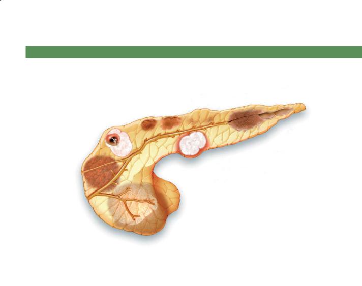
744 MAYO CLINIC GASTROINTESTINAL IMAGING REVIEW
SOLID PANCREATIC MASSES
Metastases
Solid and cystic epithelial neoplasm
Ductal adenocarcinoma
Lymphoma |
D.F. |
|
© MAYO |
|
2013 |
Islet cell tumor
Focal pancreatitis
Figure 8.79 (Used with permission of Mayo Foundation for Medical Education and Research.)
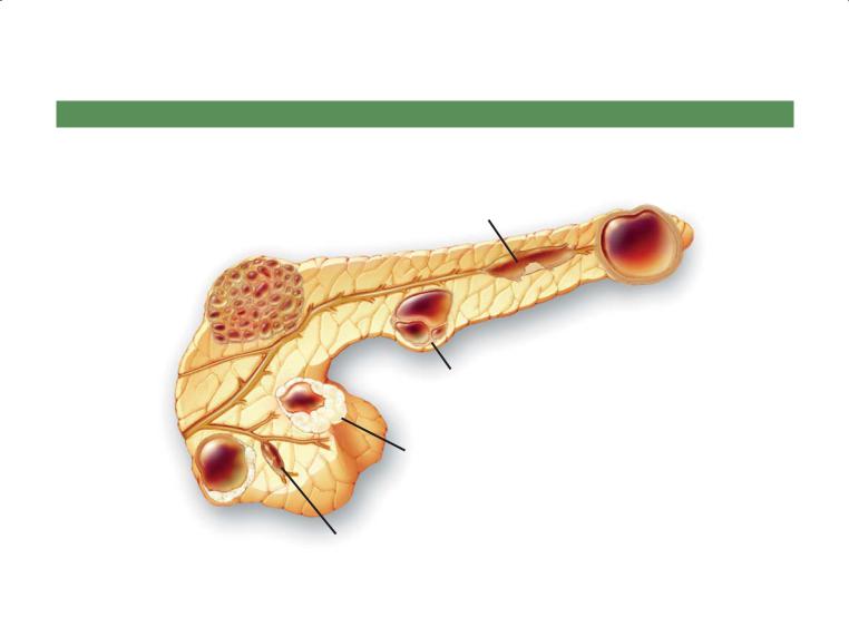
8. PANCREAS 745
CYSTIC PANCREATIC MASSES
Main duct IPMN
Serous cystadenoma
Pseudocyst
D.F.
© MAYO Mucinous cystadenoma/
2013
cystadenocarcinoma
Cystic islet cell tumor
Solid and papillary |
|
epithelial neoplasm |
Side-branch IPMN |
IPMN, intraductal pancreatic mucinous neoplasm
Figure 8.80 (Used with permission of Mayo Foundation for Medical Education and Research.)
746 MAYO CLINIC GASTROINTESTINAL IMAGING REVIEW
QUESTIONS
Multiple Choice (choose the best answer)
8.1.Which of the following is the most likely diagnosis for the findings in the figure?
a.Acute pancreatitis
b.Chronic pancreatitis
c.Ductal adenocarcinoma
d.Islet cell tumor
e.Solid and cystic papillary epithelial neoplasm (SPEN)
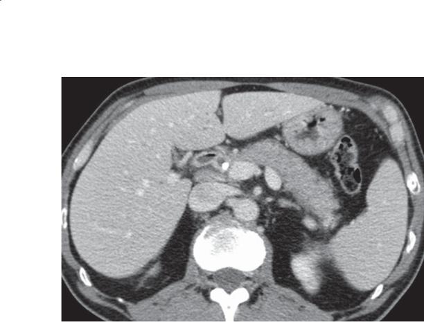
8. PANCREAS 747
8.2.Which of the following is most likely to be associated with the condition shown in the figure?
a.Increased level of immunoglobulin G4
b.Increased level of carcinoembryonic antigen
c.Increased level of cancer antigen 19-9
d.Increased level of α-fetoprotein
e.Increased level of cancer antigen 125
8.3.For the case shown in Question 8.2, what is the most appropriate management?
a.Subtotal pancreatectomy
b.Pancreatic enzyme replacement
c.ERCP with pancreatic ductal stenting
d.Steroid treatment
e.Chemotherapy
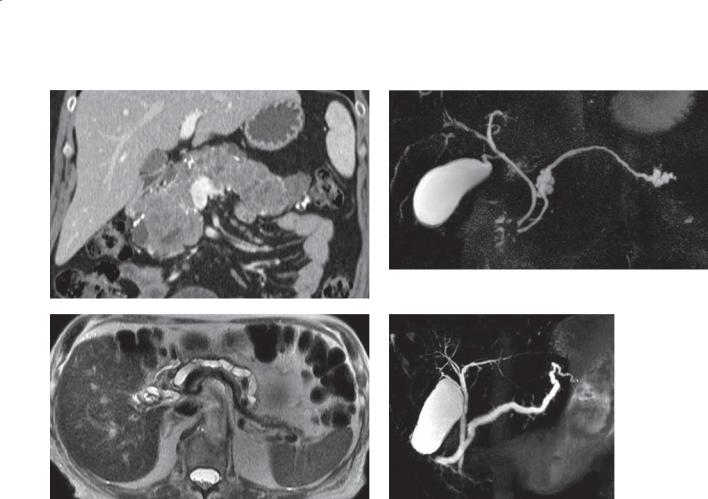
748 MAYO CLINIC GASTROINTESTINAL IMAGING REVIEW
A B
C D
Match images A through F with the responses listed below.
8.4. |
______ Possible cause of acute pancreatitis |
8.6. |
______ Renal cell carcinoma |
8.5. |
______ Polypeptide hormones |
8.7. |
______ Major papilla mucin |
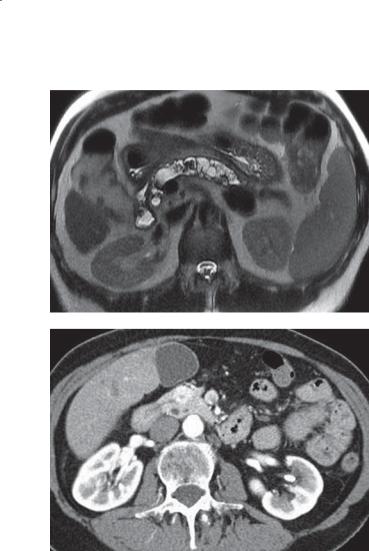
8. PANCREAS 749
E
F
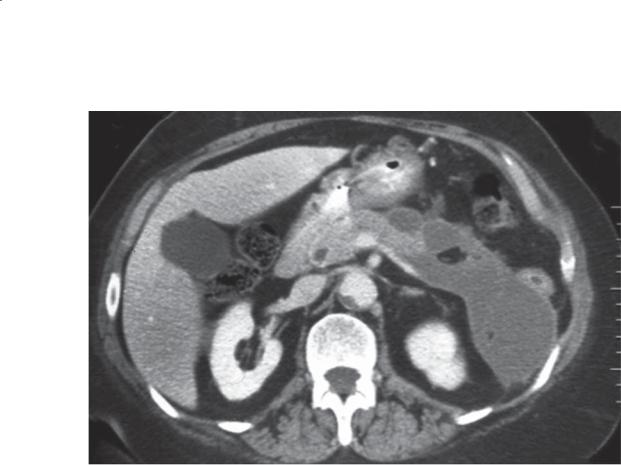
750 MAYO CLINIC GASTROINTESTINAL IMAGING REVIEW
8.8.Which of the following is the most likely diagnosis for the findings in the figure?
a.Pancreatic cancer
b.Chronic pancreatitis
c.Acute pancreatitis
d.Side branch intraductal papillary mucin-producing neoplasm (IPMN)
e.Main duct IPMN

8. PANCREAS 751
ANSWERS
8.1.Answer c.
A hypovascular mass is present within the pancreatic head-uncinate region. Abnormal tissue extends toward the superior mesenteric artery. Intrahepatic bile duct dilatation is present. These findings are typical for a ductal adenocarcinoma of the pancreas with obstruction of the extrahepatic bile duct and of the pancreatic duct (not seen) and extension beyond the pancreas to encase the superior mesenteric artery.
Acute pancreatitis usually presents with diffuse pancreatic enlargement with a peripancreatic effusion. Chronic pancreatitis usually presents with pancreatic parenchymal atropy, a dilated and beaded-appearing pancreatic duct, and ductal calcifications.
Islet cell tumors are usually hypervascular, and they do not typically cause vascular encasement. Calcification can be seen within these tumors.
Solidandcysticpapillaryepithelialneoplasmsareusually large tumors of variable enhancement and attenuation found in young women. Vascular encasement is not a usual feature.
8.2.Answer a.
Th e pancreas is diffusely enlarged, and there is a suggestion of a hypoattenuating halo about the pancreas. This finding is highly suggestive of autoimmune pancreatitis. Patients who have this condition typically have an increased level of immunoglobulin G4 in their serum, and this can be a helpful confirmatory finding. An increased level of carcinoembryonic antigen is a nonspecific tumor marker that can occur in patients with cancers of the gastrointestinal system, cervix, lung, ovary, breast, and urinary tract.
Cancer antigen 19-9 is a tumor marker usually found in patients with pancreatic cancer, but other gastrointestinal tumors can cause an increase of this serum marker.
Th e α-fetoprotein level is increased in hepatocellular carcinomas and germ cell tumors.
An increased level of cancer antigen 125 is found in patients with ovarian, endometrial, fallopian, lung, breast, and gastrointestinal tumors.
8.3.Answer d.
Patients with a confirmed diagnosis of autoimmune pancreatitis usually are given steroids, and the majority have good results.
Pancreatic enzyme replacement is usually given to patients with pancreatic insufficiency—usually due to chronic pancreatitis or prior pancreatic resection.
Pancreatic ductal stenting is most often performed in patients with chronic pancreatitis and a focal stricture causing upstream pancreatic ductal dilation/pseudocyst and pain.
Chemotherapy is reserved for patients with malignant pancreatic tumors.
8.4.Answer d.
Figure d shows pancreatic divisum. The pancreatic duct is dilated and empties into the duodenum at the minor papilla, above the level of the major ampulla (at the distal end of the common bile duct). In pancreatic divisum, the 2 pancreatic segments (dorsal and ventral) do not join with the main pancreatic duct emptying into the minor papilla via the duct of Santorini. The duct of Wirsung (ventral pancreas supplying the distal pancreatic head and uncinate process) empties into the major papilla (not seen). Some authorities believe that there is a higher incidence of pancreatitis in patients with pancreatic divisum. In this patient, the pancreatic duct is dilated, likely due to restricted flow of the pancreatic duct through the minor papilla.
8.5.Answer f.
Figure f shows insulinoma. A focal hyperenhancing mass in present within the pancreatic head. Patients with insulinoma usually present with typical features of a low glucose level. Most often, the role of the radiologist is to identify the location of this usually benign, polypeptide-producing, hypervascular neoplasm that eventually needs operative resection for cure.
8.6.Answer a.
Figure a shows von Hippel-Lindau disease. Affected patients can have diffuse cystosis of the pancreas and islet cell tumors. In addition, they are known to have a high incidence of renal cell cancers, hemangioblastomas, and other tumors.
8.7.Answer c.
Figure c shows a main duct intraductal papillary mucin-producing neoplasm (IPMN). These tumors are considered premalignant or frankly malignant and are located within the main pancreatic duct. They produce mucin that distends the duct and is responsible for the radiologic features of this condition. Mucin, when visualized at ERCP to be flowing from the major papilla, is considered pathognomonic of this condition.
Figure b shows a side branch IPMN. In this patient, the main pancreatic duct is of normal caliber, but there are 2 dilated pancreatic ductal side branches within

752 MAYO CLINIC GASTROINTESTINAL IMAGING REVIEW
the pancreatic neck and tail regions. These side branch neoplasms have the appearance of a cluster of grapes and can be multiple. Because these have malignant potential, they are usually followed with periodic imaging to assess for growth and potential operative resection.
Figure e shows chronic pancreatitis. The main pancreatic duct is dilated, tortuous, and beaded. There is a lowintensity filling defect within the dilated pancreatic duct, representing a stone. Pancreatic parenchymal atrophy is present. These findings are consistent with chronic pancreatitis.
8.8.Answer c.
Th ere is a large fluid collection in the region of the pancreatic body and tail, without visualization of the pancreas in this region. This is a case of necrotizing pancreatitis involving the pancreatic body and tail. Pancreatic cancer would have a focal mass with visualization of the pancreas about the tumor. Chronic pancreatitis is associated with parenchymal atrophy and ductal dilatation—none of which is present in this case. Side branch and main duct IPMNs are cystic neoplasms within the pancreatic parenchyma or with a dilated main pancreatic duct—which are not visible in this patient.