C H A P T E R 5
COLON
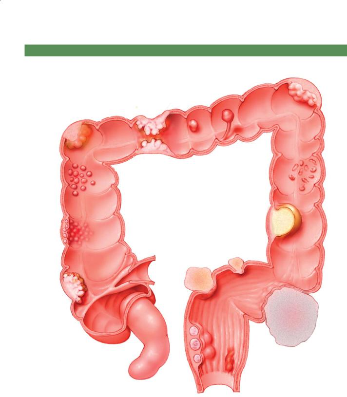
Cases 5.1–5.59 Inflammatory and Ulcerative Diseases
Ulcerative Colitis
Crohn Colitis
Diverticulitis
Appendicitis
Other
Cases 5.60–5.127 Masses and Filling Defects
Benign Tumors
Malignant Tumors
Nonneoplastic
Cases 5.128–5.136 Obstruction and Ileus
Cases 5.137–5.144 Disorders of Defecation
Cases 5.145–5.151 Miscellaneous
This page intentionally left blank
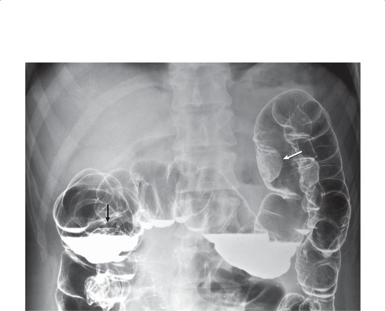
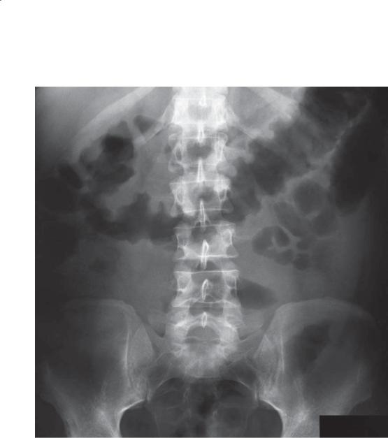
Findings
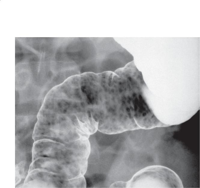
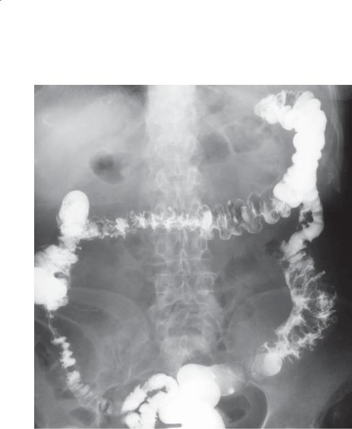
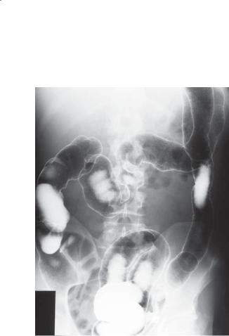
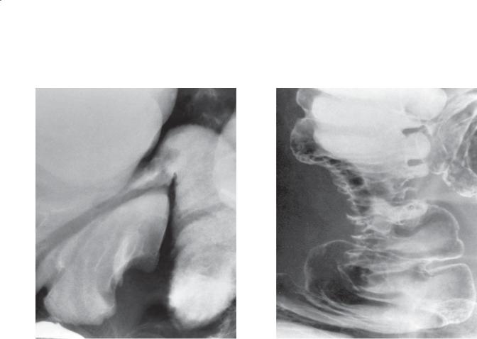
Double-contrast barium enema. The entire colon is ahaustral, with a diffuse, granular-appearing mucosa. The air-filled terminal ileum is dilated.
Di erential Diagnosis
1.Ulcerative colitis
2.Infectious colitis
Diagnosis
Ulcerative colitis
Discussion
Ulcerative colitis is a disease of unknown origin. Granulomas are conspicuously absent. Ulceration of the mucosa is nearly always present in patients with active inflammation.
Clinically, many patients initially experience the sudden onset of bloody diarrhea, abdominal cramping, and fever. The initial diagnosis often can be suggested at proctosigmoidoscopy. Extraintestinal manifestations of this disease include erythema nodosum, pyoderma
gangrenosum, primary sclerosing cholangitis, cholangiocarcinoma, arthritis, sacroiliitis, spondylitis, and iritis. Spondylitis activity has no relationship to the activity of the colon inflammation. Patients with HLAB27 histocompatibility antigen are more likely to have spondylitis and iritis develop than patients without the marker.
Radiologically, the following are critical for diagnosis:
1.Th e involved mucosa on an air-contrast barium enema has a granular (sandpaper-like) or stippled appearance.
2.Th e distribution of the disease is usually one of rectal involvement with disease affecting the more proximal colon in a continuous manner, without skip areas.
3.Circumferential bowel wall symmetry is maintained. Mucosal ulcerations affect both sides of the bowel with equal severity.
Infectious colitides can present with similar radiographic features. Stool cultures are always required at initial evaluation.
Disease type: Inflammatory and Ulcerative Diseases
326 MAYO CLINIC GASTROINTESTINAL IMAGING REVIEW
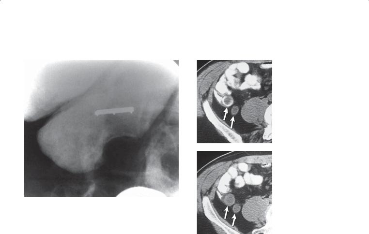
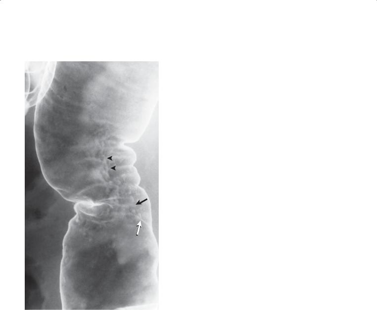
Findings
CASE 5.2. Double-contrast barium enema. Coarsely granular mucosal ulcerations are visible in the right colon.
CASE 5.3. Double-contrast barium enema. The colon is affected with a coarsely granular mucosal pattern. Numerous polypoid filling defects also are present.
Di erential Diagnosis
Ulcerative colitis
Diagnosis
Ulcerative colitis
Discussion
Polypoid changes may develop in any stage of ulcerative colitis. In patients with severe, acute inflammation, pseudopolyps are likely to be present. Pseudopolyps refer to islands of normal colonic mucosa surrounded by denuded ulcerative mucosa. Inflammatory polyps refer to regions of inflamed, elevated mucosa surrounded by granular mucosa. Inflammatory polyps may be sessile or pedunculated, and they more often are found in patients with less severe inflammation than in those with pseudopolyps.
Disease type: Inflammatory and Ulcerative Diseases
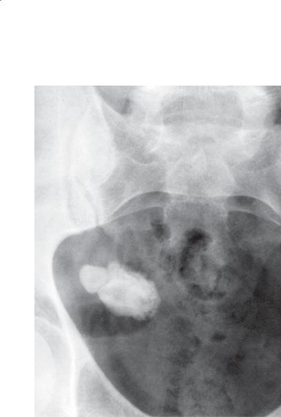
Findings
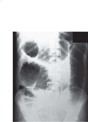
Abdominal radiograph. The colon is distended with gas (adynamic ileus pattern), and multiple polypoid masses protrude into the colonic lumen.
Di erential Diagnosis
1.Adynamic ileus
2.Colonic obstruction
3.Toxic megacolon
Diagnosis
Toxic megacolon
Discussion
Toxic megacolon is a serious complication that can occur in either ulcerative colitis or Crohn colitis (although it is most frequent in patients with ulcerative colitis). This condition occurs during a severe episode of acute inflammation. Dilatation (>6 cm) and adynamic ileus often occur because the inflammatory changes have extended into the muscular layers and usually to the serosa of the colon. Extensive mucosal
disease can be visualized by identifying nodularity and polypoid changes within the colonic lumen. These polyps usually represent pseudopolyps. Bowel wall and haustral thickening are due to intramural edema and congestion.
Colonic perforation can occur in toxic megacolon, and it is associated with a high morbidity and mortality. Perforation may be suggested by identifying either intramural pneumatosis or pneumoperitoneum. Iatrogenic intervention is a common cause of perforation. In patients with this condition, a contrast enema or colonoscopy should be avoided. Even inflation of a rectal balloon in patients with a fragile, narrowed, and inflamed rectum can easily result in perforation.
Toxic megacolon always occurs with adynamic ileus. Patients usually have fever, leukocytosis, and a tender abdomen. A single supine radiograph in adynamic ileus can simulate a distal colonic obstruction because of the posterior location of the rectosigmoid. If clinical symptoms suggest an obstruction, a supine or decubitus view is often helpful for demonstrating air filling of the rectosigmoid.
Disease type: Inflammatory and Ulcerative Diseases
328 MAYO CLINIC GASTROINTESTINAL IMAGING REVIEW
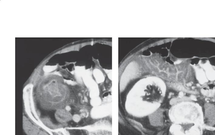
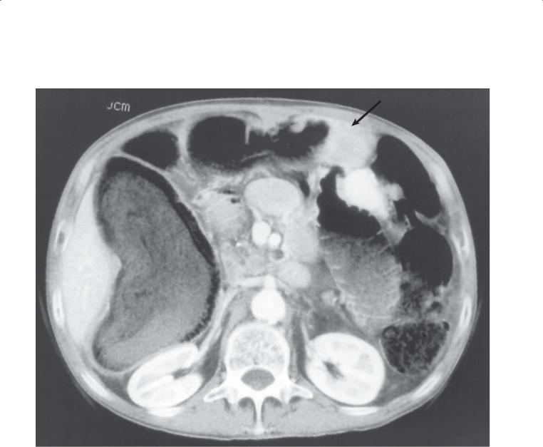
Findings
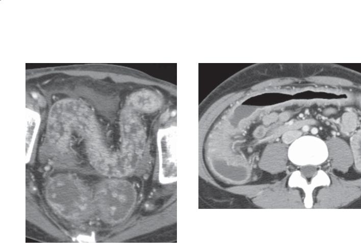
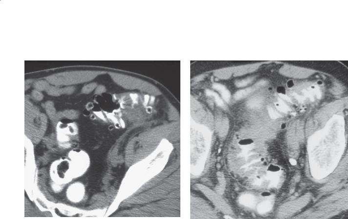
CASE 5.5. Contrast-enhanced CT. Innumerable enhancing polypoid filling defects are present throughout the rectosigmoid colon. The wall of the colon is hyperenhancing and slightly irregular in contour.
CASE 5.6. Contrast-enhanced CT. The wall of the colon is diffusely thickened. Multiple engorged and prominent vessels are present about the thick-walled colonic segments.
Di erential Diagnosis
1.Ulcerative colitis
2.Polyposis syndrome
Diagnosis
Acute changes of ulcerative colitis (case 5.5 has innumerable inflammatory pseudopolyps)
Discussion
Detection of inflammatory diseases of the colon at CT depends on the severity of the disease and the quality of the examination. Usually, mild disease confined to the mucosa is not detectable at CT. Severe disease with inflammatory changes extending to the serosal surface causes bowel wall thickening and often mucosal hyperenhancement and soft tissue stranding in the pericolonic fat (as in these cases).
Disease type: Inflammatory and Ulcerative Diseases
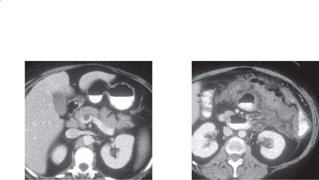
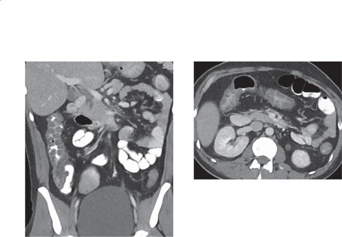
CASE 5.7
A
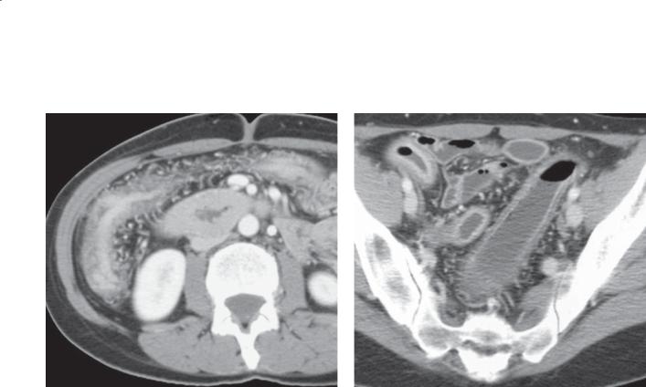
Findings
Contrast-enhanced CT. A and B. Diffuse thickening and hyperenhancement of the wall of the colon and terminal ileum. Engorgement of the vasa recti.
Di erential Diagnosis
1.Acute colitis
2.Crohn disease
3.Ulcerative colitis
4.Infectious colitis
5.Pseudomembranous colitis
Diagnosis
Ulcerative colitis
5. COLON 329
B
Discussion
Ulcerative colitis can be suspected in patients with continuous, symmetric bowel wall thickening. Thickening of the terminal ileum in this patient raises the possibility of Crohn disease, but backwash ileitis was found at colonoscopy. Hyperenhancement of the bowel wall and vascular engorgement of the pericolonic tissues indicate active inflammatory changes.
Disease type: Inflammatory and Ulcerative Diseases
330 MAYO CLINIC GASTROINTESTINAL IMAGING REVIEW
CASE 5.8
Findings
Double-contrast barium enema. Active ulcerations (stippled and linear) are present in the sigmoid colon. The rectum has a featureless mucosa with some mild loss of distensibility.
Di erential Diagnosis
1.Ulcerative colitis
2.Infectious colitis
3.Crohn colitis
Diagnosis
Ulcerative colitis, healing pattern
Discussion
Rectal involvement is usually present in patients with ulcerative colitis, and in this patient it can be assumed to have occurred in the past because of the reduced distensibility of this bowel segment. Patients using corticosteroid enemas may have a normal-appearing rectal mucosa with active disease in the more proximal colon. This distribution of disease can also occur in patients with ulcerative colitis who have not used corticosteroid enemas because of segmental mucosal healing and repair. The distribution of mucosal healing is the same as that of active inflammation in patients with ulcerative colitis—beginning in the rectum and progressing proximally in a continuous manner. Therefore, when the healing process begins, the rectum nearly always appears normal first, with variable degrees of inflammation present in the more proximal colon. The most severe disease usually is found proximally in a colonic segment near the leading edge of ascending inflammation.
Disease type: Inflammatory and Ulcerative Diseases
CASE 5.9
Findings
Double-contrast barium enema. Multiple nodules are present in the descending colon in this patient with a 22-year history of ulcerative colitis. The nodules abut each other and have flattened edges.
Di erential Diagnosis
1.Infl ammatory polyps in ulcerative colitis
2.Carcinoma
3.Lymphoma
4.Dysplasia
Diagnosis
Mucosal dysplasia
5. COLON 331
Discussion
Endoscopic biopsy showed that the nodules were formed of atypical colonic glands crowded within the lamina propria. This pattern is often found in dysplastic nodules. Patients with ulcerative colitis are at increased risk for development of colon cancer. Those patients at greatest risk have colonic epithelial dysplasia. Detection of dysplasia in a colitic colon should prompt endoscopic biopsy and possibly prophylactic colectomy. Unfortunately, most mucosal dysplasia is not visible radiographically and requires random endoscopic biopsy for detection.
Radiographically, dysplasia appears as a solitary nodule or multiple nodules. A close grouping
of nodules with opposed flattened edges is most commonly (50% of time) associated with dysplasia. Inflammatory polyps may be indistinguishable from dysplastic nodules. Colitic carcinomas usually are associated with luminal narrowing. Lymphoma is not commonly associated with ulcerative colitis.
Disease type: Inflammatory and Ulcerative Diseases
332 MAYO CLINIC GASTROINTESTINAL IMAGING REVIEW
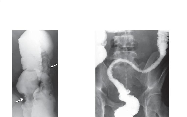
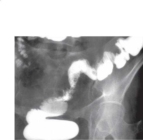
Findings
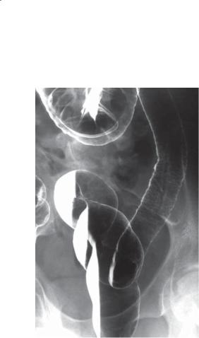
CASE 5.10. Double-contrast barium enema. An annular segment of narrowing (arrow) is present in the sigmoid colon. Granular mucosal changes also are present.
CASE 5.11. Double-contrast barium enema. A stricture is present in the region of the ahaustral splenic flexure of the colon.
Di erential Diagnosis
Colitic cancer
Diagnosis
Colitic cancer
Discussion
Patients with ulcerative colitis have an increased risk for development of colorectal cancer (colitic cancer). The risk depends on the extent and duration of inflammation. Patients with pancolitis are at higher risk of colon cancer than patients with only limited
disease. The risk of cancer developing begins after a patient has had the disease for 10 years, and it increases with time. The actual cancer risk varies from study to study—ranging from a 3% to 10% risk of malignancy after 10 years to a 13% to 25% risk after 25 years of ulcerative colitis. The clinical activity of the disease has little effect on the overall cancer risk.
Mucosal dysplasia is the precursor of carcinoma. Most cancers complicating ulcerative colitis are annular constricting lesions. Some carcinomas develop as flat, infiltrating tumors—extending into the submucosal tissues and causing either a smooth or an abruptly tapered stricture. About one-fourth of all cancers present as strictures. Multiple cancers are common. Polypoid masses are rare. Generally, these malignancies are high-grade, aggressive tumors.
Benign strictures also can develop in ulcerative colitis as a result of muscular hypertrophy or of severe inflammation. Because it is impossible to differentiate benign from malignant strictures radiographically, they should be regarded with suspicion.
Disease type: Inflammatory and Ulcerative Diseases
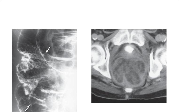
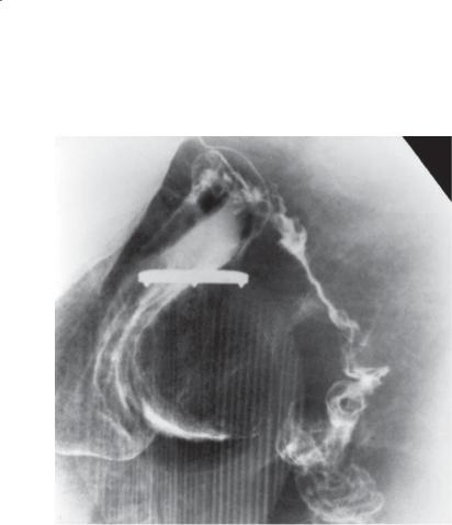
CASE 5.12
Findings
Single-contrast post-evacuation barium enema. The colon is ahaustral and shortened. Typical changes of ankylosing spondylitis also are present.
Di erential Diagnosis
1.Chronic ulcerative colitis
2.Cathartic abuse
Diagnosis
Chronic ulcerative colitis
5. COLON 333
Discussion
Long-standing ulcerative colitis often results in a colon devoid of the normal haustral markings and a diffusely shortened and often narrowed colonic
lumen. The colon usually appears featureless and rigid. Hypertrophy of the circular and longitudinal muscle fibers is now believed to be the most frequent cause of these changes.
Spondylitis often precedes the inflammatory bowel disease, whereas the symptoms of peripheral joint disease occur concurrently or after the onset of bowel symptoms.
Disease type: Inflammatory and Ulcerative Diseases
334 MAYO CLINIC GASTROINTESTINAL IMAGING REVIEW
CASE 5.13
A B
Findings
Contrast-enhanced CT. Coronal (A) and axial (B) views show diffuse thickening of the colon with mucosal hyperenhancement. The vasa recti are engorged. No haustrations are visible in the transverse colon. An enhancing polyp can be identified on B in the left transverse colon.
Di erential Diagnosis
1.Acute ulcerative colitis
2.Crohn colitis
3.Infectious colitis
4.Pseudomembranous colitis
Diagnosis
Acute ulcerative colitis
Discussion
Multiple areas of luminal narrowing are present within the right colon (A). These can be difficult to differentiate from infiltrative tumor. Deep biopsy or surgical resection may be necessary for differentiation. The lack of haustrations in the right and transverse colon indicates chronic ulcerative colitis. Mucosal hyperenhancement indicates active inflammation. Crohn colitis is usually not this symmetric and uniform, and often terminal ileal disease is also present. Pseudomembranous colitis usually has thicker colonic wall and preserved but thickened haustrations. A chronic infectious colitis could have this appearance; therefore, cultures are indicated if the diagnosis is not established.
Disease type: Inflammatory and Ulcerative Diseases
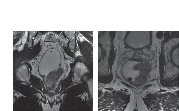
CASE 5.14
A
Findings
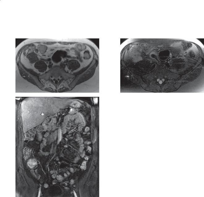
Contrast-enhanced MRI. A. Coronal T1-weighted image with fat saturation. The transverse colon is ahaustral, and there are numerous small enhancing polyps on its wall. Similar enhancing polyps are seen within the ileum. B. Axial T2-weighted image. The colon is thick-walled and contains filling defects, and there are engorged vasa recti.
Di erential Diagnosis
1.Active ulcerative colitis
2.Active Crohn disease
3.Infectious colitis
4.Ischemic colitis
5.Pseudomembranous colitis
Diagnosis
Active ulcerative colitis
5. COLON 335
B
Discussion
Th e diffusely thick colonic wall indicates a colitis. The ahaustral pattern usually indicates long-standing disease. The mucosal hyperenhancement and enhancing polyps are consistent with active, acute inflammation. Polyps in acute ulcerative colitis are usually inflammatory polyps; however, pseudopolyps (islands of remaining normal mucosa), dysplastic polyps, and malignant polyps have been described.
Their finding usually indicates the need for endoscopic assessment. Patients with ulcerative colitis and pancolitis can have backwash ileitis (<20% of patients), but the presence of backwash ileitis has no implications for successful ileal pouch creation. The ileal polyps seen in this case represented edematous lymphoid hyperplasia at endoscopy.
Disease type: Inflammatory and Ulcerative Diseases
336 MAYO CLINIC GASTROINTESTINAL IMAGING REVIEW
CASE 5.15
Findings
Contrast-enhanced CT. Diffuse thickening of the wall of the colon. The wall contains fat attenuation tissue.
Di erential Diagnosis
1.Chronic ulcerative colitis
2.Crohn colitis
Diagnosis
Chronic ulcerative colitis
Discussion
Fat attenuation in the wall of the colon usually indicates long-standing disease that is inactive. The pancolitis in this case favors ulcerative colitis. There is mild soft tissue stranding in the pericolonic tissues in the ascending colon. At colonoscopy, mild changes of acute colitis were present in this patient with known chronic ulcerative colitis.
Disease type: Inflammatory and Ulcerative Diseases
CASE 5.16
Findings
Abdominal radiograph. The haustral folds are diffusely thickened throughout the transverse colon.
Di erential Diagnosis
1.Pseudomembranous colitis
2.Infl ammatory bowel disease
3.Infectious colitis
4.Ischemic colitis
Diagnosis
Inflammatory bowel disease: acute Crohn colitis
5. COLON 337
Discussion
Most patients with acute colitis present with similar radiographic findings in the involved portions of the colon. Intramural edema and hemorrhage cause wall and haustral thickening (as in this case). Inflammatory polyps can be visible within the gas-filled colon
in patients with inflammatory bowel disease
(cases 5.4, 5.5, and 5.6). The splenic flexure is often an isolated segment of involved colon in elderly patients with ischemic colitis (case 5.45). A contrast enema usually is contraindicated in patients with acute colitis because of the risk of perforation. It can be performed safely after the acute episode has resolved. Proctoscopy is often useful for revealing the mucosal changes of ulcerative colitis or pseudomembranous colitis.
Disease type: Inflammatory and Ulcerative Diseases
338 MAYO CLINIC GASTROINTESTINAL IMAGING REVIEW
CASE 5.17
Findings
Double-contrast barium enema. A prominent lymphoid pattern is present in the sigmoid colon. Some of the nodules are mildly enlarged (>4 mm).
Di erential Diagnosis
1.Prominent lymphoid pattern
2.Early Crohn disease
3.Polyposis syndrome
Diagnosis
Early Crohn disease
Discussion
Th e earliest change of Crohn colitis is submucosal granulomatous inflammation. Enlarged lymphoid follicles are often seen radiographically. These mucosal elevations usually have poorly defined borders and may contain a small central umbilication. The early changes of Crohn colitis are best depicted with the doublecontrast technique. A lymphoid pattern can be seen in 10% to 15% of normal adults (cases 5.115 and 5.116). These nodules are uniformly small. In some patients, it may be impossible to differentiate a normal lymphoid pattern from early Crohn disease. In these patients, either repeat double-contrast colon examination in several months or endoscopic biopsy is helpful.
Disease type: Inflammatory and Ulcerative Diseases
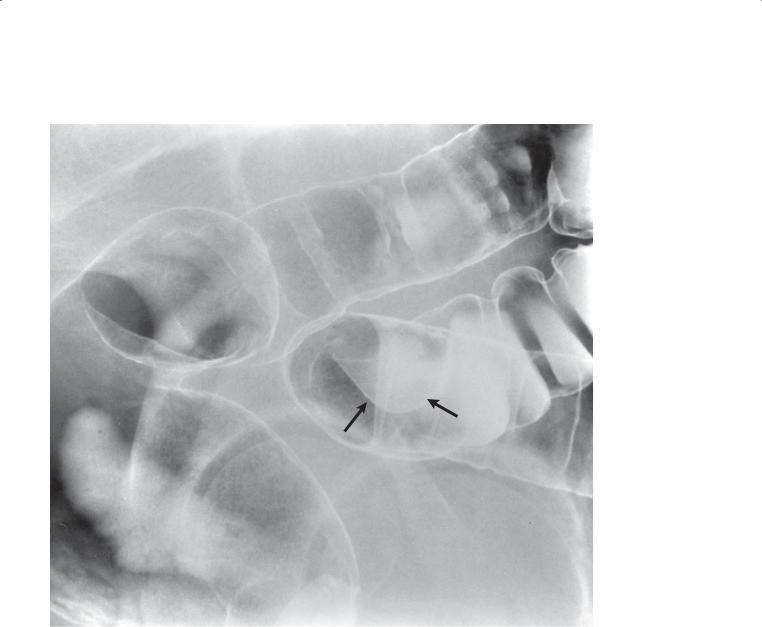
CASE 5.18
Findings
Double-contrast barium enema. Multiple discrete (aphthous) ulcers (arrows) are present within the descending colon. A halo of edema surrounds each ulceration. Many of the ulcers have become confluent, producing a linear ulceration (arrowheads). The intervening mucosa between ulcers appears normal. The distribution of ulcerations is circumferentially asymmetric.
Di erential Diagnosis
1.Crohn colitis
2.Infectious colitis
Diagnosis
Crohn colitis: aphthous and confluent ulcerations
5. COLON 339
Discussion
Aphthous ulcers develop from enlarged lymphoid follicles that ulcerate centrally. These ulcers are very shallow lesions and usually are not seen in profile. Multiplicity of lesions is common. The central barium collection often varies in size and is always surrounded by a halo of edema. Although the aphthous ulcer is characteristic of Crohn disease, it is not diagnostic. These lesions also have been reported in infectious colitides, including Yersinia and amebic colitis.
Disease type: Inflammatory and Ulcerative Diseases
340 MAYO CLINIC GASTROINTESTINAL IMAGING REVIEW
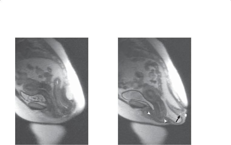
CASE 5.19
A B
C
Findings
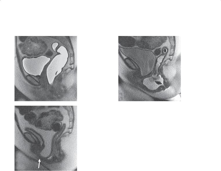
Unenhanced and enhanced MRI. A. T1-weighted image. Marked thickening of the wall of the rectosigmoid with pericolonic soft tissue stranding.
B. T2-weighted image (with fat saturation). Thickening of the wall of the rectosigmoid with high-signal intensity changes in the pericolonic fat. C. Gadoliniumenhanced T1-weighted image (with fat saturation). Diffuse mural enhancement with submucosal edema. D. T2-weighted image (with fat saturation). Focal
fluid collection (arrow) in the left perianal region. E. Contrast-enhanced CT. A fluid collection (arrow)
is present in the left perianal region with surrounding inflammatory reaction.
Di erential Diagnosis
1.Crohn colitis
2.Ulcerative colitis
3.Other types of acute colitis
Disease type: Inflammatory and Ulcerative Diseases
5. COLON 341
D E
Diagnosis
Crohn colitis with perianal abscess
Discussion
Th e extensive colonic inflammatory changes in this patient make it difficult to make a precise diagnosis. Any type of acute colitis could have this appearance.
The perianal abscess is a characteristic feature of Crohn disease (in addition to perianal fistula, abscess, and creeping fat)—and a very helpful discriminator. MRI is a preferred method for evaluating patients with perianal abscess and fistulas.
Disease type: Inflammatory and Ulcerative Diseases
342 MAYO CLINIC GASTROINTESTINAL IMAGING REVIEW
CASE 5.20
A B
Disease type: Inflammatory and Ulcerative Diseases

Findings
Double-contrast barium enema. A. Segmental regions of narrowing are present in the left transverse colon and in the descending colon. Polypoid mucosal (inflammatory polyps) changes and deep linear and thornlike ulcers are present in the affected segments. Circumferential asymmetry is present in the splenic flexure as a result of fibrosis and scarring in the medial wall of the colon. B. The mucosa in the rectum is normal, but there is a rectovaginal fistula (arrow) arising from the anterior wall of the distal rectum.
Di erential Diagnosis
1.Crohn colitis
2.Ischemic colitis
3.Infectious colitis
Diagnosis
Crohn colitis
Discussion
Advanced Crohn colitis is characterized by discontinuous and asymmetric disease (as in
5. COLON 343
this case). The rectum is often uninvolved. Deep ulcerations can appear as longitudinal and transverse collections of barium. These ulcers may crisscross, producing a cobblestone mucosal appearance. Solitary deep ulcers may appear thornlike or as
a collar button. Inflammatory polyps often are present. Colonic strictures are due to transmural inflammation and fibrosis. These strictures do not have the same malignant potential as do those in patients with ulcerative colitis (cases 5.10 and 5.11).
Fistulas and perianal disease are common in Crohn disease. Fistulas can occur from the colon to another bowel loop(s), vagina, bladder, skin, or retroperitoneal organs.
Th e segmental disease, rectal sparing, fistula, and deep ulcerations makes ulcerative colitis unlikely. Ischemic colitis due to low-flow states usually occurs in an older population, most commonly affecting the splenic flexure (cases 5.45, 5.46, and 5.47). Various infectious colitides (Actinomyces, Yersinia,
Campylobacter, Salmonella, Shigella, Chlamydia, Clostridium, Escherichia coli, Neisseria gonorrhoeae, and others) can affect the colon.
Disease type: Inflammatory and Ulcerative Diseases
344 MAYO CLINIC GASTROINTESTINAL IMAGING REVIEW
|
TABLE 5.1 |
|
Features of Ulcerative Colitis and Crohn Colitis |
|
|
|
|
|
Type of Colitis |
|
|
|
|
FEATURE |
ULCERATIVE |
CROHN |
|
|
|
Signature mucosal lesion |
Granularity |
Apthous ulcer |
|
|
|
Rectal involvement |
Yes |
No (perianal disease) |
|
|
|
Bowel wall symmetry |
Symmetric involvement |
Asymmetric involvement |
|
|
|
Depth of involvement |
Mucosal |
Transmural |
|
|
|
Extent |
Continuous from rectum toward cecuma |
Discontinuous, skip lesions |
|
|
|
Ileal disease |
Backwash ileitis, patulous ileocecal valve, |
Present in 80% of patients |
|
granular ileal mucosa |
|
|
|
|
Cancer risk |
Marked increase with extent and duration |
Increased, but less than with |
|
of disease >10 y |
ulcerative colitis |
|
|
|
Complications |
Colitis cancer |
Obstruction, fistula(e), abscess |
a Th e exception is during the healing phase of ulcerative colitis because healing occurs from rectum to cecum (therefore, the rectum and distal colon could appear normal with active disease more proximally).
Disease type: Inflammatory and Ulcerative Diseases
CASE 5.21
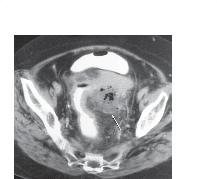
Findings
CASES 5.21 AND 5.22. Contrast-enhanced CT. Focal thickening of the wall of the sigmoid colon. Soft tissue stranding and thickening are present within the adjacent soft tissues.
Di erential Diagnosis
1.Diverticulitis
2.Colon cancer
Diagnosis
Diverticulitis
5. COLON 345
CASE 5.22
Discussion
Th ese cases illustrate a mild form of diverticulitis. Soft tissue stranding alone about the colon represents the earliest radiographic findings at CT. Bowel wall thickening in combination with soft tissue stranding also can be seen. In patients with these findings, treatment with antibiotics usually is successful. Differentiation of diverticulitis from cancer is not
always possible at CT. Follow-up CT, barium enema, or endoscopy can be used to exclude carcinoma.
Disease type: Inflammatory and Ulcerative Diseases
346 MAYO CLINIC GASTROINTESTINAL IMAGING REVIEW
CASE 5.23
CASE 5.24
Disease type: Inflammatory and Ulcerative Diseases
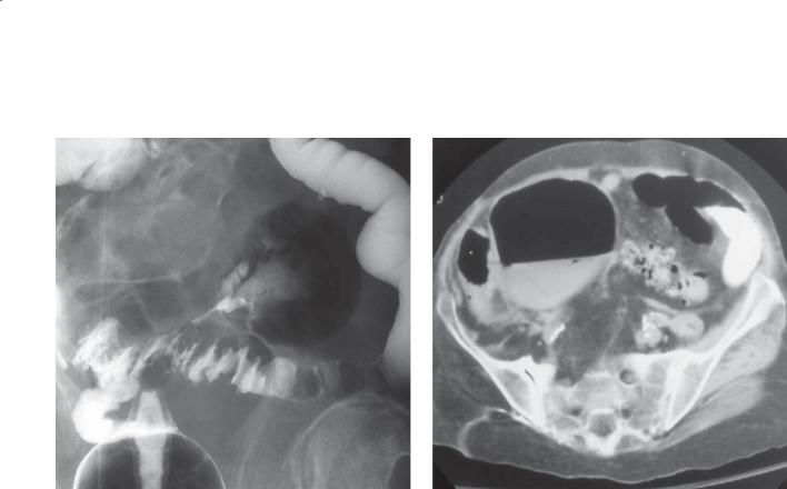
CASE 5.25.
CASE 5.23.
CASE 5.25
Findings
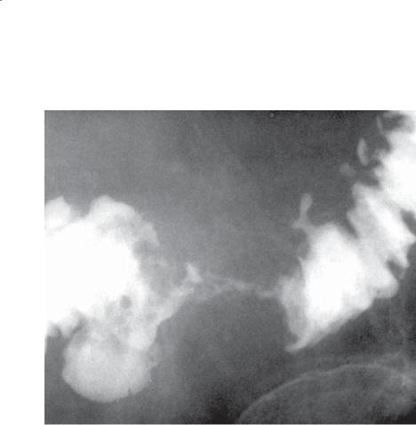
Single-contrast barium enema. A segmental region of fold thickening is present in the mid-sigmoid colon. A tract (arrow) of barium extends inferiorly from the involved sigmoid. The mucosa is intact through the involved region.
CASE 5.24. Single-contrast barium enema. The sigmoid colon is narrowed. Normal mucosal folds are present across this relatively long segment of involved colon.
Mass effect (arrow) is present along the superior sigmoid.
Single-contrast barium enema. A short focal segment of narrowing is present in the sigmoid colon. The margins of the focal abnormality are abrupt.
Normal mucosal markings are difficult to identify.
Di erential Diagnosis
1.Diverticulitis
2.Serosal metastases
3.Carcinoma
Diagnosis
Diverticulitis
5. COLON 347
Discussion
Diverticulitis is a result of inflammation of a solitary diverticulum and subsequent perforation and extravasation of colonic contents into pericolonic tissues. Usually the pericolonic inflammation is walled off. Rarely, peritonitis may develop. Diverticulitis may resolve spontaneously, often responding to antibiotic therapy and a soft diet. In some patients, the disease may be more severe; if an intramural or pericolonic abscess develops, it may require percutaneous or surgical drainage. Seeding of the mesenteric veins with microorganisms can result in sepsis, hepatic abscess, or mesenteric vein thrombosis. Fistulas can develop and communicate with the bladder, vagina, bowel loops, and other locations or organs.
Radiographic findings at contrast enema include narrowing of the colonic lumen (usually a longer segment is involved than in colon cancer), thickening of the mucosal folds (from the adjacent inflammation), mass effect with an intact overlying mucosa (due to the abscess), and extravasation of contrast material into the abscess cavity. Careful assessment of the mucosa is critical for excluding cancer.
Disease type: Inflammatory and Ulcerative Diseases
348 MAYO CLINIC GASTROINTESTINAL IMAGING REVIEW
Findings
CASE 5.26. Contrast-enhanced CT. Near the junction of the sigmoid and descending colon is a focal region of bowel wall thickening, luminal narrowing, and mesenteric soft tissue stranding. A small low-density mass (arrow) is present within the wall of the colon.
CASE 5.27. Contrast-enhanced CT. The wall of the sigmoid colon is thickened, and a small low-density mass (arrow) and soft tissue stranding are present in the sigmoid mesentery. Extensive sigmoid diverticulosis is present.
Di erential Diagnosis
1.Diverticulitis
2.Colon cancer
Diagnosis
Diverticulitis
Discussion
Th ese cases exemplify diverticulitis with small abscesses. In case 5.27, the abscess is located in the mesentery; in case 5.26, it is intramural. In patients with pericolonic soft tissue stranding and a small (<3 cm) abscess, treatment with antibiotics usually is
successful. If surgical intervention is deemed necessary, a single-stage sigmoid resection with primary reanastomosis often is possible. In both of these cases, the abscesses are too small for percutaneous drainage.
Disease type: Inflammatory and Ulcerative Diseases
CASE 5.28
Findings
Contrast-enhanced CT. Intraluminal contrast material has extravasated into the bowel wall (arrow). Bowel wall thickening and mesenteric soft tissue stranding also are present.
Di erential Diagnosis
1.Diverticulitis
2.Perforated colon cancer
3.Crohn disease
Diagnosis
Diverticulitis
5. COLON 349
Discussion
At CT, visualization of an intramural sinus tract or mesenteric abscess is considered diagnostic of diverticulitis. Perforated carcinoma, however, can
present with a pericolonic fluid collection (abscess) (cases 5.97 and 5.98). Patients with perforated colon cancer also can have symptoms that mimic those of diverticulitis. If operation is not performed, it may be prudent to perform a barium enema or colonoscopy to assess the colonic mucosa and exclude carcinoma.
Disease type: Inflammatory and Ulcerative Diseases
350 MAYO CLINIC GASTROINTESTINAL IMAGING REVIEW
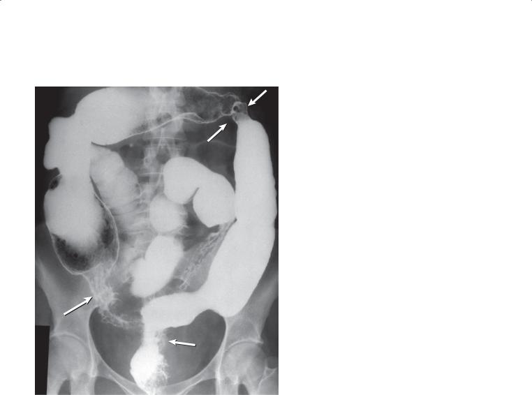
CASE 5.29
Findings
Single-contrast barium enema. Along the medial aspect of the sigmoid colon, there is an abnormal collection of barium within the wall of the colon. The collection communicates with the colonic lumen. Diverticulosis also is present.
Di erential Diagnosis
1.Diverticulitis
2.Crohn disease
Diagnosis
Diverticulitis with intramural tracking
Discussion
In a patient with an intramural collection of fluid, distinguishing Crohn disease from diverticulitis requires assessment of the underlying colonic mucosa. In diverticulitis the mucosa is normal, whereas in Crohn disease the mucosa is often ulcerated and contains nodular features. In this patient, there is
no indication of an abnormal underlying mucosa. Normal mucosal lines are seen throughout this colonic segment, as is diverticulosis.
Disease type: Inflammatory and Ulcerative Diseases
Findings
Contrast-enhanced CT. Bowel wall thickening, mesenteric soft tissue stranding, and a pericolonic abscess (arrow) involve the sigmoid colon.
Di erential Diagnosis
1.Diverticulitis
2.Perforated colon cancer
3.Crohn disease
Diagnosis
Diverticulitis
Discussion
CT can be helpful for evaluating patients with suspected diverticulitis. The advantage of this
technique is its ability to directly image the entire bowel wall and extraluminal tissues. CT findings of diverticulitis include bowel wall thickening, soft tissue stranding in the pericolonic fat, and a pericolonic abscess (fluid or gas collection). An actual abscess is found in fewer than half of the patients.
Percutaneous drainage of large abscesses can be helpful preoperatively. After much of the infection and inflammation have subsided with drainage and antibiotic therapy, a single-stage sigmoid resection often can safely be performed. More immediate operation is required if peritoneal signs are present, there is evidence of peritoneal spillage or abscess rupture, or the abscess is poorly defined and multiloculated.
Disease type: Inflammatory and Ulcerative Diseases
352 MAYO CLINIC GASTROINTESTINAL IMAGING REVIEW
CASE 5.31
A B
Findings
Contrast-enhanced CT. A and B. Th ickening of the wall of the sigmoid colon. Within the sigmoid mesentery there is a poorly defined collection of soft tissue and air.
Di erential Diagnosis
1.Diverticulitis with phlegmon
2.Perforated colon cancer
3.Foreign body perforation of sigmoid colon
Diagnosis
Diverticulitis with phlegmon
Discussion
Th e finding of extraluminal sigmoidal fluid and gas indicates perforation of the colon into the retroperitoneum. The thickened sigmoid wall could
be due to inflammation or tumor. Clinical correlation is needed, but diverticulitis would be much more common than a perforated colon cancer. Carcinoma could be excluded with endoscopy after resolution of the infection.
Identification of an extracolonic fluid and gas collection does not necessarily indicate that the abscess can be successfully drained percutaneously. Generally, an abscess that can be successfully drained is well circumscribed and of fluid attenuation. In this patient, the extraluminal collection of fluid and gas is not
well contained and the fluid and soft tissue is widely dispersed by mesenteric fat. Some radiologists might refer to this as a phlegmon (diffuse inflammation of the soft tissues due to infection), indicating that it does not appear to be percutaneously drainable. An operative procedure with removal of the widely disseminated infection is indicated. Optimally, the involved sigmoid segment would also be removed.
Disease type: Inflammatory and Ulcerative Diseases
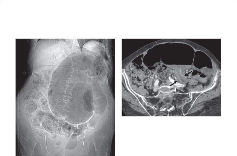
CASE 5.32
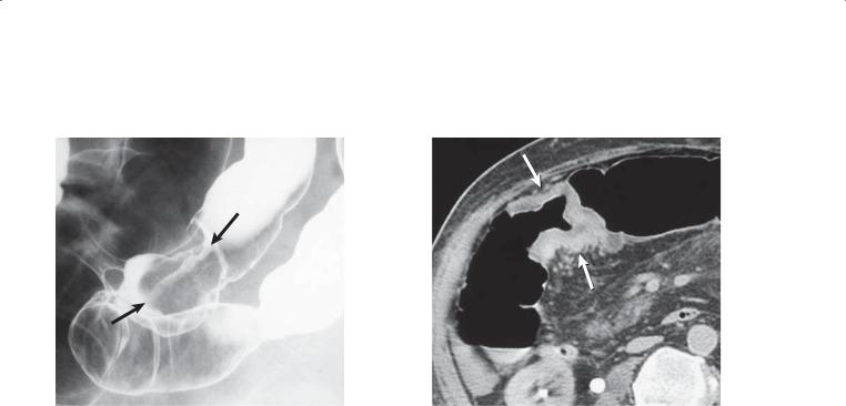
Findings
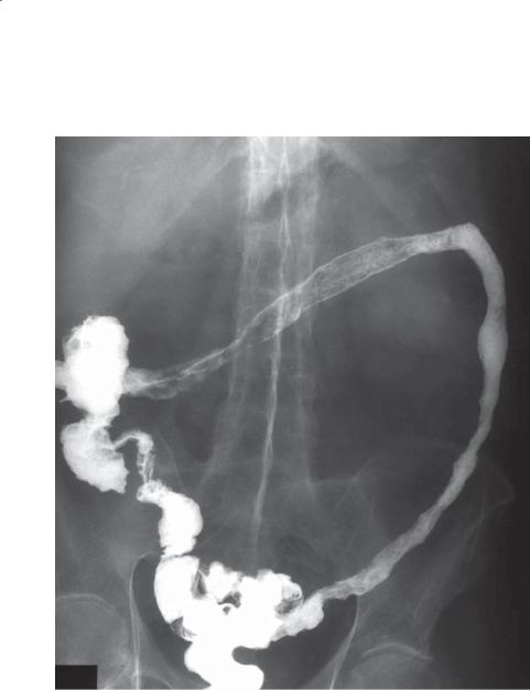
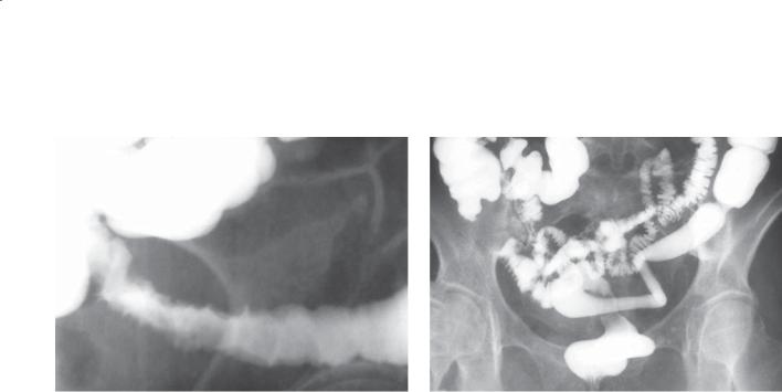
CASE 5.32. Single-contrast barium enema. The folds of the sigmoid colon are thickened and tethered. A barium-filled tract arises from the superior aspect of
the sigmoid and partially fills a well-defined gas-filled cavity. A fistulous tract also was present between the sigmoid and the destroyed right hip (not shown).
CASE 5.33. Contrast-enhanced CT. A large, thin-walled cavity containing air and fluid is present in the pelvis. This cavity was found to arise from the sigmoid on adjacent images.
Di erential Diagnosis
1.Diverticulitis with abscess
2.Giant sigmoid diverticulum
Diagnosis
Giant sigmoid diverticulum
5. COLON 353
CASE 5.33
Discussion
A giant sigmoid diverticulum develops as a result of diverticulitis. A persistent tract forms between the colonic lumen and the abscess cavity. The cavity develops a fibrous well-defined capsule over time.
Usually, a ball-valve communication is present which allows air and contrast material into the cyst but incomplete emptying. Sometimes these cysts can reach gigantic proportions, occupying a large volume of the abdomen. Operative resection of the diverticulum and affected sigmoid usually is performed for cure and to prevent perforation, torsion, and infarction.
Disease type: Inflammatory and Ulcerative Diseases
354 MAYO CLINIC GASTROINTESTINAL IMAGING REVIEW
TABLE 5.2
Findings and Treatment of Diverticulitis
CT findings
Bowel wall thickening
Pericolonic soft tissue stranding
Pericolonic or intramural fluid collection (abscess)
Contrast enema findings
Luminal narrowing Intact mucosal folds
Mass effect (from adjacent inflammatory mass with or without abscess) Extravasation of contrast material into pericolonic cavity
Treatment
Soft tissue stranding, small (3 cm) abscess: antibiotics
Well-defined abscess (>3 cm): percutaneous drainage
Poorly defined or multiloculated abscess: operation
Disease type: Inflammatory and Ulcerative Diseases
CASE 5.34
Findings
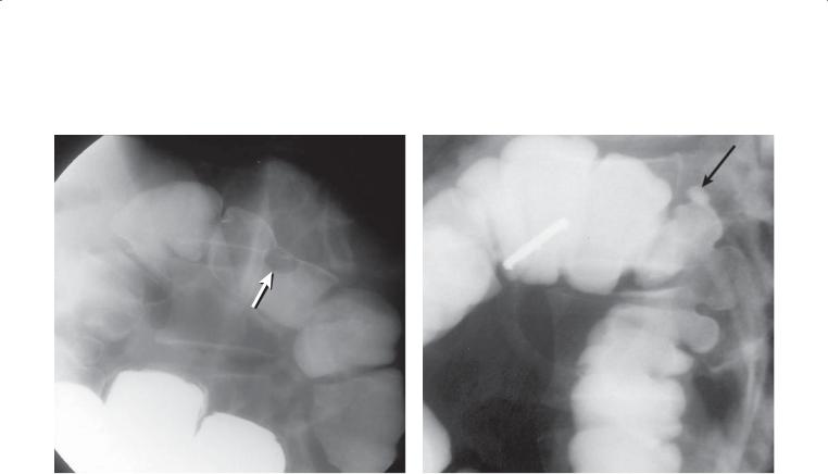
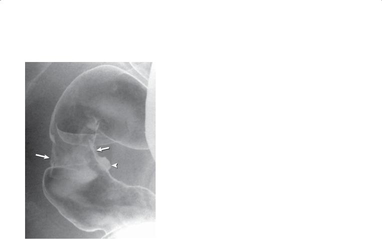
CASE 5.34. Single-contrast barium enema. A smoothsurfaced filling defect is present at the base of the cecum. The appendix did not fill.
CASE 5.35. Double-contrast barium enema. Mass effect is present along the lateral aspect of the mid ascending colon. There are thickened and tethered folds in this region.
Di erential Diagnosis
1.Appendicitis
2.Serosal metastasis
Diagnosis
Appendicitis (retrocecal location in case 5.35)
5. COLON 355
CASE 5.35
Discussion
Appendicitis is caused by obstruction of the appendiceal lumen, with secondary dilatation, infection, inflammation, ischemia, and possible perforation. Clinically, patients usually complain of generalized abdominal discomfort that localizes to the right lower quadrant. Signs of peritoneal irritation or generalized peritonitis may be present. Early recognition of appendicitis is important because the
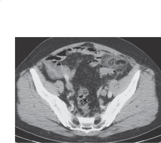
mortality associated with the disease is low, 0.1%, if the appendix is only inflamed but increases to more than 13% with an appendiceal abscess not initially treated with appendectomy. The appendix can be located almost anywhere in the abdomen, and demonstration of the appendix in an unusual location may provide an explanation for confusing clinical signs and symptoms.
Disease type: Inflammatory and Ulcerative Diseases
356 MAYO CLINIC GASTROINTESTINAL IMAGING REVIEW
Findings
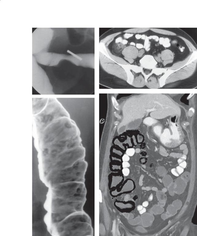
CASE 5.36. Contrast-enhanced CT. The appendix is distended and thick-walled.
CASE 5.37. Contrast-enhanced CT. Soft tissue stranding and fluid are present about the fluid-distended appendix.
CASE 5.38. Contrast-enhanced CT. An enhancing tubular structure is present in the right lower quadrant. There is soft tissue stranding and fluid in the surrounding fat.
CASE 5.39. Contrast-enhanced CT. A hyperenhancing tubular structure is present posterior to the colon with a small amount of surrounding soft tissue stranding.
Di erential Diagnosis
1.Appendicitis
2.Mucocele
Diagnosis
Appendicitis
Discussion
Th e earliest changes of appendicitis include thickening of the walls of the appendix (which is often distended with water-attenuation material) and soft tissue stranding in the periappendiceal fat. Usually patients with appendicitis present with abdominal pain, fever, and leukocytosis. Patients with a mucocele usually do not have symptoms suggesting an acute inflammatory condition.
Disease type: Inflammatory and Ulcerative Diseases
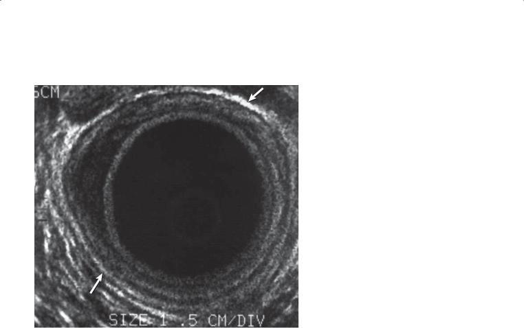
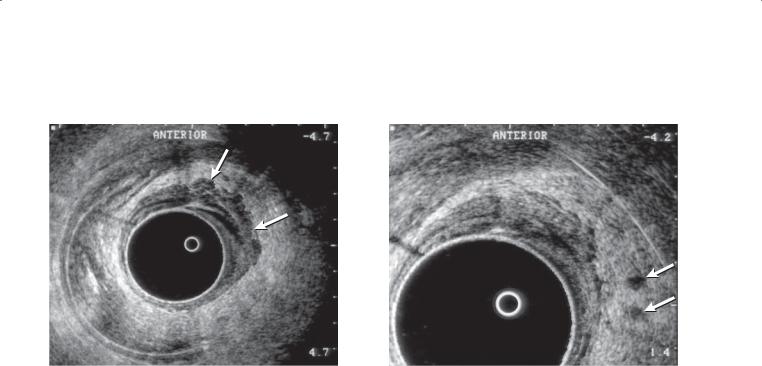
CASE 5.40
A
Findings
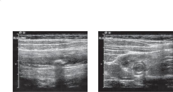
Transabdominal sonography. A. Dilated (8 mm), fluid-filled appendix containing an echogenic, posteriorly shadowing appendicolith is visible on this longitudinal sonogram of the right lower quadrant. B. Transverse view of the appendix also shows
the dilated fluid-filled appendix. The wall of the appendix is thickened (2 mm). The appendix was not compressible.
Di erential Diagnosis
Acute appendicitis
Diagnosis
Acute appendicitis
5. COLON 357
B
Discussion
Ultrasonography can be useful for directly visualizing the appendix in patients with equivocal clinical findings. Sonography is recommended as the firstline imaging test for children, ovulating women, and pregnant women. It is less useful in obese patients, patients whose abdomen is too tender for compression, and patients with overlying bowel gas.
Graded compression of the right lower abdomen is performed to empty the cecum and right colon of gas and fluid. The appendix usually is visualized at the base of the cecum. Normally, the appendiceal wall does not exceed 2 mm in thickness. Often the normal appendix is not visible, but an appendix that is abnormally distended (≥6 mm diameter) or has a thickened wall (>2 mm) is considered pathologic. A periappendiceal fluid collection can often be identified if the appendix is perforated.
Crohn disease can affect the appendix as part of the spectrum of granulomatous ileocolitis. Occasionally, the disease is localized solely to the appendix. It is usually not possible to distinguish acute appendicitis from Crohn appendicitis.
Disease type: Inflammatory and Ulcerative Diseases
358 MAYO CLINIC GASTROINTESTINAL IMAGING REVIEW
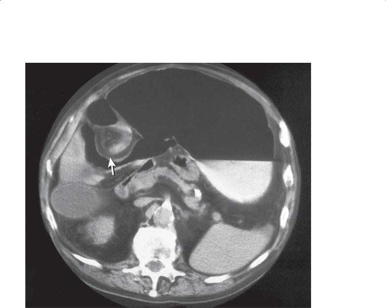
CASE 5.41
Findings
Unenhanced CT. A soft-tissue–density mass is present in the pelvic mesentery with surrounding
inflammatory stranding. There is thickening of the wall of the cecum adjacent to this inflammatory mass.
Di erential Diagnosis
Acute appendicitis
Diagnosis
Acute appendicitis (phlegmonous periappendiceal change)
Discussion
Th e appendix and periappendiceal region can be directly visualized at CT. CT findings of appendicitis include appendiceal mural thickening and enhancement, periappendiceal and mesenteric soft tissue stranding or a soft tissue mass, an appendicolith, thickening of the wall of the colon, fascial thickening, and a periappendiceal fluid collection (abscess). Direct visualization of the appendix is possible in a majority of patients.
CT also is useful for separating appendiceal abscess into three categories: 1) phlegmon, 2) well-defined abscess, and 3) poorly defined multicompartmentalized abscess. Phlegmons are best treated with antibiotics, well-defined abscess with percutaneous drainage,
and multicompartmentalized abscess with operative drainage. Interval, elective appendectomy may be required in patients treated nonoperatively.
Disease type: Inflammatory and Ulcerative Diseases
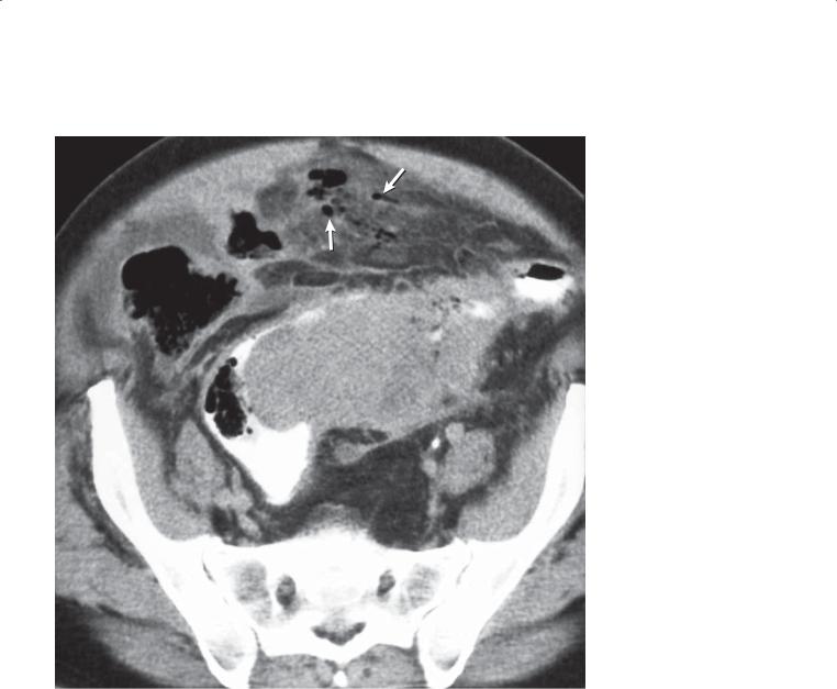
CASE 5.42
Findings
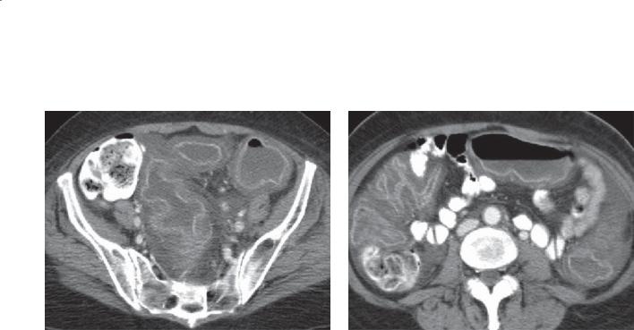
CASE 5.42. Contrast-enhanced CT. A large mass is present in the right side of the pelvis; the center is the attenuation of water and gas and has a thick soft tissue rind. The wall of the sigmoid colon is circumferentially thickened.
CASE 5.43. Contrast-enhanced CT. A multicompartmentalized inflammatory mass is present adjacent to the cecum with multiple air-fluid levels.
Di erential Diagnosis
1.Appendiceal abscess
2.Diverticular abscess
3.Pelvic inflammatory disease with abscess
Diagnosis
Appendiceal abscess
5. COLON 359
CASE 5.43
Discussion
Gangrenous appendicitis and a pelvic abscess were present at operation in both of these cases. Case 5.42 is an example of a well-defined periappendiceal abscess that could be drained percutaneously if safe access is possible. The multicompartmental periappendiceal abscess in case 5.43 usually requires operative drainage.
Disease type: Inflammatory and Ulcerative Diseases
360 MAYO CLINIC GASTROINTESTINAL IMAGING REVIEW
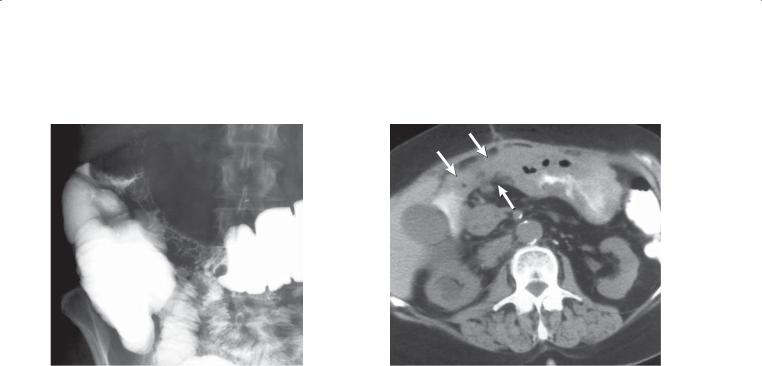
CASE 5.44
Findings
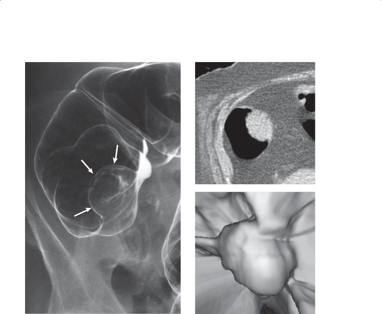
Abdominal radiograph. Two laminated calcific densities are present in the right lower quadrant.
Di erential Diagnosis
1.Appendicolith
2.Calcified mesenteric lymph node
3.Ovarian dermoid tumor
4.Calcified uterine fibroid
5.Enterolith in Meckel diverticulum
Diagnosis
Appendicolith
Discussion
Appendicoliths develop as a result of calcification about an obstructing nidus of fecal debris within
the appendiceal lumen. The calculus often has a lucent center and has an average diameter of 2 cm. Approximately 15% of all patients with appendicitis have a visible calculus. Approximately half of all symptomatic patients with a visible appendicolith already have a perforated appendix.
Other findings of appendicitis that may be present on abdominal plain radiography include an atonic gas-filled terminal ileum or cecum (sentinel loop) or a generalized adynamic ileus if peritonitis has developed. It may not be possible to exclude other differential considerations. Enteroliths often have a laminated appearance. Correlation with the clinical history is important. CT is invaluable for further evaluation of a suspicious finding.
Disease type: Inflammatory and Ulcerative Diseases
5. COLON 361
TABLE 5.3
Findings of Appendicitis
CT findings
Mural thickening >2 mm
Appendiceal distention, 6 mm diameter
Mural hyperenhancement
Periappendiceal soft tissue stranding
Periappendiceal fluid collection
Appendicolith
Sonographic findings
Mural thickening >2 mm
Appendiceal distention, 6 mm diameter
Noncompressible appendix
Pain with appendiceal compression
Periappendiceal fluid collection
Appendicolith
Contrast enema findings
Nonfilling of the entire appendix (must see bulbous tip to be normal) Filling defect at the base of the cecum, without filling of the appendix Pain with compression over the appendix
Appendicolith
Disease type: Inflammatory and Ulcerative Diseases
362 MAYO CLINIC GASTROINTESTINAL IMAGING REVIEW
Findings
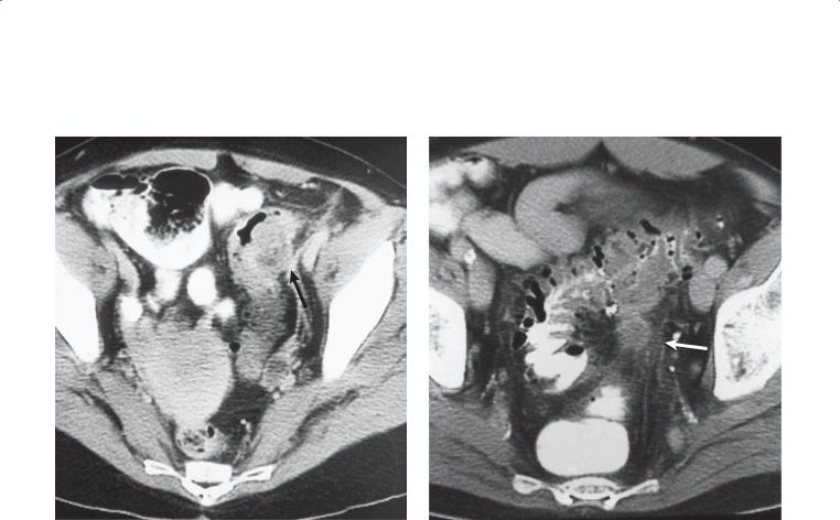
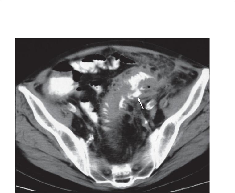
CASE 5.45. Single-contrast barium enema. Two segments (arrows) of luminal narrowing, mucosal irregularity, and ulceration are present in the region of the splenic flexure of the colon.
CASE 5.46. Single-contrast barium enema. The entire sigmoid colon is narrowed and irregular in contour.
Di erential Diagnosis
1.Crohn disease
2.Ischemic colitis
3.Infectious colitis
Diagnosis
Ischemic colitis
Discussion
Ischemia of the colon can be caused by various events, including low perfusion states and arterial or venous occlusion. The elderly are at highest risk for this condition, but younger individuals also can be affected—especially those with a vasculitis or a coagulopathy. The splenic flexure is the commonest
location for ischemic changes in patients with low perfusion states.
Normally, 3 events can ensue, depending on the degree of ischemia. 1. In mild ischemia, only mucosal sloughing occurs; after the blood supply is
reconstituted or collateral blood supply is established, the colon can return to normal. 2. Moderate ischemia affects the deeper layers of the bowel wall with stricture formation after healing. 3. Severe ischemia affects the entire bowel-wall thickness, with transmural necrosis and possible perforation.
Conventional radiographic examination of the ischemic colon usually is limited to plain abdominal radiography in the severely ischemic
patient. Adynamic ileus (case 5.135), pneumatosis coli (cases 5.125 and 5.126), pneumoperitoneum, and thickened haustral folds (often described
as thumbprinting) (case 5.47) can be seen. Mild and moderate ischemia also may be identified by findings of thickened and edematous haustral folds on plain radiography or barium enema
examination. After healing, a stricture may develop, with gradually tapering margins and a featureless mucosal pattern.
Disease type: Inflammatory and Ulcerative Diseases
CASE 5.47
A
Findings
Contrast-enhanced CT. A and B. The right and transverse colon are thick-walled, with water attenuation changes within the wall. Pericolonic fluid also is present.
Di erential Diagnosis
1.Infl ammatory bowel disease
2.Pseudomembranous colitis
3.Neutropenic colitis
4.Ischemic colitis
Diagnosis
Ischemic colitis
5. COLON 363
B
Discussion
Colonic ischemia often presents with nonspecific findings at CT, including bowel wall thickening, mesenteric soft tissue stranding, ascites, and mesenteric hemorrhage. Secondary findings of arterial or venous occlusion should be sought, as should pneumatosis coli and mesenteric venous or portal venous gas. Clinical correlation often is needed.
Disease type: Inflammatory and Ulcerative Diseases
364 MAYO CLINIC GASTROINTESTINAL IMAGING REVIEW
CASE 5.48
A B
Findings
Contrast-enhanced CT. A. A filling defect is present in the superior mesenteric vein. B. A loop of bowel (transverse and hepatic flexure of the colon) in
the upper abdomen has a thick wall, and there is considerable fluid and soft tissue stranding in the adjacent mesentery.
Di erential Diagnosis
1.Ischemic colitis due to superior mesenteric vein thrombosis
2.Intra-abdominal abscess with ascending phlebitis and thrombosis of the superior mesenteric vein
Diagnosis
Ischemic colitis due to superior mesenteric vein thrombosis
Discussion
Ischemia can be due to low flow states, arterial emboli, or venous thrombosis. Contrast-enhanced CT has the capability of directly assessing the major blood supply of the splanchnic circulation. A filling defect in either the superior mesenteric artery or vein is a critical clue to the diagnosis of ischemia. Prolonged ischemia (as present in this case) can lead to bowel perforation and abscess. This complication leads to substantial increase in morbidity and mortality. Early diagnosis and treatment are critical for reducing these complications.
Disease type: Inflammatory and Ulcerative Diseases
CASE 5.49
Findings
CASE 5.49. Single-contrast barium enema. The sigmoid colon is diffusely narrowed. The luminal contour
is irregular and the folds are markedly thickened (thumbprinting).
CASE 5.50. Single-contrast barium enema. The proximal rectum and sigmoid colon are diffusely narrowed and featureless, devoid of haustral markings.
Di erential Diagnosis
1.Radiation colitis
2.Ischemic colitis
Diagnosis
Radiation colitis
5. COLON 365
CASE 5.50
Discussion
Both patients had a history of radiation therapy. The patient in case 5.49 had recent therapy, and the
patient in case 5.50 had treatment 26 years previously. Radiation damage to the ileum and colon remains relatively common. Symptoms develop in patients after a total of 45 Gy has been administered. Most patients have a history of cervical, endometrial, ovarian, or bladder cancer. An occlusive endarteritis is the chief pathologic alteration. Acutely, edema and mucosal ulceration are present. The bowel often has a shaggy appearance, with fold thickening and luminal narrowing (case 5.49).
Radiation-induced strictures are common, developing more than 2 years after radiation treatment (case 5.50), and of variable lengths. Severe strictures (especially those in the small bowel) can cause luminal obstruction. The normal mucosal markings (haustral folds) are often absent, and there is a gradual tapering of the lumen from normal to abnormal. Patients may complain of diarrhea, cramping, or bleeding. Often the symptoms respond poorly to conservative therapy and operation is required. Surgical treatment can be difficult and associated with a high morbidity due to adherent loops, adhesions, poor tissues, and impaired healing.
Disease type: Inflammatory and Ulcerative Diseases
366 MAYO CLINIC GASTROINTESTINAL IMAGING REVIEW
CASE 5.51
Findings
Abdominal radiograph. The transverse colon is gasfilled and the haustral markings are diffusely thickened.
Di erential Diagnosis
1.Infectious colitis
2.Pseudomembranous colitis
3.Ischemic colitis
4.Acute inflammatory bowel disease
5.Neutropenic colitis
Diagnosis
Pseudomembranous colitis
Discussion
Pseudomembranous colitis is caused by the potent enterotoxins produced by Clostridium difficile, a gramnegative bacillus. Infection by this organism usually follows antibiotic therapy (originally described after lincomycin or clindamycin administration). Patients usually present with watery diarrhea. Sigmoidoscopy usually shows multiple yellowish white plaques
covering the colonic mucosa. The diagnosis can be confirmed by performing a stool assay for C difficile enterotoxin.
Radiographically, the disease often can be suggested on the basis of the plain abdominal radiograph. Haustral folds throughout the colon can appear thickened (as in this case). The disease occasionally can spare portions of the colon. Although usually involving the whole colon, it is possible for a patient with this disease to have a normal proctoscopic examination, with disease in the more proximal colon.
Inflammatory polyps may be visible within the colon in patients with inflammatory bowel disease (cases 5.4, 5.5, and 5.6). Ischemic colitis usually affects older persons and involves a segment of bowel (often the splenic flexure) rather than involving the bowel diffusely (case 5.46). A history of antibiotic use is helpful for diagnosing pseudomembranous colitis, and a history of profound neutropenia and administration of cytotoxic agents is helpful for diagnosing neutropenic colitis.
Disease type: Inflammatory and Ulcerative Diseases
CASE 5.52
A
Findings
Contrast-enhanced CT. A and B. Marked colonic and rectal (pancolonic) wall thickening with mucosal hyperenhancement. The bowel wall is of water attenuation.
Di erential Diagnosis
1.Infectious colitis
2.Pseudomembranous colitis
3.Ischemic colitis
4.Acute inflammatory bowel disease
5.Neutropenic colitis
Diagnosis
Pseudomembranous colitis
5. COLON 367
B
Discussion
Th e findings are nonspecific and could be due to any type of acute colitis. Pseudomembranous colitis was proved by stool assay for Clostridium difficile enterotoxin. The CT findings in patients with pseudomembranous colitis usually are nonspecific,
with colonic wall thickening. The wall thickening may be diffuse or segmental.
Because many patients with pseudomembranous colitis have fever, leukocytosis, and vague abdominal complaints, CT often is done to exclude an intraabdominal abscess. The findings at CT can be helpful for directing further investigation of the colon, whereas endoscopy and stool assay can show the typical yellowish white plaques and the presence
of the offending enterotoxin. Once the diagnosis is confirmed, the usual treatment is vancomycin.
Disease type: Inflammatory and Ulcerative Diseases
368 MAYO CLINIC GASTROINTESTINAL IMAGING REVIEW
CASE 5.53
A B
Findings
Contrast-enhanced CT. A and B. The wall of the colon is markedly thickened and of water attenuation. The thickened haustral folds give the colon an accordionlike appearance.
Di erential Diagnosis
1.Infectious colitis
2.Pseudomembranous colitis
3.Ischemic colitis
4.Acute inflammatory bowel disease
5.Neutropenic colitis
Diagnosis
Neutropenic colitis
Discussion
Patients with neutropenic colitis are usually undergoing chemotherapy and have a very low neutrophil count. Differentiation among the many possibilities is by history. Most patients will also have an adynamic ileus. Important predisposing factors are the ileus-induced stasis, distention, and possible ischemia; the direct cytotoxic effect on the mucosa from the chemotherapeutic medication; and the inability to mount an immunologic defense. Pathologically, mucosal and submucosal necrosis, edema, and hemorrhage are present. Perforation can occur. Abdominal plain radiographs may show an air-distended colon with thickened haustral folds, most commonly involving the cecum or right colon. Pericolonic soft tissue stranding and ascites are commonly associated.
Disease type: Inflammatory and Ulcerative Diseases
5. COLON 369
CASE 5.54
A B C
Findings
A.Single-contrast barium enema. The cecum is narrowed with an irregular contour.
B.Contrast-enhanced CT. The wall of the right colon is thickened.
C.Contrast-enhanced CT. A multiloculated, low (fluid)- density mass is present in the right lobe of the liver.
Di erential Diagnosis
1.Amebiasis
2.Colon cancer
3.Infl ammatory bowel disease
Diagnosis
Amebiasis
Discussion
Amebiasis is an infection by the protozoan Entamoeba histolytica, which is endemic throughout the world—particularly in tropical climates. Infection occurs by ingestion of the amebic cyst, which in the alkaline environment of the small bowel will shed its inner capsule and release trophozoites. Trophozoites burrow into the intestinal wall—most commonly the cecum and sigmoid colon. Multifocal or confluent ulcerations develop at the site of the bowel wall penetration. Secondary bacterial invasion of the bowel causes marked submucosal edema, bowel wall
thickening, and even hemorrhage (as in this case). A focal mass (ameboma) can develop. The protozoan infection can spread from the bowel to any part of the body by direct extension and hematogenous and lymphatic dissemination. A hepatic abscess is the usual extraintestinal site for infection (as in this case). A liver abscess can erode through the diaphragm and result in pleural, pericardial, bronchial, or lung infection. Penetration of an ulcer through the bowel wall can result in a pericolonic abscess, fistula, peritonitis, or distant intraperitoneal abscess. Longterm changes of amebiasis usually include benignappearing colonic strictures. Treatment of this disease is with antiamebic therapy. Surgical treatment of
this disease is associated with high morbidity and mortality, often without cure.
Radiologically, the cecum and sigmoid colon are affected most often. Involvement of the terminal ileum occurs in a minority of patients. Initially, the patient may have mucosal changes resembling
ulcerative colitis (granular-appearing mucosa with fine ulcerations and thickened and edematous haustra). Deeper and more extensive ulcerations may be seen as the disease progresses. The cecum often becomes nondistensible and conical in shape. Amebomas can be indistinguishable from colon cancer. Multiplicity of lesions, ulceration elsewhere in the colon, young age, and travel history may all be helpful for differentiating this disease from carcinoma.
Disease type: Inflammatory and Ulcerative Diseases
370 MAYO CLINIC GASTROINTESTINAL IMAGING REVIEW
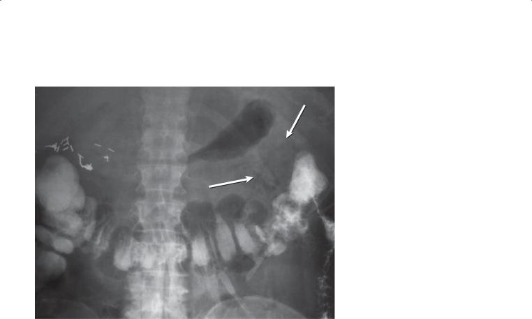
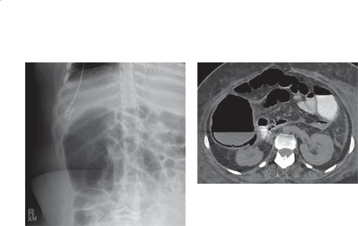
CASE 5.55
Findings
Abdominal radiograph. An extraluminal gas (arrows) collection (multiple small bubbles) is present in the region of the pancreatic bed. Luminal narrowing and thickened folds are present in the descending colon, which contains contrast material from a recent barium enema.
Di erential Diagnosis
Pancreatic abscess
Diagnosis
Pancreatic abscess
Discussion
Imaging procedures are not necessary in all patients for making the diagnosis of acute pancreatitis. Imaging tests are invaluable for confirming a suggested diagnosis and for detecting a complication of acute pancreatitis (including a pancreatic abscess). CT
and sonography have replaced abdominal plain radiography and intraluminal contrast studies when the diagnosis of acute pancreatitis is suggested. In some patients, however, symptoms may be nonspecific and abdominal plain radiography is used to assess for
the presence of adynamic ileus, obstruction, or tube placements.
A pancreatic effusion is commonly associated with acute pancreatitis. Most often the effusion is located within the left anterior pararenal space and lesser sac. The mass effect from this fluid can displace the stomach anteriorly and the colon inferiorly. As the volume of the pancreatic effusion increases, it can
extend inferiorly, adjacent to the descending colon and along the transverse mesocolon. Inflammatory changes (thickened folds, spasm, narrowing) of the transverse and descending colon can be seen. The splenic flexure of the colon usually is involved with inflammatory changes because of its proximity to the pancreatic
tail. A pancreatic effusion also can dissect within the leaves of the small bowel mesentery and cause ascites in patients with severe disease. The colon cutoff sign has been regarded as a classic radiographic finding of acute pancreatitis. Patients with this sign have gaseous distention of the right and transverse colon, with little gas visible beyond the splenic flexure (as seen
in this case). It can be impossible to exclude colonic obstruction on plain radiographs, and a contrast enema or CT may be required.
Disease type: Inflammatory and Ulcerative Diseases
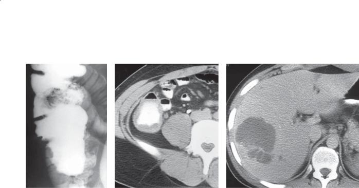
CASE 5.56
A
Findings
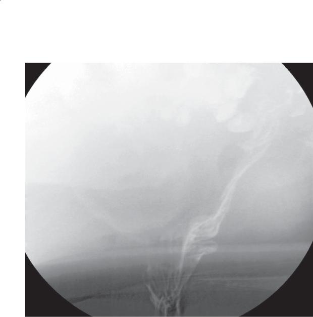
A.Contrast-enhanced CT. An abscess is present in the pancreatic bed. An inflammatory reaction and two tracts (arrows) to the splenic flexure of the colon can be identified.
B.Sinogram. A drainage catheter in the pancreatic abscess cavity is injected with contrast material. A fistula is present between the abscess and the splenic flexure of the colon.
Di erential Diagnosis
Pancreatic abscess with pancreatico-colonic fistula
Diagnosis
Pancreatic abscess with pancreatico-colonic fistula
5. COLON 371
B
Discussion
A pancreatic abscess is a life-threatening complication of acute pancreatitis. Usually, patients with a pancreatic abscess have underlying pancreatic necrosis. CT
and sonography are sensitive imaging methods for detecting peripancreatic fluid and pancreatic necrosis—which may or may not be infected. The
finding of gas in the pancreatic bed is highly suggestive of infection but is not diagnostic. Correlation with clinical information and percutaneous aspiration of fluid are important steps for determining the proper diagnosis. Fistula formation between the pancreatic bed and splenic flexure of the colon is a relatively common complication of pancreatitis and can lead to a pancreatic abscess. Knowledge that a fistula is present is critical for planning proper therapeutic intervention. Colonic resection or percutaneous drainage may be necessary. If the fistula is not repaired operatively, longterm catheter drainage often is required.
Disease type: Inflammatory and Ulcerative Diseases
372 MAYO CLINIC GASTROINTESTINAL IMAGING REVIEW
CASE 5.57
Findings
Single-contrast barium enema. Multiple wide-mouth sacculations are present along the antimesenteric border of the transverse colon.
Di erential Diagnosis
1.Scleroderma
2.Crohn disease
Diagnosis
Scleroderma
Discussion
Scleroderma causes patchy replacement of the muscular layers of the colon with collagen and elastic fibers. Intimal proliferation of the feeding arteries with possible ischemia also can occur.
Radiographically, the antimesenteric border of the colon may develop sacculations or pseudodiverticula as a result of the limp supporting tissues in the wall of the colon. The mesenteric side is not affected because the tissues and vessels in this region continue to support the bowel wall. Haustral markings may be lost, and redundancy (due to dilatation and elongation) may be present. Localized areas of narrowing also may be seen as a result of ischemia.
Th e main differential consideration is Crohn colitis, with multiple areas of asymmetric bowel wall involvement and pseudodiverticula (case 5.20). The asymmetric changes in Crohn disease usually are segmental, with normal intervening colon. The pseudodiverticula in patients with Crohn disease
may affect either the mesenteric or the antimesenteric border of the colon. The clinical history is often revealing.
Disease type: Inflammatory and Ulcerative Diseases
Findings
Single-contrast barium enema. Segmental, haustral fold thickening is present in the transverse colon. Sclerosis of the left sacrum and ileum is present.
Di erential Diagnosis
1.Ischemic colitis
2.Acute radiation colitis
3.Pancreatitis
4.Crohn disease
5.Mastocytosis
Diagnosis
Mastocytosis
Discussion
Th is finding is nonspecific and could be the result of several diseases causing bowel wall edema or
inflammation or infiltration. This patient was found to have mastocytosis.
Mastocytosis is a rare condition of abnormal deposition of mast cells, often within the skin (urticaria pigmentosa) and less commonly within other organs (liver, spleen, bones, alimentary tract). Gastrointestinal symptoms usually include nausea, vomiting, and diarrhea. The incidence of peptic ulcer disease is increased in mastocytosis, presumably due to histamine-mediated acid secretion. Malabsorption also can occur with diffuse small bowel involvement. Many patients experience an intolerance to alcohol, which can exacerbate symptoms.
Pathologically, cellular infiltration (mast cells) and edema usually are present within the bowel wall. The small bowel is most commonly affected, but potentially any portion of the gut can be involved.
Radiographically, fold thickening and distortion are usually present (as in this case). Occasionally, a diffuse, fine nodular pattern of sandlike lucencies is seen.
Urticarial lesions also have been described. Skeletal sclerosis also may be a helpful clue (as in this case).
Disease type: Inflammatory and Ulcerative Diseases
374 MAYO CLINIC GASTROINTESTINAL IMAGING REVIEW
CASE 5.59
Findings
Contrast-enhanced CT. A fat-attenuation mass with a peripheral rim of higher attenuation and surrounding inflammatory changes is present in the mesentery anterior to the sigmoid colon. There is sigmoid diverticulosis, but the wall of the adjacent colon is not thickened.
Di erential Diagnosis
1.Epiploic appendagitis
2.Diverticulitis
3.Omental torsion
4.Mesenteric panniculitis
Diagnosis
Epiploic appendagitis
Discussion
Epiploic appendages are peritoneal outpouchings that arise from the serosal surface of the colon and usually measure 2 to 5 cm in length. Epiploic appendagitis
is caused by torsion of the epiploic appendages with secondary ischemia. The most common part of the colon involved by acute appendagitis is the sigmoid colon. The disorder usually is self-limited and treated symptomatically with pain relief. This disorder usually is differentiated from diverticulitis by absence of bowel wall thickening and the epicenter of the inflammatory process centered away from the colon. Central fat within the inflammatory mass is typical. Mesenteric panniculitis usually is centered in the root of the mesentery. Omental torsion usually involves
a larger fatty region and often is located on the right side of the abdomen.
Disease type: Inflammatory and Ulcerative Diseases

|
|
5. COLON 375 |
|
|
|
|
TABLE 5.4 |
|
Inflammatory Diseases of the Colon |
CASE |
|
|
|
|
Ulcerative colitis |
Circumferential, symmetric involvement starting in the rectum |
5.1–5.15 |
|
and progressing proximally. Granular mucosa acutely; rigid, |
|
|
shortened colon chronically |
|
|
|
|
Crohn disease |
Asymmetric involvement with skip areas, aphthous ulcers, or |
5.16–5.20 |
|
cobblestone appearance. Usually rectal sparing. Perianal disease |
|
|
is common |
|
|
|
|
Diverticulitis |
Long-segment colonic thickening and inflammatory changes in |
5.21–5.33 |
|
region with diverticula. Abscess should be sought |
|
|
|
|
Appendicitis |
Acute abdomen, lumen 6 mm in diameter, wall >2 mm in |
5.34–5.44 |
|
diameter with mural enhancement, periappendiceal stranding |
|
|
or fluid collection, appendicolith |
|
|
|
|
Ischemic colitis |
Low-perfusion states and arterial or venous occlusion. Often |
5.45–5.48 |
|
seen at splenic flexure. Pneumatosis and portal venous gas |
|
|
should be sought |
|
|
|
|
Radiation colitis |
Acute changes include fold thickening and shaggy appearance. |
5.49 and 5.50 |
|
Rigidity and strictures are seen chronically. Restricted to |
|
|
radiation port |
|
|
|
|
Pseudomembranous colitis |
Clostridium difficile infection after antibiotic therapy. Usually |
5.51 and 5.52 |
|
pancolitis with marked mural thickening |
|
|
|
|
Neutropenic colitis |
Usually patients with leukemia or lymphoma undergoing |
5.53 |
|
chemotherapy. Usually right-sided colonic involvement |
|
|
|
|
Infectious colitis |
Variable colonic involvement, travel history, stool cultures |
5.54 |
|
|
|
Pancreatitis |
Inflammatory thickening and narrowing of transverse and |
5.55 and 5.56 |
|
proximal descending colon adjacent to pancreatic tail |
|
|
|
|
Scleroderma |
Sacculations or pseudodiverticula on antimesenteric border of |
5.57 |
|
colon |
|
|
|
|
Mastocytosis |
Small bowel most commonly affected. Usually fine nodular |
5.58 |
|
pattern or thickened folds. Look for skeletal sclerosis |
|
|
|
|
Epiploic appendagitis |
Caused by torsion of epiploic appendices with secondary |
5.59 |
|
ischemia. Pericolonic fat stranding with no colonic wall |
|
|
thickening. Central fat is typical |
|
|
|
|
Disease type: Inflammatory and Ulcerative Diseases
376 MAYO CLINIC GASTROINTESTINAL IMAGING REVIEW
CASE 5.60 |
CASE 5.61 |
CASE 5.62 |
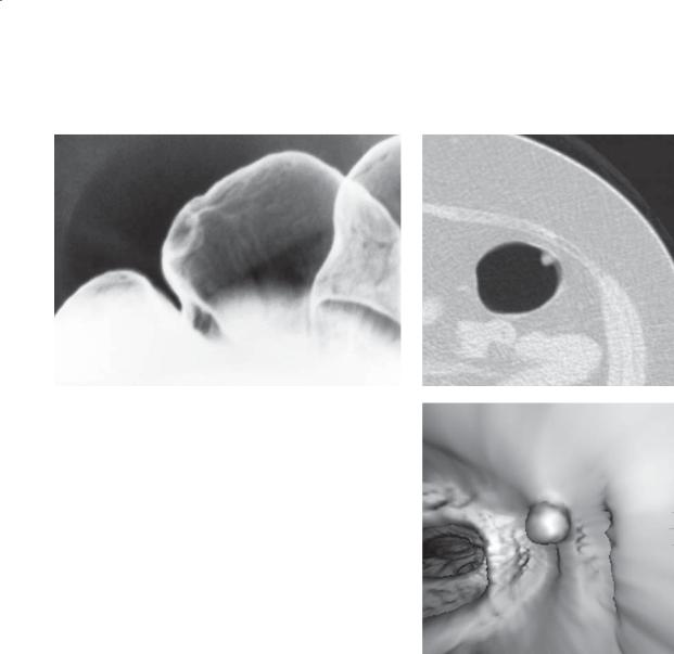
Findings
CASE 5.60. Double-contrast barium enema.
A pedunculated filling defect is present in the mid transverse colon.
CASE 5.61. CT colonography. Pedunculated filling defect in mid sigmoid colon.
CASE 5.62. CT colonography. Pedunculated filling defect in rectum.
Di erential Diagnosis
Polyp, pedunculated
Diagnosis
Polyp, pedunculated
Discussion
Polyps of the colon can be classified by histologic type: hyperplastic (retention), adenomatous, and hamartomatous. There are no reliable radiographic findings to distinguish adenomatous polyps from the other types. Hyperplastic polyps generally are small (<1 cm diameter), and these small lesions are believed not to have malignant potential. Adenomatous polyps can be further classified into
three histologic subtypes: tubular, tubulovillous, and
villous adenomas. Most authorities now believe that the majority of adenocarcinomas of the colon arise from preexisting adenomas. Polyps with a higher percentage of villous features are at a higher risk for malignant transformation than are tubular adenomas. Polyp diameter is also a key factor in assessing the risk of malignancy in an adenomatous polyp because the larger the polyp, the higher the risk. Muto et al tabulated these data as follows:
TABLE 5.5
Malignant Potential of Colorectal Adenomas
% Malignant Potential, By Size
Histologic Type |
<1 cm |
1-2 cm |
>2 cm |
Tubular |
1.0 |
10.2 |
34.7 |
Tubulovillous |
3.9 |
7.4 |
45.8 |
Villous |
9.5 |
10.3 |
52.5 |
|
|
|
|
Adapted from Muto T, Bussey HJR, Morson BC: The evolution of cancer of the colon and rectum. Cancer. 1975 Dec;36(6):2251–70. Used with permission.
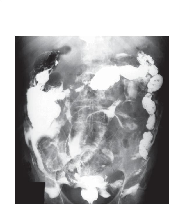
Hamartomatous polyps are associated with PeutzJeghers syndrome (case 5.71) and with juvenile polyposis (case 5.76).
Disease type: Masses and Filling Defects
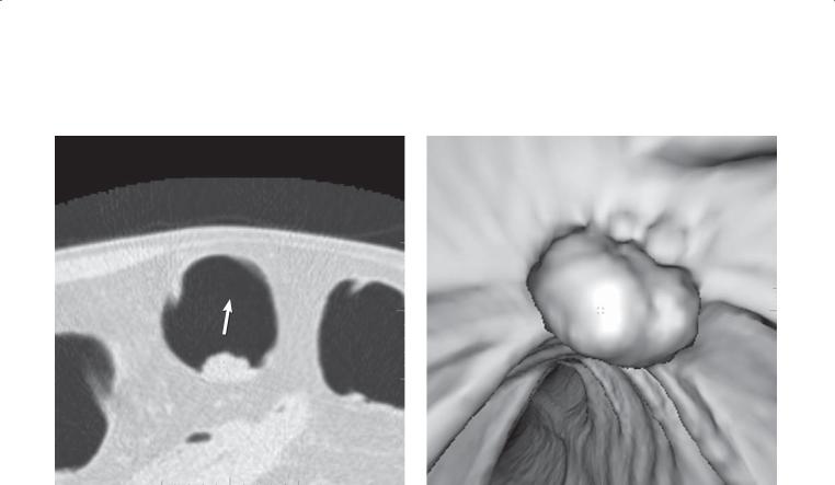
Findings
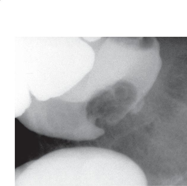
CASE 5.63. Double-contrast barium enema. A sessile filling defect is present in the colon.
CASE 5.64. CT colonography. A and B. A small halfsphere filling defect is present in the colon on both the 2- and 3-dimensional endoluminal views.
Di erential Diagnosis
1.Polyp
2.Stool
Diagnosis
Polyp
Discussion
Polyps are commonly multiple, and synchronous lesions are seen in 25% to 50% of patients. Nearly half of all polyps are located in the rectosigmoid, 20% in the right colon, and 29% to 35% in the transverse and descending colon. Polyps at CT should be homogeneous soft tissue attenuation, whereas stool
often is inhomogeneous internally, containing air or fat attenuation.
Disease type: Masses and Filling Defects
378 MAYO CLINIC GASTROINTESTINAL IMAGING REVIEW
CASE 5.65
A B
Navg
CT colonography. A. A 2-cm filling defect is present in the colon. B. Three-dimensional endoluminal view shows the filling defect as a lobulated mass, distinct from the nearby haustral fold.
Di erential Diagnosis
1.Polyp
2.Cancer
3.Stool
Diagnosis
Large adenomatous colon polyp
Th e incidence of malignancy increases with increasing polyp size and is less than 1% in polyps less than 1 cm in diameter, 10% in polyps 1 to 2 cm in diameter, and 40% in polyps more than 2 cm in diameter.
Disease type: Masses and Filling Defects
Findings
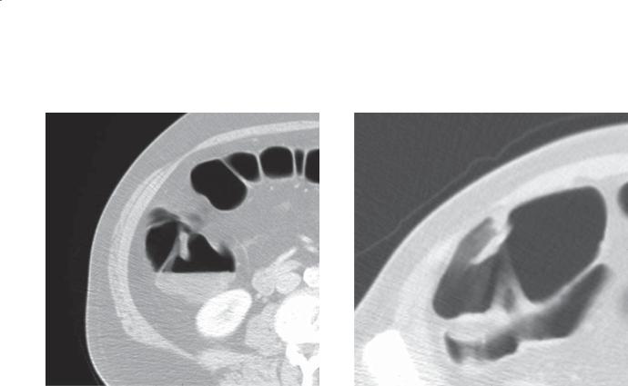
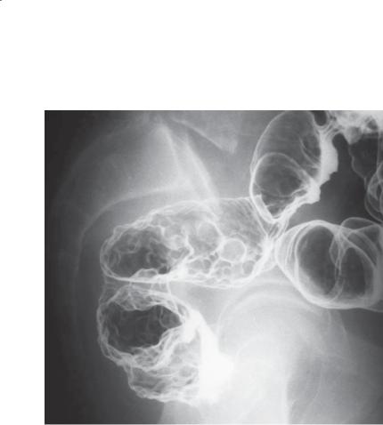
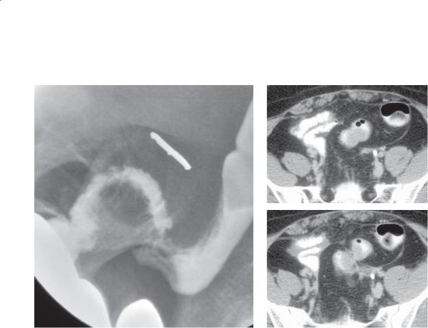
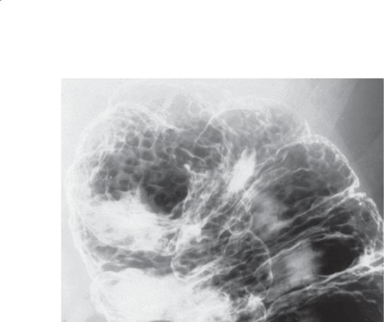
CASE 5.66. Single-contrast barium enema. A large polypoid mass is present in the base of the cecum. Barium fills the interstices of the mass. Its surface appearance resembles a cauliflower or raspberry.
CASE 5.67. CT colonography. A. Axial 2-dimensional image. An irregular-shaped filling defect is present in the ascending colon. B. Three-dimensional endoluminal image. The surface of the lesion is markedly irregular.
Di erential Diagnosis
1.Villous adenoma
2.Retained stool
Diagnosis
Villous adenoma
Discussion
Villous adenomas can be recognized if the typical surface features are visible. Characteristically, barium fills the interstices of the tumor between the individual fronds. Unfortunately, it is usually not possible to detect villous features within small polyps. Usually, polyps must be nearly 2 cm in diameter before the typical features are recognizable. Villous tumors usually are soft and compressible, and rectal lesions can easily be missed on digital examinations. Because villous tumors have a high risk of malignancy, they should be removed.
Disease type: Masses and Filling Defects
380 MAYO CLINIC GASTROINTESTINAL IMAGING REVIEW
Findings
CASE 5.68. CT colonography. A cigar-shaped filling defect is present within the ascending colon lumen.
CASE 5.69. CT colonography. A flat, somewhat lobulated filling defect is present within the ascending colon.
Di erential Diagnosis
1.Flat polyp
2.Stool
Diagnosis
Flat polyp (adenoma)
Discussion
Th e definition of a flat polyp varies. Flat polyps are often defined as those that are at least twice as wide as their height and not more than 3 mm above the flush surface. Other definitions include a spectrum of types varying from slightly elevated to depressed to flat. Generally, flat polyps are believed to more likely contain cancer than usual polyps. Flat polyps can be difficult to detect and can be either adenomatous or hyperplastic. They are most commonly found in the
right colon. Focal regions of soft tissue wall thickening identified on 2-dimensional axial images can be an important clue for the presence of a flat polyp.
Disease type: Masses and Filling Defects

CASE 5.70
A
Findings
Single-contrast barium enema. A. A large pedunculated polyp is present in the sigmoid colon. B. Almost 8 years later, a large sessile carcinoma has developed at the site of the previous polyp.
Di erential Diagnosis
Adenoma to carcinoma transformation
Diagnosis
Adenoma to carcinoma transformation
Discussion
Today, most researchers believe that the majority of colon cancers arise from preexisting polyps. This case supports that hypothesis. The role of radiologists is to detect all polypoid colonic lesions with the hope that they can be removed successfully before malignant degeneration. Risk factors for the development of colorectal polyps include increasing age, history of previous polyps, and family history of colon polyps or carcinoma. Patients with ulcerative colitis also have a higher incidence of colon cancer developing.
B
Th e American Cancer Society Guidelines for the Early Detection of Colon Cancer (January 26,
2012) recommend that, beginning at age 50 years,a men and women follow 1 of the following 5 testing options:
1.Yearly fecal occult blood test or fecal immunochemical test
2.Flexible sigmoidoscopy every 5 years
3.Double-contrast barium enema every 5 years
4.Colonoscopy every 10 years
5.CT colonography every 5 years
aPersons known to be at increased risk for colorectal cancer (because of inflammatory bowel disease, personal or family history of polyps or cancer, familial syndromes such as familial adenomatous polyposis or hereditary nonpolyposis colorectal cancer) need to begin screening at an early age and may need more frequent screening.
Note: A digital rectal examination is not an acceptable substitute for the above-recommended tests. Fecal tests (fecal occult blood and fecal immunochemical) do not detect polyps, and multiple samples must be used.
Disease type: Masses and Filling Defects
382 MAYO CLINIC GASTROINTESTINAL IMAGING REVIEW
CASE 5.71
Disease type: Masses and Filling Defects

Findings
Double-contrast barium enema. Multiple large polyps (arrows) are present in the colon. The patient had multiple mucocutaneous pigmentations at physical examination.
Di erential Diagnosis
Peutz-Jeghers syndrome
Diagnosis
Peutz-Jeghers syndrome
Discussion
Peutz-Jeghers syndrome is a disease of mucocutaneous pigmentation and gastrointestinal polyposis. Patients
5. COLON 383
usually present with symptoms of abdominal cramping, rectal bleeding, melena, or anemia. Cramping often is due to transient intussusceptions within the small bowel.
Gastrointestinal polyps in Peutz-Jeghers syndrome most frequently are found in the small bowel (95%) (cases 4.8 and 4.9), but they also can be identified in the colon and rectum (30%) and stomach (25%). The polyps can vary in size and usually are less numerous in the stomach and colon. Polyps in the stomach and small bowel are hamartomatous, but colonic polyps usually are adenomatous (and potentially malignant).
Alimentary tract malignancies develop in 2% to 3% of patients with Peutz-Jeghers syndrome.
Disease type: Masses and Filling Defects
384 MAYO CLINIC GASTROINTESTINAL IMAGING REVIEW
B
Disease type: Masses and Filling Defects
CASE 5.74.
Findings
CASE 5.72. Double-contrast barium enema. Multiple polypoid filling defects are present throughout the colon. These findings are characteristic of a polyposis syndrome.
CASE 5.73. Radiograph of the mandible. Multiple osteomas (arrows) are visible arising from the mandible.
Contrast-enhanced CT. A and B. Multiple filling defects are present throughout the colon, including a very large filling defect that occupies most of the lumen of the rectosigmoid colon.
CASE 5.75. Contrast-enhanced CT. A large mesenteric soft tissue tumor occupies a large portion of the abdomen.
Di erential Diagnosis
Polyposis syndrome—familial adenomatous polyposis syndrome
Diagnosis
Polyposis syndrome—familial adenomatous polyposis syndrome
5. COLON 385
Discussion
Familial adenomatous polyposis is the commonest of the polyposis syndromes, characterized by large numbers of adenomatous polyps that often carpet the
entire colon. Patients often present with rectal bleeding in their early 30s; if colectomy is not performed,
they will die of adenocarcinoma of the colon in their early 40s. Family screening should be performed in sporadic cases. Radiographically, innumerable polyps are identified throughout the entire colon and may be found in the small bowel and stomach. Occasionally, the polyps may conglomerate and appear as bizarre filling defects in the colon.
Gardner syndrome is a variant of familial polyposis, with various mesenchymal-derived extraintestinal manifestations. Soft tissue tumors in patients
with Gardner syndrome include 1) sebaceous cysts; 2) benign mesenchymal tumors, such as fibromas, lipofibromas, lipomas, leiomyomas, and neurofibromas; 3) malignant mesenchymal tumors;
and 4) fibrous tissue proliferation producing desmoid tumors, keloids, peritoneal adhesions, and mesenteric and retroperitoneal fibrosis. Large abdominal desmoid tumors (as in case 5.75) usually occur after total colectomy. Localized areas of dense bone (osteomas) commonly are present, often involving the skull, mandible, and maxilla. Long bone cortical thickening and dental abnormalities also are common.
Disease type: Masses and Filling Defects
386 MAYO CLINIC GASTROINTESTINAL IMAGING REVIEW
CASE 5.76
Findings
Double-contrast barium enema. Multiple variablesized polyps are present in the colon.
Di erential Diagnosis
Polyposis syndrome—unknown type
Diagnosis
Juvenile polyposis syndrome
Discussion
Juvenile polyposis is usually a disorder of children which includes solitary or multiple hamartomatous polyps. Histologically, these polyps have abundant connective tissue stroma containing mucin-filled cystic spaces lined with epithelium. Sporadic cases usually are associated with only a few polyps, whereas the hereditary form is associated with multiple polyps (juvenile polyposis coli). In the hereditary disease, polyps can be found throughout the alimentary tract. Juvenile polyps may coexist with adenomatous polyps in patients with familial adenomatous polyposis syndrome. The commonest symptoms and signs are rectal bleeding, anemia, and anal prolapse of a polyp.
Disease type: Masses and Filling Defects
|
|
|
5. COLON 387 |
|
|
|
|
|
|
TABLE 5.6 |
|
Polyposis Syndromes |
|
|
|
|
|
|
HISTOLOGIC TYPE |
NAME |
USUAL LOCATION |
ASSOCIATED DISORDERS |
|
|
|
|
Adenoma |
Familial polyposis |
Colon |
None |
|
Gardner syndrome |
Colon |
Osteomas, desmoids, fibromas, |
|
|
|
ampullary cancers, epidermoid cysts |
|
Turcot syndrome |
Colon |
Central nervous system tumors, |
|
|
|
thyroid tumors |
|
Hereditary nonpolyposis |
Colon (proximal) |
Endometrial tumors, ovarian tumors |
|
colon cancer syndrome |
|
|
|
(Lynch syndrome) |
|
|
|
|
|
|
Hamartoma |
Peutz-Jegher syndrome |
Small bowel |
Mucocutaneous pigmentation; |
|
|
|
malignancies of the ovary, breast, |
|
|
|
endometrium, pancreas |
|
Juvenile polyposis |
Colon |
Various congenital abnormalities |
|
Cronkhite-Canada |
Stomach, colon |
Alopecia, onychodystrophy, |
|
syndrome |
|
hyperpigmentation |
|
Cowden syndrome |
Colon |
Malignancies of the breast, thyroid |
|
|
|
|
Hyperplastic |
Juvenile |
Colon |
|
|
|
|
|
Disease type: Masses and Filling Defects
388 MAYO CLINIC GASTROINTESTINAL IMAGING REVIEW
CASE 5.77
A B
Findings
Double-contrast barium enema. A. A pedunculated, smooth-surfaced polyp (arrows) is present along the medial aspect of the ascending colon. B. The polyp (arrows) has changed shape with a change in position and distention. It now appears as a broad-based submucosal mass.
Di erential Diagnosis
1.Pedunculated polyp
2.Lipoma
Diagnosis
Lipoma
Discussion
Th e smooth surface of this lesion indicates its submucosal origin. Its changeable shape favors a lipoma, which was verified histologically. Colonic lipomas are the most frequent submucosal tumors of the colon. They are slow-growing, benign neoplasms that are most commonly found in the right side of the colon. Most lipomas are asymptomatic and found incidentally. Occasionally, lipomas can be the lead point for an intussusception, and patients may present with abdominal pain and rectal bleeding (case 5.79).
A filling defect (usually >3 cm) with a very smooth surface is most often seen at contrast enema. The tumor is pliable and readily changes shape with compression or with collapse of the colonic lumen. Within the distended colon, the mass often is round, elliptical, or ovoid. It is common for the tumor to elongate and become sausage-shaped within the evacuated colon. The ileocecal valve also may become lipomatous. Often the valve appears enlarged, with a very smooth surface. Pliability and distensibility are key features of a lipomatous tumor which distinguish it from other submucosal lesions.
Disease type: Masses and Filling Defects
CASE 5.78
Findings
Double-contrast barium enema. A 3-cm smoothsurfaced filling defect (arrows) in the sigmoid colon. The mass appears to make obtuse angles with the normal colon wall, but it does not displace the lumen of the colon and appears intraluminal in location.
Di erential Diagnosis
1.Lipoma
2.Gastrointestinal stromal tumor
3.Serosal metastasis
Diagnosis
Lipoma
5. COLON 389
Discussion
Lipomas are the commonest submucosal tumor of the colon. Although they most often occur in the right colon, they can be found anywhere. The smooth surface and changeable shape are characteristic findings. Stromal tumors rarely arise in the colon, but when they do the rectum is the commonest location. Serosal metastasis indicates a tumor that has spread to
the peritoneum. A history of a primary tumor, multiple lesions, and the presence of tethering of the colon are helpful additional features that strongly suggest this diagnosis.
Disease type: Masses and Filling Defects
390 MAYO CLINIC GASTROINTESTINAL IMAGING REVIEW
CASE 5.79
Findings
Contrast-enhanced CT. A. An intraluminal mass (arrow) is present within the right colon. The mass is of fatty density. B. At a slightly more caudal level, a ring of mesenteric fat (arrow) is seen within the colon.
Di erential Diagnosis
Intussuscepting colonic lipoma
Diagnosis
Intussuscepting colonic lipoma
Discussion
Most patients with a colonic lipoma are asymptomatic; however, occasionally large intraluminal lipomas cause intussusception, ulceration, or hemorrhage. Lipomas have no risk of malignant degeneration. If the lesion is asymptomatic, no treatment is required.
Disease type: Masses and Filling Defects
CASE 5.80
A
C
Findings
A.T1-weighted MRI. A 4-cm low-intensity soft tissue mass is seen at the base of the cecum.
B.T2-weighted MRI. The mass is of high-signal intensity.
C.Coronal, contrast-enhanced MRI. The mass enhances after administration of contrast material.
Di erential Diagnosis
1.Appendiceal neoplasm: adenocarcinoma, carcinoid
2.Endometrioma
3.Metastasis
5. COLON 391
B
Diagnosis
Appendiceal carcinoid
Discussion
Carcinoid tumors of the appendix are derived from primitive neuroendocrine cells in the gastrointestinal tract. Most appendiceal carcinoid tumors are small (<2 cm) and incidentally found at the time of appendectomy or pelvic surgery. These small tumors are cured with resection. Larger carcinoid tumors (>2 cm), as in this case, are rare but can spread to the serosal surface and metastasize to both lymph
nodes and liver (with carcinoid syndrome). Treatment is an appendectomy for small tumors and a right hemicolectomy for large tumors.
Disease type: Masses and Filling Defects
392 MAYO CLINIC GASTROINTESTINAL IMAGING REVIEW
B
Findings
CASE 5.81. Double-contrast barium enema. A polypoid mass (arrows) is present in the mid ascending colon. No contrast material passed proximal to the obstructing mass.
CASE 5.82. CT colonography. A and B. A large polypoid soft tissue attenuating mass is present within the hepatic flexure of the colon.
Di erential Diagnosis
1.Colon carcinoma
2.Large colon polyp
Diagnosis
Polypoid colon cancer
Discussion
Colon cancer is the most frequent cause of large bowel obstruction. An enema with contrast material or CT colonography is a rapid and accurate method to determine the cause of the obstruction. The soft tissue attenuation distinguishes it from a lipoma. Its intraluminal location favors a mucosal origin. Its large size makes carcinoma most likely. Colitis has been reported to occur proximal to an obstructing lesion in some patients. High-grade obstruction and a competent ileocecal valve also can lead to increased intraluminal pressure, decreased venous flow, and possible colonic ischemia.
Disease type: Masses and Filling Defects
5. COLON 393
CASE 5.83
A B
Navg 
Findings |
Discussion |
CT colonography. A and B. Focal, lobulated thickening |
Colon cancer at CT colonography can present as a |
of a haustral fold. |
focal area of haustral fold thickening. Often the surface |
|
|
of the tumor is irregular in contour and distinctly |
Di erential Diagnosis |
thicker than neighboring folds. The focal thickening is |
1. |
Colon cancer |
fixed and usually can be identified on both supine and |
2. |
Asymmetric haustral fold thickening |
prone views. |
Diagnosis
Colon cancer
Disease type: Masses and Filling Defects
394 MAYO CLINIC GASTROINTESTINAL IMAGING REVIEW
Findings
CASE 5.84. Double-contrast barium enema. A nearly annular, ulcerative mass (arrows) is present in the mid sigmoid colon. The lesion has margins that are abrupt and shouldered, resembling a napkin ring.
CASE 5.85. Contrast-enhanced CT. A short segment of circumferential bowel wall thickening (arrows) is present in the hepatic flexure of the colon. The wall thickening abruptly meets the normal colonic wall.
Di erential Diagnosis
Annular carcinoma
Diagnosis
Annular carcinoma
Discussion
Advanced colon cancers often appear as annular, constricting lesions. Most tumors are less than 6 cm long. These tumors are thought to arise from a flat, plaquelike growth that extends circumferentially around the bowel lumen. Most small cancers are indistinguishable from benign polyps. Thus, most polyps 1 cm or more in diameter are removed.
Disease type: Masses and Filling Defects
Findings
CASE 5.86. CT colonography. Circumferential thickening (arrows) is present about the rectum. The margins between the wall thickening and perirectal fat are poorly defined.
CASE 5.87. CT colonography. A and B. Axial images. A long segmental region of bowel wall thickening is
present in the sigmoid colon. The soft tissue windows are optimal for visualizing the abnormality.
Di erential Diagnosis
1.Annular cancer
2.Segmental luminal collapse
Diagnosis
Annular cancer
Discussion
Rectal cancers can be difficult to diagnose at CT colonography, especially those in the distal rectum, which can mimic the collapsed anus. Rectal tumors also can be obscured by an inflated balloon-tipped enema tube. Generally, the rectum is best seen with the patient in the prone position. Larger, more extensive neoplasms (as in case 5.87) can mimic a segmental region of collapse. Viewing the segment in 2 views, with use of glucagon (a spasmolytic) or by reinflating and rescanning, may be helpful to demonstrate the fixed nature of the tumor. Cancers often are best depicted with soft tissue windows, whereas polyps are most conspicuous with lung windows.
Disease type: Masses and Filling Defects
396 MAYO CLINIC GASTROINTESTINAL IMAGING REVIEW
CASE 5.88
A B
Findings
Low-dose unenhanced CT colonography. A. Supine. Focal bowel wall thickening in the mid sigmoid colon. B. Prone. Persistent, focal sigmoid colon bowel wall thickening.
Di erential Diagnosis
1.Annular colon cancer
2.Diverticulosis, changes from diverticulitis
Diagnosis
Diverticulosis, benign wall thickening
Discussion
Th is case has findings that mimic a colon cancer with fixed focal bowel wall thickening on supine and prone views. Bowel wall thickening associated with diverticulosis is usually less pronounced, and often multiple diverticula can be seen to extend beyond the wall thickening. This patient had colonoscopy twice to be sure that a colon cancer was not present. Each time the lumen appeared normal, and biopsy specimens at the level of narrowing were benign.
Although radiologists should always be concerned that findings such as this represent a cancer, occasionally diverticulosis with muscular hypertrophy and intramural fibrosis can have a similar appearance (tumor simulator).
Disease type: Masses and Filling Defects
Findings
CASE 5.89. Double-contrast barium enema. A mass (arrows) is present along the lateral wall of the mid ascending colon. The mass contains a frondlike surface.
CASE 5.90. Contrast-enhanced CT. The rectal lumen is markedly distended, and the rectal wall is unevenly thickened. Multiple intraluminal soft tissue masses are present within the rectum.
Di erential Diagnosis
Villous adenoma, probably containing carcinoma
Diagnosis
Villous adenoma containing invasive carcinoma
Discussion
Villous adenomas have the highest incidence of malignant transformation of all adenomatous polyps. The villous nature of these lesions can be suspected by identifying a lacelike or raspberrylike surface contour. It is unusual to visualize the villous features of a colonic tumor at CT. Usually, only localized bowel wall thickening or a focal mass is seen. In these patients, the large size and the morphologic features of the mass were helpful for predicting the histologic type.
Disease type: Masses and Filling Defects
398 MAYO CLINIC GASTROINTESTINAL IMAGING REVIEW
CASE 5.91
Findings
Barium enema. There are at least 4 regions (arrows) of colonic narrowing: cecum, ascending colon, splenic flexure, and rectosigmoid. The colon is diffusely ahaustral and appears shortened.
Di erential Diagnosis
1.Chronic ulcerative colitis
2.Chronic ulcerative colitis with multiple cancers
Diagnosis
Chronic ulcerative colitis with multiple cancers
Discussion
Th is patient had a long history of ulcerative colitis. At operation, multiple colon cancers were present, as was peritoneal carcinomatosis. Patients with long-standing (>10 years) ulcerative colitis have an increased risk for development of colon cancer. These cancers can
be multicentric (as in this patient) and appear as either typical annular cancers (case 5.10) or regions of tapered narrowing (case 5.11).
Approximately 5% of patients with an ordinary colon carcinoma have another synchronous cancer. Therefore, it is important to examine the entire colon—even after the discovery of an unsuspected lesion. In one series, less than one-third of synchronous lesions were palpable at operation. Only if a patient has a high-grade obstruction from the cancer should the proximal colon not be examined at barium enema. In that case, CT colonography
can be helpful to examine the colon proximal to the obstruction. Polyps more commonly are found in patients with either a synchronous polyp or cancer or a history of a previous polyp or cancer. Metachronous cancers, those developing at a later date in patients with a previous history of colorectal cancer, occur in about 5% of patients.
Disease type: Masses and Filling Defects
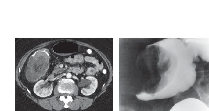
CASE 5.92
Findings
CASE 5.92. Contrast-enhanced CT. A soft tissue mass is present in the right colon with an enhancing lead point mass and some internal fat and enhancing vessels.
CASE 5.93. Single-contrast barium enema. An obstructing polypoid mass is acting as the lead point for the reducible intussusception.
Di erential Diagnosis
1.Colocolic intussusception
2.Ileocolic intussusception
Diagnosis
Colon carcinoma with colocolic intussusception
5. COLON 399
CASE 5.93
Discussion
At operation, a colon cancer was removed. Colon cancer is the most frequent cause of a colocolic intussusception in adults. The radiographic appearance at barium enema and CT usually
is diagnostic. As in the small bowel, a sausageshaped filling defect is seen within a shortened colon. A coiled-spring appearance often is visible at the proximal end of the mass at contrast enema. Sometimes it is possible to fully reduce the
intussusception and to identify the offending mass at contrast enema. In many patients, the cancer is located in the cecum. Visualization of mesenteric fat and occasionally vessels within the mass is pathognomonic (as in case 5.92) of an intussusception at CT.
Disease type: Masses and Filling Defects
400 MAYO CLINIC GASTROINTESTINAL IMAGING REVIEW
CASE 5.94
Findings
Endorectal ultrasonography. The normal bowel wall layers are visible except for a localized area of hypoechoic bowel wall thickening at the 9- to 10-o’clock position. The outermost echogenic layer (muscularis propria and perirectal fat interface) is
intact (arrows) overlying this mass. (Adapted from Jochem RJ, Reading CC, Dozois RR, Carpenter HA, Wolff BG, Charboneau JW: Endorectal ultrasonographic staging of rectal carcinoma. Mayo Clin Proc. 1990 Dec;65(12):1571–7. Used with
permission of Mayo Foundation for Medical Education and Research.)
Di erential Diagnosis
Localized rectal cancer (confined to bowel wall)
Diagnosis
Localized rectal cancer (confined to bowel wall)
Discussion
Th e intact outermost echogenic layer indicates that the tumor is confined to the bowel wall (Dukes stage B1 lesion). Endorectal sonography routinely can display the 5 normal layers of the bowel. The innermost echogenic layer represents the balloon-mucosal interface; the next hypoechoic layer, the muscularis mucosa; the middle echogenic layer, the submucosa; the fourth hypoechoic ring, the muscularis propria; and the outer echogenic ring, the muscularis propriaperirectal fat interface. The outer echogenic layer is the key ring for distinguishing tumors confined to the bowel wall from those extending into the perirectal fat (mesorectal extension). Endorectal sonography also
is sensitive for detecting perirectal lymph nodes, but it is nonspecific for distinguishing metastatic from inflammatory nodes.
Disease type: Masses and Filling Defects
5. COLON 401
CASE 5.95
A B
AS
R L
PI
Findings
MRI. A. T1-weighted axial image. Focal mass thickens the rectal wall and narrows the lumen. The margin between the muscularis propria and perirectal fat is indistinct, and there is extension of soft tissue into the mesorectum. B. T1-weighted coronal image. Distance between the tumor and the mesorectal fascia is less than 5 mm. The tumor extends for a distance of 4 cm. AS, anterior superior; L, left; PI, posterior inferior; R, right.
Di erential Diagnosis
1.Rectal cancer, resectable
2.Rectal cancer, nonresectable
3.Rectal cancer, resectable, needs preoperative radiation therapy
Diagnosis
Rectal cancer, resectable, needs preoperative radiation therapy
Discussion
MRI staging of rectal cancers can be very useful so that the appropriate treatment plan can be offered. Two key findings need to be reported for these patients: the
T stage and the distance of the tumor to the mesorectal fascia. In addition, the distance from the anus is also helpful for preoperative planning if a low rectal tumor is present.
Th e T staging criteria are as follows:
T1 Tumor invades the submucosa
T2 Tumor invades the muscularis propria
T3 Tumor penetrates the muscularis propria and extends into the perirectal tissues (mesorectum)
T4 Tumor directly invades other organs or structures
Th e key for differentiating T2 tumors from T3 tumors is the ability to visualize the interface between the muscular layer and perirectal fat. T2 tumors have a clean margin between the fat and the bowel wall. T3 tumors blur this interface with a nodular or rounded margin (as in this case).
Total mesorectal excision is the treatment of choice because it is associated with lower mortality from rectal cancer. If the mesorectal fascia is involved, preoperative radiation therapy is often administered before surgical excision. If the tumor is located within 5 mm of the mesorectal fascia (or closer), involvement is likely; therefore, reporting this distance is clinically important.
Disease type: Masses and Filling Defects
402 MAYO CLINIC GASTROINTESTINAL IMAGING REVIEW
CASE 5.96
A B
Findings
Endorectal ultrasonography. A. The anterolateral aspect of the rectal wall (from 11-o’clock to 3-o’clock position) is abnormally thickened, with loss of the normal echogenic bowel layers. In addition, the mass (arrows) can be seen to extend deeply into the perirectal fat through the muscularis propria-perirectal fat interface. B. Coned-in view shows 2 small hypoechoic lymph nodes (arrows) in the perirectal fat at the level of the invasive rectal mass.
Di erential Diagnosis
Invasive colon cancer with extension into mesorectal fat
Diagnosis
Invasive colon cancer with extension into mesorectal fat
Discussion
Colorectal cancers appear as hypoechoic or isoechoic masses that disrupt the bowel wall layers. The staging accuracy of endorectal ultrasonography is 85% to 90%. Evaluation of lymph nodes remains a challenge because reactive lymph nodes can be enlarged and discrimination of benign from malignant nodes
is impossible. Endoscopic ultrasonography can be helpful for detecting recurrent tumor adjacent to the anastomosis, a finding not visible at endoscopy.
Disease type: Masses and Filling Defects
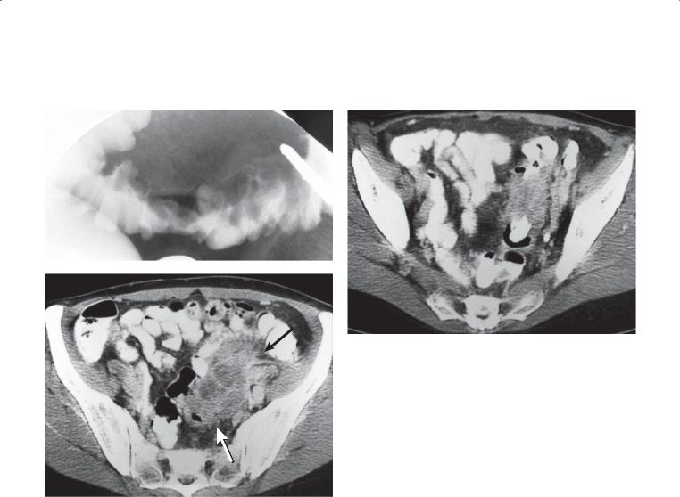
CASE 5.97
A
C
Findings
A. Single-contrast barium enema. The sigmoid lumen is narrowed, and there is a lobulated mass deforming the superior sigmoid contour. The lobulated contours are worrisome for neoplasm, but mucosal destruction or ulceration is not identified.
B and C. Contrast-enhanced CT. A mixed-density (fluid and soft tissue) mass (arrows) is present within the perisigmoidal tissues.
Di erential Diagnosis
1.Diverticulitis
2.Perforated colon cancer
Diagnosis
Perforated colon cancer
5. COLON 403
B
Discussion
Perforated colon cancers are unusual, constituting 2.5% to 8% of all colorectal cancers. The importance of this entity lies in the fact that patients may present with signs suggesting diverticulitis and receive suboptimal therapy. In addition, curative resection is less likely and operative mortality is higher. In cases of gross perforation with peritoneal contamination (found in a minority of patients), the 5-year survival rate is only 7%.
A contrast enema usually does not show the site of colonic perforation. Common findings on contrast enema include a long area of segmental narrowing and extrinsic mass effect or obstruction. Mucosal ulceration can be difficult to evaluate because of the associated inflammatory changes. Findings at CT
include a colorectal mass associated with an abscess the density of fluid or phlegmon. Metastatic disease often is identified. Endoscopy often is necessary to obtain a tissue diagnosis.
Disease type: Masses and Filling Defects
404 MAYO CLINIC GASTROINTESTINAL IMAGING REVIEW
CASE 5.98
Findings
Contrast-enhanced CT. A large soft tissue mass narrows and deforms the sigmoid colon. Contrast material is visible within the sigmoid lumen. Soft tissue stranding is present throughout the perisigmoidal fat. Extraluminal gas (arrows) is present in the soft tissues anterior to the sigmoid colon.
Di erential Diagnosis
1.Diverticulitis
2.Perforated colon cancer
Diagnosis
Perforated colon cancer
Discussion
Differentiating a perforated colon cancer from a complicated case of diverticulitis with pericolonic phlegmon or abscess is often difficult. The marked wall thickening and short segment of involved colon in this case favor perforated cancer over complicated diverticulitis. Often, however, it is necessary to recommend endoscopy after antibiotic therapy
for presumed cases of diverticulitis to exclude an underlying cancer.
Disease type: Masses and Filling Defects
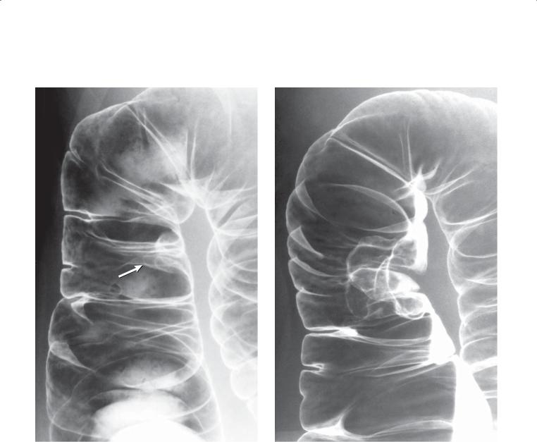
CASE 5.99
A
Findings
Double-contrast barium enema. A. A sessile polyp (arrow) is present along the medial wall of the ascending colon. The polyp was removed
endoscopically. B. Follow-up barium enema 3 years later shows a polypoid mass at the site of the previous polyp.
Di erential Diagnosis
1.Colon polyp with growth
2.Colon polyp with recurrence at index site
Diagnosis
Villous adenoma with recurrence as adenocarcinoma
5. COLON 405
B
Discussion
Among patients who have undergone curative resection for colon cancer, 30% to 50% have a recurrence within 2 years. Locally recurrent colorectal cancers can occur at sites of surgical anastomosis, within pericolic or pelvic fat near the surgical site, or at previous sites of endoscopic removal. Local recurrence developing at the anastomosis is best evaluated
with an intraluminal study (ie, barium enema, CT colonography, or endoscopy). Extraluminal and distant disease are best detected at CT. The usual radiographic appearance of an anastomotic recurrence is either a polypoid mass or irregular and eccentric narrowing of the lumen.
Disease type: Masses and Filling Defects
406 MAYO CLINIC GASTROINTESTINAL IMAGING REVIEW
CASE 5.100
A
B
Findings
Contrast-enhanced CT. A and B. An opaque staple line is seen at the colorectal anastomosis. Soft tissue (arrow) is present adjacent to the anastomosis, and there is abnormal soft tissue in the presacral space (arrowhead).
Di erential Diagnosis
Recurrent colorectal cancer
Diagnosis
Recurrent colorectal cancer
Discussion
Incomplete resection of rectal cancer can lead to recurrent tumor at or near the anastomosis and in the presacral space. In this case, recurrent tumor is present in both locations. Histologic confirmation can be obtained with a CT-directed biopsy.
Disease type: Masses and Filling Defects
CASE 5.101
Findings
Contrast-enhanced CT. The rectum has been removed. A large soft tissue mass is present in the old rectal bed and presacral region. The heterogeneous mass contains calcification.
Di erential Diagnosis
1.Recurrent rectal cancer
2.Metastases
Diagnosis
Recurrent colorectal cancer
5. COLON 407
Discussion
CT findings of recurrent tumor in the pelvis usually include an expanding presacral soft tissue mass with indistinct margins. Local invasion, lymphadenopathy, and distant metastases (liver, lung, retroperitoneum) can be seen. Recurrent neoplasm is most likely in a patient with a history of rectal cancer. Percutaneous biopsy with CT is helpful for confirming the diagnosis.
Disease type: Masses and Filling Defects
408 MAYO CLINIC GASTROINTESTINAL IMAGING REVIEW
CASE 5.102
A B
C
Findings
A. Positron emission tomography (PET) image. Sagittal image of the body demonstrates increased uptake in the presacral region and in the upper retroperitoneum.
B and C. Fused PET-CT images. High-intensity uptake is seen in the presacral region and left adrenal gland.
Di erential Diagnosis
Metastatic colorectal cancer
Diagnosis
Metastatic colorectal cancer
Discussion
PET imaging using fluorine-18–labeled deoxyglucose (FDG) has avid uptake for the detection of colorectal cancer metastases (and other highly metabolic organs). The ability to image the whole body is a strength of PET imaging. Because of the high cost of PET-CT, its use in the evaluation of patients with colorectal metastases is currently limited (as recommended by the Agency for Healthcare Research and Quality) to the following:
Patients with equivocal conventional imaging findings To determine the site of recurrence in patients with
an increased level of carcinoembryonic antigen when conventional work-up fails to unequivocally identify metastasis
In the preoperative assessment of liver metastasis before surgical resection
Disease type: Masses and Filling Defects
5. COLON 409
CASE 5.103
A B
Findings
A.Single-contrast barium enema. A long segment of circumferential narrowing is present in the right transverse colon. The mucosa through much of the involved region appears distorted but intact.
B.Unenhanced CT. The wall of the gastric antrum is thickened. A contiguous soft tissue mass (arrows)
extends from the stomach and engulfs (on lower slices) the colon. The right kidney is obstructed.
Di erential Diagnosis
1.Gastric cancer with extension to the colon
2.Colon cancer with extension to the stomach
3.Peritoneal carcinoma involving colon and stomach
Diagnosis
Gastric cancer with extension to the colon
Discussion
Metastatic disease to the colon can occur by several routes: intraperitoneal spread, direct extension, and hematogenous dissemination. Meyers described the patterns of malignant spread by each route.
Understanding these patterns allows the radiologist to
diagnose metastases and to predict the possible origin of the primary tumor.
Direct extension of malignancies is most common in patients with ovarian or uterine carcinoma, prostate cancer, and renal carcinoma. Pelvic malignancies often cause mass effect on the anterior wall of the rectum and on the inferior aspect of the sigmoid.
The affected bowel can be narrowed and nodular and contain tethered folds. Prostatic tumors often involve only the anterior aspect of the rectum, but they also can extend circumferentially around the rectum, causing annular narrowing resembling a primary rectal cancer. Renal adenocarcinoma usually does not incite a desmoplastic response, as occurs in most other metastases. These tumors (either primary or locally recurrent tumors) can invade the colon and appear as a large intraluminal, nonobstructing polypoid mass. Gastric (as in this case) and pancreatic malignancies often invade the transverse colon by extending into the gastrocolic ligament and transverse mesocolon. Classically, gastric malignancies cause narrowing, mass effect, and tethering of the superior aspect of
the transverse colon. Pancreatic malignancies usually involve the inferior aspect of the transverse colon.
Disease type: Masses and Filling Defects
410 MAYO CLINIC GASTROINTESTINAL IMAGING REVIEW
CASE 5.104
Findings
Single-contrast barium enema. The sigmoid colon is narrowed from mass effect, and mucosal tethering is present along its superior surface. This patient had ovarian cancer.
Di erential Diagnosis
Serosal metastases to the colon
Diagnosis
Serosal metastases to the colon
Discussion
Intraperitoneal metastases implant on peritoneal surfaces in regions where the ascitic flow is arrested. The 4 common sites of intraperitoneal seeding are 1) the pouch of Douglas, the most dependent and common location in the abdomen; 2) the ileocecal region, a region where fluid and cells collect after cascading down the small bowel mesentery into the right lower quadrant; 3) the superior aspect of the sigmoid colon; and 4) the right paracolic gutter, the main channel for fluid traveling from the pelvis into the upper abdomen.
Hematogenous spread to the colon is relatively infrequent. Breast cancer can invade the submucosal tissues and resemble linitis plastica in the affected segment. Narrowing and lack of distensibility are the commonest findings.
Disease type: Masses and Filling Defects
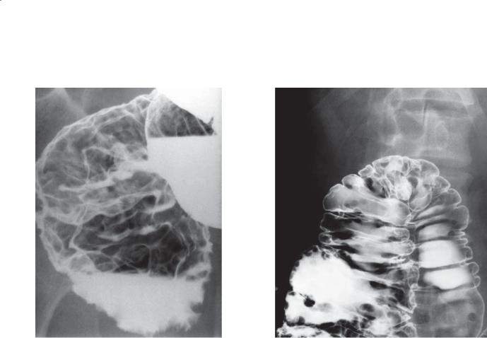
CASE 5.105
Findings
CASE 5.105. Double-contrast barium enema. Multiple smooth-surfaced filling defects are present in the rectum.
CASE 5.106. Double-contrast barium enema. Multiple smooth-surfaced filling defects are present throughout the colon.
Di erential Diagnosis
1.Lymphoma
2.Colitis cystica profunda
3.Polyposis syndrome
Diagnosis
Non-Hodgkin lymphoma
5. COLON 411
CASE 5.106
Discussion
Primary lymphoma of the colon usually occurs in patients older than 50 years and most often among men. Symptoms usually include abdominal pain, weight loss, weakness, anorexia, and fever.
Radiologically, early diffuse lymphoma presents with submucosal nodules that involve only a limited portion of the colon. The cecum and rectum are the sites most often involved. Later, innumerable diffuse filling defects with concomitant haustral thickening are seen.
Colitis cystica profunda is limited to the rectum. Polyposis syndromes have well-defined filling defects arising from the mucosa. Usually these conditions affect younger patients.
Disease type: Masses and Filling Defects
412 MAYO CLINIC GASTROINTESTINAL IMAGING REVIEW
Findings
CASE 5.107. Double-contrast barium enema. A broadbased sessile mass is present at the junction of the sigmoid and descending colon.
CASE 5.108. Single-contrast barium enema. The ileocecal valve is markedly enlarged, and the folds of the terminal ileum are thickened.
Di erential Diagnosis
1.Colon carcinoma
2.Lymphoma
Diagnosis
Lymphoma
Discussion
Localized lymphomas can have 3 radiographic presentations: a localized polypoid mass indistinguishable from a polypoid carcinoma (as in these cases), a region of narrowing resembling
carcinoma (usually without evidence of obstruction), and an aneurysmal segment of colon with luminal dilatation and mucosal ulceration. Intussusception can occur with bulky intraluminal masses.
Disease type: Masses and Filling Defects
CASE 5.109
A
Findings
Contrast-enhanced CT. A. A localized soft tissue mass (arrow) is present in the medial wall of the cecum with several small lymph nodes in the cecal mesentery. B. The rectal wall is diffusely thickened and several enlarged lymph nodes are visible in the perirectal fat.
Di erential Diagnosis
1.Lymphoma
2.Metastases
3.Multicentric colon cancer
Diagnosis
Multifocal lymphoma
5. COLON 413
B
Discussion
When lymphoma affects the bowel, it usually presents as focal or diffuse bowel wall thickening. Perforation is an important complication of this disease and develops because of the lack of the usual desmoplastic reaction that occurs with most other tumors. This complication has been reported in 9% to 47% of patients and is often clinically silent. Identification of gas or fluid within
a lymphomatous mass may indicate the presence of perforation. CT is excellent for determining the
amount of bowel wall thickening, extent of disease, and presence of associated adenopathy.
Disease type: Masses and Filling Defects
414 MAYO CLINIC GASTROINTESTINAL IMAGING REVIEW
CASE 5.110
Findings
Double-contrast barium enema. Multiple round submucosal filling defects are present within the rectum.
Di erential Diagnosis
1.Lymphoma
2.Colitis cystica profunda
3.Ulcerative colitis with multiple inflammatory polyps
4.Polyposis syndrome
Diagnosis
Colitis cystica profunda
Discussion
Colitis cystica profunda is a disease of unknown cause characterized by multiple mucin-filled cysts within the mucosa and submucosa of the rectum and, rarely, the sigmoid colon. Patients may complain of rectal
bleeding, diarrhea, abdominal pain, and prolapse of the rectum. Large cysts can cause obstruction.
Radiographically, multiple round submucosal cystlike filling defects are seen in the rectum. Occasionally the cysts can simulate a large mass and resemble a villous tumor (case 5.89).
Colitis cystica superficialis is a condition in which the mucosa is diffusely involved with mucus-filled bullae. It is believed to be of no clinical significance, except that it may be associated with pellagra and celiac disease.
Lymphoma could have a similar appearance but rarely is confined to the rectum. Similarly, evidence of more proximal disease would be likely in a patient with ulcerative colitis. A polyposis syndrome is never confined to the rectum, and the filling defects are sharply defined because of their mucosal origin.
Disease type: Masses and Filling Defects
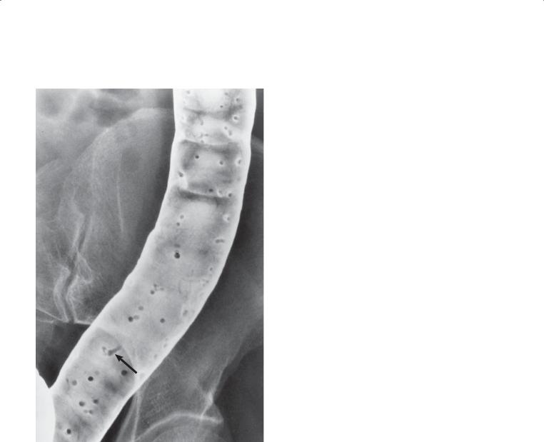
Findings
Double-contrast barium enema. Multiple polypoid filling defects are present in the colon. Some of the polyps are elongated (arrow). The background mucosal pattern is otherwise normal.
Di erential Diagnosis
Postinflammatory polyps
Diagnosis
Postinflammatory polyps
Discussion
Th is is the typical appearance of postinflammatory polyps in this patient with known ulcerative colitis. Postinflammatory polyps develop during the healing
phase in patients with either Crohn colitis or ulcerative colitis. In the process of epithelial regeneration, these polyps develop hyperplastic epithelium, granulomatous tissue, or epithelial hyperplasia. The polyps are often long and thin (wormlike) and may branch. They often are referred to as filiform polyps (from the Latin word filum, meaning “thread”). The background mucosa is nearly always normal in appearance without evidence of acute inflammation.
Polypoid changes are usually multiple and may involve the colon either locally or diffusely. Rarely, a masslike collection of postinflammatory polyps can simulate a villous adenoma or carcinoma. These giant postinflammatory polyps are usually found in the right colon and are associated with other areas of typical filiform polyps.
Disease type: Masses and Filling Defects
416 MAYO CLINIC GASTROINTESTINAL IMAGING REVIEW
A
B
Findings
CASE 5.112. Single-contrast barium enema. A round filling defect (arrows) is present in the sigmoid colon. Sigmoid diverticulosis is present. The filling defect has a bright white ring of barium around it.
CASE 5.113. CT colonography. A. Two-dimensional axial soft tissue window. Heterogeneous density material (arrow) fills a diverticulum and protrudes into the lumen. B. Three-dimensional endoluminal image.
A polypoid filling defect is present.
Di erential Diagnosis
1.Pseudopolyp
2.Polyp
Diagnosis
Pseudopolyp
Discussion
Diverticulosis is a common finding, especially among older persons. Stool may fill a diverticulum and remain lodged within it despite proper cathartics and cleansing enemas. When viewed en face, the stool-filled diverticulum can simulate a polyp. Identifying the rim of barium around the pseudopolyp is key to excluding a real polyp. If tangential views are possible, stool often can be seen within the diverticulum.
At CT, with 2-dimensional images, the filling defect can be seen to reside within a diverticulum and protrude into the lumen. The filling defect may be homogeneous or inhomogeneous in internal attenuation. Three-dimensional endoluminal images are usually not helpful for differentiating stool from polyps.
Disease type: Masses and Filling Defects
5. COLON 417
CASE 5.114
A B
Findings
Single-contrast barium enema. A. An approximately 2-cm filling defect (arrow) is present in the splenic flexure of the colon. B. A single diverticulum (arrow) is present at the site of the prior filling defect.
Di erential Diagnosis
Inverted colonic diverticulum
Diagnosis
Inverted colonic diverticulum
Discussion
An inverted diverticulum can simulate a polyp and may be indistinguishable from a polyp on a barium enema or CT colonography examination. Complete distention of the colon minimizes its occurrence.
Disease type: Masses and Filling Defects
418 MAYO CLINIC GASTROINTESTINAL IMAGING REVIEW
Findings
CASE 5.115. Double-contrast barium enema. Multiple tiny submucosal nodular filling defects are uniformly distributed in the colon.
CASE 5.116. Double-contrast barium enema. A central collection of barium (umbilication) is visible within several of the nodules.
Di erential Diagnosis
1.Normal lymphoid pattern
2.Polyposis syndrome
3.Crohn disease
4.Diffuse lymphoma
5.Retained stool
Diagnosis
Normal lymphoid follicular pattern
Discussion
Th e lymphoid follicular pattern is a common finding in normal children and in 10% to 15% of normal adults. The double-contrast technique usually is needed to identify the subtle textural changes of this entity. The key finding is the small size (<4 mm diameter) of the filling defects that are uniformly distributed in the colon. Usually the right side of the colon is affected more than the left. A polyposis syndrome (cases 5.71 through 5.76) usually has filling defects of larger and more variable size, and some are pedunculated. The lymphoid nodules and aphthous ulcers in Crohn disease are usually somewhat larger and often in a patchy segmental distribution (cases 5.17 and 5.18). Diffuse lymphoma has submucosal nodules of larger (>4 mm) and variable size (cases 5.105 and 5.106). Retained stool has angular margins, whereas lymphoid nodules are smooth and rounded.
Umbilication of lymphoid nodules (as in case 5.116) can be a normal finding. The main differential consideration is Crohn colitis with multiple aphthous ulcers (case 5.18). It may be impossible to exclude inflammatory bowel disease on the basis of the radiographic findings alone. Clinical correlation is always suggested, and follow-up studies to assess for disease progression or endoscopic biopsy also can be helpful.
Disease type: Masses and Filling Defects
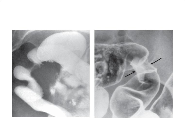
Findings
CASE 5.117. Single-contrast barium enema. There is an extrinsic mass on the anterior wall of the rectosigmoid. Tethered colonic folds are associated with the mass.
CASE 5.118. Double-contrast barium enema. A focal region of annular constriction (arrows) is present in the mid sigmoid colon.
Di erential Diagnosis
1.Serosal metastases
2.Colon carcinoma
3.Endometriosis
Diagnosis
Endometriosis
Discussion
In a woman in her reproductive years without a prior malignancy, endometriosis is an important diagnostic consideration with the radiographic findings shown here. Endometriosis was found at operation.
Endometriosis is the third most common cause of benign filling defects in the colon (after adenomas and lipomas). Intestinal implants are estimated to occur in 4% of all women between the ages of 20 and 40 years. Endometrial tissue extruded from the fallopian tubes can implant on the peritoneal surface, usually in the pouch of Douglas or on the sigmoid colon (most commonly on its inferior surface). The implants invade muscular and submucosal bowel layers and often stimulate local smooth muscle proliferation
of the colon, secondary inflammatory changes, and a variable degree of fibrosis. Symptoms include constipation, pelvic pain, diarrhea, and, rarely, rectal
bleeding. Typically, the symptoms are cyclical, but not always.
Radiographically, an intramural or extrinsic filling defect is present with an intact overlying mucosa. Sometimes the mucosa may appear pleated or tethered because of the associated desmoplastic reaction (as in case 5.117). Rarely, a constricting lesion is seen with smooth margins and an intact mucosa (case 5.118).
Disease type: Masses and Filling Defects
420 MAYO CLINIC GASTROINTESTINAL IMAGING REVIEW
CASE 5.119
Findings
Single-contrast barium enema. A small filling defect is present in the base of the cecum.
Di erential Diagnosis
1.Colon polyp
2.Lipoma
3.Mucocele
4.Endometrioma
Diagnosis
Endometriosis
Discussion
Th e radiographic appearance of an endometrioma depends on its anatomical location—serosal or submucosal. The mucosa is rarely involved, but in some cases (as in this example) the filling defect can resemble a mucosal mass. Filling defects can be as large as 2 to 7 cm in diameter.
Disease type: Masses and Filling Defects
5. COLON 421
CASE 5.120
A B
C
Findings
A. Single-contrast barium enema. The sigmoid is narrowed, and there is mass effect on its inferior border. Tethered folds are visible about the mass.
B and C. Contrast-enhanced CT. A soft tissue mass is present on the inferior aspect of the sigmoid colon. Minimal soft tissue stranding is present in the pericolonic tissues. No other pelvic mass is present.
Di erential Diagnosis
1.Endometriosis
2.Ovarian tumor
3.Pelvic inflammatory disease
4.Diverticulitis
Diagnosis
Endometriosis
Discussion
Th e soft tissue mass due to an endometrioma can be directly visualized at CT in some cases. Occasionally, it is difficult to detect. Lesions developing on the inferior aspect of the sigmoid are most likely to arise from organs in the pelvis. In females during their reproductive years, endometriosis should be a primary consideration. CT in this case was very helpful for excluding a primary pelvic tumor or changes of diverticulitis.
Disease type: Masses and Filling Defects
422 MAYO CLINIC GASTROINTESTINAL IMAGING REVIEW
CASE 5.121
A B
C
Findings
A. Single-contrast barium enema. A smooth-surfaced filling defect is present on the base of the cecum. The appendix is not seen. A compression device containing a 2-cm marker is visible.
B and C. Unenhanced CT. The appendix (arrows) is distended and fluid-filled. Linear calcification is present within part of the wall of the appendix. Findings are typical of a mucocele.
Di erential Diagnosis
Mucocele
Diagnosis
Mucocele
Discussion
A mucocele of the appendix is regarded by some authors as a mucus-distended appendix due to luminal obstruction by a fecalith, foreign body, adhesions, or volvulus. Other authors include tumorous conditions of the appendix. These conditions include focal or diffuse mucosal hyperplasia, mucinous cystadenoma, and mucinous cystadenocarcinoma. Carcinoid tumors of the appendix are not classified with the epithelialderived adenoma and adenocarcinoma tumors. They rarely are associated with appendiceal mucoceles.
At contrast enema, a mucocele usually presents with findings of a smooth filling defect on the base of the cecum. CT has improved the ability to diagnose a mucocele. Findings at CT include a thin-walled, cystic, and tubular structure with internal contents of low density. Wall calcification is common. CT also is helpful in assessing for metastases in patients with an appendiceal cystadenocarcinoma.
Disease type: Masses and Filling Defects
CASE 5.122
Findings
Single-contrast barium enema. A large smoothsurfaced filling defect is present on the base of the cecum. A compression device containing a 2-cm marker is visible.
Di erential Diagnosis
1.Mucocele
2.Lipoma
3.Appendiceal abscess
4.Lymphoma
5.Endometrioma
Diagnosis
Mucocele: appendiceal adenocarcinoma
5. COLON 423
Discussion
Patients with appendiceal tumors often (50%) present clinically with symptoms of acute appendicitis. Approximately 15% of these tumors are discovered incidentally on histologic examination of the appendix. Metastatic mucinous cystadenocarcinoma often results in diffuse peritoneal carcinomatosis that produces large quantities of mucinous material that can calcify. This condition is often referred to as pseudomyxoma peritonei.
Lipomas are soft and deformable at fluoroscopy. Younger patients are most likely to have appendicitis or endometriosis—and the clinical presentation also is different. Lymphoma is most often found in older patients.
Disease type: Masses and Filling Defects
424 MAYO CLINIC GASTROINTESTINAL IMAGING REVIEW
CASE 5.123
Findings
Contrast-enhanced CT. A large tubular, water- attenuation–filled stricture is present in the pelvis. The stricture arises from the base of the cecum.
Di erential Diagnosis
1.Appendicitis
2.Mucocele
3.Meckel diverticulum
4.Duplication cyst
Diagnosis
Mucocele
Discussion
Mucoceles can occasionally be large. The appendiceal shape and attachment to the cecum are key findings to suggest a mucocele. Appendicitis is unlikely without inflammatory changes in the periappendiceal fat and without appendiceal wall thickening.
Meckel diverticulum arises from the small bowel, not the cecum. A duplication cyst usually has no communication or attachment to the bowel.
Disease type: Masses and Filling Defects
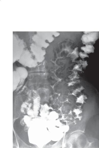
Findings
Single-contrast barium enema. Multiple intramural cystic gas collections are present throughout most of the left colon.
Di erential Diagnosis
Pneumatosis cystoides coli
Diagnosis
Pneumatosis cystoides coli
Discussion
Pneumatosis coli refers to intramural air within the colon. Intramural air within the small bowel is referred to as pneumatosis intestinalis or pneumatosis cystoides intestinalis (if the air collections are cystlike). Patients with pneumatosis cystoides coli have endotheliallined gas-filled cysts within the bowel wall which
do not communicate with the lumen. The cysts may vary in size, reaching up to several centimeters in diameter. The cause is unknown, but some patients have associated collagen vascular disorders, ischemia, diabetes, or a history of trauma. This condition often is discovered incidentally in patients without symptoms.
Th e radiologic diagnosis is dependent on recognizing the radiolucent collections of gas within the bowel wall. Often the collections are rounded and grapelike. The left side of the colon is affected more often than the right (excluding the rectum).
Once the gas density of the collections is recognized, no further diagnostic considerations are necessary. The intraluminal protrusions may simulate polyps, lymphoma, segmental colitis (Crohn colitis), and ischemic colitis. Colitis cystica profunda usually affects the rectum, and the filling defects are not the lucency of gas (case 5.110).
Disease type: Masses and Filling Defects
426 MAYO CLINIC GASTROINTESTINAL IMAGING REVIEW
Findings
CASE 5.125. Abdominal radiograph. A linear band of intramural gas (arrows) is present within the right colon. This patient had recently undergone colonoscopy with biopsy.
CASE 5.126. Contrast-enhanced CT. Linear and cystic intramural pneumatosis is present in the right colon (arrows).
Di erential Diagnosis
Pneumatosis coli
Diagnosis
Pneumatosis coli
Discussion
Pneumatosis coli can be due to the same underlying conditions as are found in pneumatosis intestinalis (cases 4.78 through 4.81). The intramural gas may appear cystic (case 4.79), but often it is linear (as in these cases). Patients receiving corticosteroid therapy, those with an underlying collagen vascular disease, those who have had a recent mucosal biopsy, or those with a history of chronic obstructive pulmonary disease are most likely to have this condition. These benign causes usually do not require any particular treatment. Ischemic colitis is the most important condition to exclude clinically, because emergency operation would be required to prevent perforation.
Intraluminal gas is always in a nondependent location, whereas pneumatosis can be present in any intramural location, including the dependent portions of the bowel.
Disease type: Masses and Filling Defects
CASE 5.127
A
Findings
A.Abdominal plain film (supine). Linear collection of gas is along the lateral margin of the ascending colon.
B.Unenhanced CT. Linear collection of gas is within the bowel wall (seen best along the dependent bowel).
Di erential Diagnosis
1.Pneumatosis coli—ischemia
2.Pneumatosis coli—benign
Diagnosis
Pneumatosis coli—benign
5. COLON 427
B
Discussion
Differentiation of benign pneumatosis from pneumatosis due to ischemia is often not possible radiographically unless there are additional findings to suggest ischemia (eg, superior mesenteric artery or vein thrombus, marked bowel wall thickening). Radiologists must call the referring physician and determine whether the patient has an acute abdomen (ischemia) or a benign abdominal examination. Ischemia demands urgent operative intervention.
Disease type: Masses and Filling Defects
428 MAYO CLINIC GASTROINTESTINAL IMAGING REVIEW
TABLE 5.7
Colonic Masses and Filling |
|
CASE |
Defects |
|
|
|
|
|
Benign tumors |
|
|
Polyp |
Pedunculated, sessile, or flat, multiple polyps in polyposis |
5.60–5.76 |
|
syndromes |
|
Lipoma |
Commest submucosal tumor, pliable (changes shape with |
5.77–5.79 |
|
compression or luminal changes) |
|
|
|
|
Malignant tumors |
|
|
Adenocarcinoma |
Annular, polypoid, or villous, 5% synchronous tumors |
5.81–5.87, |
|
|
5.89–5.101 |
Metastases |
Usually multiple masses, history of malignancy, direct tumor |
5.102–5.104 |
|
extension, serosal implants, or hematogenous or lymphatic spread |
|
Lymphoma |
Solitary, multiple or diffuse, look for adenopathy |
5.105–5.109 |
|
|
|
Other benign conditions |
|
|
Colitis cystica profunda |
Multiple smooth, round filling defects in rectum only |
5.110 |
Postinflammatory polyps |
Wormlike filiform polyps, in either ulcerative colitis or Crohn |
5.111 |
|
disease. Background mucosa normal |
|
Pseudopolyps
Normal lymphoid follicular pattern
Endometriosis
Usually caused by stool or inverted diverticulum, angular margins and inhomogeneous internal attenuation
Small (<4 mm) filling defects uniformly distributed in colon, common in children and occurs in 10%-15% of adults
Intramural or extrinsic filling defect(s) in young women, common on inferior surface of sigmoid colon
5.112–5.114
5.115 and
5.116
5.117–5.120
Mucocele |
Smooth filling defect at base of cecum, appendix obstructed by |
5.121–5.123 |
|
appendicolith or tumor |
|
Pneumatosis coli |
Linear or cystlike intramural gas collections, often benign cause, |
5.124–5.127 |
|
but ischemia should be ruled out |
|
|
|
|
Disease type: Masses and Filling Defects
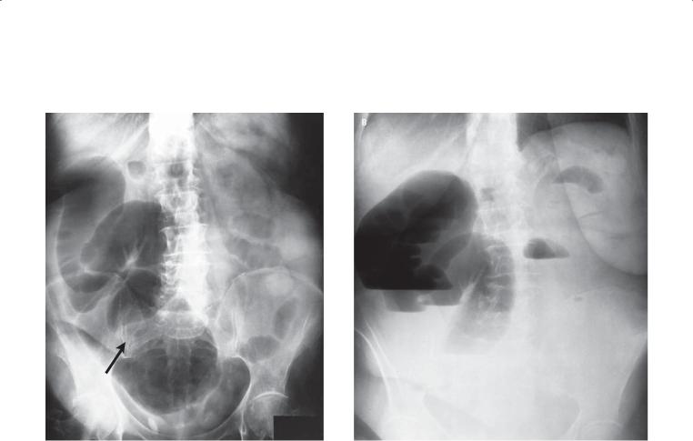
CASE 5.128
A
Findings
A.Supine abdominal radiograph. The cecum, ascending, and proximal transverse colon are distended with gas. No gas is seen distal to the mid transverse colon (arrow). Mildly dilated small bowel loops are present.
B.Upright radiograph. Multiple air-fluid levels are seen in the right side of the colon and in two small bowel loops.
Di erential Diagnosis
Mechanical colonic obstruction
Diagnosis
Mechanical colonic obstruction
Discussion
Th ese findings are typical of a mechanical colonic obstruction involving the transverse colon. At operation, an incarcerated loop of transverse colon was found to be obstructed within a midline incisional hernia.
Mechanical colonic obstruction is most often due to carcinoma. Diverticulitis, volvulus, mass effect from an extracolonic pelvic neoplasm, fecal impaction, and, rarely, a hernia or adhesion also can cause large bowel obstruction. The radiographic findings include dilatation of the colon to the level of obstruction. The colon may be filled with gas (as in this case), fluid, or stool. The small bowel is also often dilated if the ileocecal valve is incompetent. If
the valve remains competent, the colon will continue to distend. Perforation can occur with prolonged and marked cecal dilatation. A contrast enema or CT is often necessary to determine the cause of the obstruction. Only a small amount of barium should be passed beyond a high-grade obstruction so that inspissated barium does not cause a problem later (including obstipation and possible obstruction). Chronic obstruction can be recognized on abdominal radiographs by the presence of haustral thickening and the presence of gray and granularappearing fluid within the colon proximal to the obstruction.
Disease type: Obstruction and Ileus
430 MAYO CLINIC GASTROINTESTINAL IMAGING REVIEW
CASE 5.129
Findings
Contrast-enhanced CT. The colon is dilated to the level of the mid transverse colon. A soft tissue mass (arrow) encases the colon and causes partial colonic obstruction (normal-caliber descending colon).
Di erential Diagnosis
1.Mechanical colonic obstruction
2.Primary colon cancer
3.Metastases
Diagnosis
Mechanical colonic obstruction due to metastatic gastric cancer
Discussion
CT is helpful for identifying dilated bowel, the level of obstruction, and in many cases the cause of
obstruction. This patient with gastric cancer had tumor spread along the greater omentum to the transverse colon—a common route for gastric cancer to spread locally.
Disease type: Obstruction and Ileus
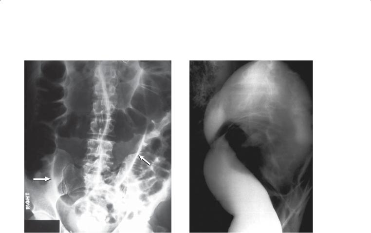
CASE 5.130
Findings
CASE 5.130. Abdominal radiograph. The entire colon is moderately distended with gas, and there is a dilated, inverted U-shaped loop (arrows) arising from the pelvis.
CASE 5.131. Water-soluble enema. The sigmoid colon gradually tapers and narrows to a region of high-grade obstruction. The colon proximal to the obstruction is dilated. Beaking (twisting) of the colon is visible at the level of the obstruction.
Di erential Diagnosis
Sigmoid volvulus
Diagnosis
Sigmoid volvulus
5. COLON 431
CASE 5.131
Discussion
Sigmoid volvulus occurs as a result of a redundant sigmoid colon on a loose mesentery that twists on itself and obstructs. Patients are often older and have a history of chronic constipation. Acute abdominal pain, distention, and vomiting often occur.
Radiographically, the diagnosis usually is made by identifying a prominent bean-shaped, dilated loop of colon arising from the pelvis on abdominal plain
radiographs. Gas often is present in the colon proximal to the sigmoid twist. If the diagnosis is equivocal, the obstruction can be confirmed with a contrast enema.
It is often helpful and possible to pass a rectal tube across the obstruction into the dilated sigmoid loop. This will decompress the volvulus and provide the opportunity for elective operative repair of the sigmoid (reduction sigmoid colocolostomy). If tube
decompression is not possible, then urgent operation is required to prevent sigmoid infarction and perforation. Recurrence of the volvulus is common without operative repair.
Disease type: Obstruction and Ileus
432 MAYO CLINIC GASTROINTESTINAL IMAGING REVIEW
CASE 5.132
A B
Finding
A.Scout radiograph at CT. A large loop of gas distended colon is present in left and mid abdomen. The descending colon is moderately distended.
B.Contrast-enhanced CT. The dilated loop is sigmoid colon, and 2 limbs of the sigmoid (arrows) are seen to abut each other, narrow, and twist (seen on images on lower part of body, not shown).
Di erential Diagnosis
1.Sigmoid volvulus
2.Dolichosigmoid
3.Obstructing colon cancer
Diagnosis
Sigmoid volvulus
Discussion
Sigmoid volvulus is a type of closed-loop obstruction in which a redundant loop of sigmoid colon twists on itself, obstructing the closed loop and the proximal colon. Twisting can be severe enough to compromise blood flow and lead to ischemia and perforation. Treatment is temporizing rectal tube decompression (if possible) and subsequent sigmoid resection at operation. Dolichosigmoid is a term sometimes used to describe a redundant sigmoid loop. This anatomy predisposes a patient to volvulus. An obstructing colon cancer is unlikely in this case because a colonic mass is not identified, and usually the entire colon proximal to the obstructing cancer is dilated. On CT, the cecum is seen to be normally positioned.
Disease type: Obstruction and Ileus
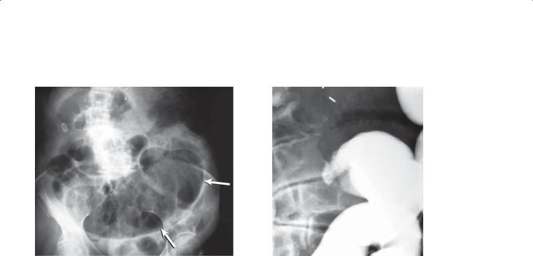
CASE 5.133
A
Findings
A.Supine abdominal radiograph. A dilated, gas-filled viscus (arrows) fills a large portion of the pelvis and extends slightly into the left upper abdomen. This organ has the shape of an abnormally positioned right colon. Dilated small bowel loops also are present throughout the abdomen. The transverse and left colon are not identified.
B.Single-contrast barium enema (spot radiograph, proximal right colon). There is gradual narrowing of the colon to the point of obstruction in the right colon.
Di erential Diagnosis
Cecal volvulus
Diagnosis
Cecal volvulus
5. COLON 433
B
Discussion
Cecal volvulus occurs as a result of a redundant right colonic mesentery, allowing all or part of the right colon to twist on itself. As the colon twists, it moves into an abnormal location within the abdomen, often in the midline or within the left upper quadrant. The twist causes mechanical obstruction of the colon and often vascular insufficiency of the affected loop.
Patients may have a history of intermittent abdominal pain, perhaps due to periodic twisting and untwisting of the colon. Treatment invariably is operative because the risk of cecal perforation is high, and if perforation does occur it is associated with a high morbidity and high mortality.
Th e initial radiologic examination is usually abdominal radiography. Typically, a dilated (>9 cm) cecum is identified in an abnormal location, often the left upper quadrant. Depending on the duration of symptoms, the colon distal to the obstructing twist has little or no remaining gas within it. Dilated, gasfilled small bowel often is identified. The abdominal radiograph should be scrutinized for evidence of perforation (free air and a rapidly decompressing cecum) and of ischemia (portal venous gas and cecal intramural pneumatosis) (cases 4.75 through 4.83 and 5.125 and 5.126). The diagnosis can be rapidly and easily confirmed with a contrast enema. The finding of obstruction in the right colon with a beaklike appearance at the site of the twist is typical.
Disease type: Obstruction and Ileus
434 MAYO CLINIC GASTROINTESTINAL IMAGING REVIEW
CASE 5.134
Findings
Contrast-enhanced CT. A dilated gasand contrastfilled viscus (cecum) fills the upper abdomen. A twisted or whorled appearance of the mesentery is present in the right upper abdomen (arrow). Intestines encircle the mesenteric vessels.
Di erential Diagnosis
1.Midgut volvulus
2.Cecal volvulus
Diagnosis
Cecal volvulus
Discussion
Usually the diagnosis of volvulus is suggested on the basis of abdominal radiographic findings (case 5.133)
and confirmed with an intraluminal examination with contrast material. A volvulus may be found in the course of a CT examination in patients with
indeterminate, intermittent abdominal pain. Midgut volvulus can be suspected when a rotational anomaly of the gut is identified (jejunum in the right upper quadrant) and the superior mesenteric artery is located to the right of the superior mesenteric vein (reversal of the normal anatomical relationships). Cecal volvulus can be more difficult to identify. The cecum is located in an abnormal position in the mid or left abdomen. In the region of the right colon, both bowel and mesentery are twisted and encircle each other (as in this case). Equivocal cases may require a contrast enema to confirm the diagnosis.
Disease type: Obstruction and Ileus
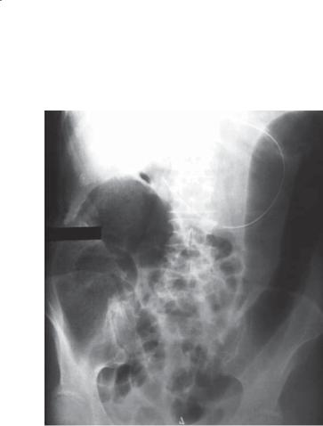
Findings
Abdominal radiograph. Gas is present in mildly dilated loops of small and large bowel. A continuous column of gas fills the colon. The cecum is located normally in the right lower quadrant.
Di erential Diagnosis
1.Adynamic ileus
2.Distal mechanical colonic obstruction
Diagnosis
Adynamic ileus
Discussion
Adynamic ileus of the colon is a condition in which the colon becomes atonic and often dilated. There are numerous causes of this condition, including postoperative and postanesthesia states, trauma, adjacent inflammation or infection, various medications, various systemic illnesses, and other
conditions. Clinically, patients may have poorly localized abdominal distress and pain, a distended abdomen, and diminished bowel sounds.
Radiographically, the colon usually is distended with air and fluid. Gas fills the colon as a continuous column; however, often the low descending and sigmoid colon are not air-filled as a result of their dependent location in the peritoneal cavity. Radiographs with the patient decubitus or prone can be helpful for moving gas into portions of the colon in question. In adynamic ileus, gas will fill the entire colon lumen because there is no obstruction. If a contrast-enema study is performed, no obstruction will be identified.
Th e main differential consideration is mechanical colonic obstruction (cases 5.128 through 5.134). If a continuous column of gas throughout the colon is not present, then a limited contrast enema (contrast material is administered up to the level where a
continuous air column is present) can be performed to assess for a distal colonic obstruction.
Disease type: Obstruction and Ileus
436 MAYO CLINIC GASTROINTESTINAL IMAGING REVIEW
CASE 5.136
A B
Findings
Supine (A) and upright (B) abdominal radiographs. The cecum (arrow) is mobile, dilated, and positioned in the right and middle abdomen. Air-fluid levels are present in the cecum and descending colon. Two gasfilled small bowel loops are visible inferior and lateral to the cecum. There is a continuous column of gas throughout the colon.
Di erential Diagnosis
1.Cecal ileus
2.Cecal volvulus
Diagnosis
Cecal ileus
Discussion
Cecal ileus can develop in patients with a mobile cecum (10% of the general population) and an underlying colonic adynamic ileus. The mobile cecum rotates anteromedially in the abdomen and gradually distends (to a diameter in excess of 9 cm). Because of the nondependent cecal location when patients are supine for prolonged periods, gas does not empty into the less dilated distal colon. Recognition of this entity is important because excessive cecal distention for longer than 2 or 3 days has a significant risk of cecal
perforation and possible death. Cecal perforation occurs more frequently in patients with cecal ileus than in those with cecal volvulus, mechanical colonic obstruction, or typical colonic ileus, probably because patients with obstruction of the colon (cecal volvulus, mechanical colonic obstruction) usually present with symptoms that demand an urgent diagnosis and treatment. Patients with cecal ileus often have nonspecific abdominal complaints, and cecal
distention may go unnoticed for days. The duration of cecal distention (longer than 2–3 days) is an important factor in determining the risk of perforation. Actual cecal size (≥9 cm) does not correlate well with risk of perforation. If conservative therapy (rectal tubes and cathartics) is not successful, more aggressive therapy with either a surgically placed cecostomy tube or colonoscopic decompression should be instituted. Colonoscopic decompression may have to be repeated several times until the ileus resolves.
Radiographically, there is a continuous column of gas throughout most of the colon (colonic ileus) with disproportionate dilatation of the cecum, which is abnormally positioned, often in the mid abdomen.
There is no obstruction at the time of a contrast enema. Cecal volvulus usually presents radiographically
with absence of gas in the colon distal to the twist and obstruction in the right colon at contrast enema.
Disease type: Obstruction and Ileus
|
|
5. COLON 437 |
|
|
|
|
TABLE 5.8 |
|
Colonic Obstruction and Ileus |
|
CASE |
|
|
|
Mechanical colonic obstruction |
Most commonly caused by carcinoma, can be caused by |
5.128 and 5.129 |
|
diverticulitis, volvulus, extrinsic mass, fecal impaction, |
|
|
hernia, or adhesions |
|
|
|
|
Sigmoid volvulus |
Inverted U-shaped loop extends into upper abdomen, |
5.130-5.132 |
|
beaking at level of obstruction |
|
|
|
|
Cecal volvulus |
Dilated cecum extending into left upper quadrant, colon |
5.133 and 5.134 |
|
distal to obstruction decompressed. Contrast enema with |
|
|
beaking at site of obstruction |
|
|
|
|
Adynamic ileus |
Distended air and fluid-filled colon with a continuous |
5.135 and 5.136 |
|
column to the rectum, often occurs postoperatively or |
|
|
with narcotic medications |
|
|
|
|
Disease type: Obstruction and Ileus
438 MAYO CLINIC GASTROINTESTINAL IMAGING REVIEW
CASE 5.137
A B
Findings
Dynamic MRI proctogram. A. Single-shot fast-spin echo sagittal image of the pelvis shows abnormal pelvic floor descent at rest. B. Image during straining shows prolapse of the rectal mucosa out of the anal canal (arrow) and abnormal descent of the uterus and bladder (arrowheads).
Di erential Diagnosis
Rectal prolapse (uterine prolapse and cystocele)
Diagnosis
Rectal prolapse (uterine prolapse and cystocele)
Discussion
Rectal prolapse is protrusion of the rectal mucosa through the anus. Patients present with constipation, incontinence, and rectal ulcerations. Rectal prolapse is six times more common in females. The exact cause is unknown, but it often is associated with long-standing constipation. If the protruding rectal mass is visible with straining on physical examination, proctography is not necessary for diagnosis. As the disease progresses, the rectum may no longer spontaneously retract after defecation, and patients may have to replace it manually. Among patients with rectal prolapse, 10% to 20% have uterine or bladder prolapse, and 35% have an associated cystocele. Rectopexy often is required for treatment. An incarcerated rectal
prolapse is rare, but it may require emergency resection if the viability of the bowel is in question.
Disease type: Disorders of Defecation
5. COLON 439
CASE 5.138
A B
C
Findings
Dynamic MRI proctogram. A. Single-shot fast-spin echo sagittal image of the pelvis at rest shows the uterus is surgically absent. The bladder and rectum appear in normal position. B. Image obtained during attempted defecation shows abnormal pelvic floor descent with a large anterior bulge of the rectum. The bladder also bulges posteriorly toward the vagina. None of the rectal contrast material could be expelled despite multiple attempts. C. After defecation, with Valsalva maneuver. The rectum and bladder are empty. Abnormal pelvic floor descent. A peritoneocele (arrow) exists with pelvic fat filling the rectovesical space.
Di erential Diagnosis
Rectocele (and cystocele)
Diagnosis
Rectocele, cystocele, peritoneocele
Discussion
A rectocele is a bulge of the anterior wall of the rectum. Rectoceles may present without any other abnormalities or as part of a generalized weakness of the pelvic floor coexisting with cystoceles, enteroceles, uterine prolapse, or rectal prolapse. Women with multiple deliveries, difficult deliveries, perineal tears, episiotomies, or hysterectomies are at increased risk for development of a rectocele. Patients often present with constipation, difficulty evacuating stool with straining, and the sensation of a mass in the vagina during a bowel movement. Some women find that pressing against the back wall of the vagina helps to empty
the rectum. Rectoceles are diagnosed with barium defecating proctography or, more recently, with MRI proctography. Conservative treatment for rectoceles includes stool softeners or a pessary. In more extreme cases, surgical repair may be necessary.
Disease type: Disorders of Defecation
CASE 5.140.
CASE 5.139.
440 MAYO CLINIC GASTROINTESTINAL IMAGING REVIEW
CASE 5.139 |
CASE 5.140 |
A |
A |
B B
Findings
Defecating proctogram, lateral view. A. Squeeze image. Normal anorectal angle and
puborectalis muscle impression. The vagina contains contrast material anteriorly. B. During evacuation and straining. A large enterocele containing multiple contrast-filled small bowel loops has entered the space between the rectum (posteriorly) and the vagina (anteriorly). The pelvic floor has descended to an abnormally low position.
MRI proctogram. A. During defecation. Abnormal descent of the pelvic floor. B. After defecation with Valsalva maneuver. The pelvic floor has descended further, and an enterocele has developed within the rectovesical space.
Di erential Diagnosis
Enterocele
Diagnosis
Enterocele
Discussion
An enterocele refers to herniation of a peritoneal sac along the ventral rectal wall into the cul-de-sac. If the small bowel loops have not been opacified, separation of the opacified vagina and upper rectum during straining suggests this abnormality. An enterocele can contain sigmoid colon, omentum and mesenteric fat, and small intestine. This can be associated with vaginal and rectal prolapse and other pelvic floor abnormalities.
Disease type: Disorders of Defecation
CASE 5.141
Findings
Defecating proctogram. A small amount of rectal intussusception is present during defecation.
Di erential Diagnosis
Rectal intussusception
Diagnosis
Rectal intussusception
5. COLON 441
Discussion
Rectal intussusception is concentric invagination of the rectal wall that starts in the upper or mid rectum and progresses toward the anal canal. If the intussusception reaches through the anal sphincter, it results in rectal prolapse (case 5.137). Patients often present with constipation and incomplete defecation. Surgical treatment with perineal rectopexy may be necessary in some cases.
Disease type: Disorders of Defecation
442 MAYO CLINIC GASTROINTESTINAL IMAGING REVIEW
CASE 5.142
Findings
Defecating proctogram. No change in the anorectal angle or perineal descent between the images taken at rest and at evacuation. The puborectalis muscle impression is prominent in both images.
Di erential Diagnosis
1.Spastic pelvic floor syndrome (anismus, pelvic floor dyssynergia)
2.“Stage fright”
Diagnosis
Spastic pelvic floor syndrome (anismus, pelvic floor dyssynergia)
Discussion
Spastic pelvic floor syndrome involves the inability to relax the puborectalis muscle, which explains the lack of change in the two images shown here. Retraining of pelvic floor muscles is required to overcome this problem. Coaching and giving the patient enough time (and privacy) may be required to ensure that embarrassment has not led to “stage fright,” in which the patient is unable to relax these muscles at the time of examination.
Disease type: Disorders of Defecation
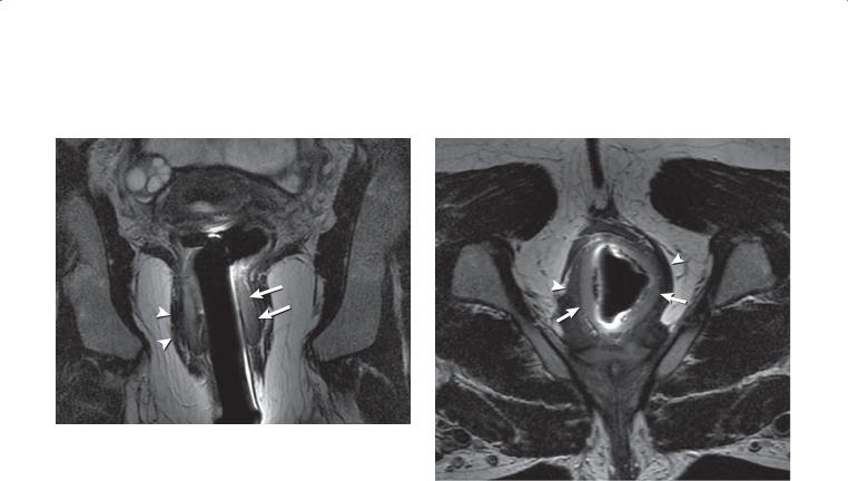
CASE 5.143
A
Findings
MRI of the pelvis using an endorectal coil placed across the anal sphincters in the coronal (A) and axial (B) planes. There is marked thickening of the internal anal sphincter (arrows).
Di erential Diagnosis
Chronic rectal prolapse
Diagnosis
Chronic rectal prolapse
5. COLON 443
B
Discussion
Th is patient had known rectal prolapse at defecation. The internal anal sphincter (IAS) can usually be recognized by its high-signal intensity, and it usually enhances briskly if intravenous contrast material
is administered. An abnormally thin IAS usually indicates primary degeneration or scleroderma. A thick IAS often is associated with chronic injury (prolapse, solitary rectal ulcer syndrome). The IAS is the main mechanism for maintaining fecal continence. The external anal sphincter (arrowheads) is of
lower signal intensity, has interspersed fat, and is in continuity with the puborectalis muscle. MRI can be useful to assess for sphincter atrophy and tears that can contribute to fecal incontinence. This information in combination with a dynamic study of the pelvic floor during defecation can provide a complete examination of patients in whom pelvic floor dysfunction is suspected.
Disease type: Disorders of Defecation
444 MAYO CLINIC GASTROINTESTINAL IMAGING REVIEW
CASE 5.144
Findings
Double-contrast barium enema. A circumferential ulceration (arrows) is present in the mid rectum, with a focal ulcer crater (arrowhead) anteriorly.
Di erential Diagnosis
1.Rectal carcinoma
2.Metastases
3.Solitary rectal ulcer syndrome
Diagnosis
Solitary rectal ulcer syndrome
Discussion
Th e solitary rectal ulcer syndrome is found in patients with chronic defecation difficulties and abnormalities of the rectal wall. Patients also may complain of rectal bleeding. Defecation abnormalities include either repeated episodes of rectal intussusception or failure of the puborectalis muscle to relax normally (spastic pelvic floor syndrome). Pathologically, one or more rectal ulcers may be present, but they are not necessary for the diagnosis. In addition, thickening of the muscularis mucosa and fibroblastic proliferation within the lamina propria are specific features.
Findings at barium enema include thickened rectal folds, mucosal granularity, rectal stricture, and ulceration. Intussusception, rectal prolapse, failure of the puborectalis muscle to relax, and abnormal perineal descent may be visible at defecating proctography.
Differential considerations include annular neoplasms (case 5.84) and metastases to the colon. The diagnosis of solitary rectal ulcer syndrome should be considered in patients with a long history of straining at stool. Defecating proctography or MR proctography is the method of choice for making the diagnosis.
Disease type: Disorders of Defecation
|
|
5. COLON 445 |
|
|
|
|
TABLE 5.9 |
|
Disorders of Defecation |
|
CASE |
|
|
|
Rectal prolapse |
Mucosa extends beyond anal opening at evacuation |
5.137 |
|
|
|
Rectocele |
Anterior bulge of rectal wall |
5.138 |
|
|
|
Enterocele |
Abnormal descent of small bowel loops into rectal fossa |
5.139 and 5.140 |
|
|
|
Rectal intussusception |
Can be normal in 30% of patients |
5.141 |
|
|
|
Spastic pelvic floor syndrome |
Lack of change of puborectalis muscle and pelvic floor |
5.142 and 5.143 |
(anismus, pelvic floor dyssynergia) |
between rest and evacuation |
|
|
|
|
Solitary rectal ulcer syndrome |
Traumatic ulceration from recurrent prolapse or |
5.144 |
|
intussusception |
|
|
|
|
Disease type: Disorders of Defecation
446 MAYO CLINIC GASTROINTESTINAL IMAGING REVIEW
CASE 5.145
Findings
Single-contrast barium enema. The right and transverse colon are relatively ahaustral; the left colon appears normal.
Di erential Diagnosis
1.Cathartic colon
2.Ulcerative colitis
3.Ischemic colitis
Diagnosis
Cathartic colon
Discussion
Cathartic colon is a term used to describe the radiographic changes that occur after prolonged use of irritant cathartics such as cascara, senna, phenolphthalein, and castor oil. The proximal colon
usually is affected initially, but left-sided involvement also can occur.
Findings include lack of haustrations (most common in the right colon), shortening, and inconsistent luminal constrictions. It is believed that the strictures are due to muscular hypertrophy. Observation of these areas at fluoroscopy or on sequential radiographs shows the slow, changeable
nature of these narrowed segments. The terminal ileum may appear gaping and resemble “backwash” ileitis. This entity usually can be differentiated from ulcerative colitis by obtaining a history. In addition, the distal colon appears to be of normal size and contour in cathartic colon. Shortening of the colon in ulcerative colitis usually is associated with lack of distensibility and narrowing (case 5.12). In cathartic colon, the wall remains distensible. Chronic changes from ischemia can result in a featureless colon segment, but usually there is luminal narrowing, sacculations, and a history consistent with ischemia.
Disease type: Miscellaneous
CASE 5.146
Findings
Water-soluble contrast enema. A large amount of contrast material is present within the peritoneal space. Contrast material is present in the colon and also outlines the serosal surface of many small bowel loops.
Di erential Diagnosis
Peritoneal extravasation of contrast material
Diagnosis
Peritoneal extravasation of contrast material
5. COLON 447
Discussion
Th is patient had a polyp fulgurated on the day before this examination. The colonic defect was repaired at operation.
If a colonic perforation is suspected, a watersoluble agent should be used instead of barium. Barium is difficult to remove surgically and can elicit a granulomatous reaction in the peritoneal cavity.
Disease type: Miscellaneous
448 MAYO CLINIC GASTROINTESTINAL IMAGING REVIEW
CASE 5.147
A B
CASE 5.148
A B
Disease type: Miscellaneous
Findings
CASE 5.147. Abdominal radiograph. A and
B. Retroperitoneal gas is present in a perirectal and paraspinal location. The majority of the gas resides in the pelvis.
CASE 5.148. A. Single-contrast barium enema.
A collection of contrast material is present around the rectum. Transverse striations are present within this collection. B. Unenhanced CT. A linear collection of barium is present within the rectal wall.
Di erential Diagnosis
Rectal perforation
Diagnosis
Rectal perforation
Discussion
Th e patient in case 5.147 recently had a colonoscopic examination with perforation of the sigmoid. Surgical repair was required. The patient in case 5.148 had an enema tip improperly placed.
Perforations after a barium enema are unusual, occurring in fewer than 1 in 25,000 examinations.
5. COLON 449
An improperly placed enema tip and an overinflated rectal balloon (particularly in patients with an abnormal, nondistensible rectum, ie, ulcerative colitis or a rectal cancer) are the most common causes. If an enema tip is difficult to insert, it is prudent to perform a rectal examination to determine the anorectal angle, to determine whether a rectal tumor is present, and to lubricate the anal canal. Balloon inflation should be performed so as not to distend the rectum. If patients complain of pain, the balloon should be deflated until it can be retained comfortably.
Colonoscopic perforations are reported to occur in approximately 1 in 1,000 examinations, and the mortality rate is approximately 1 in 10,000. These colonic leaks may not be appreciated at the time of the procedure. Pain often develops, and patients seek
medical care within a few hours after the examination. Usually, a plain abdominal radiograph shows peritoneal, retroperitoneal, or intramural gas (cases 5.125 and 5.126) if a significant leak has developed. CT is more sensitive than plain radiography for detecting small amounts of extraluminal gas or pneumatosis. A water-soluble enema sometimes is required to clarify an equivocal finding or to show the site of the leak.
Disease type: Miscellaneous
450 MAYO CLINIC GASTROINTESTINAL IMAGING REVIEW
Disease type: Miscellaneous

Findings
CASE 5.149. Single-contrast barium enema. The rectum is narrowed and straightened. The soft tissues in the pelvis are hyperlucent.
CASE 5.150. Enhanced CT. Excessive fatty tissue is present in the pelvis with associated soft-tissue stranding. The fatty tissue has elevated the prostate anteriorly and narrowed the rectum and bladder from side to side.
Di erential Diagnosis
Pelvic lipomatosis
Diagnosis
Pelvic lipomatosis
5. COLON 451
Discussion
Pelvic lipomatosis is a condition of unknown origin with fat and fibrous tissue proliferation in the pelvis. The fibrofatty tissue envelops the rectum and bladder. Patients may complain of backache, constipation, or dysuria. Males are affected more often than females.
Abdominal plain radiography shows a hyperlucent pelvis. Contrast studies show a narrowed and less distensible rectum that resembles a straight tube rising out of the pelvis. The luminal contours and mucosal pattern are otherwise normal. The presacral space often is widened by fat. Similar fatty compression of the bladder often occurs, resulting in a teardrop shape. The ureters may be displaced and mildly to moderately dilated. Clinically significant ureteral or rectal obstruction is uncommon.
CT is the most helpful examination for evaluating patients with suspected pelvic lipomatosis and for excluding other soft tissue abnormalities. The density of the deforming tissues (fat) is diagnostic of this condition.
Disease type: Miscellaneous
452 MAYO CLINIC GASTROINTESTINAL IMAGING REVIEW
CASE 5.151
A B
Findings
Double-contrast barium enema. A. Multiple spiculations and collections of barium are present in the partially collapsed descending colon. B. Air-
distended view of the same colonic segment is normal and without evidence of ulceration.
Di erential Diagnosis
1.Innominate grooves
2.Ulcerative colitis
3.Infectious colitis
Diagnosis
Innominate grooves
Discussion
Innominate lines (grooves) represent collections of barium within the crevices of the normally collapsed colon. The lines are seen only within the partially filled colon and disappear with complete distention (as seen in this case). Their importance lies in differentiating these normal lines from mucosal ulcerations found in patients with inflammatory bowel disease or infectious colitis. Real mucosal ulcers persist and are well shown on views of the colon with air distention.
Disease type: Miscellaneous
|
|
5. COLON 453 |
|
|
|
|
TABLE 5.10 |
|
Miscellaneous Colon |
|
CASE |
Conditions |
|
|
|
|
|
Cathartic colon |
Lack of haustrations in right colon. History of laxative abuse |
5.145 |
|
|
|
Rectal perforation |
Intramuscular, transverse striations. Peritoneal, outlines bowel |
5.146–5.148 |
|
loops and space |
|
|
|
|
Lipomatosis |
Hyperlucent pelvis. Straightening and narrowing of colon |
5.149 and 5.150 |
|
and bladder |
|
|
|
|
Innominate grooves |
Granular mucosa in partially distended colon. Disappears with |
5.151 |
|
full distention |
|
|
|
|
Disease type: Miscellaneous
454 MAYO CLINIC GASTROINTESTINAL IMAGING REVIEW
|
TABLE 5.11 |
Diff erential Diagnoses |
|
Solitary Filling Defects |
Narrowing |
Ileocecal valve |
Colonic spasm |
Adenocarcinoma |
Diverticulitis |
Lipoma |
Carcinoma |
Endometrioma |
Metastases |
Lymphoma |
Crohn colitis |
Appendicitis |
Ulcerative colitis |
Mucocele |
Ischemia |
Metastases |
Pancreatitis |
Multiple Filling Defects |
Radiation enteritis |
Endometriosis |
Stool or bubbles or pseudopolyps |
Cathartic colon |
Polyps |
Pelvic lipomatosis |
Carcinomas |
Thickened Folds |
Lymphoid follicular pattern |
Inflammatory polyps |
Diverticulitis |
Polyposis syndrome |
Pseudomembranous colitis |
Lymphoma |
Neutropenic colitis |
Metastases |
Ulcerative colitis |
Pneumatosis coli |
Crohn colitis |
Amebiasis |
Ischemic colitis |
Colitis cystica profunda |
Pancreatitis |
|
Appendicitis |
|
Mastocytosis |
|
Radiation enteritis |
|
Crohn colitis |
Polypoid adenocarcinoma
Villous adenoma
COLONIC FILLING DEFECTS
Lymphoma
Pedunculated
polyp
Sessile Annular polyp
adenocarcinoma
Postinflammatory Polyposis polyps syndrome
Lipoma
Lymphoid follicular pattern
Peritoneal metastases
D.F.
© MAYO 2013
Internal Colitis hemorrhoid
cystica profunda
Figure 5.152 (Used with permission of Mayo Foundation for Medical Education and Research.)
456 MAYO CLINIC GASTROINTESTINAL IMAGING REVIEW
QUESTIONS
Multiple Choice (choose the best answer)
5.1.Which of the following is the most likely diagnosis for the findings in the figure?
a.Cecal volvulus
b.Sigmoid volvulus
c.Cecal ileus
d.Pseudomembranous colitis
e.Toxic megacolon
5. COLON 457
5.2.Which of the following is most often associated with the condition shown in the figure?
a.Increased level of carcinoembryonic antigen
b.Hypoalbuminemia
c.Increased amylase level
d.Positive result for Clostridium difficile enterotoxin
e.Positive result of Hemoccult stool test
5.3.For the case described in Question 5.2, what treatment or examination would be recommended next?
a.Barium enema
b.Operation, colon resection
c.Conservative support
d.TIPS placement
e.Antibiotic therapy
458 MAYO CLINIC GASTROINTESTINAL IMAGING REVIEW
A B
D
C
Match images A through F with the responses listed below.
5.4. |
_____ 20% of patients have colonic involvement only |
5.6. |
_____ Premenopausal women |
5.5. |
_____ Treated with antibiotic therapy if uncomplicated |
5.7. |
_____ Usually prevented with screening |
460 MAYO CLINIC GASTROINTESTINAL IMAGING REVIEW
5.8.Which of the following conditions is most likely in the 68-year-old man with the findings in the figure?
a.Lymphoma
b.Polyposis syndrome
c.Nodular lymphoid hyperplasia
d.Crohn disease
e.Pseudomembranous colitis
462 MAYO CLINIC GASTROINTESTINAL IMAGING REVIEW
from preexisting adenomatous polyps. Screening and removal of adenomas can prevent most colon cancers from developing.
Figure b, from a patient with appendicitis, shows soft tissue stranding about a thick-walled appendix.
Figure d is pneumatosis coli with linear gas visible within the wall of the colon. Diagnostic considerations should include ischemia or other cause of benign pneumatosis such as corticosteroid therapy. An urgent call to the attending physician is required to exclude ischemia, which would present with an acute abdomen.
5.8.Answer a.
Diffuse filling defects of variable size are present within the colon. In an elderly mam, diffuse lymphoma is the most likely cause. Polyposis syndromes would be expected at an earlier age. Nodular lymphoid hyperplasia causes filling defects that are smaller and more monotonous in size. Crohn disease usually presents with apthous ulcers within intervening normal mucosa. Pseudomembranous colitis usually presents with diffuse fold and wall thickening without polypoid filling defects.