


C H A P T E R 6
LIVER
Cases 6.1–6.17 |
Di use Diseases |
Cases 6.18–6.76 |
Masses |
|
Cysts and Abscesses |
|
Benign Tumors |
|
Malignant Tumors |
Cases 6.77–6.79 |
Miscellaneous |

CASE 6.1
A
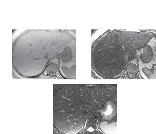
Findings
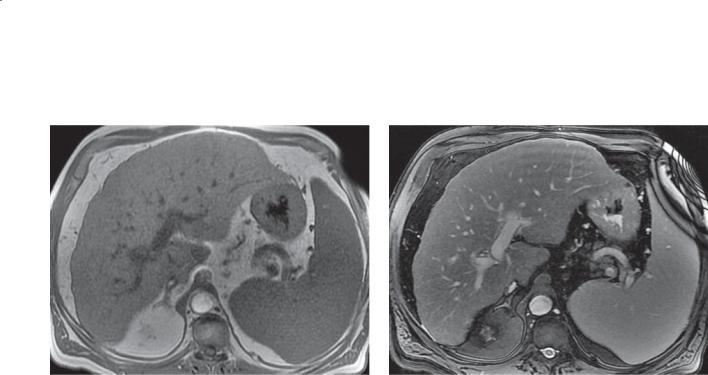
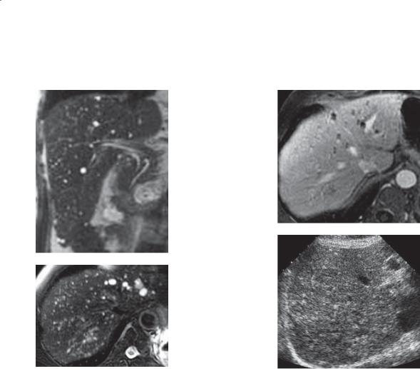
A.T1-weighted MRI. The right hepatic lobe is atrophic, and there is hypertrophy of the left hepatic lobe. Splenomegaly is seen. Excessive collaterals are visible within the upper abdomen.
B.T2-weighted MRI. The surface contour of the liver
is slightly nodular.
Di erential Diagnosis
Cirrhosis
Diagnosis
Cirrhosis
6. LIVER 465
B
Discussion
Th e morphologic changes that can occur in the liver with cirrhosis are helpful for making the diagnosis. Clinicians may not suspect diffuse hepatic disease, especially if patients present with nonspecific complaints. These cross-sectional imaging findings may be the first clue to the diagnosis. Atrophy of the right hepatic lobe with enlargement of the left hepatic lobe and caudate lobe are characteristic findings of hepatic cirrhosis. Caudate lobe enlargement is believed to be more common in patients with alcohol-induced cirrhosis. Nodular changes in the surface contour can also be helpful for making the diagnosis and indicate macronodular cirrhosis.
Pathologically, cirrhosis is divided into 2 types: micronodular and macronodular. Micronodular cirrhosis (Laënnec cirrhosis) has innumerable tiny (<3 mm) nodules. Alcoholic cirrhosis is the
commonest cause. Macronodular cirrhosis (case 6.2) is also known as postnecrotic cirrhosis and has variable-sized regenerative nodules that can be several centimeters in diameter. Viral hepatic cirrhosis is the commonest cause of this subtype.
Complications of cirrhosis include hepatic failure, portal venous hypertension, portal venous thrombosis, ascites, hepatocellular carcinoma, bacterial peritonitis, and renal failure.
Disease type: Di use Diseases
466 MAYO CLINIC GASTROINTESTINAL IMAGING REVIEW
CASE 6.2
A B C
Findings
A.T1-weighted in-phase MRI. Multiple nodules of similar intensity are present throughout the liver. The nodules give the liver surface an irregular border.
B.T1-weighted out-of-phase MRI. There is no change in the signal intensity of the nodules between the in-phase and out-of-phase images. Well-defined septations are present between nodules.
C.T2-weighted MRI. The nodules are all homogeneously of low-signal intensity, and there are high-intensity septations between nodules.
Di erential Diagnosis
Macronodular cirrhosis
Diagnosis
Macronodular cirrhosis
Discussion
Viral hepatitis is the most likely underlying cause of this cirrhosis because of the visible macronodules in the surface contour. Many authorities believe that MRI is the most sensitive imaging test for evaluating patients suspected to have diffuse liver disease. In-phase and out-of-phase T1-weighted images are
helpful to evaluate for the presence of fat within these nodules, because the presence of fat usually indicates dysplasia. In this patient, there is no signal change between images A and B, an indication of the lack of intracellular fat. Additional imaging with contrastenhanced sequences would be helpful to exclude the presence of hepatocellular carcinoma.
Th e visible tissue between the nodules is fibrous septations. These provide an alternative route for blood flow instead of through the acinar sinusoids. They are comprised pathologically of granulation tissue (edema, capillaries, inflammatory cell infiltration, and fibrosis), in amounts related to the activity of the cirrhotic process.
Disease type: Di use Diseases
CASE 6.3
A
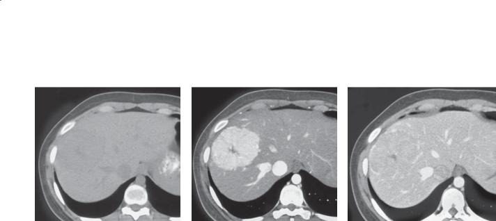
Findings
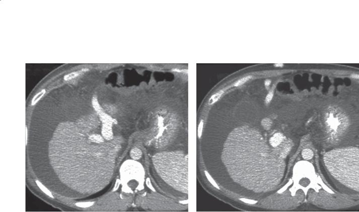
Contrast-enhanced CT. A and B. The liver is small and has a finely nodular contour. A recanalized umbilical vein leads from the left branch of the portal vein through the falciform ligament to a location posterior to the anterior abdominal wall.
Di erential Diagnosis
Cirrhosis with recanalized umbilical vein
Diagnosis
Cirrhosis with recanalized umbilical vein
6. LIVER 467
B
Discussion
Th e umbilical vein closes soon after birth, and it never reopens. A recanalized umbilical vein actually is an enlarged collateral vein that runs adjacent to the obliterated umbilical vein carrying hepatofugalflowing blood. Some authors refer to this vein
as the paraumbilical vein. This vein normally is insignificant, but it can be enlarged in patients with portal hypertension. It runs caudad from the liver along the falciform ligament to the umbilicus. In some patients, large collaterals develop in the region of the umbilicus. When these enlarged collaterals are visible on physical examination, they are referred to as caput medusae. Additional collaterals usually form to provide a connection with the systemic venous system (usually with pelvic veins).
Disease type: Di use Diseases
468 MAYO CLINIC GASTROINTESTINAL IMAGING REVIEW
CASE 6.4
A B
C
D
E
Disease type: Di use Diseases

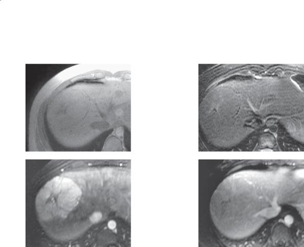
Findings
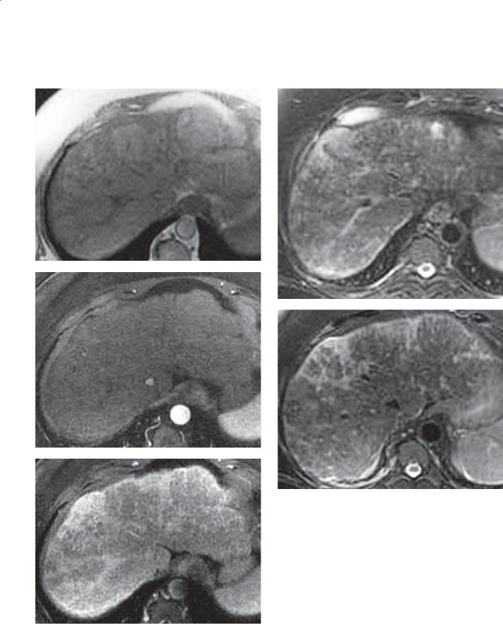
A.T1-weighted MRI. Multiple, low signal intensity, linear regions are within the periphery of the liver.
B.T2-weighted MRI. Multiple patchy areas of high signal intensity are throughout the liver.
C.Arterial-phase dynamic, enhanced MRI. There is no abnormal enhancement, and the liver surface is lobulated.
D.Venous-phase dynamic, enhanced MRI. Inhomogenous enhancement is seen throughout the liver.
E.Delayed-phase dynamic, enhanced MRI. The areas of low signal intensity identified on the T1-weighted image are enhancing.
Di erential Diagnosis
1.Diff use hepatocellular carcinoma
2.Cholangiocarcinoma
3.Cirrhosis with confluent fibrosis
Diagnosis
Cirrhosis with confluent fibrosis
6. LIVER 469
Discussion
Confluent hepatic fibrosis usually manifests as a wedge-shaped region extending from the porta hepatis to the capsule. It most commonly involves the anterior segment of the right lobe and the medial segment of the left lobe. Capsular retraction is seen in 90% of cases. An ill-defined band of abnormal signal (low intensity on T1-weighted images and high signal on T2-weighted images) is characteristic of confluent hepatic fibrosis.
Differentiating confluent hepatic fibrosis from diffuse or infiltrating hepatocellular carcinoma or cholangiocarcinoma can be difficult. Usually a
tumor is expansive and bulges the hepatic contour. Hepatocellular carcinoma enhances heterogeneously during the arterial phase of contrast enhancement. Bile duct dilatation usually accompanies a cholangiocarcinoma. In patients with confluent hepatic fibrosis, there is no venous invasion and the size of the area does not change over time.
Disease type: Di use Diseases
470 MAYO CLINIC GASTROINTESTINAL IMAGING REVIEW
CASE 6.5
Findings |
Discussion |
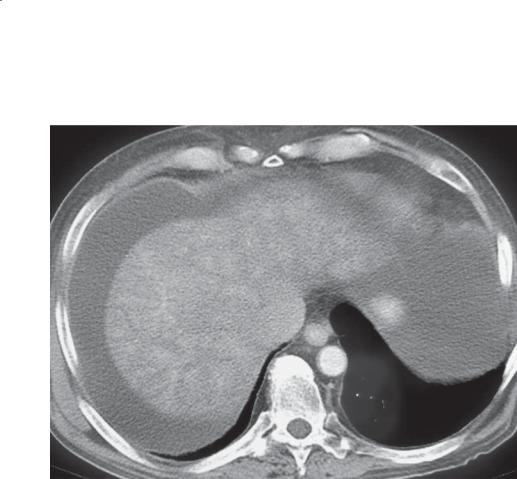
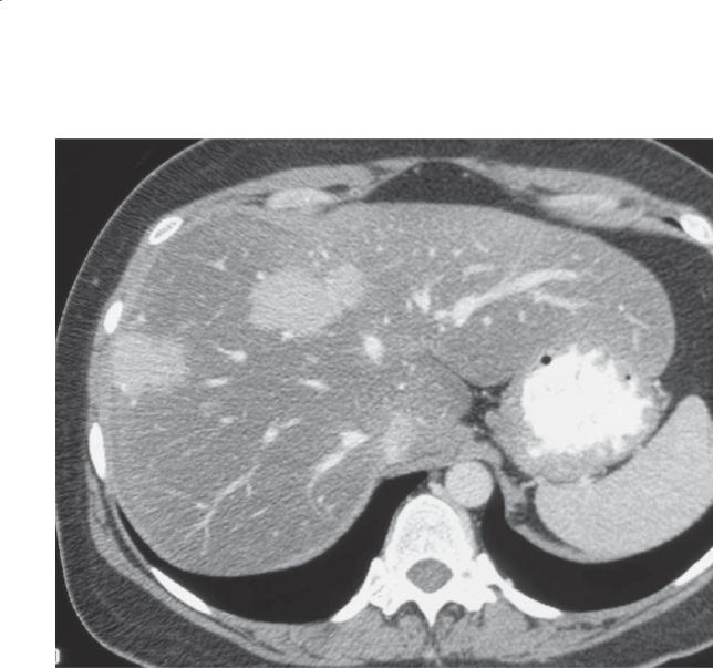
Contrast-enhanced CT. The liver enhances |
Cardiac failure and constrictive pericarditis lead to |
heterogeneously. The inferior vena cava is dilated. |
increased central venous pressure, increased hepatic |
Ascites. |
venous pressure, sinusoidal engorgement, diminished |
|
hepatic arterial flow, and hepatocellular hypoxia. If |
Di erential Diagnosis |
the increased central venous pressure persists for long |
1. Hepatic cirrhosis |
periods, changes of cirrhosis develop. Liver enzyme |
2. Cardiac cirrhosis |
values can be increased, but frank liver failure develops |
3. Budd-Chiari syndrome |
rarely. Because the cardiac findings usually overshadow |
|
the liver failure, the diagnosis usually is apparent. |
Diagnosis |
CT findings of hepatic congestion include reflux |
Cardiac cirrhosis |
of contrast material into dilated hepatic veins and |
|
the inferior vena cava. Enhancement of the liver is |
|
often mottled and resembles early changes of Budd- |
|
Chiari syndrome (case 6.13). Other findings include |
|
cardiomegaly, hepatomegaly, intrahepatic periportal |
|
lucency, ascites, and pleural and pericardial effusions. |
|
At sonography, the Doppler waveform of the portal |
|
vein also may be abnormal. There is loss of the normal |
|
continuous flow pattern and increased pulsatility that |
|
represents direct transmission of the fluid wave from |
|
the heart through the hepatic sinusoids. |
Disease type: Di use Diseases
6. LIVER 471
CASE 6.6
A B
C
Findings
A.T1-weighted in-phase MRI. The liver is diffusely higher in signal intensity than normal.
B.T1-weighted out-of-phase MRI. There is marked signal dropout throughout the liver.
C.T2-weighted MRI. The liver is normal in appearance.
Di erential Diagnosis
Diffuse fatty infiltration
Diagnosis
Diffuse fatty infiltration
Discussion
Fatty infiltration of the liver is associated with various conditions, including excessive alcohol consumption, obesity, use of parenteral nutrition, diabetes, corticosteroid use, and nonalcoholic steatohepatitis.
Fatty changes also can be present in 25% of healthy persons. Fatty infiltration of the liver may be diffuse or focal. MRI accurately differentiates fatty infiltration from tumors.
Nonalcoholic fatty liver disease is common and associated with a considerable risk for development of end-stage liver disease, diabetes mellitus, and cardiovascular disease. On T1-weighted images, fatty infiltration appears as regions of increased signal intensity. Chemical shift imaging (using in-phase
[A] and out-of-phase [B] T1-weighted images) exploits the difference in resonance frequency between the protons present in fat and water. Loss of signal intensity of hepatic parenchyma between the in-phase and out- of-phase images indicates the presence of microscopic (intracellular) lipid. The presence of normal signal intensity on the T2-weighted image is reassuring that an underlying mass is absent.
Disease type: Di use Diseases

472 MAYO CLINIC GASTROINTESTINAL IMAGING REVIEW
CASE 6.7
A B
C
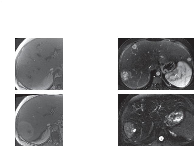
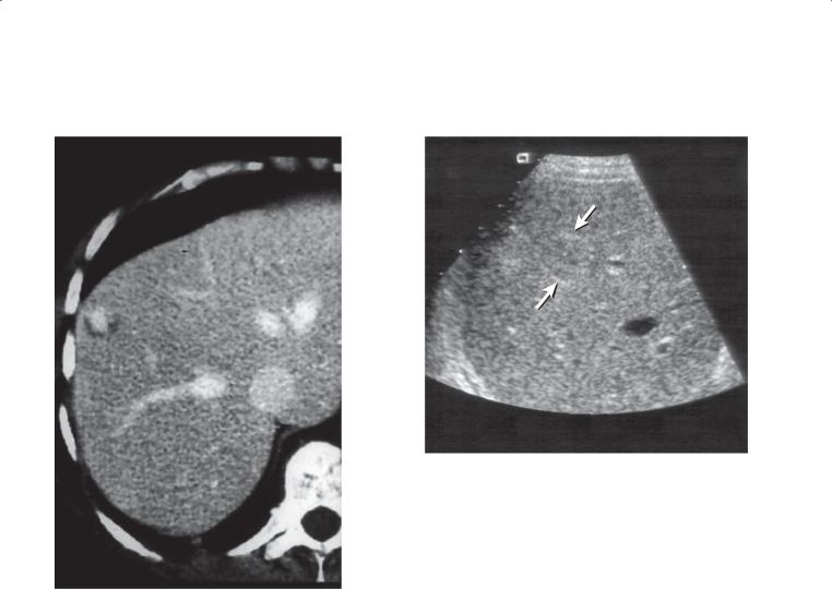
Findings
A. Contrast-enhanced CT. There is a focal lowattenuation mass within the medial segment of the left lobe of the liver and a similar-appearing smaller region within the lateral segment of the left lobe.
B and C. T1-weighted MRI. The 2 regions are of high signal intensity on the in-phase image (B) and of low signal intensity on the out-of-phase image (C).
Di erential Diagnosis
1.Focal fatty infiltration of the liver
2.Liver metastases
Diagnosis
Focal fatty infiltration of the liver
Discussion
Focal steatosis most often is found in the liver tissue adjacent to the fissure of the ligamentum teres, but it can occur anywhere, be any size, and be solitary or multiple. Focal steatosis is assumed to be related to regional differences or disturbances in hepatic blood flow. This mechanism explains why focal fatty changes are common in patients with trauma or ischemic insults to the liver. They can appear as a focal hepatic low-attenuation mass at CT and can mimic a primary or metastatic liver tumor(s).
Noncontrast-enhanced scans are most helpful for CT diagnosis. Normally, the attenuation of the liver is similar or slightly higher than that of paraspinal muscle. Fatty infiltration of the liver results in attenuation values of the liver to decrease below the normal 50 to 75 Hounsfield units.
Th e findings of focal fat at MRI are the same as those of diffuse fatty infiltration (case 6.6). At
sonography, a fatty liver is diffusely echogenic. The degree of echogenicity is roughly equivalent to the level of steatosis. Fat causes increased attenuation of the ultrasound beam and poorer visualization of deeper structures.
Disease type: Di use Diseases

6. LIVER 473
CASE 6.8
A
B
Findings
Hepatic sonogram. A and B. The liver is diffusely echogenic. A geographic hypoechoic region is present within the right lobe of the liver.
Di erential Diagnosis
1.Hepatic neoplasm
2.Fatty infiltration of the liver with focal normal liver
Diagnosis
Fatty infiltration of the liver with focal normal liver
Discussion
Fatty infiltration of the liver is a relatively common finding. It can present as diffuse or focal fatty infiltration. Occasionally an island(s) of normal liver is present in a liver that is otherwise fat-infiltrated. The region of focal normal liver can simulate a neoplasm. The key in recognizing this entity is to identify the diffusely increased echogenicity of the underlying liver. In addition, the normal liver does not cause mass effect, and vessels often can be seen to course normally through this region.
Disease type: Di use Diseases
474 MAYO CLINIC GASTROINTESTINAL IMAGING REVIEW
CASE 6.9
Findings
T2-weighted MRI. The liver and pancreas are of diffusely low signal intensity. The spleen is of normal signal intensity.
Di erential Diagnosis
1.Hemochromatosis
2.Hemosiderosis
Diagnosis
Hemochromatosis
Discussion
Primary hemochromatosis (hereditary hemochromatosis or idiopathic hemochromatosis) is an autosomal recessive disorder. Abnormal absorption and deposition of iron occur. In primary hemochromatosis, the liver is the main organ for abnormal iron deposition, consisting of ferritin and hemosiderin. Early deposition is located in periportal hepatocytes. This progresses to perilobular fibrosis
with iron deposition in the biliary epithelium, Kupffer cells, and fibrous septa. In patients with advanced disease, the liver is cirrhotic with broad fibrous septa surrounding large areas of relatively normal liver parenchyma. Other sites of abnormal iron deposition include the pancreas and heart.
One-third of deaths from hemochromatosis are the result of hepatocellular carcinoma. Cardiomyopathy, diabetes, and arthropathy also can occur. Screening for hemochromatosis usually involves measurement of serum ferritin and transferrin saturation. Definitive diagnosis of primary hemochromatosis can be made with genetic testing or liver biopsy with quantitative determination of liver iron concentration. MRI can be used to assess for complicating hepatocellular carcinoma and, in specialized centers, to quantitate the amount of hepatic iron. Low signal intensity of the liver and pancreas with normal signal in the spleen
is characteristic. Because the liver also is involved in secondary hemochromatosis (hemosiderosis), involvement of the pancreas is a key finding.
Disease type: Di use Diseases

6. LIVER 475
CASE 6.10
A C
B D
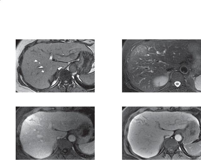
Findings
A.T1-weighted MRI. The liver and pancreas are of diffusely low signal intensity. The spleen is of normal signal intensity.
B.T2-weighted MRI. The liver and pancreas are of diffusely low signal intensity. The spleen is of normal signal intensity.
C.T2-weighted MRI. A small mass of high signal intensity is within the right hepatic lobe (arrow).
D.Diffusion-weighted MRI. A small mass of high signal intensity is within the right hepatic lobe.
Di erential Diagnosis
1.Hemochromatosis with benign cyst
2.Hemochromatosis with hepatocellular carcinoma
Diagnosis
Hemochromatosis with hepatocellular carcinoma
Discussion
Th e liver is of lower than expected signal intensity on the T2-weighted sequence (B). On T1and T2-weighted images, the paraspinal muscles are a good internal control to consider whether the liver is of abnormal signal intensity. Generally, the signal of normal liver is not lower than that of muscle. In primary hemochromatosis, iron deposition occurs within the hepatocytes rather than the Kupffer cells (reticuloendothelial system). Because the spleen
in this patient is of normal signal intensity and the pancreas is of abnormally low signal intensity, primary hemochromatosis is most likely. The mass in the
liver should represent a complicating hepatocellular carcinoma. If primary hemochromatosis is untreated, a hepatocellular carcinoma will develop in approximately one-third of patients.
Disease type: Di use Diseases
476 MAYO CLINIC GASTROINTESTINAL IMAGING REVIEW
CASE 6.11
A B
Findings
A.T1-weighted MRI. The liver is of diffusely abnormal lower signal intensity.
B.T2-weighted MRI. The liver, spleen, and bone marrow are of abnormally decreased signal intensity. The pancreas has a normal appearance. No focal masses are present.
Di erential Diagnosis
1.Hemosiderosis (secondary hemochromatosis)
2.Primary hemochromatosis
Diagnosis
Hemosiderosis
Discussion
Secondary hemochromatosis (hemosiderosis) can be caused by multiple factors, such as parenteral administration of iron (from transfusions), increased absorption of normal dietary iron, and increased
dietary iron load. Normally, iron is stored in the body as hemosiderin or ferritin in the reticuloendothelial cells of the liver, spleen, and bone marrow. Usually, no organ damage occurs. Involvement of the pancreas and heart is unusual, unless the iron load exceeds the capacity of the reticuloendothelial system.
Differentiating hemosiderosis from primary hemochromatosis is important because the treatments and natural history of the diseases are very different (cases 6.9 and 6.10). Both diseases cause a loss of signal in the liver at MRI, but, commonly, primary hemochromatosis also results in iron deposition in the pancreas and heart (and loss of parenchymal signal). Hemosiderosis spares the pancreas and heart, but iron deposition in the spleen and bone marrow usually is obvious at MRI. T2-weighted gradient echo sequences are most sensitive for visualizing iron deposition.
Disease type: Di use Diseases
6. LIVER 477
CASE 6.12
A C
B D
Findings
A.T1-weighted in-phase MRI. The liver is relatively homogeneous in signal intensity.
B.T1-weighted out-of-phase MRI. The liver is of lower signal intensity than on the in-phase image, except for a mass of high signal intensity within the posterior segment of the right hepatic lobe.
C.T1-weighted enhanced MRI. Multiple enhancing masses are present within the liver.
D.Diffusion-weighted MRI. A mass of high signal intensity is within the posterior right hepatic lobe.
Di erential Diagnosis
1.Fatty infiltration of the liver with metastases
2.Multiple hepatic adenomatosis
3.Glycogen storage disease with multiple hepatic adenomas (von Gierke disease)
Diagnosis
Glycogen storage disease with multiple hepatic adenomas (von Gierke disease)
Discussion
Th e glycogen storage diseases are inherited disorders of metabolism. Most of the disorders involve an abnormality breaking down glycogen into glucose. In glycogen storage disease type I (von Gierke disease), there is an accumulation of glycogen within the hepatocytes and proximal tubules of the kidney. Although glycogen accumulation usually results in
a hyperattenuating liver at CT, the effects of fatty infiltration predominate in this disease as a result of chronic hormonal stimulation of the liver. One of the complications of this disease is an increased incidence of hepatic cell adenomas. Unlike solitary adenomas, there is an increased incidence of hepatocellular carcinoma arising from these adenomas.
Disease type: Di use Diseases
478 MAYO CLINIC GASTROINTESTINAL IMAGING REVIEW
CASE 6.13
A B
C
Disease type: Di use Diseases

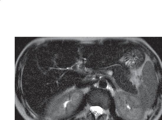
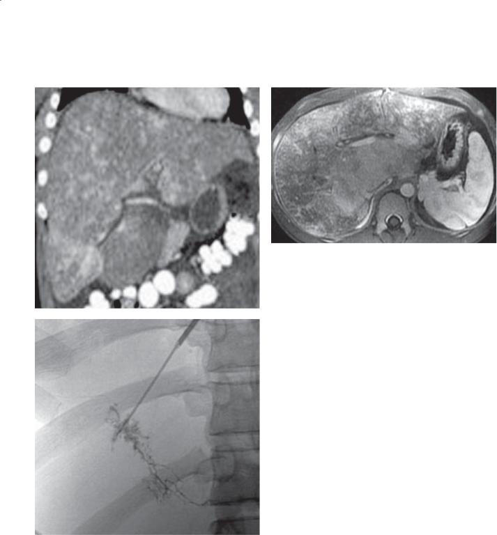
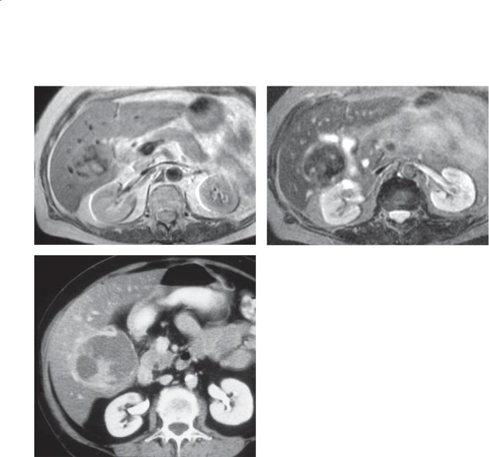
Findings
A.Coronal contrast-enhanced CT. Enlarged liver is heterogeneously perfused and has a large caudate lobe.
B.Dynamic contrast-enhanced MRI. The liver is heterogeneously perfused and has caudate lobe enlargement. The caudate lobe is homogeneously perfused compared with the rest of the liver.
C.Hepatic vein angiogram. Occluded hepatic vein
has multiple intrahepatic collaterals demonstrating a spiderlike appearance.
Di erential Diagnosis
1.Budd-Chiari syndrome
2.Hepatic congestion from cardiac failure
3.Portal vein thrombosis
Diagnosis
Budd-Chiari syndrome
Discussion
Budd-Chiari syndrome refers to a group of conditions with hepatic venous outflow obstruction. Obstruction can occur at the hepatic vein level or within the supradiaphragmatic portion of the inferior vena cava. The clinical symptoms of acute Budd-Chiari syndrome are usually ascites, abdominal pain, vomiting, and hypotension. Other biochemical evidence of hepatic failure also often is present. Primary Budd-Chiari syndrome refers to a membranous (wafer-thin to several-centimeter–thick membrane) obstruction of
6. LIVER 479
the hepatic veins. This cause is common in the Far East, Israel, and South Africa. Membranous obstruction is associated with hepatocellular carcinoma in 20% to 40% of patients. Secondary Budd-Chiari syndrome is most common in North America and Europe and can be classified further as occurring at the central and sublobular venous level or involving the major hepatic veins. Various drugs or treatments are implicated for sublobular venous occlusion, whereas a coagulopathy is often the cause of large vein occlusion.
Angiography is the standard for diagnosis. Pressure measurements within the inferior vena cava and hepatic veins are helpful to exclude a membranous obstruction. A wedged hepatic venogram that shows the spider-web pattern of intrahepatic collaterals
is pathognomonic of sublobular venous occlusion (as shown in C). The CT and MRI findings can vary
depending on the duration and extent of the condition. Findings in acute and subacute Budd-Chiari syndrome include hepatomegaly, ascites, acute hyperdense hepatic venous thrombus, patchy enhancement (often with normal enhancement of the central portion of the liver and caudate lobe), portal vein thrombosis
(in approximately 20% of cases), and compression of the inferior vena cava. Chronically, the hepatic veins may become invisible, and the peripheral portions of the liver (with hypertrophy of the caudate lobe) often atrophy. If a single hepatic vein remains patent, intrahepatic collateral veins may be visible carrying blood from the segments with occluded veins to the sole patent outflow vein.
Disease type: Di use Diseases

480 MAYO CLINIC GASTROINTESTINAL IMAGING REVIEW
CASE 6.14
A B
Findings
Unenhanced CT. A. The liver is diffusely hyperattenuating. It is considerably higher in attenuation than either the spleen or paraspinal muscle. B. Cardiomegaly with high-attenuation changes involving atelectatic lung in the lung bases.
Di erential Diagnosis
1.Hemochromatosis
2.Amiodarone therapy
3.Previous thorium dioxide therapy
4.Glycogen storage disease
5.Gold therapy
Diagnosis
Amiodarone therapy
Discussion
Th ere are few imaging findings in patients with drug-induced liver disease. Several drugs have been implicated in liver disease or failure. These drugs cause at least 40% of hospital admissions for acute hepatitis and 2% to 5% of all hospital admissions.
The most commonly implicated drugs in liver disease include halothane, acetaminophen, α-methyldopa, and phenytoin.
Amiodarone is an antiarrhythmic cardiac medication in which the drug or its iodine-containing metabolites are concentrated in the liver—leading to an abnormally high attenuation of liver parenchyma. There also can be hyperattenuation within the lung parenchyma and secondary interstitial lung disease.
Large amounts of iron stores also can lead to a hyperattenuation of the liver parenchyma in patients with primary and secondary hemochromatosis. Thorium dioxide, a discontinued vascular contrast agent, is concentrated in Kupffer cells and can lead to abnormal liver attenuation. Glycogen storage disease leads to an accumulation of glycogen within hepatocytes. The high glycogen content leads to high liver attenuation. Some patients with glycogen
storage disease have concomitant fatty infiltration that actually leads to a lower than normal liver attenuation (case 6.12). Gold used for therapy in rheumatoid arthritis is accumulated by the reticuloendothelial system and causes an increase in liver attenuation.
Disease type: Di use Diseases
CASE 6.15
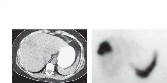
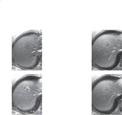
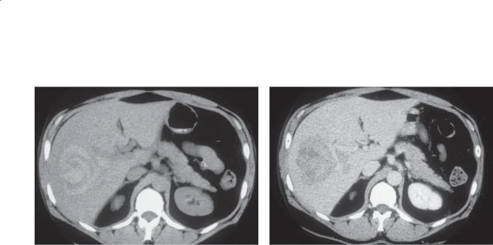
Findings
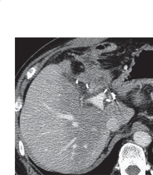
Contrast-enhanced CT. There is a large central lowattenuation region in the liver. A straight border exists between the normal and the low-attenuation region. Multiple surgical clips are present in the hepatic porta and gallbladder fossa. The patient had a history of radiation therapy for cholangiocarcinoma in this region.
Di erential Diagnosis
1.Radiation-induced liver disease
2.Focal fatty infiltration
Diagnosis
Radiation-induced liver disease
6. LIVER 481
Discussion
Radiation can cause liver changes in patients treated for biliary, liver, esophageal, gastric, pancreatic, or breast malignancies. Radiation hepatitis can develop in patients receiving more than 12 Gy as a single dose or more than 40 Gy in fractionated doses.
Findings at CT are usually a straight, sharply defined zone of different attenuation that corresponds to the treatment port. The irradiated area is usually of lower attenuation than normal liver because of the edema and fatty infiltration that occur. With time, the sharply defined zone may become more indistinct, and hepatic atrophy can develop. At sonography, the acutely irradiated zone appears hypoechoic. At MRI, acute radiation injury is of low signal intensity on the T1-weighted images and of increased signal intensity on the T2-weighted images. Chronically, fatty change often develops in these regions of injury.
Disease type: Di use Diseases
482 MAYO CLINIC GASTROINTESTINAL IMAGING REVIEW
CASE 6.16
A B
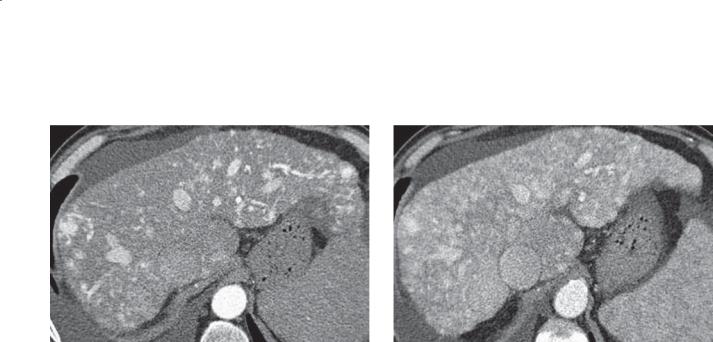
Findings
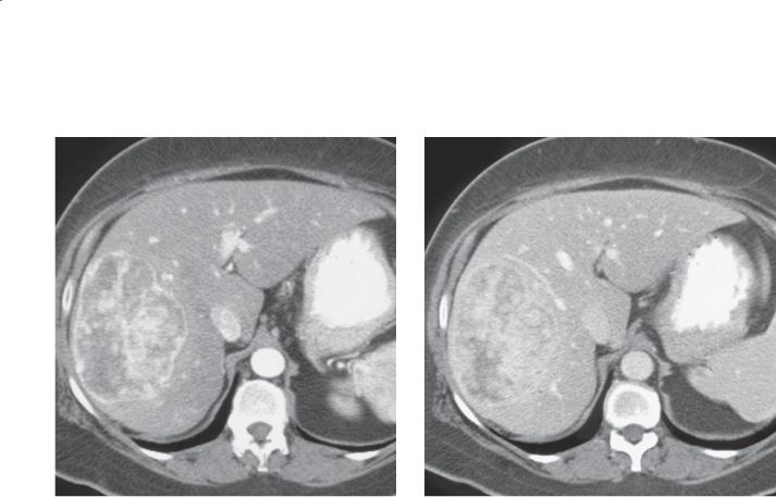
A.Arterial-phase contrast-enhanced CT. Liver is heterogeneously perfused with variable-sized nodular regions of hyperenhancement.
B.Venous-phase contrast-enhanced CT. Liver has increased heterogeneous perfusion with masslike regions of hyperenhancement.
Di erential Diagnosis
1.Diff use hepatocellular carcinoma
2.Diffuse metastases
3.Budd-Chiari syndrome
4.Hereditary hemorrhagic telangiectasia
5.Right heart failure
Diagnosis
Hereditary hemorrhagic telangiectasia
Discussion
Hereditary hemorrhagic telangiectasia (HHT) is an autosomal dominant disorder of multiple
arteriovenous malformations that lack capillaries. In many cases, telangiectasias can be identified on the patient’s face, tongue, lips, fingers, or nasal, buccal, or gastrointestinal mucosa. Bleeding from these can be serious and difficult to stop. Visceral involvement can affect lungs, brain, liver, gastrointestinal tract, and spine. Up to three-fourths of patients have liver
involvement; however, most patients are asymptomatic. Cardiac and liver failure are the commonest manifestations of liver involvement and are difficult to treat without liver transplant. Liver biopsy should be avoided in these patients.
Lesions in HHT always follow blood attenuation, unlike most hypervascular metastases that wash out and differ from blood pool attenuation, especially on delayed imaging. The hepatic veins can be identified in patients with HHT, and the peripheral or central perfusion changes of Budd-Chiari are usually absent. In right heart failure, the inferior vena cava is always dilated and the heart is enlarged. Diffuse hepatocellular carcinoma could have an appearance similar to that of diffuse metastases, but changes of cirrhosis are usually also present.
Disease type: Di use Diseases

6. LIVER 483
CASE 6.17
Findings |
Discussion |
|
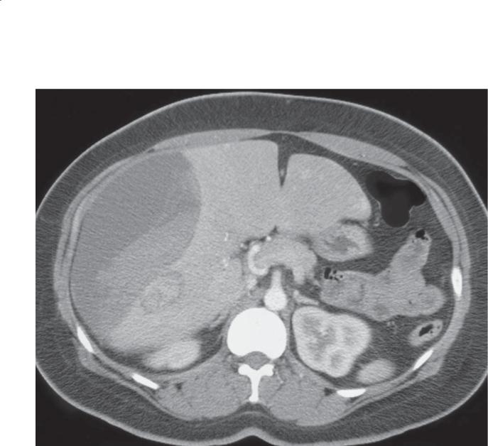
Portal venous phase contrast-enhanced CT. The liver |
In patients with breast cancer who are taking |
|
has a markedly deformed surface contour and there is |
chemotherapeutic drugs, morphologic changes |
|
ascites. |
of the liver can develop which resemble cirrhosis. |
|
|
|
Pseudocirrhosis includes a lobular hepatic contour, |
Di erential Diagnosis |
segmental volume loss, and enlargement of the |
|
1. |
Metastatic disease |
caudate lobe. Retraction of the capsular surface of the |
2. |
Hepatic cirrhosis |
liver can be seen at the site of subjacent metastases. |
3. |
Pseudocirrhosis |
These changes can develop over several months. |
|
|
Pathologically, nodular regenerative hyperplasia |
Diagnosis |
without cirrhosis has been found. |
|
Pseudocirrhosis |
|
|
Disease type: Di use Diseases

484 MAYO CLINIC GASTROINTESTINAL IMAGING REVIEW
TABLE 6.1
Di use Diseases of the Liver |
CASE |
|
|
|
|
Cirrhosis |
Nodular contour, atrophic right lobe, hypertrophic left and |
6.1–6.5 |
|
caudate lobes |
|
|
|
|
Fatty infiltration |
Diffuse or focal. Low attenuation at CT. Usually geographic with |
6.6–6.8 |
|
vessels coursing through area. Chemical shift changes at MRI. |
|
|
Hyperechoic at sonography |
|
|
|
|
Hemochromatosis |
Iron deposition in liver, pancreas, heart. Cirrhosis and hepatocellular |
6.9 and 6.10 |
|
carcinoma can occur |
|
|
|
|
Hemosiderosis |
Iron deposition in liver, spleen, marrow. No organ damage |
6.11 |
|
|
|
Glycogen storage disease |
High attenuation or fatty changes at CT. Predisposes to |
6.12 |
|
multiple adenomas |
|
|
|
|
Budd-Chiari syndrome |
Hepatic venous or inferior vena cava occlusion. Heterogeneous |
6.13 |
|
liver with normal-appearing central portion and caudate lobe |
|
|
|
|
Amiodarone therapy |
Iodine-containing antiarrhythmic. High-attenuation liver |
6.14 |
|
and lung parenchyma |
|
|
|
|
Radiation-induced disease |
Edema or fatty changes correspond to radiation port |
6.15 |
|
|
|
Hereditary hemorrhagic |
Diffuse vascular lakes and nodules within liver create |
6.16 |
telangiectasia |
heterogeneous enhancement pattern |
|
|
|
|
Pseudocirrhosis |
Cirrhotic configuration. May have linear or nodular filling defects. |
6.17 |
|
Usually a history of treatment of breast cancer with metastases to |
|
|
the liver |
|
|
|
|
Disease type: Di use Diseases
6. LIVER 485
CASE 6.18
A B
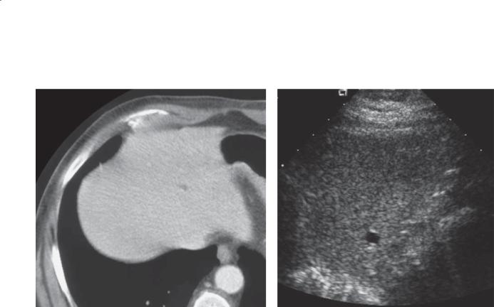
Findings
A.Contrast-enhanced CT. A tiny low-attenuation lesion is present in the superior aspect of the liver.
B.Sonogram. A tiny anechoic liver lesion with posterior acoustic enhancement is seen, corresponding to the lesion on CT.
Di erential Diagnosis
1.Tiny hepatic cyst
2.Metastases
3.Von Meyenburg complex
Diagnosis
Hepatic cyst
Discussion
Low-attenuation liver lesions are commonly found on CT of the abdomen. When the lesions are large, it is often possible to diagnose them as cysts (fluid density on CT). However, tiny lesions (such as the
one in this case) are often too small to characterize confidently and are considered indeterminate. Most of these tiny low-attenuation lesions are inconsequential liver cysts or even hemangiomas, but tiny metastases can have an identical CT appearance. In cases of high clinical concern, such as a history of malignancy, ultrasonography is often invaluable for differentiating hepatic cysts from solid liver tumors.
A liver cyst is defined as an epithelial-lined fluidfilled space. Hepatic cysts are extremely common, occurring in up to 5% of the population. The precise cause of hepatic cysts is unclear, as is the explanation of why these lesions usually do not develop until middle age. On sonographic examination, simple hepatic cysts are anechoic with a thin wall and posterior acoustic enhancement. Very rarely, a cyst may become symptomatic as a result of infection or hemorrhage into the cyst. Von Meyenburg complex (case 6.21) is
a biliary hamartoma, often containing fat. Like cysts, it has no known clinical sequelae.
Disease type: Masses
486 MAYO CLINIC GASTROINTESTINAL IMAGING REVIEW
CASE 6.19
A B C
Findings
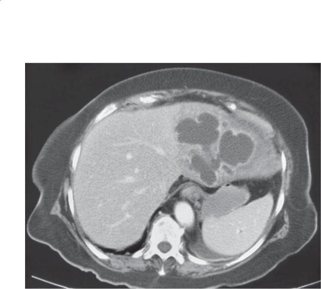
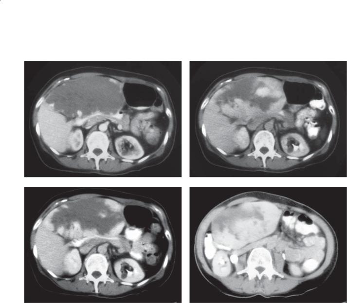
A. Unenhanced CT. Multiple cysts are present in the liver with confluent cystic change in the right lobe of the liver.
B and C. Unenhanced MRI. T2-weighted axial (B) and coronal (C) images. Multiple cysts are present in the liver. Near complete replacement of both kidneys with cysts is seen on the coronal image.
Di erential Diagnosis
1.Autosomal dominant polycystic disease
2.Multiple hepatic cysts
Diagnosis
Autosomal dominant polycystic disease
Discussion
Autosomal dominant polycystic disease (adult polycystic disease) is inherited in an autosomal dominant pattern, presenting clinically in adulthood.
The renal parenchyma is progressively replaced by cysts of varying sizes, and most patients present between ages 30 and 50 years with hypertension and renal failure. Cysts often occur in the liver (50%-75% of cases), pancreas (10% of cases), spleen (5% of cases), and other organs. No correlation exists between the severity of renal disease and the extent of liver involvement. The liver cysts in autosomal dominant polycystic disease rarely cause hepatic dysfunction. Unlike autosomal recessive polycystic disease (infantile polycystic disease), there is no association with hepatic fibrosis.
When liver function values are increased, biliary obstruction, cyst infection, or tumor should be excluded. Multiple large cysts can rarely cause compression of the portal vein, resulting in portal hypertension. Cardiovascular abnormalities associated with autosomal dominant polycystic disease include intracranial berry aneurysms (10% of cases), mitral valve prolapse (25% of cases), bicuspid aortic valve, aortic aneurysms, and aortic dissections.
Disease type: Masses
6. LIVER 487
CASE 6.20
A B
C
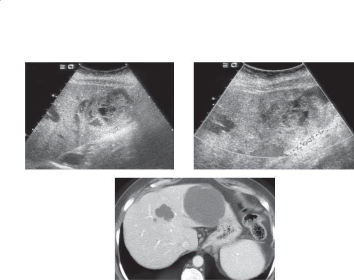
Findings
Sonogram. A and B. A complex cystic liver mass is present within the left lobe of the liver. The lesion contains significant internal echoes, consistent with a mixed solid and cystic mass. No blood flow is seen within the mass on color Doppler interrogation.
C. Contrast-enhanced CT. Two fluid-density liver masses are present, the larger of which is in the lateral segment of the left lobe of the liver.
Di erential Diagnosis
1.Infected cyst or abscess
2.Hemorrhagic cyst
3.Cystic hepatocellular carcinoma
4.Cystic metastasis
Diagnosis
Hemorrhagic liver cyst
Discussion
Th is patient underwent CT of the abdomen for acute abdominal pain. The superiority of ultrasonography over CT for the evaluation of cystic lesions is clearly shown in this case. Aspiration of this complex cyst revealed hemorrhagic products and resulted in relief of the pain. Because hepatic cysts have an epithelial lining and invariably recur, aspiration is not indicated for asymptomatic cysts.
An infected hepatic cyst could have a similar sonographic appearance, but the patient would likely present clinically with a fever and an increased
leukocyte count. A cystic liver tumor can be excluded, given the lack of an enhancing rim at CT and lack of internal blood flow at sonography.
Disease type: Masses
488 MAYO CLINIC GASTROINTESTINAL IMAGING REVIEW
CASE 6.21
A C
D
B
Findings |
Diagnosis |
A.T2-weighted coronal MRI. Innumerable tiny masses of high signal intensity are throughout the liver.
B.T2-weighted axial MRI. Innumerable tiny masses of high signal intensity are seen.
C.T1-weighted axial enhanced MRI. The tiny lesions seen on the T2-weighted images have become isointense, and the larger lesions are nonenhancing masses (some containing a rim of enhancement).
D.Sonogram. Multiple hyperechoic, tiny masses are throughout the liver.
Di erential Diagnosis
1.Diff use metastases and benign cysts
2.Multiple benign cysts
3.Multiple biliary hamartomas (von Meyenburg complexes) and benign cysts
4.Multiple vascular malformations through the liver and benign cysts
5.Diffuse candidiasis, microabscess
Multiple biliary hamartomas (von Meyenberg complexes) and benign cysts
Discussion
Von Meyenburg complexes (also known as biliary hamartomas) are composed of a cluster of proliferated bile ducts embedded in a fibrous stroma. These small (usually <1 cm) benign lesions are detected incidentally in 1% to 5% of reported autopsies and are often multiple. They are of no clinical significance except that they
can be easily mistaken for metastases on CT, MRI, and ultrasonography. Von Meyenburg complexes are usually uniformly hypoechoic or hyperechoic on ultrasonography but may contain echogenic foci with ringdown artifact related to the presence of cholesterol crystals in the dilated tubules. Von Meyenburg complexes usually can be differentiated from metastases at MRI with very high signal on T2-weighted images. No enhancement or a rim
of enhancement is visible on post-gadolinium images. This rim enhancement is thought to be caused by compressed liver parenchyma surrounding the biliary hamartoma.
Disease type: Masses

6. LIVER 489
CASE 6.22
A B
Findings
Coronal (A) and axial (B) MRCP. Numerous small cystic lesions encase the bile ducts throughout the liver. The common bile duct and intrahepatic ducts are normal. The liver has a cirrhotic configuration, and the left and caudate lobes are enlarged. A small amount of ascites is seen around the liver.
Di erential Diagnosis
1.Peribiliary cysts
2.Caroli disease
3.Multiple simple hepatic cysts
4.Von Meyenburg complexes
Diagnosis
Peribiliary cysts
Discussion
Peribiliary cysts develop in patients with severe liver disease and dilated intrahepatic peribiliary glands.
They can occur as discrete cysts, clustered cysts, or strings of cysts (as in this case) along the portal tracts. The development of peribiliary cysts seems to be related to disturbances in hepatic circulation. These cysts most commonly occur in the setting of
cirrhosis, portal venous hypertension or thrombosis, hepatic transplantation, cholangitis, or autosomal dominant polycystic disease. Peribiliary cysts usually are asymptomatic and rarely result in biliary duct dilatation. The intrahepatic and extrahepatic bile ducts remain normal in uncomplicated
cases. Peribiliary cysts can be identified by CT, MRI, or ultrasonography. Cholangiographicenhanced CT can be very helpful in some cases to differentiate peribiliary cysts from intrahepatic bile duct dilatation or Caroli disease (cases 7.43 through 7.45). Other hepatic cystic lesions such as von Meyenburg complexes (case 6.21) or simple hepatic cysts (case 6.18) arise independently from the bile ducts.
Disease type: Masses

490 MAYO CLINIC GASTROINTESTINAL IMAGING REVIEW
TABLE 6.2
Liver Cysts |
|
CASE |
|
|
|
Simple cyst |
Well-circumscribed, water attenuation, no enhancement |
6.18 |
|
|
|
Polycystic disease |
Multiple benign cysts, some hemorrhagic—kidneys, liver, pancreas |
6.19 |
|
|
|
Hemorrhagic cyst |
Complex cyst. High attenuation at CT, high-signal intensity |
6.20 |
|
T1-weighted MRI, mixed solid and cystic at sonography |
|
|
|
|
Von Meyenburg complexes |
Tiny water attenuation masses resemble tiny simple cysts. |
6.21 |
|
May be fat attenuation. Larger lesions have enhancing rim |
|
|
|
|
Peribiliary cysts |
Multiple tiny cysts clustered, or discrete cysts adjacent to biliary |
6.22 |
|
tree; usually associated with cirrhosis |
|
|
|
|
Disease type: Masses
6. LIVER 491
CASE 6.23
Findings
Contrast-enhanced CT. Several low-attenuation masses with hyperattenuating rings are present within the lateral segment of the left lobe of the liver.
Di erential Diagnosis
1.Hepatic abscess
2.Cystic hepatocellular carcinoma
3.Cystic metastases
4.Biliary cystadenoma
Diagnosis
Hepatic abscess
Discussion
Pyogenic hepatic abscesses most commonly are due to diverticulitis, appendicitis, or cholecystitis. Other causes include ascending cholangitis with biliary obstruction, hepatic arterial septicemia from bacterial endocarditis
or pneumonitis, direct extension from an adjacent infection, trauma or penetrating injuries with infection, or infected tumor. Escherichia coli is the most common organism cultured in adults, whereas Staphylococcus organisms are most common in children.
Many hepatic abscesses communicate and can be drained by a single drainage catheter. These types of abscesses may have the appearance of a cluster of water density masses (as in this case). The presence of air within a hepatic abscess is unusual. Definitive
diagnosis usually requires percutaneous aspiration and assessment of the fluid for organisms.
Not all hepatic abscesses are pyogenic in origin. Amebic abscess caused by the organism Entamoeba histolytica is a common infection worldwide. The presentation is usually nonspecific, but patients may present with a unilocular water-density mass with a low-density ring (case 6.24). Echinococcal disease is discussed in cases 6.25 through 6.27.
Disease type: Masses
492 MAYO CLINIC GASTROINTESTINAL IMAGING REVIEW
CASE 6.24
Findings
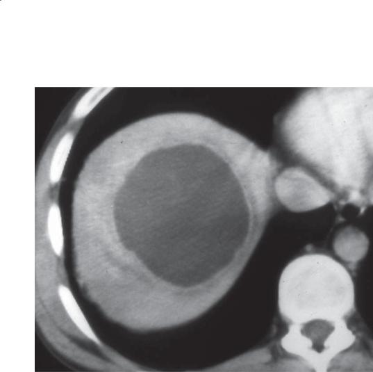
Contrast-enhanced CT. There is a large waterattenuation mass within the liver. The mass is well demarcated. A low-attenuation ring partially surrounds the mass.
Di erential Diagnosis
1.Amebic abscess
2.Cystic hepatocellular carcinoma
3.Cystic metastases
4.Biliary cystadenoma
5.Pyogenic abscess
Diagnosis
Amebic abscess
Discussion
Entamoeba histolytica affects nearly 10% of the world’s population. Most people are asymptomatic carriers. It is commonly found in the stool of homosexual men.
The life cycle of this parasite begins by oral ingestion
of the cystic form. Invasive trophozoites develop from the cyst as they pass through the digestive tract. Most invade the bowel in the cecum. Invasion can result
in a local infection with formation of an ameboma, amebic appendicitis, thickening of the wall of the cecum, and fistulization. Distal spread of the parasite to liver or lungs occurs when it gains access to either the splanchnic veins or the lymphatics. The hepatic abscess usually is solitary and most often affects the right lobe. Symptoms often are nonspecific and include right upper quadrant pain. Serologic testing is helpful because stool specimens usually are negative.
Th is case shows the typical appearance of an amebic abscess—solitary water-density mass with a lowattenuation rim. The fluid obtained on percutaneous aspiration of an amebic abscess is thick, dark reddish brown, and the consistency of anchovy paste. Major complications include pleuropulmonary involvement (consolidation, abscess, effusion, empyema, fistula), abscess rupture into the peritoneal space or pericardial sac, and renal involvement.
Disease type: Masses
CASE 6.25
A
Findings
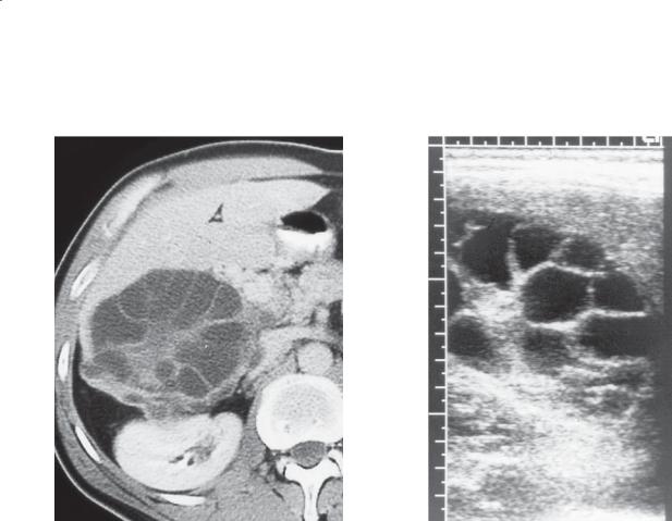
Contrast-enhanced CT (A) and hepatic sonogram (B). A multiloculated mass is present within the right lobe of the liver. Multiple smaller cysts are seen within a large mass.
Di erential Diagnosis
1.Echinococcal hepatic abscess
2.Pyogenic abscess
3.Biliary cystadenoma
Diagnosis
Echinococcal hepatic abscess
6. LIVER 493
B
Discussion
Echinococcal disease is most prevalent in Greece, Uruguay, Argentina, New Zealand, and Australia— areas where dogs are used to herd livestock. The disease is caused by two different types of tapeworms:
Echinococcus granulosus and E multilocularis. For E granulosus (as in this case), dogs are the primary host. The tapeworm lives in the dog and sheds eggs that, when ingested by humans, activate an embryo that invades the intestinal wall and enters the splanchnic venous system. Usually the embryo is carried to the liver, where it develops into a growing cyst containing larvae. For E multilocularis (alveolaris), cats and rodents are the primary host. Human infection follows the same course, except the larvae proliferate outside the hepatic cyst and penetrate liver tissue. They induce a granulomatous reaction about the cyst.
Disease type: Masses

494 MAYO CLINIC GASTROINTESTINAL IMAGING REVIEW
CASE 6.26 |
CASE 6.27 |
Findings
CASE 6.26. Contrast-enhanced CT. There is a peripherally calcified water-attenuation mass in the liver. Within the mass are several cysts that are lower attenuation than the remaining internal fluid.
CASE 6.27. Contrast-enhanced CT. Multiple low-density masses are present throughout the liver. The largest mass in the left lobe appears to be septated.
Di erential Diagnosis
1.Echinococcal cyst
2.Autosomal polycystic disease
3.Multiple simple hepatic cysts
Diagnosis
Echinococcal cyst
Discussion
Th e lesion in case 6.26 is typical for an echinococcal cyst with the visible daughter lesions and peripheral calcification. The lesions in case 6.27 are less typical and show that echinococcal disease also can present as a unilocular mass or as multiple masses. The larger masses in case 6.27 contain daughter cysts but only
appear to be septated; in this case, a high level of clinical suspicion would be required to make the correct diagnosis.
Most patients are infected during childhood and remain asymptomatic until adulthood. The cyst(s) enlarges slowly at about 1 cm per year. Serologic tests are the most helpful to establish the diagnosis. Complications include rupture of the cyst into the biliary system or peritoneal cavity.
At CT, Echinococcus granulosus presents as either a unilocular or a multilocular cyst. The walls can be of variable thickness. Daughter cysts usually are located in the periphery of the mother cyst, and they usually are of lower attenuation. Calcification of the wall is common. E multilocularis presents with geographic, infiltrating regions of hypoattenuation. Invasive, poorly defined solid masses are present.
Th erapy of E granulosus cysts is usually with a combination of surgery and antibiotics. Recently, percutaneous therapy has been described with the addition of either hypertonic saline or alcohol. There is a risk of anaphylaxis if the cyst fluid escapes into the peritoneal cavity; however, new devices have been developed to prevent this problem.
Disease type: Masses

6. LIVER 495
CASE 6.28 |
CASE 6.29 |
Findings
CASE 6.28. Contrast-enhanced CT. Multiple small, lowattenuation masses are present in the liver and spleen.
Sonogram. Multiple bull’s-eye lesions are present in the liver.
Di erential Diagnosis
1.Hepatosplenic candidiasis
2.Lymphoma
3.Leukemic infiltration
Diagnosis
Candidiasis
Discussion
Candidiasis is the commonest systemic fungal infection, usually affecting immunocompromised patients. Patients receiving chemotherapy and those with AIDS are particularly at risk. Diagnosis can be
difficult because only about 50% of affected patients have a positive blood culture.
Findings at sonography include the following:
1.Bull’s-eye pattern that includes a hyperechoic center with a hypoechoic rim
2.Wheel within a wheel, which describes a three-layer lesion consisting of a central hypoechoic nidus surrounded by a hyperechoic rim surrounded by another hypoechoic rim
3.Uniformly hypoechoic liver due to progressive fibrosis
4.Echogenic liver caused by scar formation
Findings at CT are usually multiple small, hypoattenuating masses. Occasionally, scattered calcification can be identified. Increased periportal fibrosis can be identified as hyperattenuating periportal regions seen on delayed enhancement scanning.
Disease type: Masses
496 MAYO CLINIC GASTROINTESTINAL IMAGING REVIEW
TABLE 6.3
Hepatic Abscesses |
|
CASE |
|
|
|
Pyogenic abscess |
Solitary or multiple (clustered) water-attenuation masses |
6.23 |
|
with rim enhancement |
|
|
|
|
Amebic abscess |
Solitary, low-attenuation ring, right lobe |
6.24 |
|
|
|
Echinococcal abscess |
Echinococcus granulosus. Solitary low-attenuation mass with |
6.25–6.27 |
|
internal daughter cysts |
|
|
|
|
Candidiasis |
Multiple small, low-attenuation masses in liver and spleen. |
6.28 and 6.29 |
|
Immunocompromised host. Bull’s-eye lesions at sonography |
|
|
|
|
Disease type: Masses
6. LIVER 497
CASE 6.30
A B C
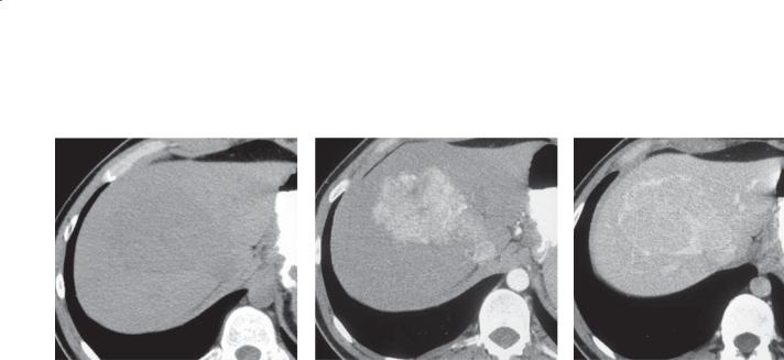
Findings
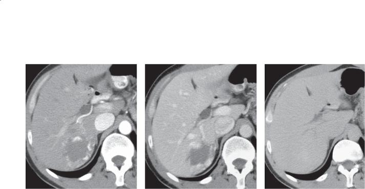
Contrast-enhanced CT. Arterial-phase (A), portal venous phase (B), and delayed (C) images of the liver show a low-attenuation lesion in the posterior right lobe of the liver with peripheral globular enhancement with progressive fill-in on later images. The enhancing tissue is isoattenuating or hyperattenuating compared with the aorta.
Di erential Diagnosis
Hepatic hemangioma
Diagnosis
Hepatic hemangioma
Discussion
Except for benign cysts, hepatic hemangiomas are the most common benign tumor of the liver, found in approximately 5% of the population. Hemangiomas can occur at any age, but frequently they are found in middle-aged women (female:male ratio, 5:1). Hemangiomas are usually solitary but can be multiple in up to one-third of patients. Approximately 90% of hemangiomas are less than 4 cm in size. These small
hemangiomas are rarely symptomatic and often are found incidentally on radiographic examinations of the abdomen. Giant cavernous hemangiomas (typically defined as larger than 4 cm, case 6.35) can rarely cause symptoms due to mass effect on adjacent structures, consumptive coagulopathy (Kasabach-Merritt syndrome), thrombosis of the vascular spaces, or even spontaneous or traumatic rupture.
Hemangiomas arise from vascular endothelial cells and consist of vascular channels of varying size supported by fibrous septae. Blood, supplied by the hepatic artery, constitutes the bulk of the internal contents.
Th e typical CT findings of a hemangioma are present in this case. The peripheral globular
enhancement that is isoattenuating or hyperattenuating compared with the aorta (because blood supply
is arterial) with progressive centripetal fill-in is considered pathognomonic. Hemangiomas also can be diagnosed confidently at MRI (case 6.32), sonography (case 6.33), and nuclear medicine-tagged red blood cell scanning (case 6.34). Atypical hemangiomas (cases 6.36 and 6.37) can be mistaken for other primary liver tumors or metastases.
Disease type: Masses
498 MAYO CLINIC GASTROINTESTINAL IMAGING REVIEW
CASE 6.31
Findings
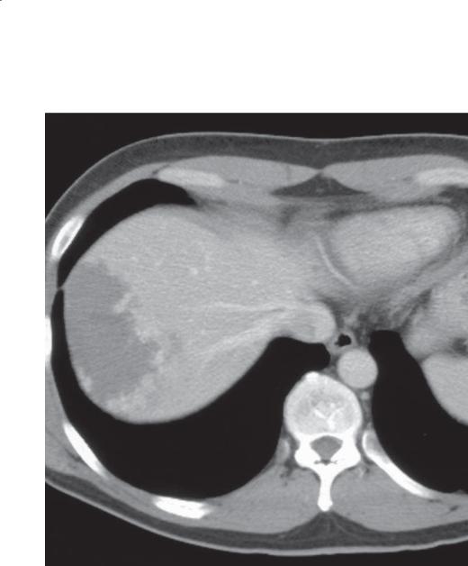
Contrast-enhanced CT. A large hypodense mass is present in the right lobe of the liver with peripheral high-attenuation globular enhancement.
Di erential Diagnosis
Hemangioma
Diagnosis
Hemangioma
Discussion
Th is case illustrates the typical enhancement pattern of a hemangioma at CT. Hemangiomas are composed of large vascular channels filled with slow-moving blood. These vascular channels are fed by the hepatic arteries and result in peripheral nodular enhancement on early phases of imaging with progressive centripetal fill-in that usually occurs after 5 to 30 minutes. The enhancement of a hemangioma should always parallel the aorta. Hemangiomas often have central areas of fibrosis (especially in larger tumors), and complete fill-in of the tumor on delayed images is not necessary for confident diagnosis.
Disease type: Masses
6. LIVER 499
CASE 6.32
A C
B
Findings
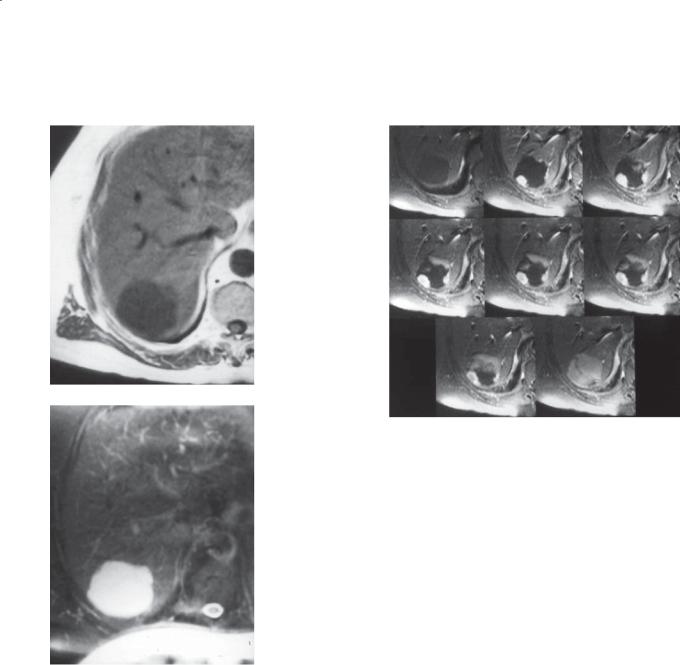
Unenhanced and post-gadolinium MRI. A. T1-weighted sequence. A mass with low signal intensity is present in the posterior right lobe of the liver. B. T2-weighted (fat-suppressed) sequence. The mass is of markedly increased signal. The signal
intensity of the mass mimics the signal intensity of the cerebrospinal fluid. C. Precontrast and multiple delayed post-gadolinium images show peripheral globular enhancement with progressive centripetal fill-in.
Di erential Diagnosis
Hemangioma
Diagnosis
Hemangioma
Discussion
MRI is a sensitive and specific imaging method for the diagnosis of hemangiomas. The markedly high signal seen on T2-weighted images (approaching that of a cyst) is due to the extremely long relaxation time of free fluid—in this case, slowly moving blood. If the signal intensity on T2-weighted images is not as high
as that of the cerebrospinal fluid, gadolinium-enhanced images can be very helpful. These images provide information similar to that of contrast-enhanced CT.
Disease type: Masses
500 MAYO CLINIC GASTROINTESTINAL IMAGING REVIEW
CASE 6.33
Findings
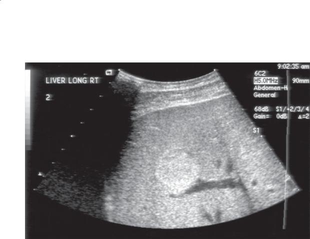
Sonogram. A well-defined, homogeneously hyperechoic mass with increased through transmission is present in the right lobe of the liver.
Di erential Diagnosis
1.Hemangioma
2.Hyperechoic metastasis
3.Focal fatty infiltration
Diagnosis
Hemangioma
Discussion
Th is mass has the typical homogeneous hyperechoic sonographic appearance of a hemangioma. Twothirds of hemangiomas have this appearance. The increased echogenicity within these lesions is related to the numerous acoustical interfaces (multiple vascular channels) within them. The increased through transmission in this case is a nonspecific finding,
but it is seen in approximately 75% of hemangiomas. Because of the extremely slow blood flow within these vascular tumors, flow will not routinely be identified within a hemangioma on color or duplex Doppler imaging. Atypical sonographic appearances of hemangiomas (case 6.37) are relatively common and often require additional imaging (CT or MRI) for
confident diagnosis. Echogenic metastases can mimic a hemangioma but often have a hypoechoic halo, raising the suspicion of malignancy. Focal fatty infiltration of the liver usually presents as a geographic hyperechoic region rather than a rounded mass.
Disease type: Masses
CASE 6.34
A
Findings
A.Unenhanced CT. A large indeterminate lowattenuation mass is present in the periphery of the liver.
B.Technetium Tc 99m–labeled red blood cell scintigraphy (1 hour delayed axial single-photon emission CT image). The mass has markedly increased tracer uptake compared with the rest of the liver.
Di erential Diagnosis
Hepatic hemangioma
Diagnosis
Hepatic hemangioma
6. LIVER 501
B
Discussion
Hemangiomas such as this one are common incidental findings on CT and ultrasonography. In this patient, the mass was evaluated with red blood cell scintigraphy because of an increased creatinine value and pacemaker (contraindications to contrastenhanced CT and MRI). Single-photon emission CT tagged red blood cell scintigraphy has an extremely high specificity (approximately 95%) for the diagnosis of hemangiomas. The sensitivity of single-photon emission CT red blood cell scintigraphy is also high for hemangiomas more than 2 cm in diameter.
Hemangiomas typically have decreased or no red blood cell-labeled tracer uptake immediately after tracer injection. Delayed imaging at 1 to 2 hours typically shows markedly increased tracer uptake within a hemangioma compared with the normal liver. Liver metastases, alternatively, typically have early tracer uptake with quick washout.
Disease type: Masses
502 MAYO CLINIC GASTROINTESTINAL IMAGING REVIEW
CASE 6.35
A C
B D
Findings
Contrast-enhanced CT. A through D. Progressively delayed post-contrast images show a very large lowdensity mass replacing the majority of the left lobe of the liver and extending into the right lobe. This mass has
peripheral globular enhancement equal to that of the aorta with progressive centripetal fill-in on delayed images.
Di erential Diagnosis
Giant hemangioma
Diagnosis
Giant hemangioma
Discussion
Giant hemangiomas usually are defined as those more than 4 cm in diameter; they constitute only 10% of all
hemangiomas. Most hemangiomas are asymptomatic, but giant hemangiomas may cause pain from mass effect on adjacent structures or, rarely, intratumoral thrombosis. Giant hemangiomas are at increased risk of bleeding with trauma and have been reported to rupture spontaneously. Giant hemangiomas can result in thrombocytopenia or an intrahepatic consumptive coagulopathy (Kasabach-Merritt syndrome). Biliary obstruction and Budd-Chiari syndrome can even occur as a result of mass effect from these large lesions.
Symptomatic or complicated giant hemangiomas often are treated with surgical enucleation. Hepatic arterial embolization of these tumors is an alternative therapy in symptomatic patients who are poor surgical candidates. Observation is reserved for asymptomatic patients.
Disease type: Masses
6. LIVER 503
CASE 6.36 |
CASE 6.37 |
Findings
CASE 6.36. Contrast-enhanced CT. A single slice from a portal-venous phase CT shows a small low-density lesion in the right lobe of the liver with central enhancement.
CASE 6.37. Sonogram. An isoechoic liver lesion with a thin echogenic rim (arrows) is present.
Di erential Diagnosis
1.Atypical hemangioma
2.Metastasis
3.Primary liver tumor
Diagnosis
Atypical hemangioma
Discussion
Approximately 15% of hemangiomas have atypical enhancement at CT, which presents a significant diagnostic challenge. These atypical hemangiomas
may present with diffuse early arterial enhancement and rapid washout (“flash” hemangioma). This enhancement pattern usually is seen in small lesions. Occasionally, as in case 6.36, hemangiomas have central enhancement with centrifugal fill-in on delayed images.
Atypical features of hemangiomas on ultrasonography are common (approximately one-third of cases) and include inhomogeneous hyperechoic masses, isoechoic or hypoechoic masses with a thin hyperechoic rim (as in case 6.37), or hypoechoic masses in the setting of a diffusely hyperechoic (fatty) liver.
MRI (especially for small lesions) or red blood cell scintigraphy (for lesions >2 cm) is often used as the primary diagnostic or problem-solving tool for atypical lesions. Radiographically indeterminate lesions can
be observed with close (6-month) follow-up imaging or with biopsy. Percutaneous biopsy with a 20-gauge needle is a safe and effective method for diagnosing hemangioma.
Disease type: Masses
504 MAYO CLINIC GASTROINTESTINAL IMAGING REVIEW
CASE 6.38
A B C
Findings
A. Unenhanced CT. An isodense mass is present in the right lobe of the liver.
B and C. Biphasic contrast-enhanced CT. A large homogeneously enhancing mass is present within the right lobe of the liver. A central scar is visible. The mass is nearly isodense with the liver on the portal venous phase of imaging with prominent peripheral enhancement.
Di erential Diagnosis
1.Focal nodular hyperplasia
2.Hepatic adenoma
3.Hepatocellular carcinoma
Diagnosis
Focal nodular hyperplasia
Discussion
Focal nodular hyperplasia (FNH) is a solid mass consisting of abnormally arranged hepatocytes, bile ducts, and Kupffer cells (hepatic hamartoma). It is the second most common liver tumor, surpassed only by hemangiomas. FNH is believed to arise as a result of
locally increased hepatic blood flow from a hepatic arteriovenous malformation. FNH is most common in women in their 30s and 40s. It is nearly always clinically silent and is discovered incidentally with cross-sectional imaging.
Th e key finding of FNH is homogeneous enhancement during the arterial phase of imaging. Visualization of a central scar (cases 6.39, 6.41, 6.42, and 6.44) is helpful.
Although FNH has no capsule, increased enhancement around the lesion can be seen (as in this case) as a result of large peripheral draining veins.
Hepatic adenoma (cases 6.45 through 6.48) can be seen in the same patient population, but it often enhances heterogeneously (evidence of prior
intratumoral hemorrhage) without a central scar. In most instances, FNH can be diagnosed with confidence. Hepatocellular carcinomas (cases 6.53 through
6.60) usually are found in patients with underlying cirrhosis and can have features similar to those of hepatic adenomas. The extremely rare fibrolamellar hepatocellular carcinoma (case 6.67) can have CT and MRI findings similar to those of FNH, including a central scar, but often other evidence of malignancy is present, including adenopathy and angioinvasion.
Disease type: Masses
6. LIVER 505
CASE 6.39
A B C
Findings
A. Unenhanced CT. An almost isodense mass is present in the periphery of the right lobe of the liver with a hypodense structure centrally.
B and C. Biphasic contrast-enhanced CT. A homogeneously enhancing mass is present on the arterial phase of enhancement with a stellate central scar. The mass is isodense with the surrounding liver during the portal venous phase of enhancement. The hypodense central scar is still visible with a thin rim of hyperenhancing veins around the mass.
Di erential Diagnosis
1.Focal nodular hyperplasia
2.Fibrolamellar hepatocellular carcinoma
Diagnosis
Focal nodular hyperplasia
Discussion
Th is case has the classic CT appearance of focal nodular hyperplasia (FNH). The intense, uniform,
early enhancement in this case is typical of FNH, as is its barely perceptible appearance on unenhanced and delayed post-contrast CT imaging. The hallmark feature of FNH is homogeneous arterial-phase enhancement, a central stellate scar, and radiating fibrous septa dividing the tumor into lobules. The central scar contains the supplying arteries and
bile ductules. The central stellate scar sometimes is more easily visualized on MRI (case 6.41). When imaging studies are inconclusive, biopsy sometimes is necessary. Histologically, the presence of abnormal vessels in a scar or septa, as well as the abnormally
organized hepatocytes, bile ductules, and Kupffer cells, is typical.
Th e presence of a central scar is not pathognomonic for FNH because it can be identified in various benign and malignant neoplasms. Only when it is found in combination with homogeneous arterial enhancement should FNH be diagnosed. Some fibrolamellar hepatocellular carcinomas also contain a scar, but their enhancement is usually inhomogeneous and other signs of a malignancy may be present.
Disease type: Masses
506 MAYO CLINIC GASTROINTESTINAL IMAGING REVIEW
CASE 6.40
A B
C
Findings
A. Sonogram. B. Color-Doppler evaluation. A large isoechoic mass is present in the right lobe of the liver. The mass is extremely vascular (especially centrally) compared with the normal surrounding liver on the color-Doppler image.
C. Contrast-enhanced CT. Corresponding arterial-phase image shows the gently lobulated homogeneously enhancing mass. The central scar is better shown on CT than ultrasonography.
Di erential Diagnosis
1.Focal nodular hyperplasia
2.Other solid liver mass
Diagnosis
Focal nodular hyperplasia
Discussion
Focal nodular hyperplasia (FNH) is often a subtle liver mass on ultrasonography, consistent with its
description as a “stealth” lesion. FNH usually is isoechoic to normal liver parenchyma and may be seen only because of subtle contour abnormalities and displacement of normal vascular structures. The central scar, when visualized, usually is hypoechoic. Doppler features of FNH are highly specific for the diagnosis because of the prominent central vessels arranged in a stellate pattern.
FNH is most commonly found incidentally in women in their 30s and 40s. It is important to
differentiate this benign lesion that does not require any treatment from other, more serious masses. Hepatic adenomas usually enhance heterogeneously and may have a capsule.
Oral contraception is not thought to cause development of FNH, but it may exert an effect on its growth. This is in contrast to the clearly proven risk for development of a hepatic adenoma with the use of oral contraceptive agents. Hepatic adenomas are also at a high risk for bleeding, in contradistinction to FNH.
Disease type: Masses
6. LIVER 507
CASE 6.41
A B
C D
Findings
Unenhanced and dynamic gadolinium-enhanced images of the liver. A. T1-weighted MRI. An isointense mass with a hypointense central scar is present in the periphery of the right lobe of the liver. B. T2-weighted MRI. The mass is nearly isointense to the liver parenchyma with a hyperintense central scar. C. Arterial-phase post-gadolinium MRI. Marked
homogeneous enhancement of the mass with a central hypointense stellate scar. Enhancing peripheral veins also are present. D. Venous-phase post-gadolinium MRI. The mass is nearly isointense compared with the surrounding liver.
Di erential Diagnosis
Focal nodular hyperplasia
Diagnosis
Focal nodular hyperplasia
Discussion
Th is case has almost all of the classic MRI findings of focal nodular hyperplasia (FNH). FNH typically is isointense on T1and T2-weighted images. The central scar is typically hypointense on T1-weighted images and hyperintense on T2-weighted images. Post-gadolinium homogeneous enhancement pattern mirrors that at contrast-enhanced CT (case 6.39).
Typical features of FNH at CT and MRI are diagnostic. Follow-up imaging can be avoided. Unfortunately, often FNH has atypical findings at unenhanced MRI (hypointense or hyperintense on either T1or T2-weighted images or both). Gadolinium enhancement then is recommended to show the homogeneous arterial perfusion of the
mass. Sulfur colloid scintigraphy can be helpful in the minority of cases that remain equivocal. Lesions must be 2 cm or more. Tracer uptake within the Kupffer cells (reticuloendothelial system) can be identified in approximately two-thirds of the masses. Hepatobiliary MRI agents (gadoxetate disodium) are usually taken up by FNH and are helpful for making a diagnosis (case 6.44).
Disease type: Masses
508 MAYO CLINIC GASTROINTESTINAL IMAGING REVIEW
CASE 6.42
A C
B D
Findings
A through D. Unenhanced and sequential postgadolinium, fat-saturated T1-weighted MRI. A slightly hypointense 3-cm mass with hypointense central
scar is seen on the pre-contrast image (A). The mass is homogeneously hyperenhancing with central
to peripheral septations on arterial-phase postgadolinium images. Gradual enhancement of the stellate central scar is visible on delayed images.
Di erential Diagnosis
Focal nodular hyperplasia
Diagnosis
Focal nodular hyperplasia
Discussion
Th is case has the typical appearance and enhancement pattern of focal nodular hyperplasia (FNH) at MRI. A central scar may be easier to detect at MRI than at CT. The scar is typically hypointense on T1-weighted images and hyperintense on T2-weighted images.
In addition, the typical lobulation (segmentation) of FNH is shown (B). Often, these tumors are segmented by fibrous septa—analogous to the segmentation found in an orange. These usually are identified most easily during the early phases of contrast enhancement. Their presence can be very helpful for suggesting the diagnosis.
Disease type: Masses

6. LIVER 509
CASE 6.43
A C
B
Findings
A and B. Coronal single-photon emission CT images from sulfur colloid scintigraphy. A rounded area of focally increased tracer uptake is seen in the lateral segment of the left lobe of the liver (arrow in A) with a smaller focus of increased uptake in the posterior right lobe of the liver (arrow in B).
C. Contrast-enhanced CT. Two hyperenhancing masses are seen on this late arterial-phase image, which correspond to the two areas of increased tracer uptake on the sulfur colloid scan.
Di erential Diagnosis
Multiple focal nodular hyperplasia
Diagnosis
Multiple focal nodular hyperplasia
Discussion
Sulfur colloid scintigraphy is often helpful for the diagnosis of focal nodular hyperplasia (FNH). FNH contains functioning reticuloendothelial cells (Kupffer cells). These cells normally take up radiolabeled sulfur colloid. When a liver mass has uptake of sulfur colloid equal to (60% of cases of FNH) or greater than (10% of cases of FNH) that of normal liver, the findings are pathognomonic of FNH. In 30% of cases of FNH, there will not be uptake of sulfur colloid, and these lesions remain indeterminate. Hepatic adenomas often contain dysfunctional Kupffer cells but sulfur colloid uptake
is in very low quantities when it occurs and rarely is isointense with liver.
FNH is multiple in 20% of cases. Multiple FNH is associated with vascular malformations in the liver (hepatic hemangiomas in 25% of cases) and in other organs.
Disease type: Masses
510 MAYO CLINIC GASTROINTESTINAL IMAGING REVIEW
CASE 6.44
A B
C D
Findings
A.T1-weighted MRI. An isointense mass with central stellate scar is present near the junction between the right and left hepatic lobes.
B.T2-weighted MRI. The mass is nearly isointense and the central scar is hyperintense.
C.T1-weighted MRI, portal venous phase, postcontrast enhancement. Homogeneous enhancement of the mass with a central hypointense scar.
D.T1-weighted image, 18-minute delayed after administration of gadoxetic acid. Hyperintense homogeneous enhancement of the mass with a hypointense central scar.
Di erential Diagnosis
1.Focal nodular hyperplasia
2.Well-differentiated hepatocellular carcinoma
3.Regenerative nodule
Diagnosis
Focal nodular hyperplasia
Discussion
Hepatocellular contrast agents can be helpful for discriminating focal nodular hyperplasia from other types of liver tumors, especially metastases. Gadoxetic acid (gadoxetate disodium) is used commonly for this purpose and to increase hepatic contrast for
the detection of small metastases. Gadoxetic acid is excreted 50% from the biliary system and 50% from the kidneys. In the liver, it is taken up by hepatocytes and excreted in the biliary system. Hepatocyte uptake occurs approximately 10 minutes after administration, and imaging can be performed successfully several hours later. Approximately 90% of patients with focal nodular hyperplasia will have uptake of gadoxetic acid (homogeneous, heterogeneous, or peripheral). Welldifferentiated hepatocellular carcinomas have been reported to have uptake of gadoxetic acid; therefore, clinical correlation with imaging findings is always necessary.
Disease type: Masses
CASE 6.45
A
Findings
Contrast-enhanced CT. A and B. Arterial and portal venous-phase images show a large, well-defined, heterogeneously enhancing mass in the right lobe of the liver with an enhancing capsule.
Di erential Diagnosis
1.Hepatic adenoma
2.Hepatocellular carcinoma
Diagnosis
Hepatic adenoma
6. LIVER 511
B
Discussion
Hepatic adenomas are rare, benign primary tumors composed of cords of hepatocytes with an abnormal architecture. These tumors are most commonly found in young women and have an increased incidence in patients using oral birth control. Hepatic adenomas often present as large solitary masses (such as in
this case), reaching up to 30 cm in diameter. They usually have heterogeneous enhancement and a welldefined capsule. No imaging methods can reliably differentiate hepatic adenoma from hepatocellular carcinoma. Because of their propensity to hemorrhage, solitary hepatic adenomas usually are removed surgically. Alternatively, if the patient is taking oral contraceptives, use of the medication may be stopped to determine whether the tumor regresses on its own.
Disease type: Masses
512 MAYO CLINIC GASTROINTESTINAL IMAGING REVIEW
CASE 6.46
A
C
Findings
A.T1-weighted MRI. A mass of mixed low and high signal intensity is present in the right lobe of the liver.
B.T2-weighted MRI. The mass is of heterogeneous intensity with a high-intensity capsule.
C.Contrast-enhanced CT. The mass and capsule are heterogeneously enhancing.
Di erential Diagnosis
1.Hepatic adenoma
2.Hepatocellular carcinoma
Diagnosis
Hepatic adenoma
B
Discussion
Hepatic adenomas usually have a heterogeneous, mosaic (multicompartment) appearance on CT and MRI. An enhancing capsule also is frequently observed. Areas of increased T1 signal intensity are often present within hepatic adenomas (as in this case) and may be caused by fat content or intratumoral hemorrhage. Although hepatocellular carcinoma can have an identical radiographic appearance, hepatic adenoma
is the favored diagnosis in young patients with no radiographic evidence or clinical history of cirrhosis.
Disease type: Masses
CASE 6.47
A
Findings
A.Unenhanced CT. A high-attenuation mass with a whorled appearance is present in the right lobe of the liver.
B.Contrast-enhanced CT. An underlying mixedattenuation mass is present on portal venous-phase imaging. The high-attenuation whorled regions on the pre-contrast image are difficult to identify on the enhanced image.
Di erential Diagnosis
1.Hemorrhagic hepatic adenoma
2.Hemorrhagic hepatocellular carcinoma
3.Hemorrhagic metastasis
Diagnosis
Hepatic adenoma, hemorrhagic
6. LIVER 513
B
Discussion
Th is 33-year-old woman who was taking oral contraceptives presented to the emergency department with acute pain in the right upper quadrant. At operation, a hemorrhagic hepatic adenoma was found. Unenhanced images are important for identifying hyperattenuating areas of acute hemorrhage associated with hepatic adenoma. Rupture of a peripheral hepatic adenoma with hemoperitoneum may be lifethreatening.
Disease type: Masses
514 MAYO CLINIC GASTROINTESTINAL IMAGING REVIEW
CASE 6.48
Findings
Contrast-enhanced CT. A large subcapsular hematoma is present along the lateral right lobe of the liver with layering blood products. A 4-cm, heterogeneously enhancing mass is present in the periphery of the right lobe of the liver, adjacent to the hematoma.
Di erential Diagnosis
1.Hepatic adenoma (with subcapsular hemorrhage)
2.Hemorrhagic hepatocellular carcinoma
3.Hemorrhagic metastasis
Diagnosis
Ruptured hepatic adenoma with subcapsular hematoma
Discussion
Even asymptomatic hepatic adenomas usually are resected, primarily to avoid potential life-threatening hemorrhages, such as in this case. Although hepatic adenomas cannot be differentiated radiographically from hepatocellular carcinoma or hepatic metastases, hepatic adenoma was correctly favored as a cause of the large subcapsular hemorrhage because the patient was a young woman taking oral contraceptives.
Disease type: Masses
CASE 6.49
Findings
Contrast-enhanced CT. Two hypervascular liver masses are present during the portal venous phase of contrast enhancement. The liver is of low attenuation compared with the spleen and paraspinal muscles.
Di erential Diagnosis
1.Multiple hepatic adenomas (in setting of glycogen storage disease)
2.Metastases
Diagnosis
Glycogen storage disease with multiple hepatic adenomas (von Gierke disease)
6. LIVER 515
Discussion
Hepatic adenomas are multiple in approximately onethird of cases. Cases of solitary or multiple hepatic adenomas are more common in women taking oral birth control pills. Hepatic adenomas also are more common in patients with glycogen storage disease, anabolic steroid use, or familial diabetes mellitus. This patient had a history of von Gierke disease (type Ia glycogen storage disease). The steatohepatitis
commonly associated with von Gierke disease accounts for the hepatomegaly and diffuse low attenuation of the liver parenchyma on CT. One or more hepatic adenomas are reported to be present in up to 40%
of patients with von Gierke disease. Malignant degeneration of hepatic adenomas into hepatocellular carcinoma can occur and should be considered if there is rapid growth of a liver lesion.
Disease type: Masses
516 MAYO CLINIC GASTROINTESTINAL IMAGING REVIEW
CASE 6.50
A B
Findings
A.Nonenhanced CT. Mass is of heterogeneous attenuation. Some of the low-attenuation tissue approximates fat attenuation.
B.Contrast-enhanced CT. Well-circumscribed, heterogeneously enhancing mass is within the left hepatic lobe. Near fat attenuation tissue is visible.
Di erential Diagnosis
1.Angiomyolipoma
2.Lipoma
3.Hepatic adenoma
4.Hepatocellular carcinoma
Diagnosis
Angiomyolipoma
Discussion
Angiomyolipomas are rare, benign mesenchymal tumors composed of varying proportions of fat, smooth muscle, and vascular elements. Typically, these tumors are asymptomatic and discovered incidentally.
Angiomyolipomas of the liver may occur as either a solitary mass (as in this case) or as multiple masses in association with tuberous sclerosis. Hepatic angiomyolipomas are much rarer than their renal counterparts and occur in only 5% of patients with tuberous sclerosis with renal angiomyolipomas.
Grossly, hepatic angiomyolipomas are wellcircumscribed masses ranging in size from 1 to
20 cm. They typically present as a hyperechoic mass on ultrasonography and can easily be mistaken for a hemangioma. Demonstration of fat within the mass is imperative for making the diagnosis. Unfortunately,
angiomyolipomas arising in the liver do not consistently have the high fat content of renal angiomyolipomas. Lipomatous tissue may make up less than 5% of hepatic angiomyolipomas. As a rule, fatty tumors of
the liver are benign (angiomyolipoma, lipoma, hepatic adenoma); however, hepatocellular carcinomas (case 6.55) or metastatic liposarcoma also can contain fat. These conditions usually have evidence of cirrhosis or a history of a liposarcoma. Percutaneous needle biopsy or surgical biopsy may be required for equivocal cases. Spontaneous hemorrhage of hepatic angiomyolipomas is less common than with their renal counterparts.
Disease type: Masses

|
|
6. LIVER 517 |
|
|
|
|
TABLE 6.4 |
|
Benign Liver Tumors |
|
CASE |
|
|
|
Hemangioma |
Globular peripheral enhancement isodense to aorta. Always follows |
6.30–6.37 |
|
blood characteristics |
|
|
|
|
Focal nodular hyperplasia |
Homogeneous arterial-phase enhancement. Central scar |
6.38–6.44 |
|
|
|
Hepatic adenoma |
Heterogeneous enhancement. Hemorrhage in symptomatic lesions. |
6.45–6.49 |
|
Oral contraceptives causative |
|
|
|
|
Angiomyolipoma |
Internal fat. Increased in tuberous sclerosis |
6.50 |
|
|
|
Cyst |
Homogeneous fluid contents without enhancement. Posterior |
6.18 |
|
acoustic enhancement |
|
|
|
|
Disease type: Masses
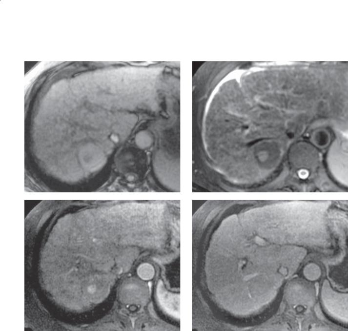
518 MAYO CLINIC GASTROINTESTINAL IMAGING REVIEW
CASE 6.51
A B
C D
Disease type: Masses

Findings
A.T1-weighted out-of-phase MRI of the liver. Cirrhotic changes in the liver with a nodular surface contour and a hyperintense mass within the posterior segment of the right hepatic lobe. The mass contains a capsule and a low-intensity nodule.
B.T2-weighted MRI. The mass is isointense to the liver except for a small hyperintense internal focus.
C.Dynamic contrast-enhanced MRI. The mass enhances homogeneously with the remainder of the liver except a small focus of hyperenhancing tissue that corresponds to the hyperintense nodule on the T2-weighted image.
D.Delayed contrast-enhanced MRI. The mass is entirely isointense with the remainder of the hepatic parenchyma.
Di erential Diagnosis
1.Dysplastic nodule
2.Dysplastic nodule with a focus of hepatocellular carcinoma
3.Regenerative nodule
4.Indeterminate hepatic mass
Diagnosis
Dysplastic nodule with a focus of hepatocellular carcinoma (early hepatocellular carcinoma)
6. LIVER 519
Discussion
Th e primary reason for screening patients with cirrhosis is to assess for a complicating hepatocellular carcinoma (HCC). A dominant “nodule” or mass should be an important clue that a problem exists. The 2 main possibilities if a mass is found are a dysplastic nodule or HCC. In the case shown here, the dominant mass is of high intensity on the T1-weighted images—a common finding for dysplastic nodules (usually due to internal fat or copper). The presence of a capsule
is unusual for a benign mass and is worrisome for a malignancy. In addition, a focus of high signal intensity on the T2-weighted images is an important finding
to differentiate benign (isointense) nodules from malignant (hyperintense) nodules. Enhancement of the abnormal nodule is further evidence that malignant transformation has occurred.
Th is case represents a nodule-within-a-nodule appearance for early HCC. Because the dysplastic nodule is larger than the enhancing tumor, this tumor likely arose as a regenerative nodule, transformed into a dysplastic nodule, and later developed a small focus of HCC. A dysplastic nodule should remain isointense on the T2-weighted and contrast-enhanced sequences. A regenerative nodule is usually isointense on both the T1and T2-weighted images. Although tumors other than HCC can develop in a cirrhotic liver, it is much less common than in a noncirrhotic liver.
Disease type: Masses
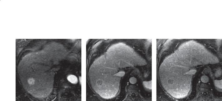
520 MAYO CLINIC GASTROINTESTINAL IMAGING REVIEW
CASE 6.52
A B C
Findings
A.Arterial-phase T1-weighted MRI. A 2-cm enhancing mass is within the liver near the dome.
B.Portal venous phase T1-weighted MRI. The mass is hypointense compared with the surrounding liver. An enhancing capsule is visible.
C.Delayed-phase T1-weighted MRI. The mass is hypointense compared with surrounding liver.
Di erential Diagnosis
1.Hepatocellular carcinoma
2.Dysplastic nodule
3.Flash hemangioma
4.Focal nodular hyperplasia
5.Hypervascular metastasis
Diagnosis
Early hepatocellular carcinoma
Discussion
Hepatocellular carcinoma (HCC) is the most common primary hepatic tumor worldwide. Most of these tumors develop as a complication of hepatic cirrhosis. In sub-Saharan Africa and Asia, the tumor is common because of the high incidence of hepatitis and exposure to aflatoxins. In the Western Hemisphere, the incidence is relatively low and the tumor usually is due to either
alcoholic liver disease or hepatitis. The traditional classification of HCC is useful only for large tumors. Before helical CT scanning, small HCC was rarely detected. With the advent of fast CT scanners and dynamic enhanced MRI, detection of smaller and earlier HCC has been possible. HCC derives its blood supply from the hepatic artery and in most cases is a hypervascular lesion. Screening of cirrhotic patients at risk for the development of HCC requires scanning during the arterial phase of contrast enhancement as well as during the portal venous and delayed phases. Hemangiomas and focal nodular hyperplasia are uncommon in cirrhotic livers. Patients with a history of a malignancy (islet cell carcinoma, carcinoid tumor, renal adenocarcinoma, melanoma) also can present with a similar-appearing lesion.
Triple-phase acquisition can be very helpful for discriminating regions of variant perfusion and dysplastic nodules from early HCC. Both dysplastic nodules and HCC can enhance during the arterial phases of contrast enhancement; however, dysplastic nodules become isointense during the portal and delayed phases of contrast enhancement. HCC washes out during
the delayed phases and will appear hypointense. Early detection is important for appropriate care. Usually, once HCC is identified and reaches 2 cm in diameter, patients are placed on the waiting list for a liver transplant, if they are transplant-eligible.
Disease type: Masses
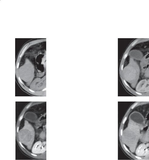
6. LIVER 521
CASE 6.53
A B
C D
Findings
A.Unenhanced CT. A large mass arises from the inferior tip of the right lobe of the liver. The mass is slightly hypoattenuating compared with normal liver.
B.Late arterial-phase contrast-enhanced CT. The mass is enhancing, and there is a hypoattenuating circumferential capsule.
C.Portal venous phase contrast-enhanced CT. The capsule is hyperattenuating compared with the hypoattenuating mass.
D.Delayed contrast-enhanced CT. The capsule and mass are isoattenuating.
Di erential Diagnosis
1.Hepatocellular carcinoma
2.Amebic abscess
3.Metastasis
Diagnosis
Focal hepatocellular carcinoma
Discussion
Advanced hepatocellular carcinoma (HCC) usually is classified as focal, multifocal, or diffuse. Focal tumors have the most favorable biologic behavior-favoring expansion rather than invasion. Typical features of a focal HCC include the presence of a capsule, fat, or a mosaic architecture.
Th e presence of a capsule about a mass is highly suggestive of focal HCC. The appearance of the capsule depends on the phase of contrast enhancement. HCC is a hypervascular lesion that usually enhances briskly during the arterial phase of contrast enhancement and washes out quickly during the portal venous phase
(arterial-venous shunting). The capsule is predominantly fibrous and enhances slowly—usually maximal enhancement occurs in the late portal venous or delayed phases of contrast enhancement. The difference in vascularity explains the changing appearance of both tumor and capsule. Capsules usually are found in welldifferentiated HCC and often indicate a more favorable prognosis than poorly marginated tumors.
Disease type: Masses
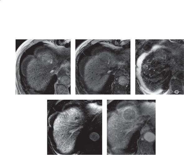
522 MAYO CLINIC GASTROINTESTINAL IMAGING REVIEW
CASE 6.54
A B C
D E
Findings
A.T1-weighted in-phase MRI. A focal mass of high signal intensity is present within the liver near the diaphragmatic dome. Speckled low-intensity changes throughout the liver parenchyma are due to iron deposition.
B.T1-weighted out-of-phase MRI. The lesion is of lower signal intensity than on the in-phase image. Only a small amount of peripheral high signal remains.
C.T2-weighted MRI. The mass is of slightly increased signal intensity compared with the liver. Ascites and respiratory artifacts are present.
D.T1-weighted MRI, contrast-enhanced arterial phase. The mass enhances during the arterial phase of enhancement.
E.T1-weighted image, delayed enhanced phase. The mass is of low signal intensity, and there is a high signal intensity capsule about the mass.
Di erential Diagnosis
1. Hepatocellular carcinoma
2.Hepatic adenoma
3.Lipoma
Diagnosis
Focal hepatocellular carcinoma—intratumoral fat
Discussion
Approximately 40% of hepatocellular carcinomas (HCCs) contain internal fat that can be detected at MRI. Because this is an unusual finding except in rare hepatic lesions (lipomas and angiomyolipomas), it
is a very helpful finding to suggest HCC. This lesion can be distinguished from a lipoma because of its high intensity on the T2-weighted image. Hepatic adenomas, benign primary hepatocellular tumors, can be indistinguishable from well-differentiated, solitary HCC. Correlation with a patient’s age (HCC in the Western Hemisphere is more common in older patients), history of cirrhosis (nearly always present in
patients with HCC), or oral contraceptive use (common among patients with hepatic adenoma) can be helpful. Dysplastic nodules can contain fat, but usually they are isointense to liver on T2-weighted images.
Disease type: Masses
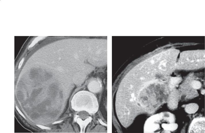
CASE 6.55
Findings
CASE 6.55. Contrast-enhanced CT. There is a large mass in the right lobe of the liver. The internal architecture of the mass is heterogeneous with a multicompartmentalized appearance.
CASE 6.56. Contrast-enhanced CT. A solitary mass with a mosaic internal appearance and hyperattenuating capsule is present in the region of the caudate lobe of the liver.
Di erential Diagnosis
1.Hepatocellular carcinoma
2.Echinococcal cyst
3.Cystic metastasis
Diagnosis
Focal hepatocellular carcinoma—mosaic appearance
6. LIVER 523
CASE 6.56
Discussion
Th ese hepatocellular carcinomas (HCCs) have the classic mosaic internal architecture. The term “mosaic” refers to a multicompartmentalized tumor with multiple discrete solid and cystic regions. In most cases, cystic regions are present in the periphery of the tumor as commonly as in the central regions. The hypoattenuating regions are often small, rounded regions but can assume various shapes. These regions correspond to areas of necrosis and hemorrhage pathologically. The mosaic appearance is highly suggestive of HCC.
Disease type: Masses
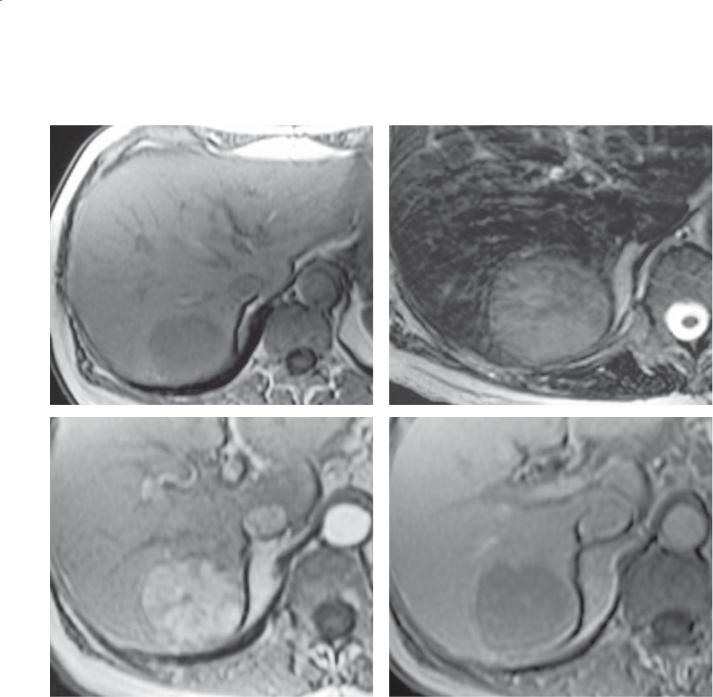
524 MAYO CLINIC GASTROINTESTINAL IMAGING REVIEW
CASE 6.57
A B
C D
Disease type: Masses
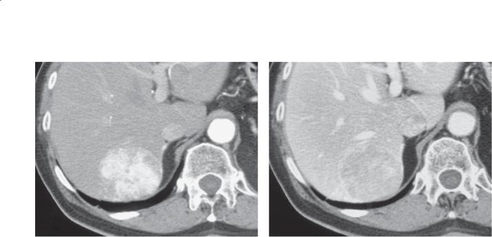
E
Findings
A.T1-weighted MRI. The well-circumscribed mass is of low signal intensity compared with the liver parenchyma.
B.T2-weighted MRI. The mass is fairly homogeneous and hyperintense.
C.Early-phase contrast-enhanced MRI. The mass enhances briskly and inhomogeneously during the early phases of contrast enhancement.
D.Delayed enhancement MRI (3 minutes). The mass is hypointense compared with normal liver.
E.Arterial-phase contrast-enhanced CT. Heterogeneously enhancing mass is in the right hepatic lobe. An enhancing capsule is visible.
F.Portal venous phase contrast-enhanced CT. The mass is now heterogeneously hypoattenuating. The capsule remains hyperattenuating.
Di erential Diagnosis
1.Hepatic cell adenoma
2.Hepatocellular carcinoma
3.Focal nodular hyperplasia
6. LIVER 525
F
Diagnosis
Focal hepatocellular carcinoma
Discussion
Th is case has classic imaging findings of a focal hepatocellular carcinoma (HCC). At unenhanced MRI, the lesion has nonspecific imaging characteristics (hypointense on T1and hyperintense on T2-weighted images). The lesion is hypervascular and has intratumoral shunting (rapid washout of contrast material) after contrast enhancement. Typical capsular enhancement is seen during portal venous-phase CT.
It may be impossible to distinguish a welldifferentiated, solitary HCC from an adenoma. Usually, the clinical history of a young woman taking oral contraceptives strongly suggests an adenoma. HCC should be strongly considered if the patient has a history of cirrhosis and is older than 50 years. Focal nodular hyperplasia should enhance homogeneously during the arterial phase of contrast enhancement and should not have a late-enhancing capsule.
Disease type: Masses
526 MAYO CLINIC GASTROINTESTINAL IMAGING REVIEW
CASE 6.58
A B
Findings
Contrast-enhanced CT. A. A poorly defined mass is present within the right lobe of the liver. A smaller, indeterminate mass is present within the left lobe.
B. Thrombus fills the main and right branches of the portal vein. The liver is cirrhotic in configuration, and multiple large varices and ascites are present.
Di erential Diagnosis
Hepatocellular carcinoma
Diagnosis
Focal hepatocellular carcinoma
Discussion
In many cases hepatocellular carcinoma (HCC) may not have a typical appearance and presents as an indeterminate hepatic mass. In these cases, the presence of secondary signs of HCC are key to suggesting the correct diagnosis. Secondary signs
include angioinvasion with portal venous or hepatic venous thrombus, morphologic changes of hepatic cirrhosis, hemochromatosis, or prior thorium dioxide exposure. In the case shown here, a hypoattenuating nondescript mass and the presence of cirrhosis and portal vein thrombosis are key findings to suggest the correct diagnosis of HCC.
Disease type: Masses
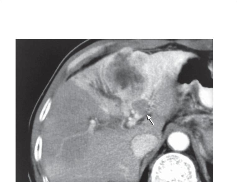
CASE 6.59
Findings
Contrast-enhanced CT. There is a mass within the lateral segment of the left lobe of the liver. The mass has components that are both hyperattenuating and hypoattenuating. About the mass is a wedge-shaped region of hyperattenuation. There is a nodular filling defect within the left lateral segmental portal vein (arrow).
Di erential Diagnosis
1.Hepatocellular carcinoma
2.Metastasis
Diagnosis
Focal hepatocellular carcinoma—transient hepatic attenuation defect
6. LIVER 527
Discussion
Local hepatic perfusion differences allow transient hepatic attenuation defects (THAD) to be seen in the liver. The presence of a THAD should prompt the search for an underlying neoplasm causing compression or obstruction of the supplying portal vein. Because the liver receives the majority of its blood supply from the portal vein, portal venous obstruction results in the recruitment of arterial blood to supply the ischemic region. The additional arterial supply explains the hyperenhancement seen during the early phases of contrast enhancement. In the case shown here, a tumor thrombus is seen to occlude the supplying portal vein. Occlusive tumor thrombus accounts for the THAD, and the presence
of angioinvasion suggests that the underlying tumor is hepatocellular carcinoma.
Disease type: Masses
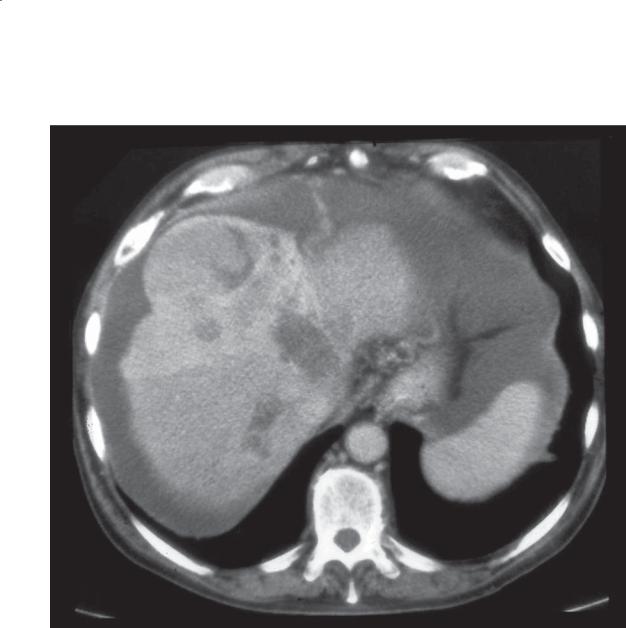
528 MAYO CLINIC GASTROINTESTINAL IMAGING REVIEW
CASE 6.60
Findings
Contrast-enhanced CT. A heterogeneously enhancing mass bulges the hepatic contour. A wedge-shaped region of hyperattenuation surrounds the mass. Filling defects expand the hepatic veins. Ascites.
Di erential Diagnosis
Hepatocellular carcinoma
Diagnosis
Focal hepatocellular carcinoma
Discussion
Th is case exemplifies the importance of secondary findings for diagnosing hepatocellular carcinoma (HCC). Although the primary tumor does not have any specific features of HCC (capsule, fat, mosaic appearance), the presence of a transient hepatic attenuation defect, ascites (due to cirrhosis), and angioinvasion of the hepatic veins (with thrombus) are typical secondary findings. These findings in combination make HCC the only real consideration. Angioinvasion can occur within the portal veins or hepatic veins or both. The same aggressive growth characteristics also can lead to biliary invasion in rare instances.
Disease type: Masses

CASE 6.61
A
Findings
A.Unenhanced CT. A large hypoattenuating mass is present within the right lobe of the liver.
B.Contrast-enhanced CT. The mass does not enhance and appears cystic.
Di erential Diagnosis
1.Abscess
2.Biliary cystadenoma
3.Cystic metastasis
4.Cystic hepatocellular carcinoma
Diagnosis
Cystic hepatocellular carcinoma
6. LIVER 529
B
Discussion
Although the typical hepatocellular carcinoma (HCC) is a hypervascular tumor, occasional tumors are cystic in appearance and mimic an abscess. The diagnosis can be suggested by the presence of secondary signs of HCC (none are seen in this case). Patients with a pyogenic abscess (case 6.23) of this size usually are symptomatic (fevers and leukocytosis). Although possible, a solitary metastasis is rarely this large.
A biliary cystadenoma (case 7.26) generally has enhancing septations.
Disease type: Masses
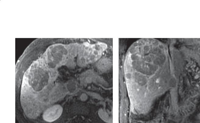
530 MAYO CLINIC GASTROINTESTINAL IMAGING REVIEW
CASE 6.62
A B
Findings
A.Contrast-enhanced T1-weighted MRI. Multiple masses of variable size are present throughout the liver.
B.Contrast-enhanced T1-weighted MRI, coronal plane. A dominant lesion is present in the dome of the liver with multiple surrounding smaller masses.
Di erential Diagnosis
1.Metastases
2.Multifocal hepatocellular carcinoma
Diagnosis
Multifocal hepatocellular carcinoma
Discussion
Th is is a common presentation for advanced hepatocellular carcinoma (HCC). Although multifocal disease can be indistinguishable from metastases, 2 findings should raise suspicion that the masses could be HCC: 1) a dominant (larger) mass with surrounding smaller satellite masses, and 2) masses that are hyperintense during the early phases of contrast enhancement compared with normal liver. In the case shown here, the MRIs were obtained during the late phase of portal venous enhancement, and the tumor
is hypointense compared with normal liver because of arterial-venous shunting that often is found in patients with HCC. Satellite masses are believed to occur from portal venous metastases that seed adjacent hepatic parenchyma.
Disease type: Masses
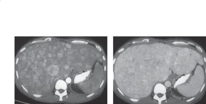
CASE 6.63
A
Findings
A.Arterial-phase contrast-enhanced CT. The liver enhances inhomogeneously. There are multiple illdefined, nodular, enhancing masses throughout the liver.
B.Portal venous phase contrast-enhanced CT. The liver remains slightly inhomogeneous in attenuation. The nodular masses are not well seen, although the contour of the liver is nodular and the left lobe is enlarged.
Di erential Diagnosis
1.Diff use hepatocellular carcinoma
2.Diffuse hypervascular metastases
3.Budd-Chiari syndrome
Diagnosis
Diffuse hepatocellular carcinoma
6. LIVER 531
B
Discussion
Diffuse hepatocellular carcinoma (HCC) is the least common presentation of HCC. The tumor usually infiltrates throughout the liver, and the
borders of the individual nodules are poorly defined. Arterial-phase scanning can be key to detection, because these tumors may be difficult to identify
on portal venous and delayed enhancement images. Diffuse hypervascular metastases (neuroendocrine tumor, renal adenocarcinoma) could have a similar appearance, but usually portions of the tumor appear masslike and have heterogeneous internal changes. Budd-Chiari syndrome (case 6.13) can present with an inhomogeneous liver—but usually the central portions of the liver (including the caudate lobe)
are spared. Biopsy may be required to confirm the presence of the tumor.
Disease type: Masses
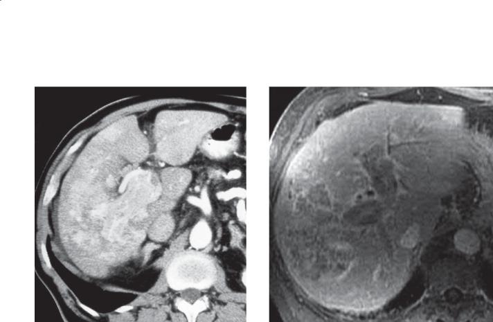
532 MAYO CLINIC GASTROINTESTINAL IMAGING REVIEW
CASE 6.64
A B
Findings
A.Arterial-phase contrast-enhanced CT. The liver enhances inhomogeneously. Hyperattenuating, enhancing soft tissue is present within the portal vein.
B.T1-weighted, contrast-enhanced MRI, delayed phase. A heterogeneously enhancing mass is within the right hepatic lobe. The portal veins are filled with similar-appearing tissue.
Di erential Diagnosis
Hepatocellular carcinoma with portal venous tumor thrombus
Diagnosis
Advanced hepatocellular carcinoma with portal venous tumor thrombus
Discussion
A liver mass in combination with portal venous thrombus is highly suggestive of the diagnosis of hepatocellular carcinoma (HCC). Portal venous thrombosis is common because of the propensity for angioinvasion by HCC. It is not always possible to determine whether the filling defect is bland or tumor thrombus. Contrast enhancement at CT (as in this case) or the presence of an arterial Doppler waveform at sonography is diagnostic for tumor
thrombus. Tumor thrombus adds to the likelihood that the hepatic mass is HCC, and it has grave prognostic significance.
Disease type: Masses
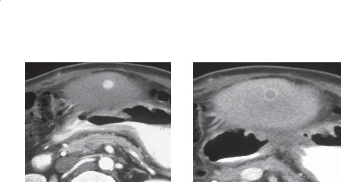
CASE 6.65
A
Findings
A.Arterial-phase contrast-enhanced CT. There is a small enhancing nodule within the left lobe of the liver. This patient had a prior right hepatectomy for hepatocellular carcinoma.
B.Portal venous phase contrast-enhanced CT. The nodule is no longer hyperattenuating, but a small capsule about the mass is hyperattenuating.
Di erential Diagnosis
1.Recurrent hepatocellular carcinoma
2.Focal nodular hyperplasia
3.Flash hemangioma
Diagnosis
Recurrent hepatocellular carcinoma
6. LIVER 533
B
Discussion
Benign tumors are unusual in a cirrhotic liver. As a general rule, any mass in a cirrhotic liver is hepatocellular carcinoma (HCC) until proved
otherwise. In this patient with a history of HCC, the likelihood that this hypervascular mass represents HCC is even greater. The characteristic late-enhancing capsule with central washout also is supportive of this diagnosis. Triphasic CT scanning during the arterial, portal venous, and delayed phases of contrast enhancement is important for optimal detection and characterization of these lesions.
Disease type: Masses
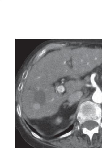
534 MAYO CLINIC GASTROINTESTINAL IMAGING REVIEW
CASE 6.66
Findings
Contrast-enhanced CT. There is a hypoattenuating mass within the right lobe of the liver with an enhancing nodule in its inferior pole. The left lobe of the liver is enlarged, and the right lobe appears atrophic.
Th is patient had a prior percutaneous radiofrequency ablation of a small hepatocellular carcinoma in the right lobe of the liver.
Di erential Diagnosis
Recurrent hepatocellular carcinoma
Diagnosis
Recurrent hepatocellular carcinoma
Discussion
CT and MRI are used routinely to assess for recurrent tumor immediately after an ablation for hepatocellular carcinoma (HCC) and at interim follow-up. A successful ablation results in a waterattenuation mass without evidence of enhancement. Hyperattenuating nodules within or adjacent to the ablation zone are consistent with recurrent or residual HCC (as seen in this case).
Disease type: Masses

CASE 6.67
Findings
Contrast-enhanced CT. A large, well-circumscribed mass replaces the medial and lateral segments of the left lobe of the liver. The mass has a central scar, and there are several peripheral regions of low attenuation.
Di erential Diagnosis
1.Hepatocellular carcinoma
2.Fibrolamellar hepatocellular carcinoma
3.Metastasis
Diagnosis
Fibrolamellar hepatocellular carcinoma
6. LIVER 535
Discussion
Fibrolamellar hepatocellular carcinoma (HCC) is a type of primary hepatocellular malignancy that usually develops in young adults without underlying cirrhosis. Because these tumors often are discovered late in the course of the disease, they are large and of advanced stage at diagnosis. At one time, the prognosis for these tumors was believed to be better than that with usual HCC—but despite heroic attempts at surgical cure, most patients die as a result of the tumor. The classic radiographic features include a large mass containing a central scar and calcification. Although these features may be present in these tumors, they also can be present in many other benign and malignant tumors. For example, approximately 10% of usual HCCs contain calcification, and most large hemangiomas have a central scar. In most patients, a large, radiographically indeterminate mass is discovered in the liver. The nature of the mass is determined after surgical removal or biopsy.
Disease type: Masses
536 MAYO CLINIC GASTROINTESTINAL IMAGING REVIEW
TABLE 6.5
Variable Appearance of Hepatocellular Carcinoma
The radiographic appearance of hepatocellular carcinoma (HCC) depends on the stage of the tumor at the time of detection and on the biologic aggressiveness of the neoplasm. The following classification and radiographic findings are helpful for understanding the varied appearance of this tumor.
|
|
CASE |
|
|
|
Early HCC |
Hyperenhancing mass, nodule within a nodule |
6.51 and 6.52 |
|
|
|
Advanced HCC |
|
|
Focal |
Internal fat, mosaic internal architecture, capsule |
6.53–6.61 |
Multifocal |
May simulate metastases, dominant mass with satellites |
6.62 |
Diffuse |
Innumerable masses |
6.63 |
|
|
|
Secondary findings |
|
|
Portal vein or hepatic vein |
|
6.58–6.60, 6.64 |
thrombosis |
|
|
Intratumoral shunting |
6.59 and 6.60 |
|
|
Hemochromatosis |
6.10 |
|
|
Prior thorium dioxide exposure |
6.72 |
|
|
|
|
Disease type: Masses
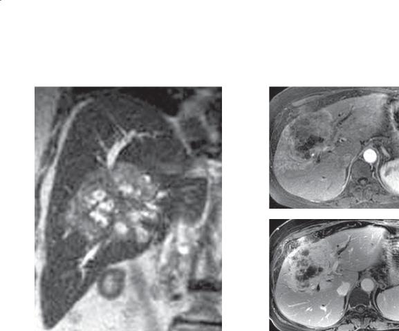
6. LIVER 537
CASE 6.68
A B
C
Findings
A.T2-weighted coronal MRI. Mass of hetereogeneous signal intensity is in the central region of the liver with peripheral biliary dilatation to the level of the mass.
B.Arterial-phase contrast-enhanced T1-weighted MRI. The mass is slightly enhanced.
C.Delayed-phase contrast-enhanced T1-weighted MRI. Intense enhancement with areas of central necrosis. Peripheral bile duct dilatation is present to the level of the mass.
Di erential Diagnosis
1.Cholangiocarcinoma
2.Hepatocellular carcinoma
3.Metastases
Diagnosis
Intrahepatic cholangiocarcinoma
Discussion
Cholangiocarcinoma arising from an intrahepatic bile duct can become large before becoming clinically apparent because of the large reserve capacity of
the liver. In some cases, the mass remains small and chronically obstructs the bile duct and lobar atrophy occurs. In the case shown here, the mass is a large polypoid-type tumor that has displaced central liver tissue. Dilated ducts are seen to obstruct at the level of the mass. Its central location and associated bile duct dilatation make cholangiocarcinoma most likely.
Risk factors for cholangiocarcinoma include primary sclerosing cholangitis, choledochal cyst, familial polyposis syndrome, congenital hepatic fibrosis, infection with Opisthorchis (formerly Clonorchis) sinensis (liver fluke), and prior exposure to thorium dioxide.
Additional examples of cholangiocarcinoma are found in cases 7.29 through 7.37.
Disease type: Masses
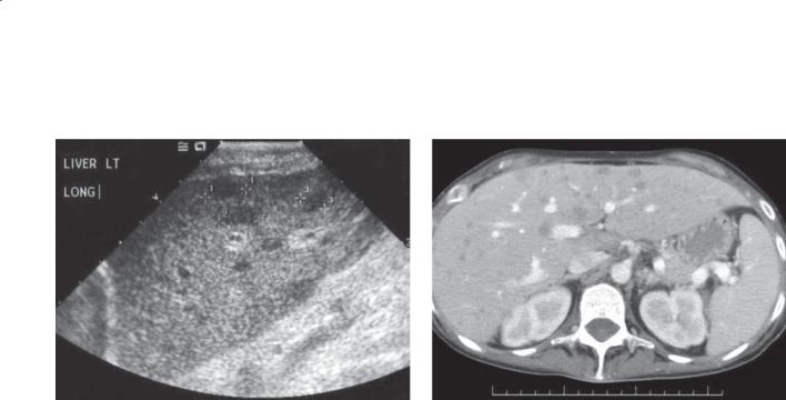
538 MAYO CLINIC GASTROINTESTINAL IMAGING REVIEW
CASE 6.69
A B
Findings
A.Longitudinal sonogram. Multiple hypoechoic masses are present in both lobes of the liver.
B.Contrast-enhanced CT. Multiple low-density lesions are seen in both lobes of the liver, corresponding to the hypoechoic masses on ultrasonography.
Di erential Diagnosis
1.Hepatic metastasis
2.Hepatic lymphoma
3.Fungal abscesses (candidiasis)
Diagnosis
Hepatic lymphoma
Discussion
Liver biopsy led to the diagnosis of non-Hodgkin lymphoma in this case. The liver is a common secondary site for lymphomatous involvement, which is detected at autopsy in 60% of patients with Hodgkin lymphoma and 50% of patients with non-Hodgkin
lymphoma. Secondary hepatic lymphoma typically presents radiologically as multiple small, hypoechoic lesions at sonography and multiple small, lowattenuation lesions at CT (as in this case). Secondary lymphomatous involvement of the liver is often difficult to detect radiographically because lymphomatous implants are often tiny and diffuse. Bulky abdominal adenopathy may favor the diagnosis of secondary hepatic lymphoma, but biopsy often is required to exclude metastatic disease or hepatic candidiasis (cases 6.28 and 6.29).
Primary lymphoma of the liver is extremely rare, constituting only 0.4% of all extranodal non-
Hodgkin lymphomas and 0.016% of all non-Hodgkin lymphomas. The most common presentation of primary hepatic lymphoma is a solitary mass. Hepatic resection with additional chemotherapy or radiation therapy is advocated for these rare tumors. There appears to be a relationship between chronic liver disease and the development of primary hepatic lymphoma. This relationship is strongest in patients with human immunodeficiency virus and hepatitis C.
Disease type: Masses
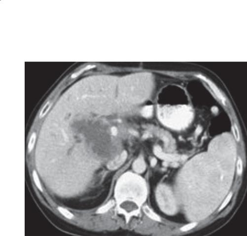
6. LIVER 539
CASE 6.70
Findings
Contrast-enhanced CT. Centrally necrotic hepatic mass is near the hepatic porta with irregular and nodular regions of enhancement in its periphery.
Di erential Diagnosis
1.Necrotic periportal lymphadenopathy
2.Primary hepatocellular carcinoma
3.Metastasis
4.Gallbladder carcinoma
5.Intrahepatic lymphoma
Diagnosis
Intrahepatic lymphoma, posttransplant lymphoproliferative disorder
Discussion
Th is patient had received a liver transplant within the past year, and at sonography the mass was clearly intrahepatic. The history of transplant excludes gallbladder cancer because cholecystectomy is routinely performed. If the patient had a known hepatocellular carcinoma, this would have been an important consideration. Otherwise, posttransplant lymphoproliferative disorder (PTLD)
is most likely. Other areas of adenopathy should be searched for. Metastasis is unlikely, because patients with a known malignancy would not have received a transplant within the year.
PTLD is a spectrum of abnormal lymphocyte proliferation ranging from abnormal proliferation of lymphoid cells to frankly malignant non-Hodgkin lymphoma. Nearly all patients have had a prior exposure to Epstein-Barr virus (EBV) that affects B lymphocytes, and the lymphomas that develop all contain the EBV genome. Anti–lymphocyte drugs (T-cell suppressors) required to suppress cellular immunity and rejection are responsible for activation of the B-cell lymphocytes in this disorder.
PTLD usually is detected within the first year after transplant and may be found incidentally in the course of routine imaging. Overall incidence is 2% to 10% in transplant patients (2%-4% for those with liver transplants). Radiographically, the disease
presents as a lymphoma with solitary or multiple, often necrotic, masses that may encase vessels. Treatment involves reduction of the immunosuppressants and a combination of chemotherapy, radiation, and surgery. The mortality rate approaches 50%.
Disease type: Masses
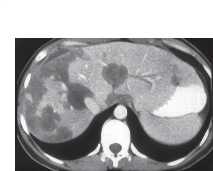
540 MAYO CLINIC GASTROINTESTINAL IMAGING REVIEW
CASE 6.71
Findings
Contrast-enhanced CT. There are multiple masses throughout the liver, greater within the right lobe and periphery of the liver. The masses appear to be growing together into confluent regions of hypoattenuation.
Di erential Diagnosis
1.Epithelioid hemangioendothelioma
2.Metastases
3.Multicentric hepatocellular carcinoma
Diagnosis
Epithelioid hemangioendothelioma
Discussion
Th is tumor is a rare malignant tumor of vascular origin. The tumor is believed to begin as multiple solitary masses that grow together to form a large confluent mass(es). Typically, the lesions are located in the periphery of the liver, and there may be capsular retraction at the level of the mass. Distinguishing
this lesion from more common metastases or hepatocellular carcinoma may not be possible.
Disease type: Masses
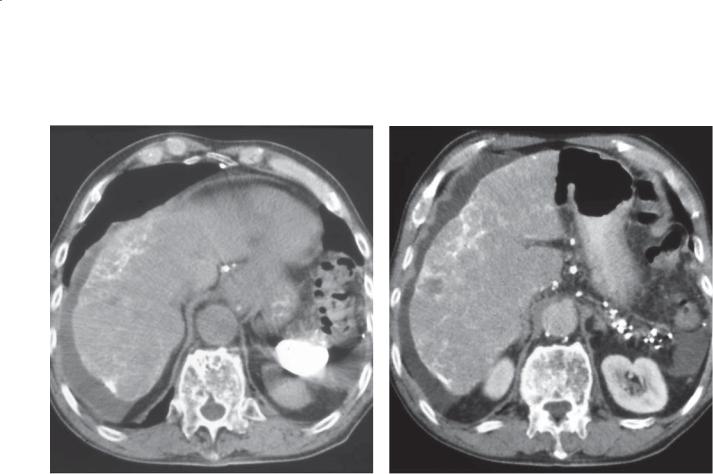
CASE 6.72
A
Findings
Unenhanced CT (A) and contrast-enhanced CT (B). The liver is of heterogeneous attenuation. Highattenuation material is seen throughout the liver
in a lacelike pattern, especially about the capsular and subcapsular regions. The spleen is small and
hyperdense. Multiple hyperdense lymph nodes are seen in the peripancreatic and gastrohepatic regions.
Di erential Diagnosis
1.Th orium dioxide-induced angiosarcoma
2.Th orium dioxide-induced cholangiocarcinoma
3.Th orium dioxide-induced hepatocellular carcinoma
4.Th orium dioxide-induced cirrhosis
Diagnosis
Th orium dioxide-induced angiosarcoma
6. LIVER 541
B
Discussion
Th orium dioxide was an intravascular contrast agent that emitted alpha radiation. The long half-life of the radioactive agent exposes patients to chronic
radiation during their lifetimes. It was used in various radiologic procedures between 1920 and the 1950s, usually neuroangiography. After leaving the vascular space, the agent is taken up by the reticuloendothelial system (liver, spleen, bone marrow). The spleen usually becomes atrophic and hyperattenuating. Uptake of
the agent in upper abdominal lymph nodes is also common. There is an increased incidence of liver cancers (angiosarcoma, hepatocellular carcinoma, and cholangiocarcinoma), cirrhosis, blood diseases, lung cancer, peritoneal tumors, and other neoplasms.
Th e patient in this case had an angiosarcoma, but other infiltrating neoplasms could have a similar appearance. Heterogeneity of the liver parenchyma is the key finding to suggest a complicating tumor. Percutaneous biopsy is necessary to confirm the diagnosis.
Disease type: Masses
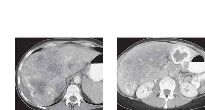
542 MAYO CLINIC GASTROINTESTINAL IMAGING REVIEW
CASE 6.73
A B
Findings
Contrast-enhanced CT. A and B. Multiple hypoattenuating and partially calcified masses are present throughout the liver.
Di erential Diagnosis
Metastases
Diagnosis
Calcified metastases—colon primary
Discussion
Metastases are the commonest malignant tumor to involve the liver. One-fourth to one-half of all patients who die of cancer have liver metastases. Colon metastases are the commonest, and they usually present as expansile, hypoattenuating masses. Well-differentiated colon metastases
that produce mucin may calcify (as in this case). Stomach and small bowel adenocarcinoma can present similarly—but calcify less often. Breast and lung cancers usually are smaller and often diffuse or infiltrative. Pancreatic cancer metastases usually are smaller and variable in number. Patients with
pancreatic cancer do not usually live long enough for these metastases to reach a large size. Hypovascular metastases (as discussed above) usually are detected most accurately during the portal venous phase of contrast enhancement. Hypervascular metastases include tumors arising from the neuroendocrine system (carcinoid, islet cell, pheochromocytoma), melanoma, choriocarcinoma, breast cancer, thyroid cancer, and renal adenocarcinoma. If hypervascular metastasis is suspected, acquiring a scan during
the arterial phase of contrast enhancement can be helpful.
Disease type: Masses
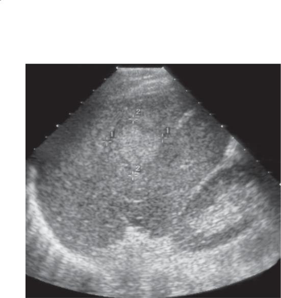
CASE 6.74
Findings
Sonogram. A 3.5-cm hyperechoic mass is present in the right lobe of the liver with a hypoechoic halo.
Di erential Diagnosis
1.Hepatic metastasis
2.Hepatocellular carcinoma
3.Hepatic adenoma
Diagnosis
Hepatic metastasis
6. LIVER 543
Discussion
Th is patient had a history of sigmoid adenocarcinoma, and needle biopsy of the liver mass was positive
for metastasis. Two common patterns for hepatic metastases on ultrasonography are multiple hypoechoic masses or multiple isoechoic or hyperechoic masses with hypoechoic halos. Although not absolutely indicative of malignancy, a hypoechoic halo around
a liver mass is an ominous sign. This halo usually is seen with metastases or hepatocellular carcinoma; however, hepatic adenomas and atypical focal nodular hyperplasia also can present with hypoechoic halos. As a general rule, hepatic masses with hypoechoic halos should be treated as malignant until proved otherwise.
Th e hypoechoic halo has been shown histologically to usually represent compressed normal liver parenchyma caused by a rapidly expanding tumor. Less often, it can be due to peripheral tumor fibrosis or vascularization.
Disease type: Masses

544 MAYO CLINIC GASTROINTESTINAL IMAGING REVIEW
CASE 6.75
A B
Findings
Contrast-enhanced CT. A. Arterial phase. Multiple hyperenhancing and cystic masses are present in the liver. B. Portal venous phase. The hypervascular masses have become isoattenuating with the liver. Splenomegaly.
Di erential Diagnosis
1.Hypervascular metastases
2.Multifocal hepatocellular carcinoma
3.Multiple benign liver tumors (flash hemangiomas, adenomas, focal nodular hyperplasia)
Diagnosis
Hypervascular metastases (metastatic carcinoid tumor)
Discussion
Hypervascular metastases, such as a carcinoid tumor, can be isoattenuating to liver on unenhanced and portal venous-phase imaging. This case shows the importance of arterial-phase imaging when evaluating for hypervascular metastases (islet cell, carcinoid, melanoma, renal cell, thyroid, breast, sarcoma). Clinical history is important in this case because other benign and malignant tumors can have a similar CT appearance. The multiplicity of lesions favors metastatic disease in this case.
Disease type: Masses
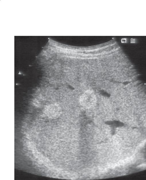
CASE 6.76
Findings
Sonogram. Rounded hyperechoic masses are seen in the right lobe of the liver. The masses have associated posterior acoustic shadowing.
Di erential Diagnosis
1.Hyperechoic metastases
2.Hemangiomas
3.Fat-containing liver tumors (lipoma, angiomyolipoma)
4.Multifocal hepatocellular carcinoma
Diagnosis
Hyperechoic metastases (renal adenocarcinoma)
6. LIVER 545
Discussion
Th e more vascular a tumor, the more likely it is to be echogenic. For this reason, metastases from renal cell carcinoma, carcinoid, sarcomas, and islet cell carcinomas tend to be hyperechoic. This patient had metastatic renal adenocarcinoma. The posterior acoustic shadowing behind these masses should be a tip-off that they are unlikely to be hemangiomas
(case 6.33). Hemangiomas often have slight increased through transmission.
Disease type: Masses
546 MAYO CLINIC GASTROINTESTINAL IMAGING REVIEW
TABLE 6.6
Malignant Liver Masses |
|
CASE |
Hepatocellular carcinoma |
Capsule, mosaic and fatty internal contents. Cirrhosis, |
6.51–6.67 |
|
usually hypervascular |
|
|
|
|
Cholangiocarcinoma |
Scirrhous or polypoid. Klatskin tumor at bifurcation |
6.68, 7.29–7.37 |
|
most common |
|
|
|
|
Lymphoma |
Multiple, diffuse tiny lesions. Usually not specific |
6.69 |
|
appearance |
|
|
|
|
Posttransplant lymphoproliferative |
Lymphoma appearance. Necrotic centrally, vascular |
6.70 |
disorder |
encasement |
|
|
|
|
Epithelioid hemangioendothelioma |
Peripheral mass, conglomerate. Capsular retraction |
6.71 |
|
|
|
Angiosarcoma |
Associated with thorium dioxide |
6.72 |
|
|
|
Metastases |
Multiple or solitary. Calcification, central necrosis, |
6.73–6.76 |
|
hemorrhage can occur |
|
|
|
|
Disease type: Masses

|
|
|
|
|
6. LIVER 547 |
|
|
|
|
|
|
|
|
|
TABLE 6.7 |
|
|
MRI Characteristics of Common Liver Tumors |
|
|
|
||
TUMOR |
T1 SIGNAL |
T2 SIGNAL |
GADOLINIUM |
COMMENT |
CASE |
|
|
|
ENHANCEMENT |
|
|
Benign cyst |
↓↓ |
↑↑ |
None |
Imperceptible rim |
6.18–6.20 |
Hemangioma |
↓↓ |
↑↑ |
Nodular, same |
Usually peripheral |
6.30–6.35 |
|
|
|
intensity as vessels |
globules of |
|
|
|
|
|
enhancement |
|
Focal nodular |
Variable |
Variable |
Early, homogeneous |
Gadoxetic acid |
6.38–6.44 |
hyperplasia |
|
|
enhancement with |
uptake, central scar |
|
|
|
|
rapid washout |
|
|
Adenoma |
↑ Internal |
↑ Heterogeneous |
Heterogeneous |
|
hemorrhage |
with hemorrhage; |
with hemorrhage; |
|
or fat; variable |
homogeneous |
homogeneous |
|
|
without hemorrhage |
without hemorrhage |
No gadoxetic acid |
6.45–6.49 |
uptake; fertile female on oral contraceptives
Hepatocellular |
↑ Fat, copper; ↑ Heterogeneous |
Arterial enhancement |
Secondary findings |
6.51–6.67 |
|
carcinoma |
↓ Usual |
|
with washout on |
of cirrhosis, PV or |
|
|
|
|
delayed images, |
HV thrombosis, PV |
|
|
|
|
larger lesions |
hypertension, capsule, |
|
|
|
|
heterogeneous |
mosaic internal |
|
|
|
|
|
architecture |
|
Cholangio- |
↓ or isointense |
↑ or isointense |
Late enhancement |
Upstream bile duct |
6.68, |
carcinoma |
|
|
|
dilatation |
7.29–7.37 |
Metastases |
↓ |
↑ |
Hypoenhancement |
No gadoxetic |
6.73–6.76 |
|
|
|
or hyperenhancement |
acid uptake |
|
|
|
|
depending on |
|
|
|
|
|
tumor type |
|
|
Abbreviations: ↑, hyperintense; ↑↑, markedly hyperintense; ↓, hypointense; ↓↓, markedly hypointense; HV, hepatic vein; PV, portal vein.
Disease type: Masses

548 MAYO CLINIC GASTROINTESTINAL IMAGING REVIEW
CASE 6.77
7
2
8 1 4A
6
3
4B
5
D.F.
© MAYO 2013
Findings
Th e liver is divided into 8 sectors according to the Couinaud classification. This classification is useful to hepatic surgeons because it divides the liver according to the portal venous and hepatic venous supply. Sector 1 corresponds to the caudate lobe. Sectors 2 (superior) and 3 (inferior) represent the lateral segment of the left hepatic lobe. Sector 4 corresponds to the medial segment of the left hepatic lobe and often is divided into a superior (A) and inferior (B) subsector. The right
lobe of the liver is divided into 4 sectors. The inferior (inferior to the main portal vein) segments are divided into anterior (sector 5) and posterior (sector 6) sectors. The superior segments are divided into anterior (sector 8) and posterior (sector 7) sectors. The anterior segment of the right lobe consists of sectors 5 and 8, and the posterior segment consists of sectors 6
and 7. (Used with permission of Mayo Foundation for Medical Education and Research.)
Disease type: Miscellaneous

6. LIVER 549
CASE 6.78
A B
Findings
A and B. Contrast-enhanced CT. Diffuse hyperenhancement within the distribution of the portal tracts that result in abnormal linear branching structures throughout the liver.
Di erential Diagnosis
1.Infectious disorders: tuberculosis, histoplasmosis, schistosomiasis, toxoplasmosis
2.Sarcoidosis
3.Drug reaction
4.Primary biliary cirrhosis
Diagnosis
Schistosomiasis
Discussion
Schistosomiasis (bilharziasis) is the second most prevalent tropical disease in the world, second only to malaria. More than 200 million people are infected. The main forms of human schistosomiasis are caused by 5 species of flatworm (blood flukes) in the genus
Schistosoma: S haematobium, S mansoni, S japonicum, S intercalatum, and S mekongi.
Adult worms mate and lay eggs. When the ova reach fresh water, miracidia are released and penetrate the snail. They asexually multiply cercariae. The cercariae leave the snail and swim to a human or
nonhuman animal, where they penetrate the skin. Once inside, cercariae travel to the portal veins, where they develop into adult worms. Humans excrete approximately 50% of the eggs. The rest are trapped in various parts of the body.
Disease occurs as a result of the immunologic reaction to Schistosoma eggs trapped in tissues. Antigens released from the egg stimulate a granulomatous reaction. In the later stages of the disease, collagen deposition and fibrosis result in organ damage that may not be reversible. The most common gastrointestinal complication is periportal fibrosis that can lead to portal hypertension and gastrointestinal hemorrhage. Imaging findings, due to the abundant periportal fibrosis, include thickening of the portal tracts and hyperenhancement during the portal venous and delayed phases of contrast enhancement (as in this case).
Many other causes of liver granulomas could present similarly. Infectious conditions are most common, including bacterial (mycobacterial infections, brucellosis, tularemia, actinomycosis), fungal (histoplasmosis, cryptococcosis, blastomycosis), parasitic (toxoplasmosis), and others. Sarcoidosis is the most important noninfectious cause. Various drugs (quinidine, sulfonamides, allopurinol, phenylbutazone) also can be responsible. Primary biliary cirrhosis is the only important primary cause of hepatic granulomas.
Disease type: Miscellaneous
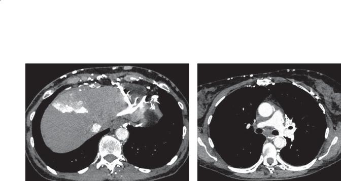
550 MAYO CLINIC GASTROINTESTINAL IMAGING REVIEW
CASE 6.79
A B
Findings
A. Contrast-enhanced CT. A focal segment (medial segment of the left hepatic lobe, quadrate lobe) of the liver is hyperattenuating with contrast material. A prominent collateral is seen adjacent to the lateral segment, and multiple collaterals are within the
anterior abdominal wall and paraspinal region. B. The superior vena cava is not seen, and multiple collaterals are within the mediastinum, about the esophagus, and in the anterior aspect of the chest wall.
Di erential Diagnosis
1.Superior vena cava obstruction with intrahepatic collaterals
2.Transient hepatic attenuation defect due to portal vein occlusion
3.Hyperenhancing mass with sump arterial perfusion
Diagnosis
Superior vena cava obstruction with intrahepatic collaterals and downhill varices
Discussion
In patients with obstruction of the superior vena cava (SVC), several different collateral pathways will develop. A venous pathway via the internal mammary vein connecting to the left portal vein via the paraumbilical vein is common. These collaterals
can result in increased vascular flow within the medial segment of the left hepatic lobe (previously known as the quadrate lobe). At CT, this finding is referred to as the “hot quadrate sign.” This can also be demonstrated with technetium Tc 99m–sulfur colloid scans of the liver and spleen and is referred to as the “focal hepatic hot spot sign.”
Th e geographic nature of the finding in the liver suggests a perfusion abnormality rather than a mass. Portal venous phase images can be assessed to ensure that the portal vein supplying the medial segment is patent and that an underlying mass is not present. Inspection of the chest images is often confirmatory that an SVC obstruction is present, most commonly due to neoplastic obstruction or prior radiation, chemotherapy, or transvenous line placement.
Disease type: Miscellaneous

|
|
|
6. LIVER 551 |
|
|
|
|
|
|
TABLE 6.8 |
|
Miscellaneous Liver Conditions |
|
CASE |
|
|
|
|
|
Liver segments (sectors) |
1: |
caudate |
6.77 |
|
2: superior subsegment, left lateral segment |
|
|
|
3: |
inferior subsegment, left lateral segment |
|
|
4A: superior subsegment, left medial segment |
|
|
|
4B: inferior subsegment, left medial segment |
|
|
|
5: anterior-inferior subsegment, right lobe |
|
|
|
6: posterior-inferior subsegment, right lobe |
|
|
|
7: posterior-superior subsegment, right lobe |
|
|
|
8: anterior-superior subsegment, right lobe |
|
|
|
|
|
|
Schistosomiasis |
Thickening of portal tracts, delayed enhancement due to |
6.78 |
|
|
abundant fibrosis |
|
|
|
|
|
|
Superior vena cava |
Hyperenhancing medial segment of left hepatic lobe, collaterals, |
6.79 |
|
obstruction |
superior vena cava obstruction, “hot quadrate sign” |
|
|
|
|
|
|
Disease type: Miscellaneous

552 MAYO CLINIC GASTROINTESTINAL IMAGING REVIEW
|
TABLE 6.9 |
Di erential Diagnoses |
|
Cystic Masses |
Solid Masses |
BENIGN |
BENIGN |
Simple cyst |
Hemangioma |
Polycystic disease |
Focal nodular hyperplasia |
Von Meyenburg complexes |
Hepatic adenoma |
Peribiliary cysts |
Lipoma |
Biliary cystadenoma |
Angiomyolipoma |
Biloma |
Regenerative nodules |
Pyogenic abscess |
Dysplastic nodules |
Amebic abscess |
Focal fatty infiltration |
Echinococcal cyst |
MALIGNANT |
|
|
MALIGNANT |
Hepatocellular carcinoma |
Biliary cystadenocarcinoma |
Cholangiocarcinoma |
Cystic cholangiocarcinoma |
Metastases |
Cystic metastases (mucinous) |
Lymphoma |
Necrotic tumors |
Posttransplant lymphoproliferative disorder |
Increased Density on CT |
|
Hemochromatosis |
|
Drugs: amiodarone, cisplatin |
|
Glycogen storage disease |
|
Wilson disease |
|
Gold therapy |
|
Hypervascular Masses |
Hyperechoic Masses |
BENIGN |
BENIGN |
Hemangioma |
Hemangioma |
Focal nodular hyperplasia |
Focal fatty infiltration |
Hepatic adenoma |
Lipoma |
Hereditary hemorrhagic telangiectasia |
Angiomyolipoma |
MALIGNANT |
MALIGNANT |
Hepatocellular carcinoma |
Hepatocellular carcinoma |
Metastases (islet cell, carcinoid, renal |
Hypervascular metastases (islet cell, carcinoid, renal |
adenocarcinoma, breast, thyroid, sarcoma) |
adenocarcinoma, breast, thyroid, sarcoma) |
|
Calcified metastases |
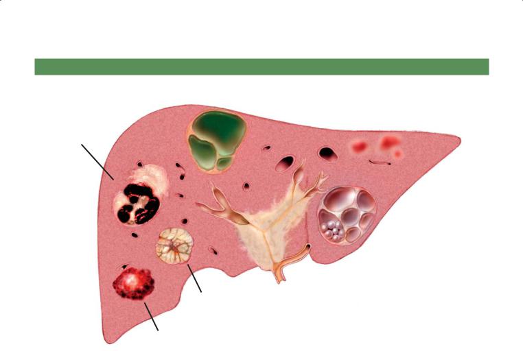
6. LIVER 553
LIVER MASSES
Biliary cystadenoma/ cystadenocarcinoma
Hepatocellular |
Metastases |
|
adenoma/ |
||
|
||
adenocarcinoma |
|
Echinococcal cyst
Hilar cholangiocarcinoma
Focal nodular
D.F. hyperplasia
© MAYO 2013
Cavernous hemangioma
Figure 6.80 (Used with permission of Mayo Foundation for Medical Education and Research.)
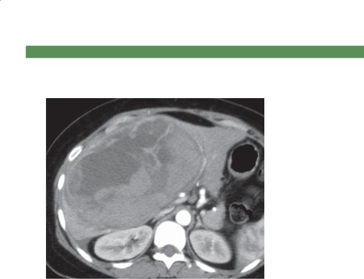
554 MAYO CLINIC GASTROINTESTINAL IMAGING REVIEW
QUESTIONS
Multiple Choice (choose the best answer)
6.1.In this case of a 25-year-old woman, which of the following is the most likely diagnosis for the findings in the figure?
a.Hemangioma
b.Focal nodular hyperplasia
c.Hepatic adenoma
d.Biliary cystadenoma
e.Colon cancer metastasis
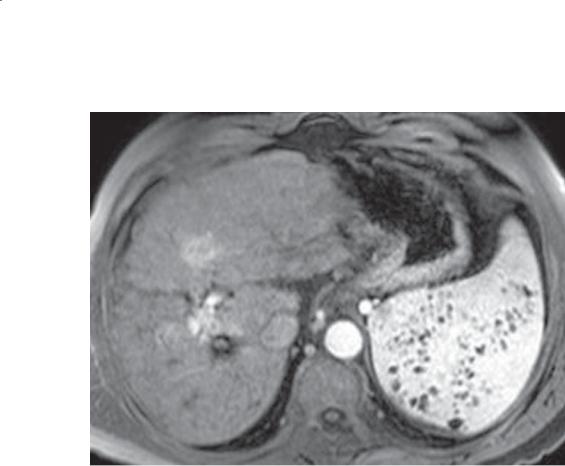
6. LIVER 555
6.2.Given the findings in the figure, what procedure could be anticipated for the patient?
a.Colonoscopy
b.Placement of a transjugular intrahepatic portosystemic shunt (TIPS)
c.Video capsule endoscopy
d.Phlebotomy
e.Positron emission tomography
6.3.Given the findings in the figure shown in Question 6.2, what symptoms would be expected in the patient?
a.Discomfort in the left upper quadrant
b.Fever
c.Hematochezia
d.Light-headedness
e.Diffi culty swallowing
556 MAYO CLINIC GASTROINTESTINAL IMAGING REVIEW
A B
C D
Match images A through F with the responses listed below.
6.4. |
_____ Increased level of α-fetoprotein |
6.6. |
_____ Diabetes mellitus |
6.5. |
_____ Breast cancer |
6.7. |
_____ Bile ductules |
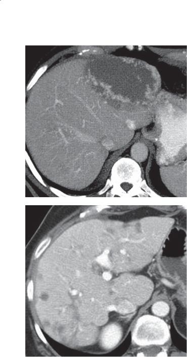
6. LIVER 557
E
F
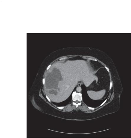
558 MAYO CLINIC GASTROINTESTINAL IMAGING REVIEW
6.8.Which of the following is the most likely cause of the abnormality shown in the figure?
a.Esophageal cancer
b.Cholecystitis
c.Amebiasis
d.Biliary cystadenoma
e.Echinococcal disease

6. LIVER 559
ANSWERS
6.1. Answer c. |
6.4. Answer b. |
Th ere is a large heterogeneously enhancing focal hepatic mass on this arterial-phase CT scan. The mass is well circumscribed. In young females, hepatic adenoma is most likely, especially if they are taking birth control medication.
Hemangiomas should have peripheral globules of blood attenuation (like the aorta) on contrast-enhanced CT.
Focal nodular hyperplasia is typically homogeneously enhancing on arterial-phase CT. In some cases, the focal stellate central scar and internal septations are visible.
Biliary cystadenoma would be water attenuation with visible septations.
Colon cancer metastasis would be intermediate attenuation and could contain internal amorphous calcifications.
6.2.Answer b.
Th is patient has an irregularly surfaced liver and splenomegaly containing multiple low-attenuation regions (Gamna-Gandy bodies). The patient has cirrhosis with long-standing portal venous hypertension. The only procedure associated with these findings is TIPS placement. This is usually performed for patients with portal venous hypertension and bleeding esophageal varices that do not respond to sclerotherapy.
Colonoscopy visualizes the colon. Video capsule endoscopy is usually used for evaluation of the small bowel. Phlebotomy is used to treat patients with hemochromatosis. Positron emission tomography is usually used to assess for metastases in patients with known neoplasms.
6.3.Answer a.
Discomfort in the left upper quadrant is due to the splenomegaly.
Although patients with cirrhosis can have spontaneous bacterial peritonitis, it is rare. There is no evidence of an infection in this patient.
Hematochezia is usually associated with colorectal bleeding. Patients with variceal bleeding usually have melena.
Light-headedness is a nonspecific symptom and could be due to several conditions.
Difficulty swallowing usually points to an esophageal problem. Varices in the esophagus usually present only with bleeding and do not cause anatomic narrowing of the esophagus that would lead to dysphagia.
Figure b shows an irregularly surfaced liver and an enhancing mass within the right hepatic lobe. Findings are due to cirrhosis with a complicating hepatocellular carcinoma. Patients with hepatocellular carcinoma often have an increased level of α-fetoprotein.
6.5.Answer f.
Figure f shows multiple small, hypoattenuating masses within the liver with some associated retraction (scarring). The appearance resembles cirrhosis, except the hypoattenuating masses are atypical. In patients with breast cancer metastases to the liver who are receiving chemotherapy, a pseudocirrhotic appearance often can develop. As the tumor shrinks, there is believed to be associated scarring of the hepatic parenchyma.
6.6.Answer a.
In figure a, the liver, pancreas, and spleen are of very low signal intensity. Hemochromatosis can be
distinguished from hemosiderosis by identification of a low signal intensity pancreas (present in primary hemochromatosis). Cirrhosis, diabetes mellitus, and heart failure will develop as a result of toxic parenchymal iron deposition in these organs. The spleen is typically of normal signal intensity, but in some patients with extreme iron overload it may also be of abnormally low signal intensity.
6.7.Answer d.
Figure d, a delayed MR scan, shows 2 hyperintense lesions within the liver. The patient received gadoxetic acid (a hepatobiliary contrast agent that is taken up by hepatocytes and excreted in the biliary system). Uptake of this agent is characteristic of focal nodular hyperplasia. Often a central scar (seen in both lesions) is visible. Pathologically, these lesions have bile ductules—a characteristic histologic finding within the central scar region.
Figure c shows focal fatty infiltration. The hepatic veins are not displaced by the low-attenuation changes in the liver, an indication of an infiltrating change rather than a mass.
Figure e shows a giant hemangioma. Peripheral, globular, confluent enhancement is isoattenuating with the blood pool.
6.8.Answer b.
Th ere is a multiloculated water-attenuation mass within the left hepatic lobe. This has the appearance of a pyogenic liver abscess, of which cholecystitis is a

560 MAYO CLINIC GASTROINTESTINAL IMAGING REVIEW
common cause. Appendicitis and diverticulitis are also common causes of a hepatic abscess. Esophageal cancer can cause metastases to the liver, but they would be expected to contain more solid tissue and to be diffusely distributed within the liver. Amebiasis characteristically
is a solitary abscess with a rim of low-attenuation liver about the central fluid component. Biliary cystadenoma is usually solitary, containing multiple septations and nodules. Echinococcal disease often has a large dominant cyst containing multiple daughter cysts.