

C H A P T E R 2
STOMACH
Cases 2.1–2.15 Inflammatory and Ulcerative Diseases
Cases 2.16–2.43 Masses and Filling Defects
Benign Tumors
Malignant Tumors
Other
Cases 2.44–2.51 |
Narrowings |
|
Benign |
|
Malignant |
Cases 2.52–2.68 |
Postoperative Stomach |
Cases 2.69–2.75 |
Miscellaneous |

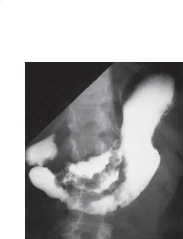
CASE 2.1
Findings
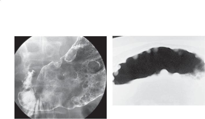
CASE 2.1. Double-contrast UGI. Thickened, lobulated folds are present in the body and antrum of the stomach.
CASE 2.2. Double-contrast UGI. Nodular fold thickening is present in the gastric antrum.
Di erential Diagnosis
1.Gastritis (eg, caused by Helicobacter pylori, alcohol, medication)
2.Zollinger-Ellison syndrome
3.Lymphoma
Diagnosis
H pylori gastritis
2. STOMACH 79
CASE 2.2
Discussion
H pylori infection is the major cause of gastritis (70%), gastric ulcers (70%), and duodenal ulcers (90%). It also has been identified as a substantial risk factor for development of gastric adenocarcinoma and mucosaassociated lymphoid tissue lymphoma. H pylori is a gram-negative rod that colonizes the stomachs of a large percentage of the US population (>50% of those older than 60 years), most of whom are asymptomatic. However, eradication of the H pylori infection with antibiotics in addition to acid blockers has been effective for curing gastritis and ulcers in up to 90% of patients. H pylori infection usually is diagnosed with serologic tests or endoscopic biopsy. Treatment of asymptomatic patients currently is not recommended because of the cost and possible adverse effects of the medical therapy.
H pylori gastritis usually presents as smooth, nonspecific fold thickening involving the body and antrum of the stomach on barium examinations. Occasionally, H pylori gastritis causes markedly thickened nodular folds indistinguishable from lymphoma or submucosal carcinoma. In these cases, endoscopy is recommended to exclude malignancy. Zollinger-Ellison syndrome (cases 2.11 and 2.12) also can cause gastric fold thickening.
Disease type: Inflammatory and Ulcerative Diseases
80 MAYO CLINIC GASTROINTESTINAL IMAGING REVIEW
CASE 2.3
Findings
Double-contrast UGI. Multiple small, round filling defects (mounds of edema) (arrows) containing a small central collection of barium (erosion or tiny ulceration) are present in the gastric antrum and body.
Di erential Diagnosis
1.Acute erosive (varioliform) gastritis
2.Barium precipitate
3.Crohn disease
4.Infectious gastritis (viral or fungal)
Diagnosis
Acute erosive gastritis
Discussion
Acute gastritis often presents with symptoms mimicking those of peptic ulcer disease. This disorder has many causes, including stress reactions, Helicobacter pylori, alcohol, aspirin, nonsteroidal
anti-inflammatory drugs (NSAIDs), chemotherapeutic agents, and several other medications. In our practice, aspirin and NSAIDs are the most common causes.
Typically, multiple small, elevated mounds of edema with central ulcerations are identified, often in line with a rugal fold. These typical erosions are often referred to as varioliform erosions.
It is important not to mistake barium precipitates for punctate erosions. Precipitates never have a surrounding mound of edema and often are randomly oriented—not along the crest of a rugal fold, as often seen with true erosions. The early aphthous ulcerations found in Crohn disease, gastric candidiasis, or viral infections may be indistinguishable from erosive gastritis. Erosions are almost always multiple. Crohn disease usually presents with additional lesions in the small bowel. Single lesions may represent an artifact or a small submucosal process, such as pancreatic
rest (case 2.39), gastrointestinal stromal tumor (cases 2.19, 2.20, and 2.21), or metastasis (case 2.37).
Disease type: Inflammatory and Ulcerative Diseases
CASE 2.4
Findings
Single-contrast UGI. An ulcer crater is located along the lesser curvature of the gastric antrum. Symmetric and smoothly contoured folds radiate to the crater. The mound of edema (arrowheads) is smooth in contour, and the crater is located centrally within the mound.
Di erential Diagnosis
Benign gastric ulcer
Diagnosis
Benign gastric ulcer
2. STOMACH 81
Discussion
Th is ulcer has some of the typical signs of benignity: a round or oval crater, folds that cross the mound of surrounding edema and extend up to the crater, a symmetric and smooth filling defect (edema) about the crater, extension of the ulcer beyond the normal gastric lumen contour, and radiating gastric folds that are smooth and symmetric (without nodularity or clubbing). Most benign ulcers occur along the lesser curvature or posterior wall of the antrum or body of the stomach. The anterior wall is affected least often. The greater curvature is a somewhat unusual location for benign ulcers. Benign ulcers along the greater curvature of the body and antrum of the stomach are often associated with ingestion of aspirin-containing medication (case 2.7).
Disease type: Inflammatory and Ulcerative Diseases
82 MAYO CLINIC GASTROINTESTINAL IMAGING REVIEW
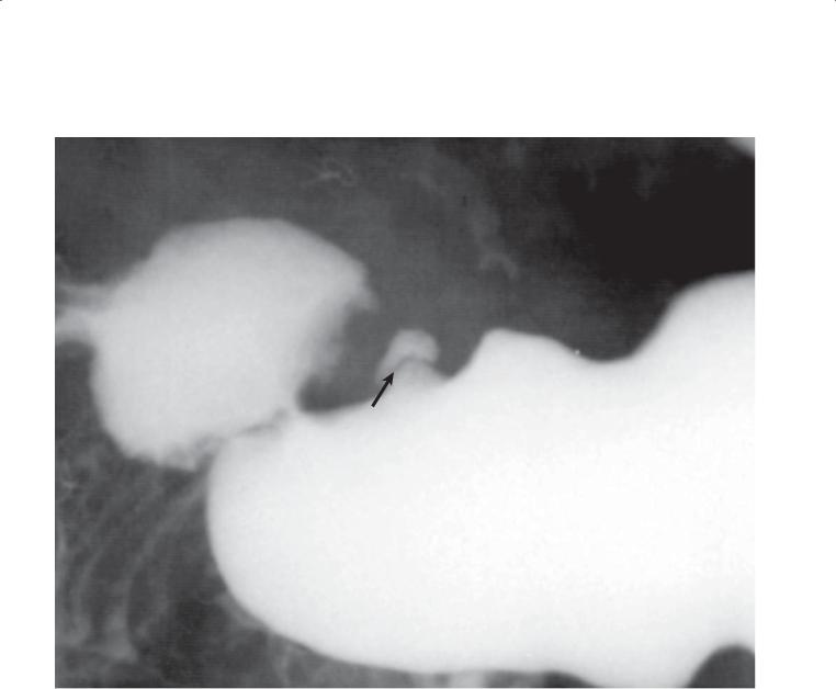
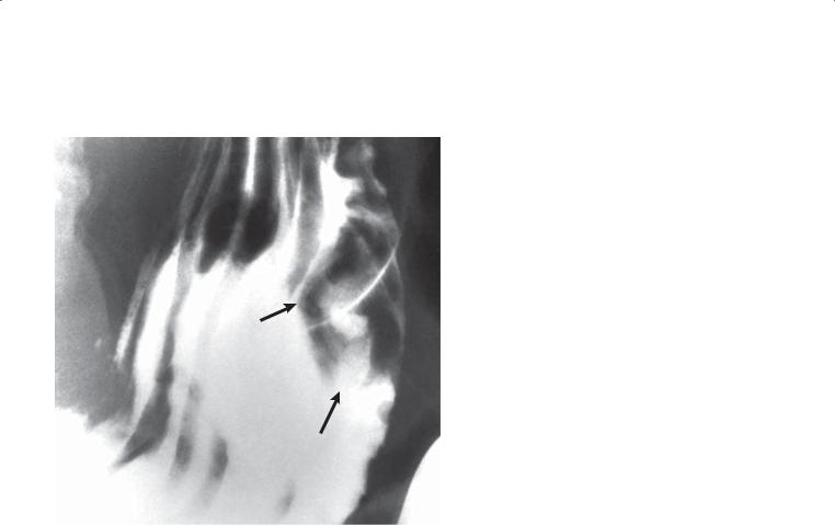
CASE 2.5
Findings
Single-contrast UGI. An ulcer crater is seen on this tangential view of the gastric antrum. A Hampton line (arrow) is seen at the base of the crater.
Di erential Diagnosis
Benign gastric ulcer
Diagnosis
Benign gastric ulcer (Hampton line)
Discussion
Gastric ulcers are common abnormalities, in large part a result of the widespread use of anti-inflammatory agents and Helicobacter pylori infection. The radiographic examination is a reliable method for distinguishing benign from malignant ulcers. Characteristics of a benign ulcer on the tangential view include a Hampton line (as seen in this case) or a smooth and symmetric ulcer collar or mound. A Hampton line represents nonulcerated acid-resistant mucosa around the ulcer crater. An ulcer collar represents edematous submucosal tissue surrounding the ulcer cavity. A collar is always wider than the thin, delicate Hampton line. Ulcers with typical radiographic features of benignity are nearly always benign and can be followed radiographically until complete healing has occurred.
Disease type: Inflammatory and Ulcerative Diseases
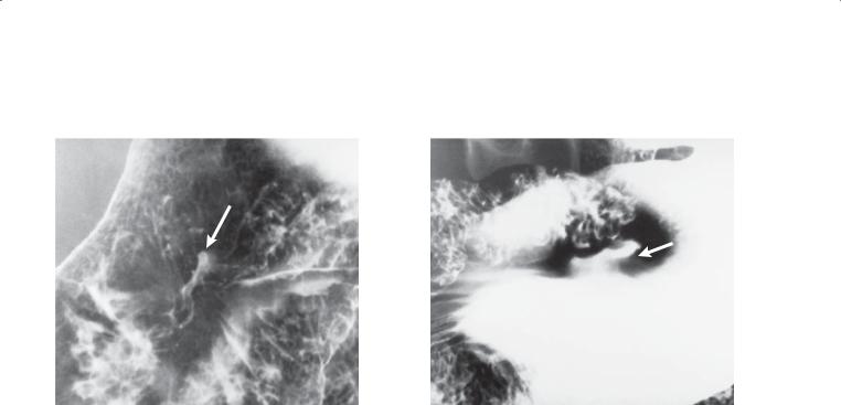
CASE 2.6
A
Findings
Double-contrast UGI. A. A linear collection of barium (arrow) is visible on the en-face view of the gastric body. Several folds radiate to the edge of the crater.
B. The elongated ulcer collection and surrounding collar (arrow) are seen when viewed in profile.
Di erential Diagnosis
Benign linear ulcer
Diagnosis
Benign linear ulcer
2. STOMACH 83
B
Discussion
An ulcer collar represents edematous submucosa about the ulcer crater. The crater extends within the submucosa because this tissue is not as acid resistant as the mucosa. A linear configuration of the crater is often seen in patients with healing ulcers. Healing ulcers may be asymmetric or “split” into 2 smaller
craters as reepithelialization occurs. The average time required for healing of an ulcer is 8 weeks. Large ulcers require more time to heal than do smaller ones. For this reason, follow-up studies should probably not
be performed less than 6 to 8 weeks after the initial diagnosis. Most ulcers leave a scar after healing has occurred. Scars often appear as a depression or pit on the mucosal surface. Radiating folds often can be seen extending up to the depression. Retraction (as a result of intramural fibrosis) of the adjacent gastric wall is also common. The finding of areae gastricae covering the scarred region confirms ulcer healing; however, this is only rarely identified on double-contrast examinations.
Disease type: Inflammatory and Ulcerative Diseases
84 MAYO CLINIC GASTROINTESTINAL IMAGING REVIEW
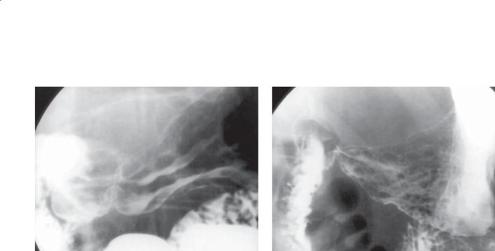
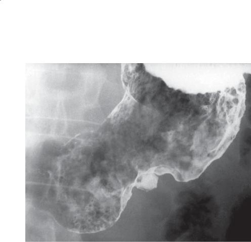
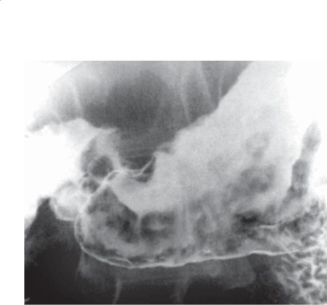
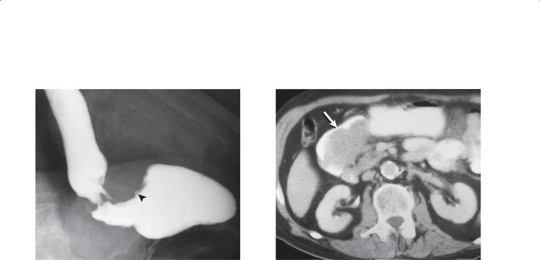
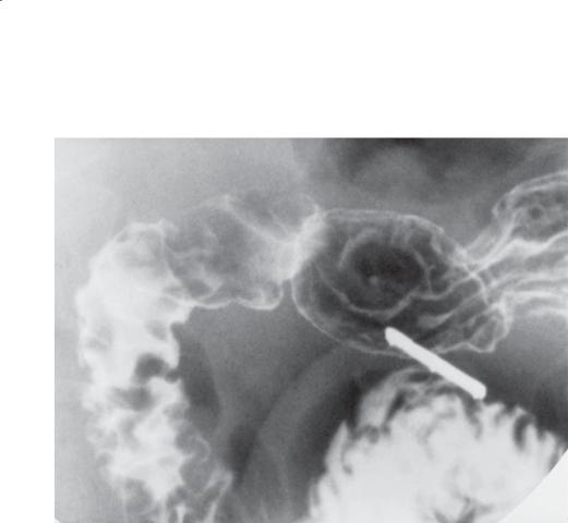
CASE 2.7
Findings
Double-contrast UGI. An ulcer crater is present on the greater curvature of the stomach. A smooth mound of edema surrounds the centrally located ulcer. The crater does not extend beyond the normal gastric lumen contour.
Di erential Diagnosis
1.Benign sump ulcer
2.Gastric adenocarcinoma
3.Intramural diverticulum
Diagnosis
Benign gastric sump ulcer
Discussion
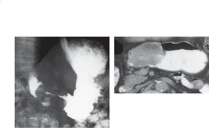
Benign ulcers of the greater curvature may not have all of the usual radiographic features associated with benignity. The location of an ulcer is not a reliable predictor of benignity or malignancy. As a general rule, however, benign ulcers are rarely located in the proximal half of the stomach along the greater curvature. Occasionally, benign gastric ulcers are found in the proximal stomach along the lesser curvature in elderly patients. Many of the ulcers in the distal stomach are benign and medicationinduced. Aspirin-induced ulcers often develop along the greater curvature because it is a dependent location where pills collect and directly cause mucosal injury. Sometimes they are referred to as sump ulcers (as in this case). Usually the mound of edema surrounding a benign ulcer is smooth-surfaced and tapers gradually, merging imperceptibly with the normal gastric wall. In this patient, the surrounding mass of edema has rather abrupt margins with the normal gastric wall (see the figure on page 150). Endoscopy is often necessary to exclude malignancy if equivocal or worrisome findings are present. A gastric intramural diverticulum usually does not have substantial surrounding mass effect and changes shape with peristaltic contractions.
Disease type: Inflammatory and Ulcerative Diseases
CASE 2.8
Findings
Single-contrast UGI. A large ulcer is present along the greater curvature of the stomach. Gastric folds extend up to the crater. The crater projects beyond the expected location of the greater curvature mucosa.
Di erential Diagnosis
1.Giant gastric ulcer
2.Postoperative deformity
Diagnosis
Benign giant gastric ulcer
2. STOMACH 85
Discussion
Giant ulcers are defined as those larger than 3 cm in diameter. The size of the crater has no bearing on
their benign or malignant nature. They are commonly associated with contained perforations. Usually, a large mass is visible in patients with giant malignant ulcers (case 2.24). Multiple ulcerations can be seen
in 12% to 20% of patients with benign gastric ulcers. Multiplicity favors a benign cause, but each ulcer should be evaluated individually. Concurrent gastric and duodenal ulcers are unusual.
Disease type: Inflammatory and Ulcerative Diseases
86 MAYO CLINIC GASTROINTESTINAL IMAGING REVIEW
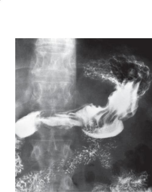
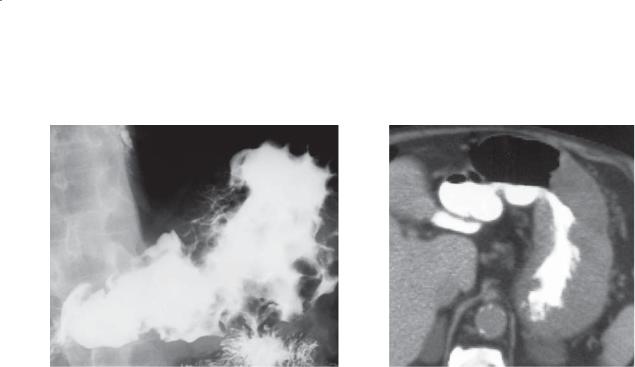
CASE 2.9
Findings
Single-contrast UGI. A large fistulous communication (arrow) exists between the greater curvature of the stomach and the left transverse colon.
Di erential Diagnosis
1.Benign gastric ulcer with fistula
2.Malignant gastric ulcer with fistula
3.Postoperative anastomosis
Diagnosis
Gastrocolic fistula due to a benign gastric ulcer
Discussion
In this patient, a penetrating benign gastric ulcer was found at operation. Major complications of gastric ulcers include bleeding, perforation, obstruction, and penetration. Bleeding is the most frequent
complication, occurring in 10% to 24% of patients with peptic ulcer disease. Most actively bleeding ulcers are assessed endoscopically. An ulcer can penetrate into any adjacent organ or tissue. Common sites include the pancreas, omentum, biliary tract, liver, and colon. Most gastrocolic fistulas are due to primary carcinomas or lymphomas arising from either the stomach or
the colon. Offending ulcers usually are located along the greater curvature, and offending benign ulcers are usually found in patients receiving aspirin or corticosteroid therapy or both. Penetration of an ulcer into the pancreas may result in pancreatitis or a pancreatic abscess.
An enteric anastomosis is unlikely because the communication does not contain enteric folds (suggesting ulceration or cavity), and intentional anastomosis to the colon is never performed.
Disease type: Inflammatory and Ulcerative Diseases
CASE 2.10
Findings
Enhanced abdominal CT. Orally administered contrast material is seen within the body of the stomach and within an ulcer crater (arrow) in the posterior gastric wall. There is a communication between the stomach (at the site of the ulcer) and into an irregularly shaped cavity within the lesser sac. Pneumoperitoneum (arrowhead) is seen anteriorly within the peritoneal cavity (postoperative in origin).
Di erential Diagnosis
1.Perforated benign gastric ulcer
2.Perforated malignant ulcer
Diagnosis
Perforated benign gastric ulcer
2. STOMACH 87
Discussion
CT can be helpful for determining the extraluminal extent of disease in a patient with a known or suspected penetrating ulcer. This information can be helpful for preoperative planning and for assessment of possible percutaneous drainage. It may not be possible to distinguish a benign from a malignant ulcer at CT. Endoscopy is often required for further evaluation.
Disease type: Inflammatory and Ulcerative Diseases
88 MAYO CLINIC GASTROINTESTINAL IMAGING REVIEW
CASE 2.11 |
CASE 2.12 |
Findings
CASE 2.11. Single-contrast UGI. Enlarged rugal folds are present in the gastric antrum. A benign ulcer is present along the lesser curvature.
CASE 2.12. Enhanced abdominal CT. The gastric wall and rugal folds are markedly thickened.
Di erential Diagnosis
1.Zollinger-Ellison syndrome
2.Lymphoma
3.Primary gastric carcinoma
4.Ménétrier disease
Diagnosis
Zollinger-Ellison syndrome
Discussion
Zollinger-Ellison syndrome is caused by a gastrinsecreting islet cell neoplasm (usually in the pancreas) that results in marked gastric hypersecretion of hydrochloric acid and severe peptic ulcer disease.
At least half of these tumors are malignant, metastasizing to regional lymph nodes and liver. Approximately one-fourth of patients have
multiple endocrine neoplasia type 1 (parathyroid adenoma, pituitary adenoma, pheochromocytoma). Patients usually are middle-aged and present with intractable peptic ulcer disease, often with associated malabsorption (due to inactivation of lipase and bile
salt precipitation from hyperacidity). Serum gastrin levels are always increased, and paradoxic increase in gastrin levels occurs after an injection of secretin. This result distinguishes Zollinger-Ellison syndrome from other diseases with increased serum gastrin levels (eg, retained gastric antrum, antral G-cell hyperplasia, short-bowel syndrome, and uremia).
Upper gastrointestinal radiographic findings include enlarged rugal folds, hypersecretion, peptic ulcers, and thickened folds in the proximal small bowel. At CT, gastric and duodenal wall thickening and hypersecretion may be seen. The primary tumor in the pancreas (or nearby organs) and evidence of metastases also may be visible.
Small gastrinomas without evidence of metastases are surgically resected. Advanced lesions are treated with a combination of histamine2-receptor blockers, chemotherapy, and hepatic artery embolization.
Multiphase CT and octreotide scintigraphy are most commonly used for identification of the primary tumor. CT and MRI are commonly used for follow-up imaging studies.
Gastric lymphoma produces disorganized, enlarged folds, whereas adenocarcinoma presents as a focal mass or diffusely narrowed stomach (linitis plastica). Neither of these diseases results in inflammatory changes in the duodenum. Thickened folds in Ménétrier disease usually are confined to the proximal stomach.
Disease type: Inflammatory and Ulcerative Diseases
CASE 2.13
Findings
Single-contrast UGI. Multiple erosions are present in the gastric antrum.
Di erential Diagnosis
1.Erosive gastritis
2.Crohn disease
Diagnosis
Crohn disease
Discussion
In this patient, Crohn disease was proved by endoscopic biopsy (chronic inflammation and granulomas). Crohn disease (regional enteritis)
2. STOMACH 89
of the stomach is uncommon, but when present it usually is associated with disease in the small bowel or colon or both. The earliest changes from Crohn disease include aphthous ulcers and fold thickening (as in this case). Continued inflammation results in confluent ulcers, cobblestoning, denuded mucosa, fibrosis, and strictures. The distal half of the stomach usually is affected, often with concomitant duodenal involvement.
Th e finding of aphthous ulcerations (superficial erosions) is nonspecific and could be due to peptic ulcer disease or medication-induced gastritis (eg, aspirin). Endoscopy and histologic examination also are often nonspecific.
Disease type: Inflammatory and Ulcerative Diseases
90 MAYO CLINIC GASTROINTESTINAL IMAGING REVIEW
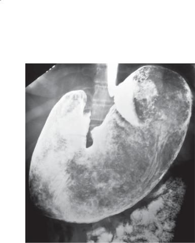
CASE 2.14 |
CASE 2.15 |
Findings
CASE 2.14. Single-contrast UGI. Markedly enlarged gastric folds are present within the proximal half of the stomach.
CASE 2.15. Unenhanced abdominal CT. The wall of the gastric fundus and body is markedly thickened,
especially along the greater curvature. The antral wall is of normal thickness. Bilateral adrenal masses are present.
Di erential Diagnosis
1.Lymphoma
2.Ménétrier disease
3.Zollinger-Ellison syndrome
4.Eosinophilic gastritis
Diagnosis
Ménétrier disease
Discussion
Ménétrier disease is a disorder of unknown cause, sometimes referred to as a hypertrophic gastropathy. Pathologically, hyperplasia of surface epithelial cells is present with abundant mucous cells and mucoid secretions. Parietal cells may be replaced by the
epithelial cells, causing achlorhydria. The process is not primarily inflammatory, but inflammatory cells may be present. Erosions and hemorrhage may cause anemia. Carcinoma has been reported in patients with this disorder, but whether it predisposes to malignancy is unknown. Treatment (antisecretory drugs and occasionally gastrectomy) is often unnecessary unless pain, bleeding, or protein loss is severe.
Radiologic findings are usually those of enlarged rugal folds, often sparing the antrum. The folds are pliable, with a distensible stomach. Folds are enlarged but organized and follow the distribution of normal rugae. Occasionally, segmental rugal enlargement is seen and presents as a polypoid mass.
Gastric folds may be enlarged without any associated clinical disorder. These folds nearly always can be effaced with adequate distention of the gastric fundus. Enlarged folds in patients with lymphoma are usually disorganized and can be nodular and irregular (cases 2.31, 2.32, and 2.33). Zollinger-Ellison syndrome with enlarged gastric folds often is associated with hypersecretion, peptic ulcers, a dilated duodenum, and thickened folds in the proximal small bowel (cases 2.11 and 2.12). Patients with eosinophilic gastritis have thickened folds within the stomach and small bowel as well as a history of allergy.
Disease type: Inflammatory and Ulcerative Diseases

|
|
2. STOMACH 91 |
|
|
|
|
TABLE 2.1 |
|
Thickened Gastric Folds |
|
CASE |
|
|
|
Helicobacter pylori gastritis |
Usually in antrum. Commonest manifestation |
2.1 and 2.2 |
|
|
|
Zollinger-Ellison syndrome |
Hypersecretion and ulcerations in stomach and duodenum. |
2.11 and 2.12 |
|
Enhancing pancreatic (or antral or duodenal) mass(es) |
|
|
|
|
Crohn disease |
Stomach is uncommon location. Aphthous ulcers and |
2.13 |
|
fold thickening |
|
|
|
|
Varices |
In patients with portal hypertension, esophageal varices also |
2.41–2.43 |
|
should be sought. Usually in cardia and fundus |
|
|
|
|
Ménétrier disease |
May have hypersecretion. Usually in cardia and fundus. |
2.14 and 2.15 |
|
Often a diagnosis of exclusion. |
|
|
|
|
Lymphoma |
Variable size and nodularity |
2.31–2.33 |
|
|
|
Disease type: Inflammatory and Ulcerative Diseases
92 MAYO CLINIC GASTROINTESTINAL IMAGING REVIEW
CASE 2.16 |
CASE 2.17 |
Findings
CASE 2.16. Double-contrast UGI. Multiple polypoid filling defects are present in the stomach.
CASE 2.17. Unenhanced abdominal CT. Multiple polyps arise from the anterior and posterior walls of the stomach.
Di erential Diagnosis
Gastric polyps
Diagnosis
Gastric polyps
Discussion
In this patient, hyperplastic polyps were found at endoscopic biopsy. Gastric polyps occur in about 2% of the population. Most polyps are either hyperplastic or adenomatous histologically. Hyperplastic polyps develop as a reactive change to chronic inflammation with mucosal proliferation and cystic dilatation
of gastric glands. Some authors refer to them as regenerative or inflammatory polyps. They are often multiple and usually small (<1 cm in diameter). They have a propensity to develop in the fundus
of the stomach, but they may be found anywhere. Hyperplastic polyps are believed to be benign, without malignant potential; however, rare malignant transformation has been reported. Innumerable hyperplastic polyps in the gastric fundus occur
in patients with familial adenomatous polyposis syndrome (fundic gland polyposis syndrome).
Adenomatous polyps are infrequent, often solitary lesions, with diameters usually exceeding 1 cm. These polyps can develop into carcinomas, but this is an unusual occurrence, generally among large (>2 cm) lesions. Overall, development of a cancer from gastric polyps is rare, perhaps occurring in 1% to 2% of patients. Endoscopic biopsy and polypectomy are safe and should be considered for solitary large polyps.
Disease type: Masses and Filling Defects

2. STOMACH 93
CASE 2.18
Findings
Double-contrast UGI. Innumerable small polyps are present throughout the stomach. They are most numerous within the fundus.
Di erential Diagnosis
1.Gastric polyps
2.Fundic gland polyposis syndrome
Diagnosis
Fundic gland polyposis syndrome
Discussion
Th is patient had known familial adenomatous polyposis (FAP) syndrome. Gastric polyps in patients with FAP are usually hyperplastic, whereas polyps elsewhere in the intestines are adenomatous. Gardner syndrome and Turcot syndrome are variations of FAP with associated manifestations outside the
gastrointestinal tract. In patients with Gardner syndrome, desmoid tumors, osteomas, epidermoid cysts, or papillary thyroid cancer can develop. Patients with Turcot syndrome have development of central nervous system tumors, including gliomas and medulloblastomas. The common association of multiple hyperplastic polyps in the gastric fundus in patients with FAP is known as fundic gland
polyposis syndrome. Gastric adenomatous polyps are uncommon, but when they occur in these patients they usually are located in the antrum and may be multiple. Gastric polyps developing in the other polyposis syndromes (Peutz-Jeghers syndrome, juvenile polyposis, and Cronkhite-Canada syndrome) are classified as hamartomas. Patients with FAP also are predisposed to adenomas and adenocarcinomas developing in the periampullary region. Colorectal cancer eventually develops in nearly all patients who have FAP without proctocolectomy.
Disease type: Masses and Filling Defects

94 MAYO CLINIC GASTROINTESTINAL IMAGING REVIEW
TABLE 2.2
Polyposis Syndromes
SYNDROME |
POLYP TYPE |
INHERITANCE |
COMMENT |
|
|
|
|
Familial |
Hyperplastic: stomach |
Autosomal dominant |
Includes Gardner syndrome and |
adenomatous |
Adenomatous: small |
|
Turcot syndrome |
polyposis (FAP) |
bowel, colon |
|
|
|
|
|
|
Gardner syndrome |
Hyperplastic: stomach |
Autosomal dominant |
FAP and extraintestinal |
|
Adenomatous: small |
|
manifestations: desmoid tumors, |
|
|
osteomas, epidermoid cysts, |
|
|
bowel, colon |
|
|
|
|
papillary thyroid cancer |
|
|
|
|
|
|
|
|
|
Turcot syndrome |
Hyperplastic: stomach |
Autosomal dominant |
FAP and central nervous system |
|
Adenomatous: small bowel, |
|
tumors: gliomas, medulloblastomas |
|
|
|
|
|
colon |
|
|
|
|
|
|
Hereditary |
Adenomatous |
Autosomal dominant |
DNA mismatch repair, mutation. 90% |
nonpolyposis colon |
|
|
of colon cancers associated with |
cancer syndrome |
|
|
microsatellite instability. Associated |
(Lynch syndrome) |
|
|
endometrium, stomach, small bowel, |
|
|
|
liver, biliary, brain, ovary, ureter, and |
|
|
|
renal pelvis cancers |
|
|
|
|
Peutz-Jeghers |
Hamartomatous: usually |
Autosomal dominant |
Mucocutaneous pigmentation, |
syndrome |
small bowel |
|
gastroduodenal and colon |
|
|
|
malignancy, extraintestinal |
|
|
|
neoplasms (gynecologic) |
|
|
|
|
Cowden disease |
Hamartomatous |
Autosomal dominant |
Mucocutaneous lesions, thyroid |
|
|
|
abnormalities, breast abnormalities |
|
|
|
|
Cronkhite-Canada |
Hamartomatous |
Sporadic |
Stomach, small bowel, colon, |
syndrome |
|
|
ectodermal changes (skin, hair, nails) |
|
|
|
|
Juvenile polyps |
Hamartomatous |
Familial—autosomal |
Classification: |
|
|
dominant; nonfamilial |
1. Isolated juvenile polyps of |
|
|
form |
childhood |
|
|
|
2. Juvenile polyposis of |
|
|
|
gastrointestinal tract |
|
|
|
3. Juvenile polyps of infancy |
|
|
|
|
Disease type: Masses and Filling Defects
CASE 2.19
A
Findings
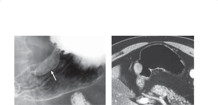
A.Single-contrast UGI. A well-demarcated, smoothsurfaced mass (arrow) is present within the gastric body.
B.Contrast-enhanced CT. A well-defined soft tissue mass arises from the distal stomach.
Di erential Diagnosis
1.Gastrointestinal stromal tumor
2.Fibroma
3.Lipoma
4.Carcinoid tumor
Diagnosis
Gastrointestinal stromal tumor
2. STOMACH 95
B
Discussion
Gastrointestinal stromal tumors (GISTs) are the most common submucosal gastric tumor. They can occur anywhere within the stomach. Depending on their growth characteristics, they can occupy an intramural location (as in this case), extend intraluminally (cases 2.20 and 2.21), or extend as an exophytic mass from the stomach (case 2.36). The smooth surface is characteristic of a submucosal tumor. A 90° angle is often formed between the edges of the mass and the normal gastric wall.
GISTs are often asymptomatic lesions that are discovered incidentally. Melena is a common complaint in patients with symptomatic lesions that are ulcerated. Abdominal pain and obstruction also can occur.
Any tumor arising from the cellular elements of the submucosa could have similar radiographic features: leiomyomas, fibromas, lipomas (case 2.22), neurogenic tumors, malignant GISTs (case 2.36), vascular tumors, and carcinoids.
Disease type: Masses and Filling Defects
96 MAYO CLINIC GASTROINTESTINAL IMAGING REVIEW
CASE 2.20 |
CASE 2.21 |
Findings
CASE 2.20. Single-contrast UGI. A large intraluminal mass is present within the stomach.
CASE 2.21. Enhanced abdominal CT. A mass arising from the gastric wall is present within the fundus.
Di erential Diagnosis
1.Gastrointestinal stromal tumor
2.Bezoar
Diagnosis
Intraluminal gastrointestinal stromal tumor
Discussion
In these patients, a large gastrointestinal stromal tumor (GIST) was removed at operation. Some submucosal tumors grow intraluminally and resemble a polypoid neoplasm (carcinoma or lymphoma) (cases 2.25, 2.26, and 2.30) or a foreign body (bezoar) (case 2.38). Large tumors such as this may cause gastric obstruction.
CT may be helpful for determining the origin and extent of exophytic tumors. Malignant GISTs and leiomyosarcomas tend to be larger (often >10 cm in diameter) than their benign counterparts, have an irregular shape, and are often inhomogeneous with regions of central necrosis (case 2.36). The presence of distant metastases and adjacent organ invasion also can be assessed at CT.
Disease type: Masses and Filling Defects
CASE 2.22
Findings
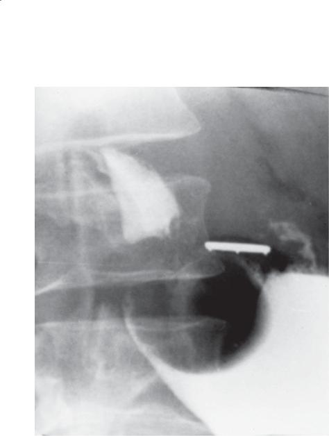
Single-contrast UGI. A well-circumscribed, round filling defect is visible in the distal gastric antrum. The mass has a smooth surface, indicating intact overlying mucosa. A 2-cm metallic marker attached to a compression device is visible adjacent to the mass.
Di erential Diagnosis
1.Gastrointestinal stromal tumor
2.Lipoma
3.Ectopic pancreatic rest
Diagnosis
Gastric lipoma
2. STOMACH 97
Discussion
Gastric lipomas may be radiologically indistinguishable from gastrointestinal stromal tumors (GISTs). Lipomas usually present as solitary intraluminal masses within the antrum and may change shape during peristalsis or compression. These tumors may be pedunculated and can prolapse into the duodenum or, rarely, obstruct the pylorus. The surface of these tumors may ulcerate. CT is diagnostic if the typical fatty density is identified within the tumor. They may be indistinguishable at fluoroscopy from a GIST or an ectopic pancreatic rest (case 2.39). The finding of a small central depression favors the diagnosis of an ectopic pancreatic rest.
Disease type: Masses and Filling Defects
98 MAYO CLINIC GASTROINTESTINAL IMAGING REVIEW
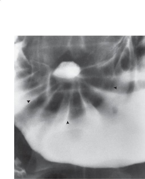
CASE 2.23
Findings
Single-contrast UGI. A small ulcer crater is present in the gastric antrum. Several abnormal folds are present adjacent to the ulcer crater. The folds have a clubbed (arrows) and fused (arrowhead) appearance.
Di erential Diagnosis
1.Gastric adenocarcinoma (malignant ulcer)
2.Benign ulcer
Diagnosis
Gastric adenocarcinoma (malignant ulcer)
Discussion
At operation in this patient, a gastric adenocarcinoma was removed. Early depressed cancers are often recognized by the characteristic changes of the converging folds about the cancer or ulcer. Malignant fold alteration usually includes clubbing, tapering, interruption (amputation), and fusion. When these findings are identified, a cancer should be suspected and an endoscopic biopsy should be performed. Benign ulcers have smooth, tapered folds that radiate to the ulcer crater.
Disease type: Masses and Filling Defects
2. STOMACH 99
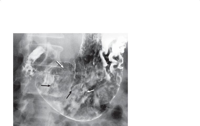
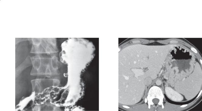
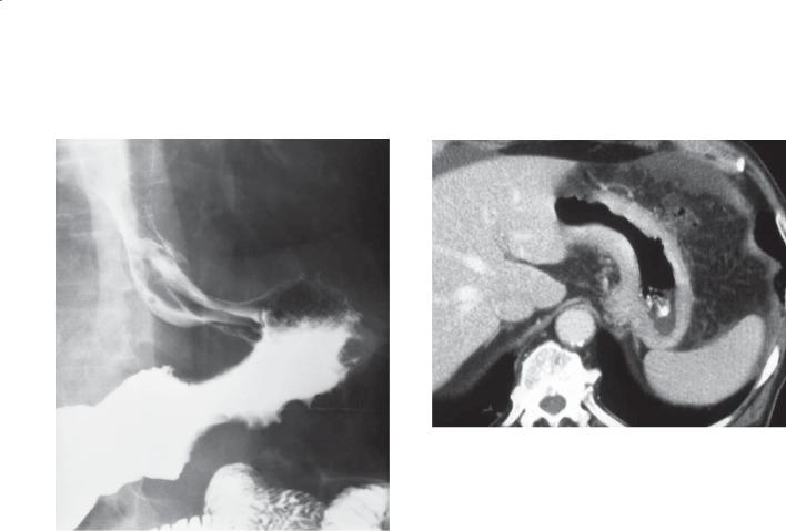
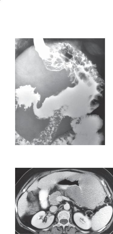
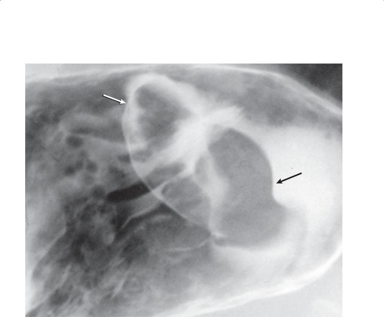
CASE 2.24
Findings
Single-contrast UGI. A large, ulcerated mass straddles the lesser curvature of the gastric body and antrum. The ulcer does not extend beyond the expected location of the normal gastric wall, and the surrounding mass has a lobulated contour.
Di erential Diagnosis
Gastric adenocarcinoma
Diagnosis
Gastric adenocarcinoma: Carman meniscus sign
Discussion
Gastric cancer remains a lethal disease. Most patients in the United States have advanced-stage disease
at diagnosis (stage III or IV) and a very low 5-year survival rate (<10% with these advanced-stage lesions).
Radiologic features of malignancy in lesions that are ulcerated include the following: 1) the tissue surrounding the ulcer is nodular—often the orifice and floor of the crater are also nodular, 2) there is abrupt transition between the surrounding tissue and the normal gastric wall (usually forming an acute angle), 3) the crater does not project beyond the expected location of the gastric wall, 4) radiating folds stop at the edge of the surrounding tissue and do not reach the crater itself, 5) the crater is asymmetrically placed within the surrounding tissue, and 6) the crater is often wider than it is deep.
Th e Carman meniscus sign is seen when the malignant ulcer straddles the lesser curvature and compression is applied apposing both surfaces of the surrounding tumor. The ulcer appears as a crescent (half-moon) on the lesser curvature, with nodular tumor surrounding the periphery of the ulcer. This sign is reported to be pathognomonic of carcinoma (see the figure on page 150).
Disease type: Masses and Filling Defects
100 MAYO CLINIC GASTROINTESTINAL IMAGING REVIEW
CASE 2.25 |
CASE 2.26 |
Findings
CASE 2.25. Double-contrast UGI. A polypoid irregularsurface filling defect (arrowhead) is present in the gastric fundus and cardia.
CASE 2.26. Enhanced abdominal CT. A large polypoid, intraluminal soft tissue mass (arrow) is present in the gastric antrum.
Di erential Diagnosis
1.Gastric adenocarcinoma
2.Gastrointestinal stromal tumor
3.Lymphoma
4.Solitary varix
5.Metastases
Diagnosis
Polypoid gastric adenocarcinoma
Discussion
Gastric adenocarcinoma was found at operation in these patients. The incidence of gastric cancer has decreased in the United States. It now is the third most frequent gastrointestinal cancer, after colorectal and pancreatic cancer, and the sixth most frequent cancer overall. A few decades ago, gastric cancer was
the most frequent gastrointestinal malignancy. There is considerable geographic variability throughout the world in the incidence of this disease. High cancer rates are found in Japan, Finland, Iceland, and Chile. Several etiologic factors are likely to contribute to the development of this disease, including high-starch diets, polycyclic hydrocarbons (found in home-smoked foods), and nitrosamines (found in processed meats and vegetables). Patients with some gastric conditions, including atrophic gastritis, pernicious anemia, postsubtotal gastrectomy, and adenomatous polyps, also are considered to have a higher risk for development of gastric malignancy.
Th e use of routine preoperative CT for patients with known gastric cancer is controversial. Some investigators have found CT unreliable for determining the true extent of disease. Nodal metastases can be particularly problematic, because normal-sized lymph nodes can contain metastases and enlarged lymph nodes may be inflammatory.
Gastrointestinal stromal tumors usually have a very smooth surface, whereas lymphomas and metastases usually present with multiple masses. A solitary varix should be considered in patients with portal venous hypertension. Endoscopy is usually required for a definitive diagnosis.
Disease type: Masses and Filling Defects
2. STOMACH 101
CASE 2.27 |
CASE 2.28 |
Findings
CASE 2.27. Single-contrast UGI. Several filling defects are seen within a nondistensible segment of the gastric body. The mass encases and narrows the stomach. Gastric folds can be seen at the level of the mass along the greater curvature.
CASE 2.28. Contrast-enhanced CT. Thickening of the wall of the gastric fundus and body is present. The luminal contour is irregular.
Di erential Diagnosis
1.Lymphoma
2.Gastric adenocarcinoma
3.Ménétrier disease
4.Metastases
Diagnosis
Polypoid, infiltrative gastric adenocarcinoma
Discussion
Gastric adenocarcinoma was found at operation. Differential considerations may be indistinguishable radiographically. The stomach often remains distensible in Ménétrier disease. Today, most patients with gastric cancers will have preoperative endoscopy for histologic confirmation. A correct diagnosis of gastric cancer usually can be made endoscopically; however, infiltrative tumors may have a positive result on biopsy in only 70% of patients. The presence of intact gastric folds indicates the submucosal location of the infiltrative process.
Resection is the only treatment that can result in cure. Many authorities favor an extensive gastric and lymph node resection. Approximately 40% of
patients with gastric cancer have advanced disease that precludes a curative resection. Many patients undergo a palliative bypass procedure to prevent obstruction or to relieve dysphagia.
Disease type: Masses and Filling Defects
102 MAYO CLINIC GASTROINTESTINAL IMAGING REVIEW
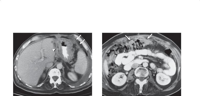
CASE 2.29
A B
Findings
Contrast-enhanced CT. A. The stomach is circumferentially thickened. The serosal contours are irregular and poorly defined. Abnormal soft tissue masses are seen about the anterior peritoneal surface (arrows). The soft tissue thickening (arrowhead) within the gastrohepatic ligament is adenopathy. A left pleural effusion is present. B. Multiple soft tissue masses (arrows) are seen within the omentum, anterior and adjacent to the transverse colon.
Di erential Diagnosis
Metastatic gastric carcinoma
Diagnosis
Metastatic gastric carcinoma
Discussion
Th e lesser and greater omentum represent tissues that are in direct contiguity with the gastric serosa. Once cancer extends to the serosa, extension to the omentum occurs in most patients. More than 90% of patients have omental involvement if the tumor has reached the serosa, whereas only approximately a third of patients with tumor limited to the muscular layer have omental tumor.
Lymphatic metastases are common within nodes along the lesser curvature (within the gastrohepatic ligament) and greater curvature. Other commonly involved nodal groups include parapancreatic, paraaortic, and nodes around the middle colic artery.
After tumor spreads beyond the serosa, cells may seed the peritoneal cavity. Usual locations for peritoneal metastases include the pouch of Douglas, sigmoid mesocolon, right paracolic gutter, and the
small bowel mesentery. Metastatic spread to the ovary is referred to as a Krukenberg tumor. Most liver and lung metastases occur as a result of hematogenous dissemination.
Direct invasion of gastric cancer to the pancreas or liver can be difficult to recognize at CT. Only if there are obvious findings of invasion should the diagnosis be made confidently.
Disease type: Masses and Filling Defects
CASE 2.30
A
Findings
A.Single-contrast UGI. The gastric antrum is markedly narrowed by a large constricting mass.
B.Enhanced abdominal CT. A large polypoid mass
is seen arising from the anterior wall of the gastric antrum.
Di erential Diagnosis
1.Gastric carcinoma
2.Lymphoma
Diagnosis
Gastric lymphoma (solitary)
2. STOMACH 103
B
Discussion
Solitary lymphoma, as in this case, can mimic gastric adenocarcinoma (cases 2.23 and 2.24). Diagnostic tissue may be difficult to obtain at endoscopic biopsy because the overlying mucosa may be intact and prevent adequate tumor sampling. For this reason, multiple biopsy specimens at sites of possible mucosal involvement need to be obtained at endoscopy.
Disease type: Masses and Filling Defects
104 MAYO CLINIC GASTROINTESTINAL IMAGING REVIEW
CASE 2.31
CASE 2.32
Disease type: Masses and Filling Defects

2. STOMACH 105
CASE 2.33
Findings
CASE 2.31. Double-contrast UGI. Marked rugal fold thickening is present throughout the stomach. Multiple nodules are also present throughout the duodenum.
CASE 2.32. Enhanced abdominal CT. Marked wall thickening is seen throughout the stomach.
CASE 2.33. Double-contrast UGI. Diffuse nodularity and fold thickening are present within the gastric body and antrum.
Di erential Diagnosis
1.Lymphoma
2.Ménétrier disease
3.Gastric adenocarcinoma
4.Metastases
Diagnosis
Gastric lymphoma (diffuse infiltrating)
Discussion
Th e amount of wall thickening in this case is characteristic of gastric lymphoma. Infiltrative lymphoma is often confined to the submucosal layer of the stomach. Ulcerations may be present. Despite
extensive disease, there is little restriction in gastric volume.
Lymphoma occasionally crosses the pylorus and affects the adjacent duodenum (as in case 2.31). Lymphoma also can occur within other regions of the alimentary tract. Because treatment and prognosis differ if the disease has spread beyond the stomach, it is often helpful to extend a UGI examination and also study the small bowel.
CT can be helpful in patients with known or suspected gastric lymphoma. The extent of
extraluminal disease (usually adenopathy) can be documented and complications such as perforation and fistulization can be detected. Perigastric adenopathy is a common finding in patients with lymphoma, but it also can be found in patients with carcinoma. Lymphadenopathy at or below the level of renal pedicles is uncommon in patients with gastric carcinoma, but it has been reported in at least onethird of patients with lymphoma.
Differential considerations include Ménétrier disease (cases 2.14 and 2.15) and linitis plastica (gastric adenocarcinoma) (cases 2.49 and 2.50). Ménétrier disease usually affects the proximal stomach, and linitis plastica narrows the gastric lumen and produces a noncompliant affected segment.
Disease type: Masses and Filling Defects
106 MAYO CLINIC GASTROINTESTINAL IMAGING REVIEW
CASE 2.34
Findings
Single-contrast UGI. Multiple smooth-surfaced filling defects (arrows) are present along the greater curvature of the gastric fundus.
Di erential Diagnosis
1.Gastric adenocarcinoma
2.Lymphoma
3.Metastases
4.Varices
Diagnosis
Gastric lymphoma (multiple submucosal masses)
Discussion
Th e stomach is the most common extranodal site for non-Hodgkin lymphoma. Disease confined to the stomach has a much better prognosis than does disseminated tumor, and it is often treated surgically rather than with chemotherapy.
Several radiographic forms of lymphoma have been identified, including infiltrative, ulcerative, polypoid, and intraluminal-fungating. There is often overlap among the radiographic forms. Radiographic features that are helpful in suggesting lymphoma instead of gastric adenocarcinoma include multiplicity of lesions, involvement of a large extent of stomach, evidence
for a submucosal origin of the tumor, extension of the tumor across the pylorus, and less luminal narrowing than expected.
Gastric varices can have a polypoid submucosal appearance (cases 2.41, 2.42, and 2.43) and can change shape during the examination.
Disease type: Masses and Filling Defects

CASE 2.35
Findings
CASE 2.35. Double-contrast UGI. A smooth-surfaced mass is present in the posterior wall of the gastric fundus.
CASE 2.36. Contrast-enhanced CT. A large, mixeddensity, predominantly exophytic mass lies between the posterior wall of the fundus and the aorta.
Di erential Diagnosis
Gastrointestinal stromal tumor
Diagnosis
Malignant gastrointestinal stromal tumor
2. STOMACH 107
CASE 2.36
Discussion
Gastrointestinal stromal tumors (GISTs) are unusual primary gastric tumors of mesenchymal origin. Diagnosis of a GIST is important because it has a much better prognosis than does gastric adenocarcinoma. These intramural tumors often extend exophytically from the stomach, as in these cases. However, they may present with a large endogastric component, depending on the direction of growth.
Th ere are no reliable radiographic criteria to differentiate benign from malignant GISTs, except that the larger the mass, the more likely it is to be malignant. CT can assist in determining the origin and extent of the tumor, as well as evaluating for possible metastases. Metastases from malignant GISTs often spread to the liver and peritoneal cavity, whereas metastases to local lymph nodes are unusual. Most submucosal tumors are removed regardless of size because even small tumors can harbor malignancy.
Disease type: Masses and Filling Defects
108 MAYO CLINIC GASTROINTESTINAL IMAGING REVIEW
CASE 2.37
Findings
Double-contrast UGI. A gastric mass (arrows) containing a central ulceration is present in the upper body of the stomach.
Di erential Diagnosis
1.Gastric adenocarcinoma
2.Lymphoma
3.Metastasis
4.Gastrointestinal stromal tumor
5.Ectopic pancreatic rest
Diagnosis
Metastatic melanoma
Discussion
Melanoma metastases have a similar radiographic appearance in the stomach and small bowel. They usually present as submucosal masses containing a central ulceration. The appearance has been described as a target or bull’s-eye lesion. Although ulcerative metastases can cause gastrointestinal bleeding, other causes of bleeding also should be searched for in symptomatic patients with cancer. Severe gastritis and benign ulcers are more common sources of bleeding in affected patients.
Differential considerations for a solitary ulcerated lesion in the stomach include gastrointestinal stromal tumor (case 2.19), primary gastric adenocarcinoma (case 2.23), lymphoma (case 2.30), or ectopic pancreatic rest (containing a central umbilication) (case 2.39). Multiple lesions favor a diagnosis of metastases or lymphoma. The clinical history of metastatic melanoma in this patient is critical in determining the most likely diagnosis.
Disease type: Masses and Filling Defects
2. STOMACH 109
CASE 2.38
Findings
Single-contrast UGI. A huge filling defect occupies the entire gastric lumen.
Di erential Diagnosis
1.Bezoar
2.Adenocarcinoma
3.Gastrointestinal stromal tumor
4.Lymphoma
Diagnosis
Bezoar
Discussion
Bezoars are concretions of ingested material. They are most commonly composed of vegetable
material (phytobezoar), hair (trichobezoar), or other substances. Various foods (persimmons, berries, other fruits, vegetables, milk), mucus, pitch, tar, antacids, shellac, and furniture polish have been
reported to form bezoars. Delayed gastric emptying, diminished gastric acid and pepsin production, excess or abnormal production of mucus, dietary content, and improper mastication are factors that can contribute to bezoar formation. Patients with prior gastric surgery are particularly prone to bezoar development (case 2.57).
Radiographic features of a bezoar include a mottled soft tissue mass on abdominal plain radiographs. A filling defect is present within the stomach on barium studies. The defect is not attached to the wall, and contrast material often collects within the interstices of the bezoar. Complications are rare but include obstruction, ulceration, hemorrhage, and perforation. Gastric tumor (carcinoma, case 2.26; lymphoma,
case 2.30; gastrointestinal stromal tumor, case 2.20) is the most important differential consideration. Free
movement of the mass within the stomach and the lack of attachment of the mass to the stomach can usually distinguish it from a true neoplasm.
Disease type: Masses and Filling Defects
110 MAYO CLINIC GASTROINTESTINAL IMAGING REVIEW
CASE 2.39
Findings
Double-contrast UGI. A 1.5-cm, round filling defect containing a central umbilication is present within the gastric antrum. The metallic marker is a measuring and compression device scored at 1-cm intervals.
Di erential Diagnosis
1.Gastric ulcer (erosion)
2.Ectopic pancreatic rest
3.Gastrointestinal stromal tumor
4.Other submucosal tumors (lipoma, fibroma, neuroma, hemangioma)
Diagnosis
Ectopic pancreatic rest
Discussion
Th is case shows the typical size, location, and appearance of an ectopic (heterotopic) pancreas. Ectopic rests of pancreatic tissue have been identified in up to 14% of all autopsy specimens. They are more common than gastrointestinal stromal tumors (GISTs). Usually they are found incidentally, but any condition affecting the pancreas also can affect ectopic tissue. Diseases including pancreatitis, pseudocyst formation, carcinoma, and adenoma have been reported.
Although the distal stomach is usually involved, other locations including the duodenum, ileum, and within a Meckel diverticulum are possible. Radiographically, they appear as umbilicated submucosal nodules, although nearly half may not contain the characteristic central depression. The umbilication is not an ulceration; it is covered by normal epithelium. Rudimentary ducts may empty into this depression. These masses are often indistinguishable from a GIST (case 2.19) or other submucosal tumors.
Disease type: Masses and Filling Defects
2. STOMACH 111
CASE 2.40
Findings |
Discussion |
|
Double-contrast UGI. A symmetric, smooth filling |
Different fundoplication procedures (Nissen 1, |
|
defect (arrows) surrounds the gastroesophageal |
Nissen 2, Hill, Belsey) are performed to correct |
|
junction. |
gastroesophageal reflux. Any of these procedures may |
|
|
|
give the radiographic appearance of a pseudotumor. |
Di erential Diagnosis |
The Nissen 2 fundoplication is commonly performed. |
|
1. |
Fundoplication |
This procedure involves wrapping a cuff of gastric |
2. |
Gastric adenocarcinoma |
fundus around the posterior esophagus and then |
3. |
Lymphoma |
suturing it together anteriorly. The cuff of fundus |
|
|
narrows the distal esophagus to prevent reflux. |
Diagnosis |
The main differential consideration is a neoplasm. |
|
Fundal pseudotumor: fundoplication |
A history of antireflux operation and the typical |
|
|
|
radiographic findings (as in this case) usually make the |
|
|
diagnosis straightforward. |
Disease type: Masses and Filling Defects

112 MAYO CLINIC GASTROINTESTINAL IMAGING REVIEW
CASE 2.41 |
CASE 2.42 |
CASE 2.43
Disease type: Masses and Filling Defects

Findings
CASE 2.41. Double-contrast UGI. Multiple, serpentine filling defects are present in the gastric fundus. A calcified mass is present in the region of the pancreas, which represents a partially calcified islet cell carcinoma.
CASE 2.42. Single-contrast UGI. A lobulated mass is present in the gastric cardia and fundus.
CASE 2.43. Contrast-enhanced CT. Enhancing vascular strictures (arrow) are present within the gastric fundus medially. Multiple additional upper abdominal collaterals also are visible.
Di erential Diagnosis
CASE 2.41
1.Gastric varices
2.Ménétrier disease
3.Lymphoma
4.Zollinger-Ellison syndrome
5.Pancreatitis
CASE 2.42
1.Gastric carcinoma
2.Lymphoma
3.Gastric varices
CASE 2.43
1. Gastric varices
Diagnosis
Gastric varices
Discussion
Case 2.41 has the typical findings of gastric varices with serpiginous fold thickening in the fundus. The gastric varices in this case were caused by splenic vein
2. STOMACH 113
occlusion from the islet cell tumor. Case 2.42 shows a rare appearance of gastric varices presenting as a polypoid mass in the gastric fundus, which resembles a carcinoma. The presence of esophageal varices or a
history of portal venous hypertension should make the examiner consider a possible gastric pseudotumor. CT (case 2.43) is invaluable for determining the vascular nature of the mass and often allows detection of the underlying disease causing the varices.
Gastric varices develop as a result of portal venous hypertension or splenic vein occlusion. Only one-third to one-half of patients with uphill esophageal varices from portal venous hypertension have gastric varices, but nearly all patients with gastric varices and portal venous hypertension have esophageal varices. This is probably the result of the subserosal location of gastric varices that in many patients cannot be detected at barium examination. Splenic vein occlusion usually is due to pancreatic disease (pancreatitis or pancreatic carcinoma), but it can be idiopathic. Esophageal varices are absent in splenic vein occlusion because blood flow travels from the short gastric veins to
the gastric fundus plexus and returns to the portal circulation by way of the left gastric (coronary) vein.
Radiographic findings include serpentine defects, multiple lobulated masses resembling a bunch
of grapes, or even a single, polypoid fundal mass (case 2.42). The double-contrast examination may be more sensitive for examining the fundus for varices, because this region is usually not accessible for adequate compression using the single-contrast technique. Examination of the esophagus for varices is important because the combined findings of esophageal and gastric varices indicate portal venous hypertension. A normal esophagus suggests splenic vein occlusion.
Disease type: Masses and Filling Defects

114 MAYO CLINIC GASTROINTESTINAL IMAGING REVIEW
TABLE 2.3
Gastric Filling Defects |
|
CASE |
|
|
|
Benign tumors |
|
|
Polyps |
Adenomatous: few and large (>1 cm). Hyperplastic: many |
2.16–2.18 |
|
and small. Fundic gland polyposis syndrome: hyperplastic |
|
|
in familial adenomatous polyposis |
|
Gastrointestinal stromal |
Smooth surface. Can ulcerate. Most common gastric |
2.19–2.21 |
tumor and leiomyoma |
submucosal tumors. Leiomyomas and gastrointestinal |
|
|
stromal tumors are indistinguishable |
|
Lipoma |
Usually indistinguishable from gastrointestinal stromal |
2.22 |
|
tumor without CT. Diagnostic fatty density on CT |
|
|
|
|
Malignant tumors
Carcinoma
Lymphoma
Can be polypoid, infiltrative, ulcerative, or scirrhous. |
2.23–2.29 |
Usually large with irregular surface |
|
Diverse presentation: solitary or multiple masses, |
2.30–2.34 |
infiltrative. Often has associated adenopathy |
|
Malignant gastrointestinal |
Smooth surface, but can ulcerate. Can present as large |
2.35 and 2.36 |
stromal tumor |
intraluminal or exophytic mass |
|
Metastases |
Melanoma, breast, lung are common. Also locally invasive |
2.37 |
|
tumors (esophagus, pancreas, and colon) |
|
|
|
|
Nonneoplastic |
|
|
Bezoar, retained food |
Moves. Contrast material fills interstices. |
2.38 |
Ectopic pancreatic rest |
Usually distal stomach. Central umbilication |
2.39 |
Fundoplication defect |
Fundus encircles gastroesophageal junction |
2.40 |
Varices |
Usually multiple serpentine filling defects in fundus. Splenic |
2.41–2.43 |
|
vein thrombosis causes isolated gastric varices. Seen with |
|
|
esophageal varices in portal hypertension |
|
|
|
|
Disease type: Masses and Filling Defects

CASE 2.44
Findings
Single-contrast UGI. Within the gastric antrum, there is symmetric, smooth, tapered narrowing.
Di erential Diagnosis
1.Scarring from chronic peptic ulcer disease
2.Granulomatous disease (Crohn disease, sarcoidosis, tuberculosis, syphilis, eosinophilic gastroenteritis)
3.Scirrhous carcinoma
Diagnosis
Crohn disease
2. STOMACH 115
Discussion
Chronic changes of Crohn disease are usually due to previous intramural inflammation and subsequent fibrosis. Because the distal stomach is most commonly involved, diffuse narrowing of this region often results in a tubular configuration resembling a ram’s horn or pseudo-Billroth I appearance. The scarring may not affect the circumference of the antrum evenly, resulting in irregular deformity of the antrum and proximal duodenum. Fistula formation and mass effect are
less frequent in the stomach than in the small bowel or colon. Without a history of peptic ulcers or other evidence of Crohn disease, it may not be possible to narrow the differential possibilities without endoscopic biopsy of this region.
Disease type: Narrowings
116 MAYO CLINIC GASTROINTESTINAL IMAGING REVIEW
CASE 2.45
Findings
Single-contrast UGI. The distal third of the stomach is narrowed, tapered, and nondistensible.
Di erential Diagnosis
1.Chronic peptic ulcer disease
2.Granulomatous disease (Crohn disease, sarcoidosis, tuberculosis, syphilis, eosinophilic gastroenteritis)
3.Gastric carcinoma
4.Metastatic breast cancer
5.Prior caustic ingestion
Diagnosis
Granulomatous gastritis (sarcoidosis)
Discussion
Th e most frequent cause of granulomatous gastritis is Crohn disease (case 2.44). Most patients with Crohn disease have concomitant disease in the small bowel. Sarcoidosis involving the stomach nearly always presents in association with disseminated disease. In fact, disease in other organs usually is critical for distinguishing sarcoidosis from the other granulomatous diseases. No specific radiologic findings are helpful for differentiating the various
granulomatous diseases. Gastric carcinoma (cases 2.49 and 2.50), metastatic breast cancer (case 2.51), and scarring from corrosive ingestion (case 2.48) could have similar radiographic changes.
Disease type: Narrowings
CASE 2.46
Findings
Single-contrast UGI. Tapered narrowing is present in the antrum of the stomach with nodular fold thickening of the distal stomach and visualized proximal duodenum.
Di erential Diagnosis
1.Granulomatous disease (Crohn disease, sarcoidosis, tuberculosis, syphilis, eosinophilic gastroenteritis)
2.Gastric carcinoma
3.Gastric lymphoma
Diagnosis
Eosinophilic gastroenteritis
2. STOMACH 117
Discussion
Eosinophilic gastroenteritis is a rare idiopathic disease of the gastrointestinal tract that virtually always affects the gastric antrum, as well as all or part of the small bowel (case 4.53). It is caused by an eosinophilic infiltrate of the mucosa and submucosa and commonly results in mucosal nodularity and narrowing of the distal stomach and diffuse or segmental nodular fold thickening in the small bowel. The disease also may rarely involve the colon.
Patients with eosinophilic gastroenteritis often present with abdominal pain, diarrhea, and
occasionally malabsorption. The gastric antrum is usually the best site for diagnostic biopsy. Most patients have peripheral eosinophilia demonstrable on blood smears. In addition, many patients have an allergy history. The clinical course is usually self-limited, and the disease often responds to corticosteroid therapy or resolves spontaneously.
Disease type: Narrowings
118 MAYO CLINIC GASTROINTESTINAL IMAGING REVIEW
CASE 2.47
Findings |
Discussion |
Double-contrast UGI. The stomach is featureless |
Atrophic gastritis is a descriptive term for chronic |
without evidence of rugal folds. |
gastritis. Chronic gastritis can be due to an |
|
autoimmune disease (type A) with autoantibodies to |
Di erential Diagnosis |
parietal cells and intrinsic factor. It most commonly |
1. Atrophic gastritis |
affects the body and fundus of the stomach. Long- |
2. Gastric overdistention |
standing Helicobacter pylori infection (type B) can |
|
also cause chronic gastritis; this type has antral |
Diagnosis |
predominance early in the disease. Many patients will |
Atrophic gastritis |
have the condition for 15 to 20 years, and therefore |
|
it often occurs in older persons. The condition is |
|
associated with an increase in the prevalence of both |
|
gastric cancer and gastric lymphoma. |
|
Radiographically, the stomach has a bald appearance |
|
due to the lack of rugal folds. Distention of the stomach |
|
with effervescent granules can obliterate some gastric |
|
folds, but other folds (especially in the region of the |
|
fundus) are almost always visible, even with optimal |
|
distention. |
Disease type: Narrowings

2. STOMACH 119
CASE 2.48
Findings
Double-contrast UGI. The antrum of the stomach is irregularly narrowed. There is an abrupt margin at the junction of the antrum and body along the greater curvature.
Di erential Diagnosis
1.Caustic gastric stricture
2.Annular carcinoma
3.Granulomatous infection
Diagnosis
Caustic stricture of the gastric antrum
Discussion
Th is patient had a history of ammonia ingestion. Caustic injury to the stomach from acid, alkali, or other chemicals usually affects the antrum more severely than the proximal stomach. The amount of gastric damage depends on the depth of the burn. Mucosal damage (first-degree burn) alone heals
without sequelae. Second-degree burns involve the submucosa and muscular layers of the stomach. Healing occurs with fibrosis and stricture formation. The secretory and peristaltic functions of the stomach also may be inhibited, depending on the amount
of damage. Third-degree burns involve transmural damage, usually resulting in inflammation in the surrounding affected tissues. Hemorrhage, sepsis, and shock can occur.
Ulceration of the stomach and duodenum can be seen 2 to 3 days after caustic ingestion. Subsequent changes include 1) large intraluminal filling defects from hematomas, 2) contraction of the distal stomach with atony, 3) rigidity, and 4) contour irregularity. Late changes include antral stenosis (as seen in this case). Abrupt annular narrowing of the gastric lumen can be confused with a carcinoma (cases 2.49 and 2.50). Associated lesions in the esophagus and duodenum, in addition to a history of caustic ingestion, can help differentiate this lesion from cancer. Granulomatous disease usually results in smooth, tapered narrowing of the antrum.
Disease type: Narrowings

120 MAYO CLINIC GASTROINTESTINAL IMAGING REVIEW
CASE 2.49 |
CASE 2.50 |
Findings
CASE 2.49. Double-contrast UGI. The wall of the stomach is markedly thickened (arrows), with narrowing and scalloping of the gastric contour.
CASE 2.50. Contrast-enhanced CT. Marked thickening and enhancement of the gastric wall are visible.
Di erential Diagnosis
1.Gastric carcinoma
2.Metastases
3.Lymphoma
Diagnosis
Scirrhous gastric adenocarcinoma
Discussion
Scirrhous carcinomas spread within the submucosa of the gastric wall, often inciting a desmoplastic reaction. Because of the narrowing and rigidity that often accompany these tumors, the radiographic appearance of the stomach has been likened to a leather bottle (linitis plastica). Gastric narrowing is not detectable in
all patients. In patients with these lesions, there may be mucosal nodularity, fold thickening, or ulceration. Scirrhous tumors often enhance after intravenous administration of contrast material. These tumors usually involve a large extent of the stomach and often spread to peritoneal surfaces. Polypoid and ulcerative adenocarcinomas usually do not avidly enhance.
Many conditions can present with similar radiographic findings. Breast cancer metastases can resemble scirrhous tumors and often narrow the gastric antrum. Omental metastases, often arising from a primary carcinoma in the transverse colon, can invade and narrow the stomach by way of the gastrocolic ligament.
Histologic confirmation of scirrhous adenocarcinoma of the stomach may be difficult to obtain by endoscopic biopsy. Located in the
submucosal tissues, scirrhous adenocarcinoma often requires a deep biopsy for diagnosis. In addition, the associated desmoplastic reaction may widely intersperse cancer cells between large areas of fibrosis.
Disease type: Narrowings
CASE 2.51
Findings
Double-contrast UGI. The body and antrum of the stomach are deformed and narrowed. Gastric folds are visible in this region of narrowing.
Di erential Diagnosis
1.Scirrhous adenocarcinoma
2.Metastatic breast cancer
3.Granulomatous diseases affecting the stomach
Diagnosis
Metastatic breast cancer
2. STOMACH 121
Discussion
Metastatic breast cancer involving the stomach may produce a linitis plastica appearance indistinguishable from that of primary scirrhous gastric adenocarcinoma (cases 2.49 and 2.50). Whenever a scirrhous carcinoma is identified in a woman, breast metastases also
should be considered. Both scirrhous carcinoma and granulomatous disease can have an identical
radiographic appearance. Endoscopic biopsy is usually required for definitive diagnosis.
Disease type: Narrowings

122 MAYO CLINIC GASTROINTESTINAL IMAGING REVIEW
TABLE 2.4
Gastric Narrowings |
|
CASE |
|
|
|
Benign |
|
|
Peptic scarring |
Nearly always antral with loss of normal antral shoulders. Duodenal |
3.8 |
|
changes are commonly associated |
|
Granulomatous disease |
Ram-horn deformity of antrum. Smooth narrowing, Crohn disease, |
2.44–2.46 |
|
sarcoidosis, tuberculosis, syphilis, eosinophilic gastroenteritis |
|
Corrosive ingestion |
Usually acid. Distal stomach |
2.48 |
|
|
|
Malignant |
|
|
Carcinoma |
Irregular luminal contour. Scirrhous: long segment with |
2.49 and 2.50 |
|
intact rugal folds |
|
Metastases |
Linitis plastica appearance. Often seen with metastatic breast cancer. |
2.51 |
|
Identical appearance to that of scirrhous adenocarcinoma |
|
Lymphoma |
Infiltrative form can cause gastric narrowing. Thickened, |
2.30–2.34 |
|
disorganized folds |
|
|
|
|
Disease type: Narrowings
2. STOMACH 123
CASE 2.52
Findings
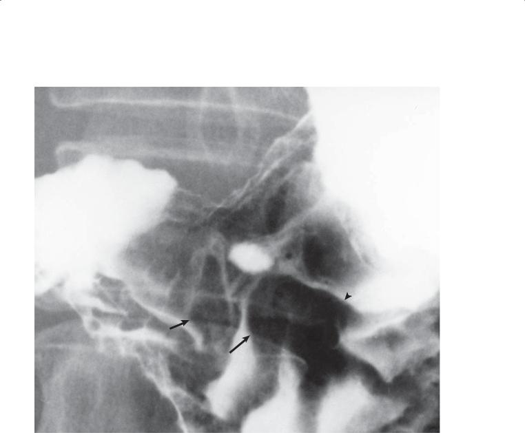
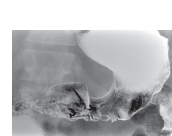
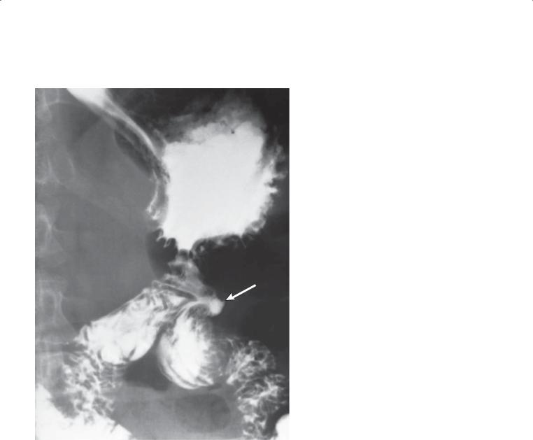
Single-contrast UGI. An ulcer crater (arrow) is present within the efferent limb, adjacent to the gastrojejunal anastomosis, in this patient with a Billroth II gastroenterostomy.
Di erential Diagnosis
Marginal ulcer
Diagnosis
Marginal ulcer
Discussion
A marginal or stomal ulcer is a perianastomotic ulcer developing after a gastroenterostomy. Most ulcers
occur in the efferent limb of the jejunum, within 2 cm of the stoma. Marginal ulcers should raise suspicion of several possible conditions: incomplete vagotomy, retained gastric antrum, Zollinger-Ellison syndrome, hypercalcemia, smoking, or ulcerogenic drug abuse (aspirin, nonsteroidal anti-inflammatory drug).
A spectrum of radiographic findings of peptic disease affecting the postanastomotic jejunum can be seen, including typical ulcer craters, giant craters resembling large diverticula, thickened jejunal folds, and rigidity of the affected jejunal segment. Despite ideal radiographic techniques, some postoperative ulcers are not detected (reports vary between 20% and 50%). Endoscopy should be recommended for
symptomatic patients with negative radiologic studies.
Disease type: Postoperative Stomach
124 MAYO CLINIC GASTROINTESTINAL IMAGING REVIEW
CASE 2.53 |
CASE 2.54 |
Findings
CASE 2.53. Contrast-enhanced CT. A dilated, fluid-filled duodenum is visible in this patient, who had a previous Billroth II gastroenterostomy.
CASE 2.54. Single-contrast UGI. The afferent limb of a Billroth II anastomosis is markedly dilated. A cause for the obstruction near the gastroenterostomy is not visible.
Di erential Diagnosis
Afferent loop syndrome
Diagnosis
Afferent loop syndrome
Discussion
Th e usual cause of afferent loop syndrome is obstruction of the afferent loop due to adhesions, recurrent ulcer, recurrent tumor, or internal herniation.
Patients with afferent loop syndrome often present with distention, pain, and nausea. Conventional barium studies occasionally identify the site of obstruction and its cause. Often, however, only nonfilling of the afferent limb is detected. CT is helpful for directly imaging the fluid-filled and dilated afferent loop. Fixed obstruction of the loop may lead to blind loop syndrome with bacterial overgrowth, vitamin B12 deficiency, and megaloblastic anemia. Perforation of the afferent limb with peritoneal spillage has been reported and may cause death.
Afferent loop syndrome also can be due to a nonphysiologic surgical anastomosis, in which food preferentially empties into the afferent limb. Preferential filling of the afferent loop and delayed emptying often can be appreciated fluoroscopically
in patients with nonphysiologic anastomoses. Both of these patients had twisting of the afferent loop around adhesions, which required surgical revision.
Disease type: Postoperative Stomach
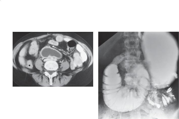
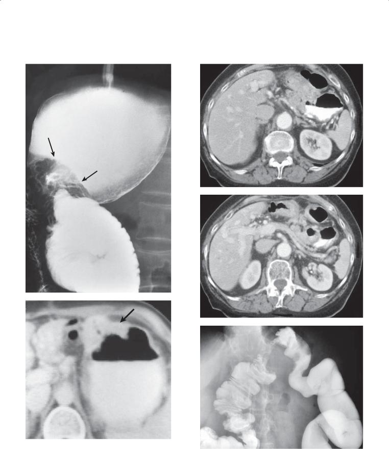
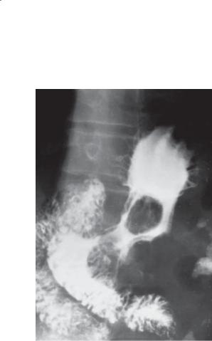
CASE 2.55
A
Findings
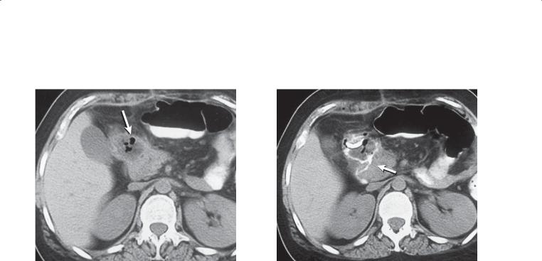
A.Unenhanced abdominal CT. An abscess (arrow) containing fluid and air is located adjacent to the duodenal stump in this patient with a recent Billroth II gastrojejunostomy.
B.Follow-up CT sinogram obtained after placement of a percutaneous locking loop catheter into the abscess cavity shows communication (arrow) between the abscess and the duodenum.
Di erential Diagnosis
1.Blown duodenal stump (duodenal anastomotic dehiscence)
2.Intra-abdominal abscess
Diagnosis
Blown duodenal stump
2. STOMACH 125
B
Discussion
Breakdown or leakage of the duodenal stump (blown duodenal stump) is a serious complication after Billroth II anastomosis. Recognition of this condition is important because it has a high mortality. Anastomotic breakdown can occur anytime between the first day and up to 3 weeks after operation. Early intervention and drainage are important for proper management.
Th e diagnosis may be suggested by the findings on an abdominal plain radiograph, by identifying extraluminal gas, or by abnormal soft tissue in the subhepatic space or about the duodenal stump. CT is excellent for evaluating patients for this complication.
The presence and location of abnormal fluid collections can be identified, and in some patients percutaneous drainage can be performed.
Disease type: Postoperative Stomach
126 MAYO CLINIC GASTROINTESTINAL IMAGING REVIEW
CASE 2.56
Findings
Single-contrast UGI. A filling defect is present within a gastric remnant. The defect is tubular, and valvulae conniventes are seen within it.
Di erential Diagnosis
1.Jejunogastric intussusception
2.Gastric remnant carcinoma
Diagnosis
Jejunogastric intussusception
Discussion
Clinically significant intussusception is an unusual complication after gastroenterostomy. Intussusception
can occur antegradely or retrogradely, acutely or chronically. In patients with acute intussusception, clinical signs and symptoms of gastric outlet obstruction can develop, including hematemesis and an upper abdominal mass or fullness. Chronic intussusception is usually intermittent and selfreducing. Symptomatic patients should be regarded as surgical emergencies because of the high mortality associated with untreated patients in whom bowel perforation develops. The efferent limb is most commonly the intussuscipiens, but the afferent or both limbs also can intussuscept. A mass acting as a lead point should be searched for in patients with an antegrade intussusception, when the jejunum is the intussusceptum.
Disease type: Postoperative Stomach

CASE 2.57
Findings
Single-contrast UGI. There is a large filling defect within the gastric remnant in this patient with a Billroth II gastroenterostomy. Contrast material was seen completely surrounding the filling defect during the examination.
Di erential Diagnosis
1.Gastric bezoar
2.Polypoid gastric cancer
3.Gastrointestinal stromal tumor
Diagnosis
Gastric remnant bezoar
2. STOMACH 127
Discussion
Bezoar formation, usually from retained vegetable matter (phytobezoar), is a relatively common complication after a gastroenterostomy. Bezoar formation often occurs in patients with a narrowed gastroenterostomy stoma and as a result of surgical vagotomy that slows gastric transit and decreases acid production.
Radiologic diagnosis is based on finding a filling defect within the gastric remnant that is not attached to the gastric wall. Often, interstices within the phytobezoar fill with barium and give it a variegated appearance. Retained food can simulate a bezoar, and it is probably prudent to reexamine the fasting patient several hours later to determine whether the finding is still present. Yeast bezoars due to yeast overgrowth
and blood clot bezoars also can occur and cause gastric outlet obstruction.
Disease type: Postoperative Stomach

128 MAYO CLINIC GASTROINTESTINAL IMAGING REVIEW
CASE 2.58 |
CASE 2.59 |
Disease type: Postoperative Stomach

Findings
CASE 2.58. Single-contrast UGI. Postoperative changes of a Billroth II gastroenterostomy are visible with a small and stenotic residual gastric remnant (arrow). Mass effect deforms the distal esophagus eccentrically.
CASE 2.59. Single-contrast UGI. A polypoid intraluminal mass is present within the gastric remnant in another patient with a Billroth II gastroenterostomy.
Di erential Diagnosis
1.Postgastrectomy carcinoma
2.Postoperative deformity
3.Gastrointestinal stromal tumor
Diagnosis
Postgastrectomy carcinoma
2. STOMACH 129
Discussion
Th ese 2 cases have different appearances of postgastrectomy carcinomas. Case 2.58 illustrates an advanced case of gastric remnant shrinkage due to a constricting, diffuse carcinoma. Detection of this type of gastric remnant cancer can be difficult. Baseline examinations are helpful to assess for subtle changes in the gastric pouch. The polypoid cancer in case 2.59 is much easier to diagnose on the UGI examination. The changes in case 2.58 are too extensive to be attributed to postoperative deformity or adhesions. No mucosal markings (gastric folds) are visible in the narrowed segment. This finding usually indicates ulceration. The filling defect in case 2.59 could be due to a gastrointestinal stromal tumor. Inspection of its contour can be helpful to differentiate submucosal (smooth surface) from mucosal (irregular surface)- based lesions.
Disease type: Postoperative Stomach
130 MAYO CLINIC GASTROINTESTINAL IMAGING REVIEW
CASE 2.60 |
CASE 2.61 |
A |
A |
B
B
C
Disease type: Postoperative Stomach

Findings
CASE 2.60. A. Single-contrast UGI. A lobulated filling defect (arrows) is present within the gastric remnant near the anastomosis. The stomach is distended because of partial mechanical obstruction from the mass.
B. Contrast-enhanced CT. A soft tissue mass corresponds to the filling defect (arrow).
CASE 2.61. A and B. Contrast-enhanced CT. A soft tissue mass at the level of the gastrojejunostomy extends anteriorly and abuts the transverse colon.
C. Single-contrast barium enema. Narrowing of the lumen of the transverse colon with mass effect along its inferior margin and tethered mucosal folds.
Di erential Diagnosis
1.Postgastrectomy carcinoma
2.Jejunogastric intussusception
Diagnosis
Postgastrectomy carcinoma
2. STOMACH 131
Discussion
Gastric carcinoma in a gastric remnant usually occurs either soon (several months) after surgical resection for gastric adenocarcinoma as a result of subtotal tumor removal or as a primary cancer developing 20 to 30 years after gastric surgery for ulcer disease. A twofold to sixfold increased risk has been reported for development of carcinoma within the gastric remnant in patients with a gastroenterostomy performed for managing gastric ulcer disease. The increased risk is believed to be related to long-standing gastritis as a result of reflux of bile acids and pancreatic secretions into the stomach. Increased cancer risk remains controversial, because some investigators have not confirmed this relationship.
Radiologic detection can be difficult. Filling defects, mucosal ulceration, and stomal or gastric pouch narrowing are the usual findings. Suspicious findings should be evaluated further with endoscopy. Examination of the gastric remnant with the aircontrast technique is recommended because the remaining pouch is rarely accessible for compression.
Disease type: Postoperative Stomach
132 MAYO CLINIC GASTROINTESTINAL IMAGING REVIEW
CASE 2.62
Findings
Single-contrast barium UGI. Postoperative changes of partial gastrectomy with Billroth II gastroenterostomy are present. There is a large filling defect within the gastric remnant. There is also narrowing and deformity of the remaining distal stomach and jejunum about the anastomosis.
Di erential Diagnosis
1.Postgastrectomy carcinoma
2.Bile reflux gastritis
3.Gastric polyp
Diagnosis
Bile reflux gastritis with inflammatory polyp
Discussion
Th e filling defect was identified at endoscopy and found to be an inflammatory polyp. The markedly edematous folds were due to bile reflux gastritis.
Gastritis after operation is common, but usually it resolves within a few weeks. Reflux of bile by way of the afferent loop in a patient with a Billroth II anastomosis also can produce gastritis and can lead to considerable pain. Bile reflux gastritis rarely is diagnosed radiographically, but it may be suggested by identifying enlarged gastric folds (as seen in the distal portion of the gastric remnant in this patient).
This patient’s inflammatory polyp was probably due to chronic inflammatory changes from bile reflux. Other types of polyps (hyperplastic and adenomatous) also can develop in a gastric remnant. The filling defect
in this case cannot be differentiated from a polypoid carcinoma. Endoscopic biopsy was necessary to make the distinction. Roux-en-Y procedures often are performed in patients with complications from a Billroth II gastroenterostomy.
Disease type: Postoperative Stomach
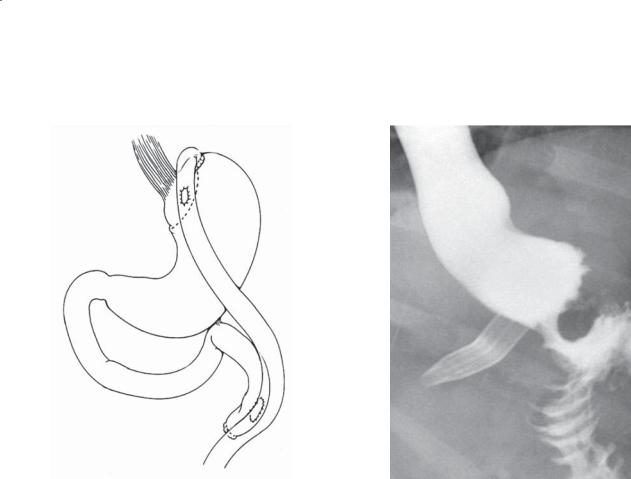
CASE 2.63
A
Findings
A.The anatomical changes that are created at the time of a gastric bypass operation. Notice the small gastric pouch, gastrojejunostomy, and jejuno-jejunostomy.
(Adapted from Carucci LR, Turner MA, Conklin RC, DeMaria EJ, Kellum JM, Sugerman HJ. Roux- en-Y gastric bypass surgery for morbid obesity: evaluation of postoperative extraluminal leaks with upper gastrointestinal series. Radiology. 2006 Jan;238[1]:119-27. Used with permission.)
B.Water-soluble UGI. A gastric pouch is visible with a freely functioning gastrojejunostomy. Surgical drain is seen adjacent to the gastric pouch. (Courtesy of Mary Ann Turner, MD, Virginia Commonwealth University Medical Center, Richmond, Virginia. Used with permission.)
Di erential Diagnosis
Normal postoperative gastric bypass examination
Diagnosis
Normal postoperative gastric bypass examination
2. STOMACH 133
B
Discussion
Obesity is a national epidemic, and traditional methods for weight loss have not proved to be optimal in the long term. Bariatric surgery is a proven method for sustained weight loss and reduction of the morbidity associated with obesity. The traditional gastric bypass operation consists of 3 parts:
1.Creation of a small gastric pouch
2.Jejunogastrostomy using a jejunal Roux limb with a small gastric stomal opening, 8-12 mm
3.Side-to-side anastomosis of the excluded limb with antegrade jejunal Roux limb
Note that during a UGI examination neither the excluded stomach nor the excluded duodenal-jejunal limb is visualized.
Disease type: Postoperative Stomach
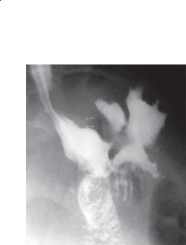
134 MAYO CLINIC GASTROINTESTINAL IMAGING REVIEW
CASE 2.64
Findings
Water-soluble UGI. Extraluminal extravasation of contrast material extends to the left of the gastric pouch in this patient who had had a gastric bypass procedure. (Adapted from Carucci LR, Turner MA, Conklin RC, DeMaria EJ, Kellum JM, Sugerman HJ. Roux-en-Y gastric bypass surgery for morbid
obesity: evaluation of postoperative extraluminal leaks with upper gastrointestinal series. Radiology. 2006 Jan;238[1]:119-27. Used with permission.)
Di erential Diagnosis
Anastomotic leak at gastrojejunostomy
Diagnosis
Anastomotic leak (gastrojejunostomy) after gastric bypass
Discussion
Anastomotic leaks are the commonest postoperative problem in patients who undergo gastric bypass. Commonly, patients are evaluated with oral watersoluble contrast 1 to 2 days after the bypass procedure to evaluate for anastomotic leak. Extravasation of contrast material or communication of contrast material with a surgically placed drain confirms this operative complication.
Th e incidence of postoperative gastric bypass leaks ranges from 1% to 5%. Most leaks occur at the level of the gastrojejunostomy. Early recognition is important because unrecognized complications can lead to death. Distal anastomotic leaks at the jejunojejunostomy are difficult to detect at fluoroscopy.
Disease type: Postoperative Stomach
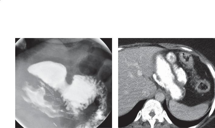
2. STOMACH 135
CASE 2.65
A
Findings
A.Upper gastrointestinal examination. A small gastric pouch is identified with a patent gastrojejunostomy. A small amount of barium is seen within the bypassed proximal stomach. (Adapted from Carucci LR, Conklin RC, Turner MA. Roux-en-Y gastric bypass surgery
for morbid obesity: evaluation of leak into excluded stomach with upper gastrointestinal examination. Radiology. 2008 Aug;248[2]:504-10. Epub 2008 Jun 6. Used with permission.)
B.Contrast-enhanced CT with oral contrast
material. A prominent staple line is present near the gastroesophageal junction with opacification of the normal stomach lumen and proximal jejunum. (Courtesy of Mary Ann Turner, MD, Virginia
Commonwealth University Medical Center, Richmond, Virginia. Used with permission.)
Di erential Diagnosis
1.Anastomotic leak
2.Staple line dehiscence
Diagnosis
Staple line dehiscence after gastric bypass
B
Discussion
Staple line dehiscence (breakdown or disruption) indicates that the staple line between the excluded gastric segment and the gastric pouch is no longer sealed. This complication of gastric bypass is
not associated with the morbidity and mortality complications of an anastomotic leak. Staple line breakdown can be early (due to surgical dehiscence) or delayed (due to stretching of the gastric pouch). Clinically, complication occurs in less than 5% of patients undergoing gastric bypass. Visualization
of contrast material entering the bypassed stomach early in the examination is key to making the proper diagnosis.
Disease type: Postoperative Stomach
136 MAYO CLINIC GASTROINTESTINAL IMAGING REVIEW
CASE 2.66 |
CASE 2.67 |
|
A |
B
Findings
CASE 2.66. Small bowel follow-through examination. A clustered group of small bowel loops is present in the left upper quadrant. These loops enter the left upper quadrant through a narrowed region where entering and exiting small bowel loops are located
(arrows). (Courtesy of Mary Ann Turner, MD, Virginia Commonwealth University Medical Center, Richmond, Virginia. Used with permission.)
A and B. Abdominal CTs. A cluster of small bowel loops is present in the left upper quadrant. The
anastomotic suture line is identified and is located in an abnormal location. (Courtesy of Mary Ann Turner, MD, Virginia Commonwealth University Medical Center, Richmond, Virginia. Used with permission.)
Di erential Diagnosis
1.Ventral hernia
2.Internal hernia
3.Mechanical small bowel obstruction
Diagnosis
Internal hernia
Disease type: Postoperative Stomach

Discussion
Internal hernias occur in about 2% of patients who have had gastric bypass and are the commonest cause of obstruction in this patient group. In many patients, a defect is created surgically in the transverse mesocolon for the retrocolic-positioned gastrojejunal loop. Small bowel loops that herniate through this defect can undergo stasis, obstruction, torsion, ischemia, and infarction.
Key findings to alert the radiologist of the presence of an internal hernia after gastric bypass include the following:
1.Clustered bowel loops (most commonly in the left or mid abdomen)
2.Visible entrance and exit limbs leading to the clustered loops
2. STOMACH 137
3.Displaced jejunojejunal suture line
4.Change in bowel configuration compared with that of prior examinations
5.Stasis within the herniated loops
Ventral hernias can potentially have a similar appearance on the small bowel follow-through examination. Turning the patient to the lateral position can exclude this type of hernia. Adhesive bands from the operation can also lead to small bowel obstruction, but a mechanical obstruction from adhesions does not have the clustered loops or suture line displacement seen in an internal hernia.
Disease type: Postoperative Stomach
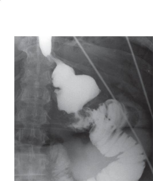
138 MAYO CLINIC GASTROINTESTINAL IMAGING REVIEW
CASE 2.68
Findings
Water-soluble UGI. Gastric bypass with a patent gastrojejunostomy. Only a short segment of dilated jejunum is visible. Contrast does not pass beyond the Roux-en-Y jejunojejunostomy. (Courtesy of Mary Ann Turner, MD, Virginia Commonwealth University Medical Center, Richmond, Virginia. Used with permission.)
Di erential Diagnosis
Obstruction of the jejunum at the jejunojejunostomy
Diagnosis
Obstruction of the jejunum at the jejunojejunostomy
Discussion
Obstruction of the jejunum at the jejunojejunostomy can occur as a result of a tight anastomosis (narrowed lumen or postoperative edema) or adhesive bands.
Intussusception causing obstruction at this site has also been reported. Because internal hernias are also an important cause for obstruction, this diagnosis should be considered whenever an obstruction
is found in patients who have had gastric bypass (cases 2.66 and 2.67).
Disease type: Postoperative Stomach

|
2. STOMACH 139 |
|
|
|
|
|
TABLE 2.5 |
|
Postoperative Stomach |
|
CASE |
|
|
|
Marginal ulcer |
Usually distal to gastrojejunal anastomosis. Can be multiple |
2.52 |
|
|
|
Afferent loop syndrome |
Dilated duodenum. May not fill at UGI. CT helpful for |
2.53 and 2.54 |
|
directly visualizing |
|
|
|
|
Blown duodenal stump |
Fluid or abscess adjacent to proximal duodenum or |
2.55 |
|
subhepatic space |
|
|
|
|
Jejunogastric intussusception |
Filling defect in postoperative stomach. Valvulae confirm |
2.56 |
|
enteric nature |
|
|
|
|
Gastric remnant bezoar |
Filling defect in postoperative stomach. No wall attachment |
2.57 |
|
|
|
Postgastrectomy carcinoma |
Filling defect, luminal irregularity, or shrinkage of gastric pouch |
2.58–2.61 |
|
|
|
Bile reflux gastritis |
Usually associated with Billroth I or II gastroenterostomy. |
2.62 |
|
Thickened folds, filling defect(s) |
|
|
|
|
Gastric bypass complications |
|
|
Anastomotic leak |
Usually at gastrojejunostomy. Extraluminal collection. |
2.64 |
|
Early complication |
|
Staple line dehiscence |
Contrast material fills bypassed gastric segment |
2.65 |
Internal hernia |
Clustered loops. Displaced suture line |
2.66 and 2.67 |
Obstruction |
Dilated jejunal loop to jejunojejunostomy |
2.68 |
|
|
|
Disease type: Postoperative Stomach

140 MAYO CLINIC GASTROINTESTINAL IMAGING REVIEW
CASE 2.69 |
CASE 2.70 |
|
A |
B
Findings
CASE 2.69. Single-contrast UGI. A moderate-sized pouch (arrow) arises from the gastric fundus posteriorly. Its narrow neck arises near the esophagogastric junction. Folds extend from the stomach into the pouch.
CASE 2.70. A and B. Contrast-enhanced CT.
An outpouching arises from the gastric fundus. The most caudal slice does not appear connected to the stomach.
Di erential Diagnosis
Gastric diverticulum
Diagnosis
Gastric diverticulum
Discussion
Diverticula of the stomach are unusual congenital entities. Most arise from the posterior surface of the gastric fundus near the esophagogastric junction. Gastric diverticula are rarely symptomatic, but diverticular bleeding has been reported.
Radiologic recognition of these entities depends on identifying mucosal folds within the diverticulum and observing its characteristic changing size and shape. A connecting neck is also a helpful finding. Occasionally, gastric diverticula can simulate an adrenal mass at CT. The use of oral contrast material can be very helpful for establishing the correct diagnosis.
Disease type: Miscellaneous
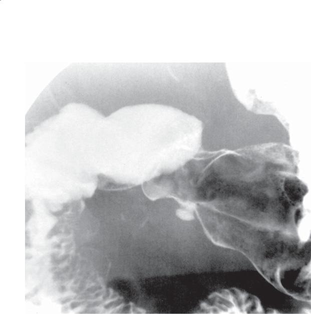
CASE 2.71
Findings
Double-contrast UGI. An intramural collection of barium is present along the greater curvature of the gastric antrum. This collection was observed fluoroscopically to change in size and shape.
Di erential Diagnosis
1.Gastric ulcer
2.Partial gastric diverticulum
Diagnosis
Partial gastric diverticulum
2. STOMACH 141
Discussion
A partial gastric diverticulum is a protrusion of mucosa into the muscular wall of the stomach without disturbing the serosa. It is rare (found in approximately 0.05% of the population) and asymptomatic. Its importance lies in differentiating it from a peptic ulcer. Most partial diverticula are located along the greater curvature of the antrum. They often have a narrow neck and change in shape during fluoroscopic visualization. Ectopic pancreatic tissue (case 2.39)
in the distal stomach is reported to be a frequent finding. The exact role this aberrant tissue plays in the development of these diverticula is unclear.
Disease type: Miscellaneous
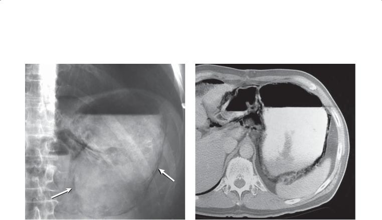
142 MAYO CLINIC GASTROINTESTINAL IMAGING REVIEW
CASE 2.72
A B
Findings
A.Abdominal radiograph. Intramural gas (arrows) is present throughout the fluidand gas-filled stomach.
B.Contrast-enhanced CT. Intramural gastric air is confirmed at CT. In addition, retroperitoneal gas is seen in the region of the lesser sac.
Di erential Diagnosis
Emphysematous gastritis
Diagnosis
Emphysematous gastritis
Discussion
Emphysematous gastritis refers to gas within the stomach wall. Infection with a gas-forming organism (eg, Escherichia coli or Clostridium perfringens) is a common cause. Affected patients are usually severely ill and often die. Other causes of intramural gastric gas include gastric obstruction (due to a pyloric or prepyloric ulcer or tumor) with associated increased intragastric pressure, severe vomiting, and mucosal disruption due to ischemia, infection, or corrosive ingestion. Clinical evaluation of the patient and a brief history are often helpful for excluding many of these causes. Patients receiving corticosteroids are particularly prone to intramural pneumatosis that has no clinical sequelae. Benign pneumatosis usually occurs in either the small bowel or the colon.
Radiologically, a gaslike lucency is identified within the gastric wall. The diagnosis often can be made successfully on the basis of abdominal
radiographs, upper gastrointestinal barium studies, or CT. It is impossible to differentiate infectious from noninfectious pneumatosis on the radiograph. The patient in this case was receiving corticosteroid medication and recovered uneventfully without operation.
Disease type: Miscellaneous
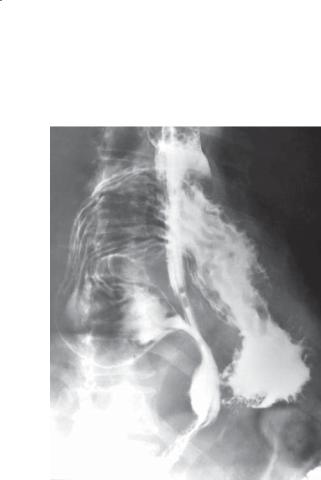
CASE 2.73
Findings
Double-contrast UGI. An intrathoracic stomach is present and has rotated 180° around the longitudinal axis of the stomach, with the greater curvature cephalad to the lesser curvature.
Di erential Diagnosis
Intrathoracic stomach with gastric volvulus
Diagnosis
Intrathoracic stomach with gastric volvulus
2. STOMACH 143
Discussion
Gastric volvulus is a rare occurrence defined as 180° torsion either along the longitudinal axis of the
stomach (organoaxial) or about the gastric mesentery (mesenteroaxial). Ischemia and gangrene can develop in either type, necessitating emergency surgical repair.
Organoaxial volvulus is the more common type, and it is often associated with a long-standing hiatal hernia, paraesophageal hernia, or eventration of the diaphragm. The intrathoracic stomach rotates so that the greater curvature is located superiorly, with the gastric body cranial to the fundus (as in this case). This type of volvulus often presents as an upside-down stomach. Acute symptoms are those of epigastric pain, retching without vomitus, and an inability to pass a nasogastric tube. Mesenteroaxial volvulus presents with the pylorus and antrum folded anteriorly and superiorly. This type often causes either partial or complete obstruction in the pyloroantral region.
Radiologic examination usually shows an intrathoracic location of the stomach and the anatomical alterations described above. It may not be possible to demonstrate filling or emptying of the stomach if obstruction is present.
Disease type: Miscellaneous
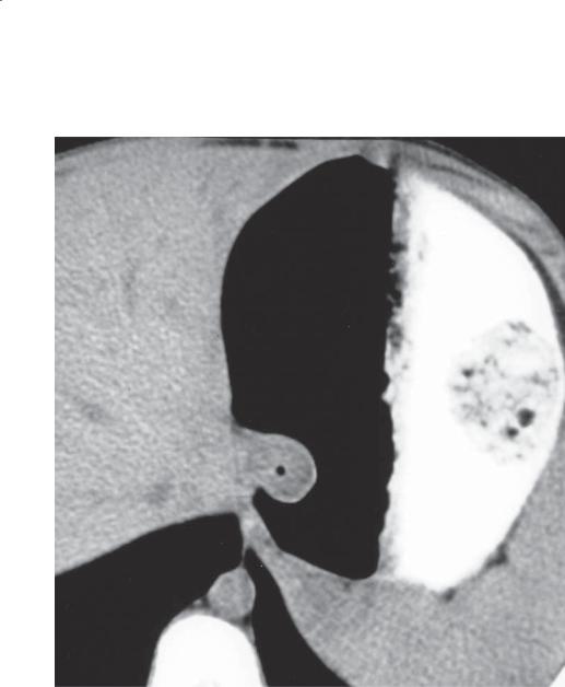
144 MAYO CLINIC GASTROINTESTINAL IMAGING REVIEW
CASE 2.74
Findings
Unenhanced left decubitus abdominal CT. The distal esophagus at the level of the gastroesophageal junction is protruding into the gastric lumen.
Di erential Diagnosis
1.Fundal pseudotumor
2.Gastrointestinal stromal tumor
3.Carcinoma of the gastroesophageal junction
Diagnosis
Fundal pseudotumor
Discussion
Th is pseudotumor can be shown to be esophageal in origin by its contiguity with the esophagus on more cephalad CT sections. In addition, the air in its lumen and its round shape confirm its esophageal nature. Protrusion of the distal esophagus into the stomach causing a filling defect is a normal variant.
Disease type: Miscellaneous

CASE 2.75
A
Findings
Enhanced abdominal CT. A. Masslike thickening of the gastric fundus (arrow) is present near the esophagogastric junction. B. After administration of
effervescent granules, the apparent mass is no longer seen.
Di erential Diagnosis
1.Pseudothickening of a normal stomach
2.Gastrointestinal stromal tumor
3.Carcinoma of the esophagogastric junction
Diagnosis
Pseudothickening of a normal stomach
2. STOMACH 145
B
Discussion
Th e gastric fundus near the esophagogastric junction nearly always appears thickened in an incompletely distended stomach. This region is particularly prominent in this patient. Adequate fundal distention with orally administered contrast material, water,
or gas usually helps exclude a mass in this region. Scanning the patient in a prone or right posterior oblique position, after administration of effervescent granules, is one of the most effective ways to ensure adequate fundal distention.
Disease type: Miscellaneous
146 MAYO CLINIC GASTROINTESTINAL IMAGING REVIEW
TABLE 2.6
Miscellaneous Stomach |
|
CASE |
Conditions |
|
|
|
|
|
Diverticulum |
Usually posterior fundus. Can simulate adrenal mass on |
2.69 and 2.70 |
|
CT without oral contrast material |
|
|
|
|
Partial diverticulum |
Simulates ulcer, usually along greater curvature of antrum, |
2.71 |
|
changes shape with peristalsis |
|
|
|
|
Emphysematous gastritis |
Air within stomach wall, often infectious cause |
2.72 |
|
|
|
Volvulus |
Upside-down stomach commonest (organoaxial). Can result |
2.73 |
|
in obstruction and ischemia |
|
|
|
|
Pseudotumor |
1. Protrusion of esophagus into stomach, a normal variant. |
2.74 |
|
No associated mass |
2.75 |
|
2. Collapsed fundus has masslike appearance, distention of |
|
|
stomach makes mass disappear |
|
|
|
|
Disease type: Miscellaneous

2. STOMACH 147
TABLE 2.7
Di erential Diagnoses
Thickened Folds
BENIGN
Gastritis (Helicobacter pylori) Peptic ulcer disease Zollinger-Ellison syndrome Ménétrier disease
Varices
Gastric Narrowings
BENIGN
Peptic scarring Corrosive ingestion
Granulomatous disease (Crohn disease, sarcoidosis, tuberculosis, syphilis, eosinophilic gastroenteritis)
MALIGNANT
MALIGNANT |
Scirrhous adenocarcinoma |
Lymphoma |
Metastases |
Carcinoma |
Lymphoma |
Metastases |
|
Ulcerative Lesions
NONNEOPLASTIC
Superficial gastric erosions
Peptic or medication-induced ulcers
TUMORS
Adenocarcinoma
Gastrointestinal stromal tumor
Lymphoma
Metastases
Filling Defects
BENIGN TUMORS
Polyp(s)
Gastrointestinal stromal tumor
Leiomyoma
Lipoma
MALIGNANT TUMORS
Adenocarcinoma
Malignant gastrointestinal stromal tumor
Leiomyosarcoma
Metastases
Lymphoma
NONNEOPLASTIC
Ectopic pancreatic rest
Bezoar
Fundoplication defect
Varices
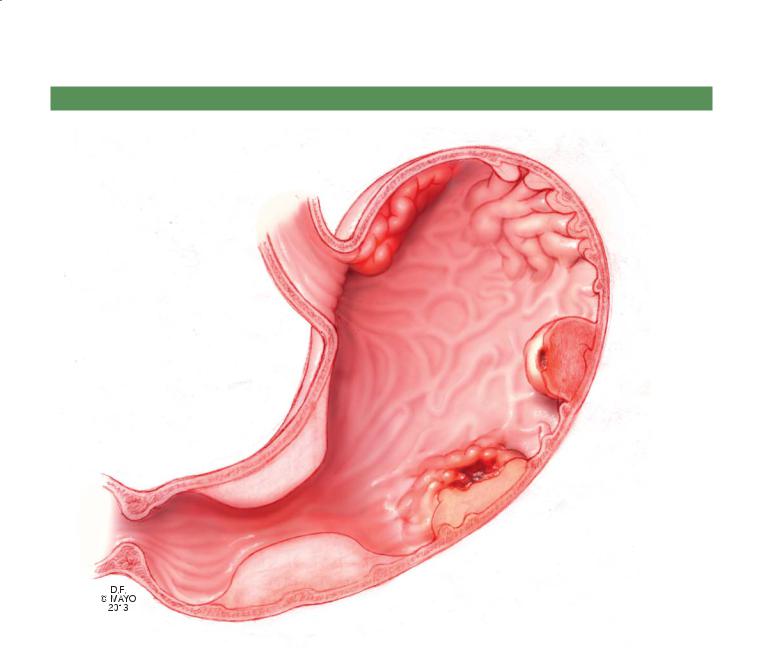
148 MAYO CLINIC GASTROINTESTINAL IMAGING REVIEW
GASTRIC FILLING DEFECTS
Ménétrier disease
(fold thickening)
Varices
Submucosal tumor |
Gastrointestinal |
with narrowing |
|
Scirrhous gastric |
stromal tumor, |
adenocarcinoma or |
leiomyoma, |
breast metastasis |
leiomyosarcoma |
Carcinoma
DFEC
Figure 2.76 (Used with permission of Mayo Foundation for Medical Education and Research.)
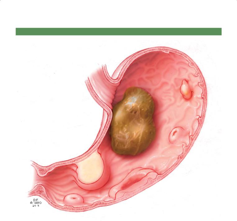
2. STOMACH 149
GASTRIC FILLING DEFECTS
Adenomatous
polyp
Bezoar
Lipoma
(submucosal) Metastatic
melanoma
(target, multiple)
Lymphoma
(solitary or multiple)
Ectopic pancreas
(central umbilication)
Figure 2.77 (Used with permission of Mayo Foundation for Medical Education and Research.)
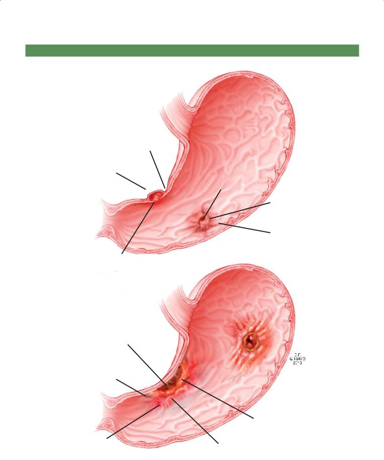
150 MAYO CLINIC GASTROINTESTINAL IMAGING REVIEW
BENIGN AND MALIGNANT GASTRIC ULCERS
Benign
Smooth mound of edema about crater
Extends beyond lumen border into gastric wall
Hampton line: normal
mucosa at ulcer base. Thickened line is ulcer collar from edematous mucosa
Malignant
Crater lies within lumen border and may be eccentrically located in mass
Irregular mass
Folds can be irregular, conjoined, and nodular
Round crater
Smooth mound of edema with central crater
Smooth folds traverse edema and end at crater edge
Malignant crater that spans lesser curvature has meniscal configuration (Carman meniscus sign)
Gastric folds extend to malignant mass but do not reach crater
Figure 2.78 (Used with permission of Mayo Foundation for Medical Education and Research.)
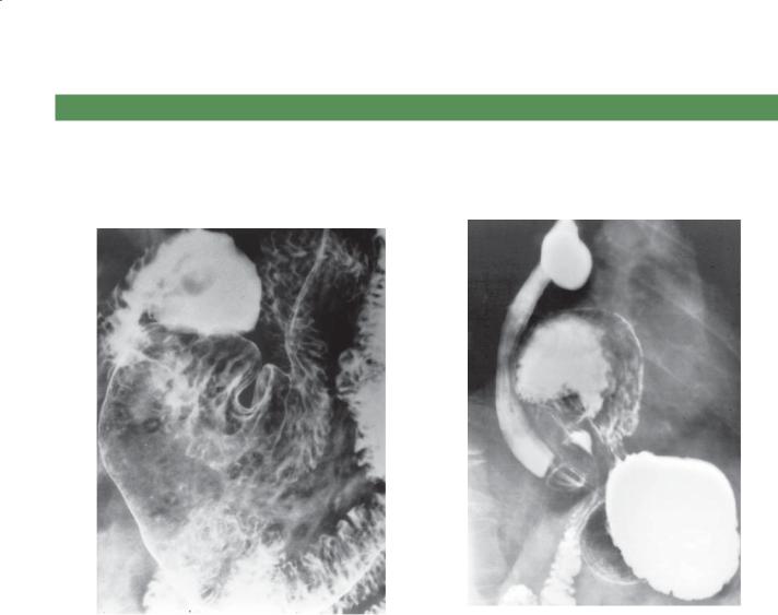
2. STOMACH 151
QUESTIONS
Multiple Choice (choose the best answer) |
2.2. Which of the following is most likely to be associated |
|
2.1. What of the following is the most likely diagnosis for |
with a complication of the condition shown in the |
|
figure? |
||
the findings in the figure? |
||
|
a. Ectopic pancreatic rest(s)
a. Vomiting
b. Superfi cial gastric erosions
b. Heartburn
c. Lymphoma
c. Pain
d. Eosinophilic gastritis
d. Dysphagia
e. Sarcoidosis
e. Constipation
2.3. For the case shown in Question 2.2, what test should be recommended next?
a. Endoscopy b. CT
c. MRI
d. Ultrasonography e. No further testing
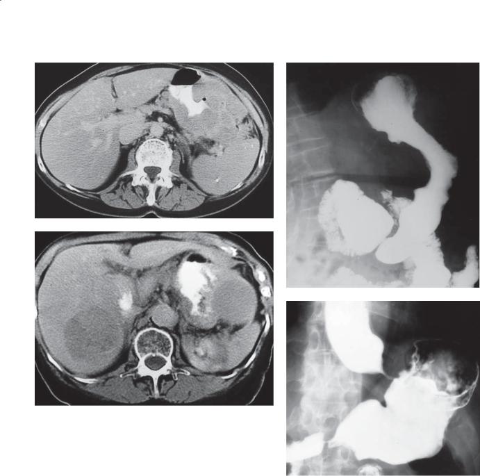
152 MAYO CLINIC GASTROINTESTINAL IMAGING REVIEW
A B
C
D
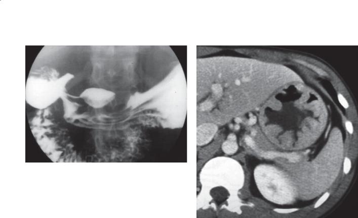
2. STOMACH 153
E F
Match images A through F with the responses listed below.
2.4._____ Indistinguishable from metastatic breast cancer
2.5._____ Adenopathy and splenomegaly may be associated
2.6._____ Likely due to chronic Helicobacter pylori infection
2.7._____ Anti-secretagogues are often used in symptomatic patients
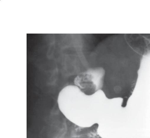
154 MAYO CLINIC GASTROINTESTINAL IMAGING REVIEW
2.8.What is the most likely symptom associated with the condition shown in this figure?
a.None
b.Epigastric discomfort
c.Nausea or vomiting
d.Weakness due to anemia
e.Hematemesis

2. STOMACH 155
ANSWERS
2.1.Answer b.
Multiple small collections of barium are seen within the gastric antrum. These collections represent small ulcerations in the mucosa. The lucent filling defect seen about these collections represents surrounding edema. The commonest causes include use of aspirin and nonsteroidal anti-inflammatory drugs. Helicobacter pylori infection could present with these findings.
Ectopic pancreatic rest is usually solitary and represents a larger filling defect with a small central collection of barium. In this case, there are too many collections and the filling defects are smaller than expected.
Lymphoma is usually a submucosal infiltrative lesion with either diffuse or localized involvement. Ulceration of a large mass can occur. In this case, there is not a mass or evidence of infiltration of the stomach wall.
Eosinophilic gastritis can present with nodular changes in the gastric antrum, but ulceration of these nodules would be unusual.
Sarcoidosis can cause narrowing of the gastric antrum from infiltration of the wall. Multiple ulcerations of the affected bowel segment would be very unusual.
2.2.Answer c.
Th is figure is an example of a paraesophageal hernia. Affected patients are at risk for strangulation and incarceration of the herniated portion of the stomach. Surgical repair is often indicated. Compromise of the blood supply to the stomach can be associated with pain in either the chest or the abdomen. With torsion of the stomach, obstruction can occur and lead to vomiting; however, in this image, contrast material is moving through the stomach into the small bowel without evidence of obstruction.
Patients who have esophageal hiatal hernias are at risk for development of gastroesophageal reflux and heartburn. This patient has a tiny esophageal hiatal hernia, but the main problem is the paraesophageal hernia, which does not typically result in reflux and heartburn.
Dysphagia is most often associated with an anatomic narrowing of the esophagus. There is no obstruction in this patient, in that contrast material easily passed into the duodenum. Similarly, without obstruction, it is unlikely that the patient would be constipated on the basis of this finding.
2.3.Answer e.
No further testing is necessary for this diagnosis. Clinicians should correlate the clinical findings and determine whether a patient is suitable for operative
repair. If so, referral to a surgeon would be the next most appropriate step.
2.4.Answer b.
Scirrhous carcinoma of the stomach is an infiltrative adenocarcinoma of the stomach that narrows the lumen of a large gastric segment, often with preservation of the overlying mucosa. Metastatic breast cancer can present in a radiologically identical manner.
2.5.Answer a.
Lymphoma of the stomach often presents with bulky diffuse gastric wall thickening (as in this case). Although adenopathy and splenomegaly (typical findings of lymphoma) do not have to be present in gastric lymphoma, these associated findings would make this diagnosis most likely.
2.6.Answer e.
Primary adenocarcinoma of the stomach (Carman meniscus sign) is shown by the ulcerated mass in the gastric antrum. The ulceration does not extend beyond the normal luminal boundaries, and the gastric folds extend to the mass but not to the ulcer. In the past, many gastric cancers likely arose due to H pylori–induced gastritis.
2.7.Answer f.
Ménétrier disease is an idiopathic disease with giant rugal hypertrophy (as shown in the figure) that may be associated with a protein-losing enteropathy. Affected patients are usually given antisecretory drugs to prevent this complication. Notice how the gastric folds are regular in shape. Lymphoma often creates similarappearing fold enlargement, but the folds are usually not as similar in shape (more disorganized in appearance) as these folds.
Figure c shows a gastrointestinal stromal tumor with a large pedunculated mass arising from the stomach with bulky metastasis in the liver.
Figure d shows a carcinoma of the gastroesophageal junction; such a tumor can resemble achalasia. There is associated mass effect within the cardia of the stomach.
2.8.Answer a.
Th e figure shows prolapsed gastric mucosa. It is considered a normal finding in patients with redundant gastric mucosa, which can prolapse through the pylorus with vigorous gastric peristalsis. No treatment is necessary. Observation of this filling defect, which changes in shape and disappears with gastric distention, is key to the diagnosis. Usually the filling defect is symmetric about the pylorus.
This page intentionally left blank