


C H A P T E R 4
SMALL BOWEL
Cases 4.1–4.39 Masses and Filling Defects
Benign Tumors
Malignant Tumors
Nonneoplastic
Cases 4.40–4.61 Di use and Segmental Diseases
Thin, Straight Folds (Type I)
Thick, Straight Folds (Type II)
Thick, Nodular Folds (Type III)
Cases 4.62–4.98 Common Small Bowel Diseases
Crohn Disease
Ischemia
Obstruction
Sprue
Cases 4.99–4.115 |
Miscellaneous |

This page intentionally left blank
CASE 4.1
Findings
Small bowel follow-through. A wellmarginated, smooth-surfaced filling defect is present in the jejunum.
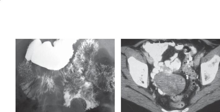
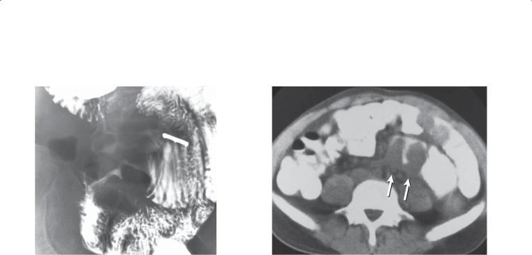
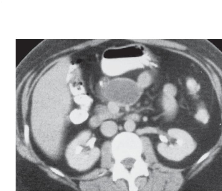
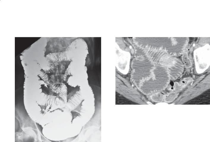
CASE 4.2. Contrast-enhanced CT. A large, solid soft tissue mass is present in the pelvis, intimately associated with the small bowel.
Di erential Diagnosis
1.Gastrointestinal stromal tumor
2.Hemangioma
3.Lipoma
4.Metastasis
5.Lymphoma
Diagnosis
Gastrointestinal stromal tumor
4. SMALL BOWEL 211
CASE 4.2
Discussion
Gastrointestinal stromal tumors (GISTs) can be classified as either benign or malignant. GISTs of the stomach are more common than GISTs of the small bowel. The radiographic appearance of these tumors depends on their location within the bowel wall. Subserosal GISTs (seen on the CT) may be undetected unless adjacent small bowel loops are displaced
by the mass. Submucosal tumors appear as typical intramural lesions (smooth-surfaced, usually with 90° margins to the normal bowel lumen) elsewhere in the gastrointestinal tract. Some may grow intraluminally and appear as a polypoid mass. Gastrointestinal bleeding is the usual symptom. The bleeding usually occurs as short, repeated episodes of melena or darkred stool. Even in the absence of active bleeding, these tumors may be detectable at CT or angiography because of their hypervascularity.
Disease type: Masses and Filling Defects
212 MAYO CLINIC GASTROINTESTINAL IMAGING REVIEW
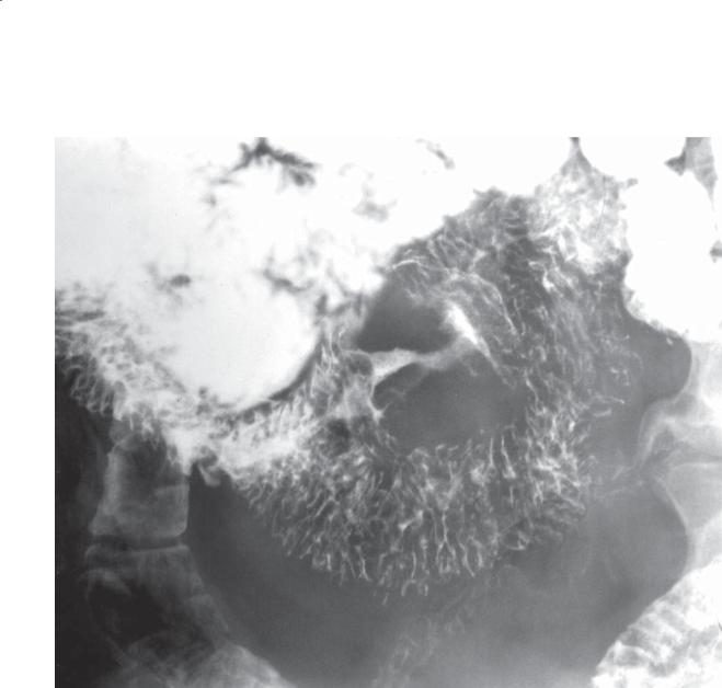
CASE 4.3
A B
Findings
Small bowel follow-through (spot radiograph).
A. A smooth-surfaced, well-defined filling defect is present in the ileum. B. The mass has changed in shape with peristalsis and compression.
Di erential Diagnosis
1.Gastrointestinal stromal tumor
2.Lipoma
3.Hemangioma
4.Metastasis
Diagnosis
Lipoma
Discussion
Lipomas are the third most frequently occurring benign small bowel tumor. They can occur anywhere in the alimentary tract. In the small bowel, they are usually distal. Most are asymptomatic. Symptoms, when present, are often due to an intussusception. Occasionally, obstruction or bleeding can develop. These tumors have no malignant potential.
Radiographically, the smooth surface and compressible nature of these masses suggest the diagnosis. The fatty attenuation of these tumors is diagnostic at CT (case 4.4).
Disease type: Masses and Filling Defects
CASE 4.4
A
Findings
A.Small bowel follow-through. Multiple well-defined, smooth-surfaced filling defects are present within several small bowel loops.
B.Contrast-enhanced CT. Multiple masses the attenuation of fat are present within several loops of small intestine. (Adapted from Ormson MJ, Stephens DH, Carlson HC. CT recognition of intestinal lipomatosis. AJR. 1985 Feb;144[2]:313-4. Used with permission.)
Di erential Diagnosis
1.Multiple lipomas
2.Intestinal liposarcoma
3.Metastases
Diagnosis
Multiple intestinal lipomas
4. SMALL BOWEL 213
B
Discussion
Multiple small bowel lesions can be found in patients with lymphoma, a polyposis syndrome, hemangiomas, neurofibromas, and metastases. At CT, the tumors
in all these other conditions are the attenuation of soft tissue. CT is especially helpful for confirming the diagnosis if a lesion that resembles a lipoma is found on a conventional barium study. If the typical fat density of these tumors is identified at CT, the diagnosis of a lipoma can be made unequivocally. Rarely, multiple lipomas are present (as in this case). No surgical therapy is necessary if patients are asymptomatic. Intestinal lipomatosis can be
distinguished from a liposarcoma by the homogeneous fat density and absence of intratumoral soft tissue density in lipomas.
Disease type: Masses and Filling Defects

214 MAYO CLINIC GASTROINTESTINAL IMAGING REVIEW
CASE 4.5
Findings
Small bowel follow-through (spot radiograph). A smooth-surfaced filling defect is present in the
small bowel. This is likely due to an intramural tumor. A compression device containing a metallic
marker is present.
Di erential Diagnosis
1.Gastrointestinal stromal tumor
2.Lipoma
3.Hemangioma
4.Metastasis
Diagnosis
Hemangioma
Discussion
Hemangiomas are rare tumors of the small bowel that usually affect the jejunum. Bleeding, intussusception, and, rarely, obstruction are the most frequently associated complications. Pathologically, multiple thin-walled vessels are seen either intraluminally or within the wall of the small bowel. Patients with Turner syndrome, tuberous sclerosis, blue rubber bleb nevus syndrome, and Rendu-Osler-Weber syndrome have an increased incidence of this disorder.
Radiologically, hemangiomas usually present as focal masses but occasionally can be a diffuse
malformation. Many are multiple, small compressible lesions and can be easily overlooked during a small bowel examination. Larger malformations may contain multiple calcified phleboliths that are detectable on a plain film radiograph or at CT.
Disease type: Masses and Filling Defects
CASE 4.6
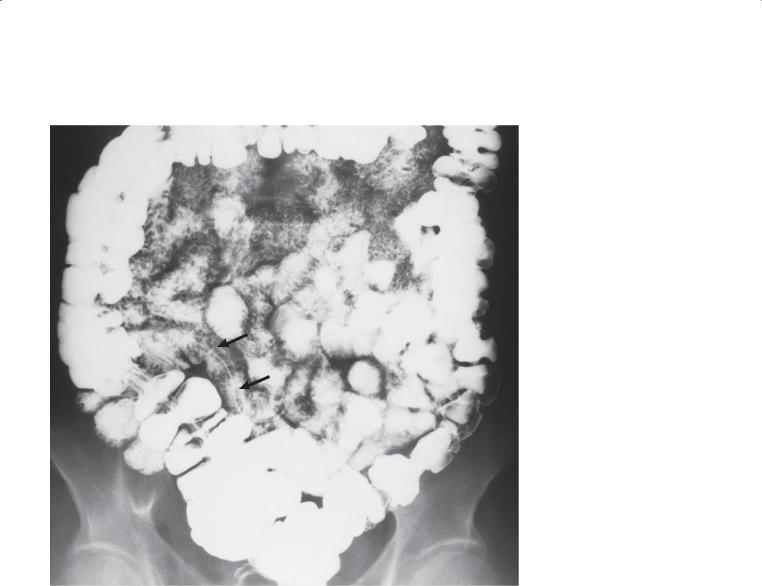
Findings
Contrast-enhanced CT enterography. There are approximately 4 small foci of abnormal enhancement within the jejunum (arrow). No active contrast extravasation is present.
Di erential Diagnosis
1.Normal jejunal enhancement
2.Hemangioma
3.Small bowel tumor
Diagnosis
Hemangioma
4. SMALL BOWEL 215
Discussion
A CT technique using intravenous contrast material and water-attenuation luminal contrast material (CT enterography technique) is required for optimal detection of sources for gastrointestinal blood loss. Hemangiomas can present with multiple calcified phleboliths within the bowel wall, but most do not. Identification of small tufts of enhancement within the bowel wall is key to the diagnosis. Small dots
of enhancement can be seen normally within the jejunum, and these should not be confused with hemangiomas. Notice how these abnormal foci are larger and appear different than punctate regions of enhancement within the jejunum.
Disease type: Masses and Filling Defects
216 MAYO CLINIC GASTROINTESTINAL IMAGING REVIEW
CASE 4.7
Findings
Small bowel follow-through. Multiple submucosal and intraluminal filling defects are present within the distal small bowel.
Di erential Diagnosis
1.Metastases
2.Lymphoma
3.Hemangiomatosis
4.Multiple lipomas
Diagnosis
Diffuse hemangiomatosis
Discussion
Diffuse hemangiomatosis is a rare cause of gastrointestinal bleeding; it usually occurs in infants. Intussusception, obstruction, and malabsorption also can cause symptoms. This condition may
be associated with other syndromes, including Klippel-Trénaunay-Weber syndrome (varicose veins, cutaneous hemangiomas, soft tissue and bone hypertrophy), Maffucci syndrome (enchondromas, subcutaneous cavernous hemangiomas), and diffuse hemangiomatosis. There is a spectrum of disease ranging from small submucosal nodules to diffuse intestinal wall involvement with associated extension into the mesentery, retroperitoneum, and other
adjacent tissues. Bowel wall phleboliths may suggest a diagnosis but are an unusual radiographic finding.
Disease type: Masses and Filling Defects
4. SMALL BOWEL 217
CASE 4.8 |
CASE 4.9 |
A |
A |
B B
Findings
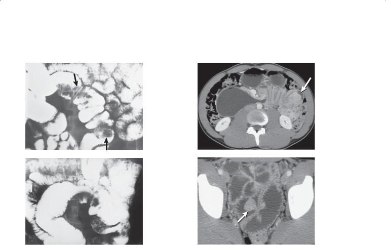
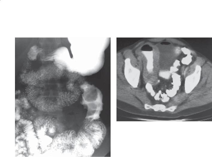
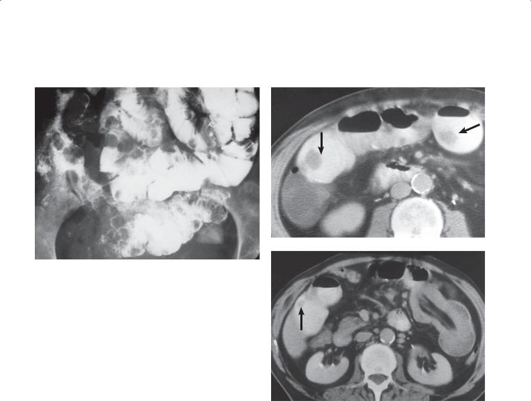
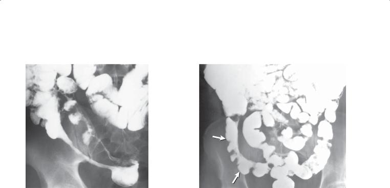
CASE 4.8. Small bowel follow-through. A. Two large lobulated polyps (arrows) are present in the mid small bowel. B. Transient intussusception with the typical coiled-spring appearance was observed. A polyp was the lead point.
CASE 4.9. Contrast-enhanced CT. A. The proximal transverse duodenum is dilated and fluid-filled. The distal duodenum has an accordion appearance, and an enhancing mass (arrow) is visible within the small bowel lumen. B. Multiple dilated and fluid-filled small bowel loops are visible in the pelvis. An enhancing intraluminal polypoid mass is visible (arrow).
Di erential Diagnosis
1.Hamartomas (Peutz-Jeghers syndrome)
2.Lymphoma
3.Metastases
Diagnosis
Hamartomas (Peutz-Jeghers syndrome)
Discussion
Hamartomas were surgically removed in these patients with Peutz-Jeghers syndrome. Hamartomatous polyps are most often found in the small bowel in patients younger than 30 years—the majority of these patients have Peutz-Jeghers syndrome. The small bowel polyps are often cauliflower-like, found in groups, and located within the jejunum. Patients may present with bleeding, pain, or obstruction from intussusception. Usually these lesions are benign, but adenocarcinomas have been reported in the gastrointestinal tract (usually stomach, duodenum, or colon). Ovarian cysts and tumors are found in a minority (5%) of female patients with Peutz-Jeghers syndrome.
Peutz-Jeghers syndrome is inherited as an autosomal dominant disease. Hamartomas most often affect the small bowel, but approximately a fourth of patients have similar polyps in the stomach. Colonic polyps
in these patients are adenomatous. Brown pigmented spots on the perioral mucous surfaces are typical.
Disease type: Masses and Filling Defects
218 MAYO CLINIC GASTROINTESTINAL IMAGING REVIEW
CASE 4.10
A B
Findings
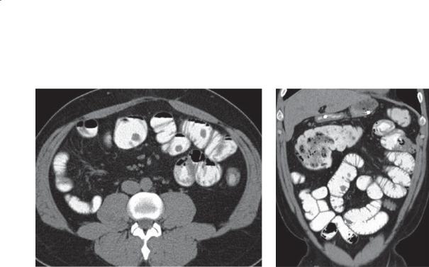
Contrast-enhanced CT. A. Axial. Multiple filling defects are present within the small bowel. B. Coronal. The filling defects are of variable size, many arising from the small bowel folds.
Di erential Diagnosis
1.Metastases
2.Small bowel polyps
3.Polyposis syndrome (Peutz-Jeghers syndrome)
Diagnosis
Peutz-Jeghers syndrome
Discussion
Metastases to the small bowel would be statistically the most common diagnosis for these findings. Review of a patient’s history is needed to narrow the differential possibilities. In this patient, Peutz-Jeghers syndrome had already been diagnosed. These polyps can lead to complications of bleeding, obstruction, intussusception, and even malignant transformation to adenocarcinoma. None of these complications are apparent on these images.
Disease type: Masses and Filling Defects
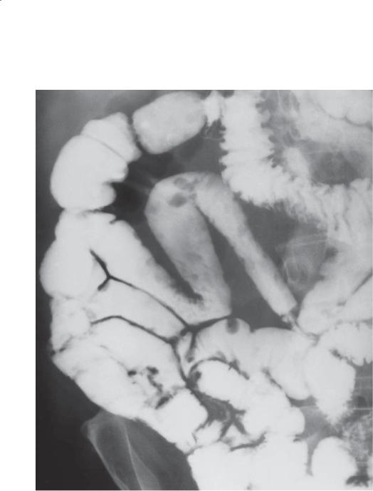
CASE 4.11
Findings
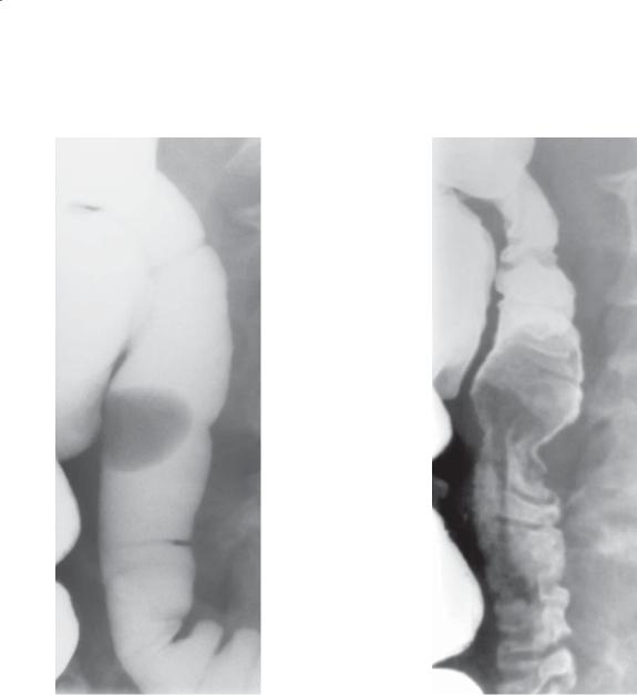
Small bowel follow-through (spot radiograph). A round, smooth-surfaced filling defect is present in the ileum.
Di erential Diagnosis
1.Gastrointestinal stromal tumor
2.Lipoma
3.Hemangioma
Diagnosis
Inflammatory fibroid polyp
4. SMALL BOWEL 219
Discussion
Th ese polyps are rare and are not true neoplasms histologically. They have been called by various names: infective granuloma, fibroma, hemangiopericytoma, neurinoma, plasma cell granuloma, and Vanek tumor. Histologically, many cell types are found, including fibroblasts, endothelial cells, histiocytes, leukocytes, and small blood vessels. This mixture of cells resembles reparative tissue. The cause of these tumors is unknown, but they can lead to intussusception and obstruction. Radiologically, they are indistinguishable from other polypoid small bowel tumors.
Disease type: Masses and Filling Defects
220 MAYO CLINIC GASTROINTESTINAL IMAGING REVIEW
CASE 4.12 |
CASE 4.13 |
Findings
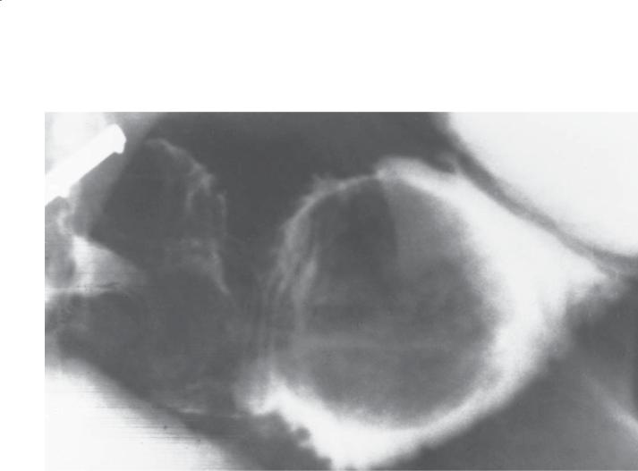
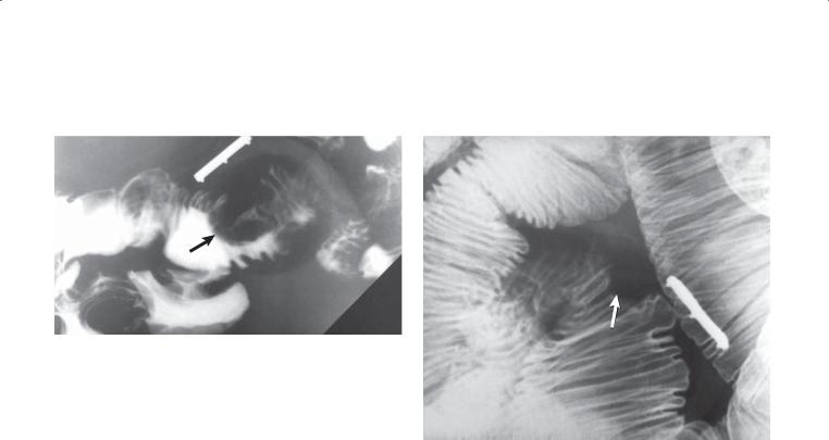
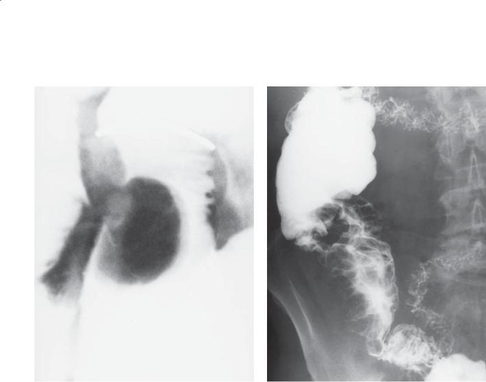
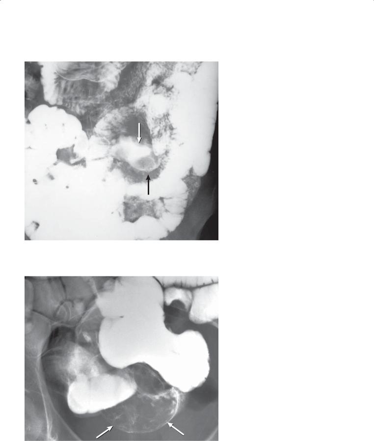
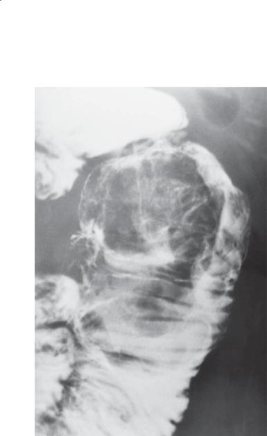
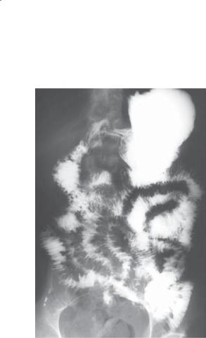
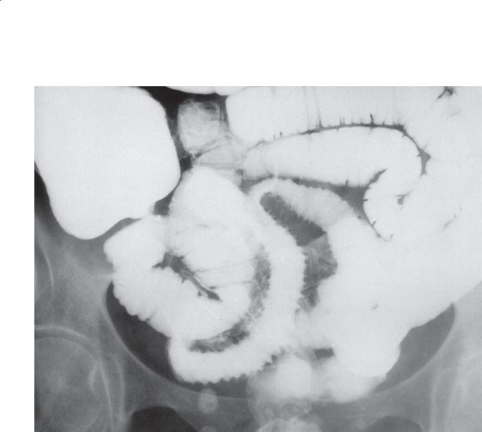
CASE 4.12. Small bowel follow-through (spot radiograph). An ulcerated submucosal mass (arrow) is present in the distal ileum.
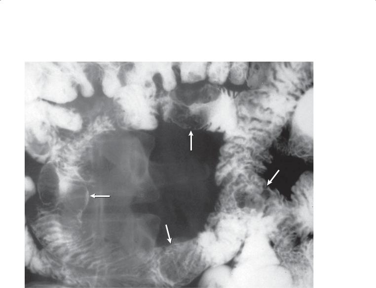
CASE 4.13. Enteroclysis (spot radiograph). Kinking of a small bowel loop and a submucosal mass (arrow) are present on this examination. A compression device with metallic marker is present.
Di erential Diagnosis
1.Gastrointestinal stromal tumor
2.Metastasis
3.Lymphoma
4.Carcinoid
Diagnosis
Carcinoid tumor
Discussion
Carcinoid tumors are neuroendocrine neoplasms derived from Kulchitsky cells that can be found throughout the intestinal tract. Their neural crest origin and biochemical behavior have led to their classification as an amine precursor uptake and decarboxylation tumor. These tumors usually produce 5-hydroxytryptamine (serotonin). 5-Hydroxyindoleacetic acid is a useful biologic
marker produced by the degradation of serotonin; it can be measured in the serum or urine of patients
with these tumors. Malignant transformation usually occurs in tumors 1 cm or more in diameter. Carcinoids usually are found in the distal small bowel; nearly 40% are within 2 feet of the ileocecal valve. About 30% of patients have more than one tumor.
Small bowel radiographic features of the primary tumor are usually those of an intramural neoplasm, usually 2 to 3 cm in diameter. Ulceration may be present. Often the primary neoplasm is not detected radiographically, and evidence of mesenteric metastases is generally found (case 4.14).
Carcinoid tumors initially spread by direct invasion through the bowel wall into the mesentery. A fibrotic reaction ensues within the mesentery, with kinking and obstruction of the bowel. Usually, obstruction is only partial, and patients may complain of symptoms attributable to partial mechanical obstruction for many years. Advanced disease with mesenteric metastases may result in separation of small bowel loops, tethering of small bowel folds, encasement, and luminal narrowing. The radiographic findings of mesenteric metastases are not specific for carcinoid tumors, and findings can be similar in other malignancies. Kinking and tethered folds also can
be caused by adhesions or a localized inflammatory process.
Disease type: Masses and Filling Defects

4. SMALL BOWEL 221
CASE 4.14
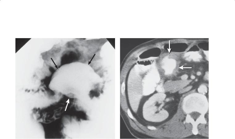
Findings
Contrast-enhanced CT. A soft tissue mesenteric mass is present with associated marked thickening and stranding of mesenteric tissues.
Di erential Diagnosis
1.Carcinoid tumor
2.Retractile mesenteritis
3.Mesenteric metastases
Diagnosis
Carcinoid tumor
Discussion
Th e combination of mesenteric and desmoplastic stranding in this case is typical of a carcinoid tumor.
CT evaluation of patients with known or suspected carcinoid tumors can be helpful. The primary tumor is often not detected, but the extent of mesenteric disease, retroperitoneal adenopathy, and hepatic metastases can be assessed. The metastases in the small bowel mesentery often have a typical starburst appearance of linear stranding radiating from a central mesenteric mass and calcification. Liver metastases are hypervascular, often containing regions of central necrosis. Retroperitoneal adenopathy is frequent but
rarely is found without hepatic or mesenteric metastases. Evidence of metastases or a known tumor is
helpful for eliminating retractile mesenteritis as a consideration. Although possible, discrete hyperenhancing masses are less common with retractile mesenteritis.
Disease type: Masses and Filling Defects
222 MAYO CLINIC GASTROINTESTINAL IMAGING REVIEW
CASE 4.15 |
CASE 4.16 |
Findings
CASE 4.15. Small bowel follow-through. A circumferential ulcerated mass involves a relatively long segment of proximal small bowel. The lumen is larger than the adjacent normal small bowel lumen, exemplifying aneurysmal dilatation.
CASE 4.16. Contrast-enhanced CT. A large lesion encases a distal small bowel loop. Marked bowel wall thickening is present. The lumen is dilated. (Adapted from Dudiak KM, Johnson CD, Stephens DH. Primary tumors of the small intestine: CT evaluation. AJR. 1989 May;152[5]:995-8. Used with permission.)
Di erential Diagnosis
1.Lymphoma
2.Malignant gastrointestinal stromal tumor
3.Metastases
Diagnosis
Lymphoma
Discussion
Small bowel lymphoma constitutes 20% of all malignant small bowel tumors. The vast majority of tumors are of the non-Hodgkin type. Usual symptoms include nausea, vomiting, weight loss, and abdominal pain. Although there are no known
predisposing factors, patients with conditions such as AIDS, celiac sprue, Crohn disease, and systemic lupus erythematosus have a higher risk of the disease.
Th ere are several radiologic classifications for lymphoma. The traditional classification includes multiple nodules (case 4.19), infiltrating form
(case 4.17), polypoid (case 4.18), and endo-exoenteric (case 4.21) with excavation and fistula formation.
An abbreviated classification includes primary form, lymphoma complicating sprue, and mesenteric nodal form. The traditional classification describes only primary small bowel lymphoma, whereas the
abbreviated classification emphasizes secondary forms of the disease.
Disease type: Masses and Filling Defects
CASE 4.17
Findings
Small bowel follow-through. A focal segment of small bowel is denuded of folds. The caliber of the small bowel remains normal throughout the length of the abnormality.
Di erential Diagnosis
1.Ischemia
2.Amyloidosis
3.Lymphoma
Diagnosis
Lymphoma
4. SMALL BOWEL 223
Discussion
Focal infiltrative lymphoma may ulcerate the mucosa (with secondary loss of bowel markings). Luminal narrowing occurs when a fibrotic reaction is present. The absence of fibrosis leads to dilatation. The pathologic process is analogous to
aneurysmal ulceration, except the lumen through the lymphomatous mass is the same diameter as that of normal small bowel. Chronic changes from ischemia usually result in a narrowed small bowel lumen
with loss of folds. Amyloidosis usually causes fold thickening or secondary ischemic changes.
Disease type: Masses and Filling Defects
224 MAYO CLINIC GASTROINTESTINAL IMAGING REVIEW
CASE 4.18 |
CASE 4.19 |
Findings
CASE 4.18. Small bowel follow-through (spot radiograph). An intraluminal polypoid mass is present within the small bowel.
CASE 4.19. Small bowel follow-through. A long segment of terminal ileum is involved with multiple submucosal, polypoid filling defects. The lumen is increased in diameter.
Di erential Diagnosis
1.Metastases
2.Lymphoma
3.Polyposis syndrome (Peutz-Jeghers syndrome)
Diagnosis
Lymphoma
Discussion
A solitary intraluminal mass (as in case 4.18) is an unusual manifestation of lymphoma. The mass is believed to arise within the submucosa of the bowel wall and, as a result of peristalsis, to form a
predominantly intraluminal mass, sometimes attached to the bowel wall by a pseudopedicle. The mass could become the lead point for an intussusception.
Multiple masses (as in case 4.19) are a more common presentation for lymphoma. Lymphoma often involves the distal ileum and can cross the ileocecal valve and affect the cecum.
Disease type: Masses and Filling Defects

CASE 4.20
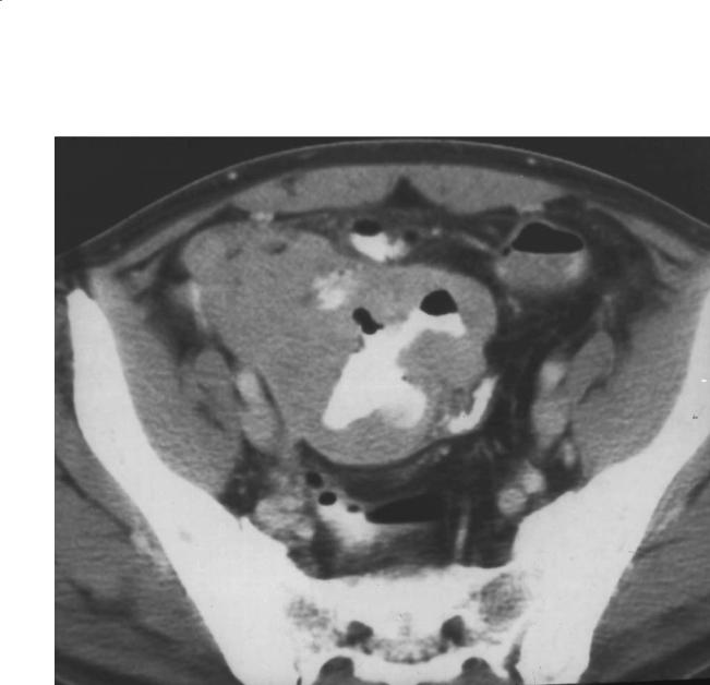
Findings
CASE 4.20. Contrast-enhanced CT. Marked wall thickening (arrow) affects a small bowel loop. In addition, adjacent adenopathy (arrowheads) encases mesenteric vessels. (Adapted from Dudiak KM, Johnson CD, Stephens DH. Primary tumors of
the small intestine: CT evaluation. AJR. 1989 May;152[5]:995-8. Used with permission.)
CASE 4.21. Contrast-enhanced CT. Centrally necrotic mesenteric lymphadenopathy is seen in the right side of the abdomen. These lymph nodes coalesce and form a mass (arrow) that displaces and narrows the terminal ileum.
Di erential Diagnosis
1.Lymphoma
2.Metastases
Diagnosis
Lymphoma
4. SMALL BOWEL 225
CASE 4.21
Discussion
Mesenteric or, less often, retroperitoneal adenopathy, or both, may be present in any form of primary small bowel lymphoma. In case 4.20, lymphoma within the bowel wall appears to be the site of origin of the tumor, and there is secondary spread to mesenteric lymph nodes.
In some patients, the bulk of the tumor is extraluminal, within mesenteric lymph nodes. These nodal masses can become large and cause narrowing, displacement, angulation, and local bowel invasion. Case 4.21 is an example of the mesenteric-nodal form of non-Hodgkin lymphoma.
Disease type: Masses and Filling Defects
226 MAYO CLINIC GASTROINTESTINAL IMAGING REVIEW
CASE 4.22
Findings
Small bowel follow-through. A focal region of ulcerative constriction (arrow) is present in the mid small bowel. The loop proximal to the stenosis is dilated, indicating partial mechanical obstruction.
Di erential Diagnosis
1.Adenocarcinoma
2.Metastasis
3.Lymphoma
Diagnosis
Lymphoma (Hodgkin type)
Discussion
Hodgkin lymphoma of the small bowel is less common than non-Hodgkin lymphoma. Unlike non-Hodgkin lymphoma, these tumors can incite a desmoplastic reaction, producing luminal narrowing and at times obstruction. Other patterns of Hodgkin lymphoma also can be seen: diffuse fold thickening and irregularity, long segmental regions of involvement, and ulceration. Aneurysmal ulceration, perforation, and fistulization are distinctly uncommon. A primary carcinoma (cases 4.24 and 4.25) could have an identical radiographic appearance.
Disease type: Masses and Filling Defects
CASE 4.23
Findings
Contrast-enhanced CT. A bulky soft tissue mass is present in the pelvis. Oral contrast material within its central portion indicates communication of the bowel lumen with its central cavity.
Di erential Diagnosis
1.Malignant gastrointestinal stromal tumor
2.Lymphoma
3.Metastases
Diagnosis
AIDS–related lymphoma
4. SMALL BOWEL 227
Discussion
Lymphomas arising from the alimentary tract can present as large bulky tumors. The presence of adenopathy can be very helpful to distinguish it from a malignant gastrointestinal stromal tumor. Patients who are severely immunocompromised are at increased risk for the development of malignancies. Patients with AIDS have an increased risk for development of opportunistic neoplasms, including Kaposi sarcoma, AIDS-related lymphoma, and several opportunistic infections. Most lymphomas in patients with AIDS are B-cell lymphomas and are aggressive.
Disease type: Masses and Filling Defects
228 MAYO CLINIC GASTROINTESTINAL IMAGING REVIEW
CASE 4.24
A B
Findings
A.Small bowel follow-through (spot radiograph). An ulcerative, annular, constrictive lesion is present in the mid small bowel. The lesion is partially
obstructive with mild dilatation of the proximal bowel. A compression device with a metallic marker
is present.
B.Unenhanced CT. A lobulated soft tissue mass (arrows) narrows the small bowel lumen and thickens its wall. (Adapted from Dudiak KM, Johnson CD, Stephens DH. Primary tumors of the small intestine: CT evaluation. AJR. 1989 May;152[5]:995–8. Used with permission.)
Di erential Diagnosis
1.Primary adenocarcinoma
2.Metastasis
3.Hodgkin-type lymphoma
Diagnosis
Primary adenocarcinoma
Discussion
Primary small bowel adenocarcinoma is most commonly found in the proximal small bowel, usually the duodenum. Patients are often symptomatic: abdominal pain, obstruction, bleeding, or anemia.
Th ere is an increased incidence of this tumor in patients with adult celiac disease and a small increased risk with regional enteritis. Radiographically, a focal region of narrowing with mucosal ulceration is usually seen on barium studies. Detection of these lesions can be challenging at CT if the tumor is less than 2 cm in diameter. Focal circumferential bowel wall thickening in the proximal small bowel is characteristic of an adenocarcinoma at CT.
Disease type: Masses and Filling Defects
4. SMALL BOWEL 229
CASE 4.25
Findings |
Discussion |
Small bowel follow-through. An apple-core lesion is |
Th e short annular nature of the lesion is characteristic |
present in the jejunum. Notice the absence of normal |
of an adenocarcinoma. |
mucosal markings throughout this lesion. The contour |
|
of the lesion is irregular, and the mucosal surface has a |
|
smudged appearance due to ulceration. |
|
Di erential Diagnosis
1.Primary adenocarcinoma
2.Metastasis
3.Hodgkin lymphoma
Diagnosis
Primary adenocarcinoma
Disease type: Masses and Filling Defects
230 MAYO CLINIC GASTROINTESTINAL IMAGING REVIEW
CASE 4.26
CASE 4.27
Disease type: Masses and Filling Defects
CASE 4.28
A
Findings
CASE 4.26. Small bowel follow-through. An ulcerated mass (arrows) is present in the proximal small bowel. Ulcerated lumen is expanded compared with adjacent small bowel loops.
CASE 4.27. Small bowel follow-through. A large intraluminal filling defect (arrows) is present within the small bowel.
Contrast-enhanced CT. A. Axial. Two solid, enhancing masses are seen in the left side of the pelvis. The larger mass contains central fluid and air, findings indicating cavitation. B. Coronal. The larger mass has an enhancing wall and fluid-containing center. A small calcification is present adjacent to the wall.
Di erential Diagnosis
1.Lymphoma
2.Gastrointestinal stromal tumor
3.Metastasis
Diagnosis
Malignant gastrointestinal stromal tumor
4. SMALL BOWEL 231
B
Discussion
Gastrointestinal stromal tumors can be divided according to their gross pathologic features into intramural, exoenteric (case 4.26), endoenteric (case 4.27), and dumbbell growths. The exoenteric growths are most common and can incorporate bowel lumen (as in case 4.26). Ulceration, necrosis, degeneration, hemorrhage, fistula, and infection often can be found in these tumors. These tumors
metastasize by hematogenous and peritoneal seeding. Nodal metastases are distinctly uncommon. In the absence of metastases, the size of the primary tumor is the most important predictor of malignancy.
Disease type: Masses and Filling Defects
232 MAYO CLINIC GASTROINTESTINAL IMAGING REVIEW
CASE 4.29 |
CASE 4.30 |
|
A |
B
Findings
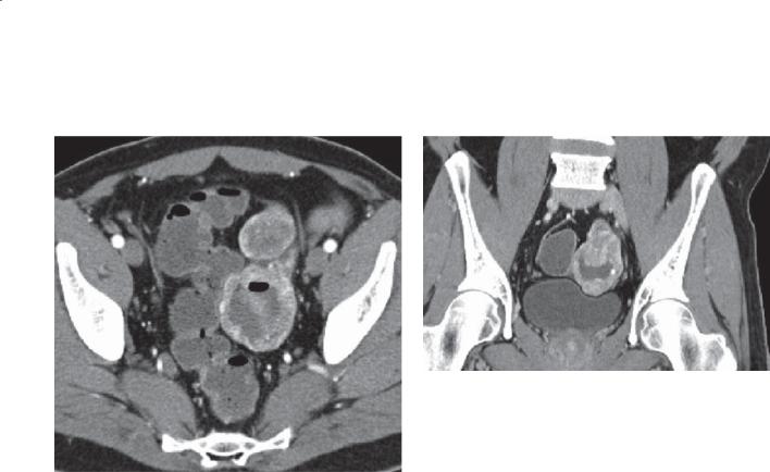
CASE 4.29. Small bowel follow-through. Multiple filling defects are present throughout the small bowel. They are smooth-surfaced and of uniform size.
CASE 4.30. Contrast-enhanced CT. A. Multiple intraluminal filling defects (arrows) are seen in several bowel loops. B. An intussusception is present on the left side of the abdomen. Intraluminal mesenteric fat and vessels are diagnostic of this condition. There
is also a small soft tissue nodule (arrow) in another bowel loop.
Di erential Diagnosis
1.Metastases
2.Polyposis syndrome
3.Lymphoma
Diagnosis
Metastases, melanoma
Discussion
Metastatic melanoma metastasizes widely and usually spreads hematogenously to the gastrointestinal tract. The small bowel is involved in 50% of cases at autopsy, followed by the colon and stomach. Hematogenous seeding of the small bowel most often occurs in patients with melanoma, breast or lung cancer, and Kaposi sarcoma. If a hematogenous metastasis grows to circumferentially engulf the bowel, it can resemble a primary adenocarcinoma (cases 4.24 and 4.25).
Melanoma metastases can present as multiple masses (as in these cases), a solitary large mass (case 4.31), or a bulky ulcerated lesion(s). Intraluminal masses can act as a lead point for an intussusception (case 4.30), causing pain, obstruction, bleeding, ischemia, or even perforation.
Disease type: Masses and Filling Defects
CASE 4.31
Findings
Small bowel follow-through. A large intraluminal, polypoid mass is present within the dilated proximal small bowel.
Di erential Diagnosis
1.Metastasis
2.Gastrointestinal stromal tumor
3.Hamartomatous polyp
4.Lipoma
5.Lymphoma
Diagnosis
Metastasis, melanoma
4. SMALL BOWEL 233
Discussion
Intraluminal (as in this case) or ulcerated (case 4.32) submucosal metastases can bleed, obstruct, or intussuscept (case 4.30). Renal adenocarcinomas have a propensity to directly invade adjacent organs and present as bulky intraluminal masses. Often, the retroperitoneal duodenum is involved. Any submucosal tumor can extrude in the lumen as a polypoid mass. Lipomas (cases 4.3 and 4.4) and gastrointestinal stromal tumors (cases 4.1 and 4.2) are the commonest primary small bowel tumors to protrude intraluminally as polypoid masses. The history of melanoma in this patient is helpful for making the correct diagnosis.
Disease type: Masses and Filling Defects
234 MAYO CLINIC GASTROINTESTINAL IMAGING REVIEW
CASE 4.32
Findings |
Discussion |
Small bowel follow-through (spot radiograph). |
Multiplicity of lesions should make metastases and |
Multiple masses (arrows) are present within a small |
lymphoma the main considerations. |
bowel loop. The central portion of some of these |
|
masses appears ulcerated. A large mesenteric mass |
|
displaces the small bowel loop around it. Tethered |
|
folds adjacent to the mesenteric mass also are present. |
|
Di erential Diagnosis
1.Metastases
2.Lymphoma
Diagnosis
Small bowel metastases
Disease type: Masses and Filling Defects
CASE 4.33
A
Findings
A.Small bowel follow-through (spot radiograph). A large ulcerated mass (arrows) is present within the small bowel. The ulceration has an aneurysmal appearance.
B.Contrast-enhanced CT. The mass (arrows) described above is seen. A rind of soft tissue surrounds the ulcer.
Di erential Diagnosis
1.Lymphoma
2.Malignant gastrointestinal stromal tumor
3.Metastases
Diagnosis
Metastases, colon carcinoma
4. SMALL BOWEL 235
B
Discussion
Some metastases arise in the bowel wall and grow into the mesentery. Some authors refer to this as exoenteric growth, in which the tumor grows and destroys the bowel wall, forming a large cavitated mass devoid of mucosal markings. Tumors most likely to present in this manner include lymphoma (cases 4.15, 4.16, 4.17, and 4.21), malignant gastrointestinal stromal tumor (case 4.28), and occasionally colon cancer metastases.
Disease type: Masses and Filling Defects
236 MAYO CLINIC GASTROINTESTINAL IMAGING REVIEW
CASE 4.34 |
CASE 4.35 |
Findings
CASE 4.34. Small bowel follow-through. The small bowel loops in the ileocecal region are displaced by a mesenteric mass. Many of the small bowel folds appear tethered and thickened in response to a mesenteric abnormality.
CASE 4.35. Contrast-enhanced CT. Soft tissue encasement throughout the leaves of the small bowel. Small bowel loops are displaced by the masses.
Di erential Diagnosis
1.Serosal metastases
2.Peritoneal mesothelioma
Diagnosis
Serosal metastases, appendiceal adenocarcinoma
Discussion
Tumors that spread by intraperitoneal seeding often implant and grow in three regions: the pouch of Douglas (the most dependent position in the pelvis), the ileocecal region, and the superior aspect of the sigmoid colon. Meyers described the anatomical regions and usual locations of metastatic implants. Serosal implants displace adjacent bowel loops, narrow bowel lumen(s), cause angulation and kinking of loops, thicken small bowel folds, and result in fold tethering (from direct invasion and mesenteric retraction). Primary tumors arising from the gastrointestinal tract or ovaries often spread by intraperitoneal seeding.
Disease type: Masses and Filling Defects
CASE 4.36
Findings
Contrast-enhanced CT. A well-circumscribed mass the attenuation of water is located within the mesentery.
Di erential Diagnosis
1.Duplication cyst
2.Mesenteric cyst
3.Blind loop obstruction
4.Diverticulum
5.Cystic neoplasm
6.Intramural hematoma
Diagnosis
Duplication cyst
4. SMALL BOWEL 237
Discussion
Duplication cysts can occur anywhere in the gastrointestinal tract, but the terminal ileum is the commonest location. They are usually in proximity to another bowel loop and may communicate with the lumen. The duplication may contain the mucosa of any bowel segment, and up to 25% contain heterotopic gastric mucosa. Most are detected early in life. Treatment is simple excision.
Disease type: Masses and Filling Defects

238 MAYO CLINIC GASTROINTESTINAL IMAGING REVIEW
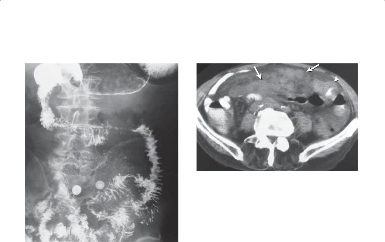
CASE 4.37
A B
Findings
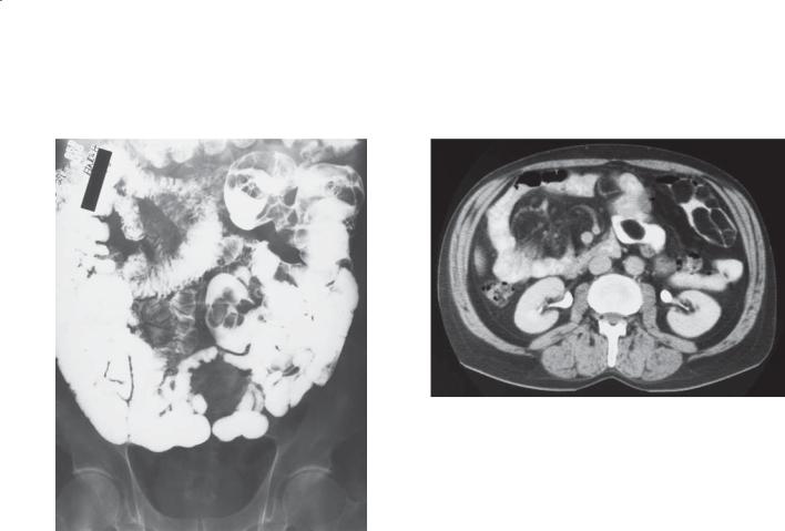
Abdominal radiograph. A. Multiple, dilated small bowel loops are present with little visible colonic gas. B. A few linear gas lucencies representing the biliary tree (arrows) and gallbladder (arrowheads) are visible in the right upper quadrant.
Di erential Diagnosis
1.Gallstone ileus
2.Postoperative biliary-enteric anastomosis with mechanical small bowel obstruction
Diagnosis
Gallstone ileus
Discussion
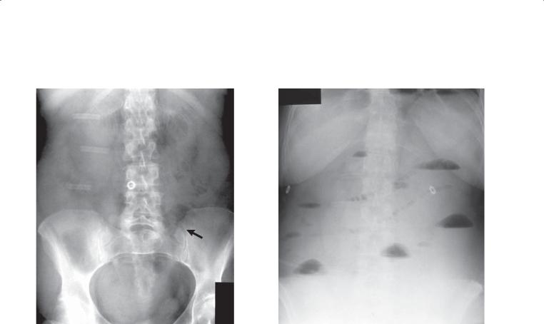
Th e combination of biliary gas and mechanical small bowel obstruction is highly suggestive of gallstone ileus if no prior biliary operation has been performed. In this case, an obstructing radiolucent gallstone causing ileal obstruction was removed at laparotomy. Gallstone ileus is a syndrome of mechanical small bowel obstruction by gallstone(s). Classically, elderly women without prior biliary or abdominal disease present with acute intestinal obstruction. Symptoms of acute cholecystitis are unusual, but many patients have a history of gallstones and recurrent cholecystitis. Gallstones can erode into the stomach, small bowel, or colon. Stones in the small bowel most often cause obstruction at
the ileocecal valve (the narrowest point). Obstructing stones usually are not spontaneously passed and require surgical lithotomy. Cholecystectomy and fistula repairs are usually performed, but not acutely.
Radiographically, the triad of air within the biliary tree, ectopic calcified gallstone, and mechanical small bowel obstruction is considered characteristic of this entity. Even the findings of biliary air and small bowel obstruction should be regarded as consistent with gallstone ileus.
Disease type: Masses and Filling Defects

CASE 4.38
Findings
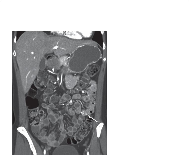
Contrast-enhanced CT. Multiple dilated small bowel loops are present in the pelvis. A peripherally calcified stone is present within the lumen of a small bowel loop. (This was the site of a transition to normal-caliber loops on other CT sections).
Di erential Diagnosis
1.Gallstone ileus
2.Calcified enterolith with small bowel obstruction
Diagnosis
Gallstone ileus
4. SMALL BOWEL 239
Discussion
CT is a sensitive technique for the detection of calcium-containing gallstones, small bowel obstruction, and biliary air. If gallstone ileus is
suspected, CT can be very helpful for confirming the diagnosis. The finding of a stone at the transition point between dilated and normal-caliber small bowel is diagnostic of gallstone ileus.
Disease type: Masses and Filling Defects
240 MAYO CLINIC GASTROINTESTINAL IMAGING REVIEW
CASE 4.39
Findings
Small bowel follow-through. An elongated filling defect (arrows) is present in the distal small bowel. A thin white line traverses the length of the filling defect.
Di erential Diagnosis
Ascariasis
Diagnosis
Ascariasis
Discussion
Th e thin white line traversing the length of the worm represents ingested barium in the worm’s enteric tract. Ascariasis is a common roundworm infection that most frequently occurs in tropical climates. Infection is acquired by ingesting contaminated water, food, or
soil. Ingested eggs hatch in the small bowel, penetrate the intestinal mucosa, and are carried to the lungs by the portal system or intestinal lymphatics. The worms perforate the alveoli, travel up the bronchi, and are swallowed. Worms grow, reproduce, and shed infectious eggs, usually within the distal small bowel. Involved organ systems include respiratory (pneumonia, bronchitis, hemoptysis, and asthma), gastrointestinal (nausea, vomiting, distention, tenderness), and biliary (jaundice, cholecystitis, cholangitis, pancreatitis, and hepatic abscess).
Radiographically, identification of the typical elongated filling defect is characteristic. Mucosal folds may be thickened. Occasionally, the mass of worms can be large enough to cause partial or complete intestinal obstruction. A worm may be identified at sonography or endoscopic retrograde cholangiopancreatography in the biliary tree or pancreatic duct.
Disease type: Masses and Filling Defects

|
|
4. SMALL BOWEL 241 |
|
|
|
|
TABLE 4.1 |
|
Filling Defects of the Small Bowel |
|
CASE |
|
|
|
Benign tumors |
|
|
Benign gastrointestinal stromal tumor |
Submucosal tumor with smooth contours, |
4.1 and 4.2 |
|
most frequent in jejunum |
|
Lipoma
Hemangioma
Pliable submucosal tumor with smooth borders, fat density at CT
Focal mass or diffuse malformation (hemangiomatosis), compressible, can contain calcified phleboliths
4.3 and 4.4
4.5–4.7
Polyp |
Solitary or multiple, can occur in patients with |
4.8–4.11 |
|
polyposis syndromes |
|
|
|
|
Malignant tumors |
|
|
Carcinoid |
Usually distal small bowel, usually 2–3 cm |
4.12–4.14 |
|
in diameter, mesenteric metastases elicit |
|
|
desmoplastic reaction and can calcify |
|
Lymphoma
Adenocarcinoma
Mostly non-Hodgkin variety, can present as focal mass, multiple nodules, infiltrative mass,
or excavated mass (aneurysmal dilatation)
Usually found in proximal small bowel, often an apple-core lesion
4.15–4.23
4.24 and 4.25
Malignant gastrointestinal |
Usually large submucosal mass, often exophytic |
4.26–4.28 |
stromal tumor |
growth, liver metastases should be sought, nodal |
|
|
involvement unusual |
|
Metastases |
Usually present as multiple masses, but may be |
4.29–4.35 |
|
solitary |
|
|
|
|
Nonneoplastic |
|
|
Gastrointestinal duplication cyst |
Terminal ileum is most common location, fluid |
4.36 |
|
density at CT |
|
Gallstone ileus
Ascariasis
Small bowel filling defect, small bowel obstruction, |
4.37 and 4.38 |
and biliary air. Most common site of obstruction is |
|
at ileocecal valve |
|
Tubular filling defect with linear barium in worm’s |
4.39 |
gastrointestinal tract |
|
Disease type: Masses and Filling Defects
242 MAYO CLINIC GASTROINTESTINAL IMAGING REVIEW
APPROACH TO DIFFUSE AND SEGMENTAL SMALL BOWEL DISEASE
Thin, straight folds |
Thick, straight folds |
|
Nodular changes |
with a dilated lumen |
|
||
|
|||
|
|
involving folds |
|
|
|
|
Type I (thin, straight folds) |
Type II (thick, straight folds) |
Type III (thick, nodular folds) |
|
Segmental |
Segmental |
|
Diffuse |
Diffuse |
Diffuse or segmental disease of the small bowel is often confusing, probably because of the infrequency with which many of these rare disorders are encountered and the relatively nonspecific radiographic appearance of many of these diseases.
If a disease diffusely or segmentally affects the small bowel, changes often occur within the wall of the bowel and alter the normal fold pattern.
Analysis of, first, the fold pattern and, second, the diffuse or segmental involvement of the small bowel
can be helpful for understanding the underlying pathologic condition and for developing a reasonable differential diagnosis. Unfortunately, this classification is somewhat arbitrary, because some diseases may have findings that overlap between designated fold types or a disease may present
with different fold patterns in different patients. Despite its limitations, this approach may be helpful as a starting point in the analysis of diffuse and segmental small bowel disease.
Disease type: Di use and Segmental Diseases
4. SMALL BOWEL 243
TYPE I FOLDS: THIN (<3 MM), STRAIGHT FOLDS WITH A DILATED LUMEN
Cases 4.40–4.43
1.Mechanical obstruction
2.Paralytic ileus
3.Scleroderma
4.Sprue
Disease type: Di use and Segmental Diseases
244 MAYO CLINIC GASTROINTESTINAL IMAGING REVIEW
CASE 4.40
A B
Findings
A.Supine abdominal radiograph. Multiple dilated, partially gas-filled small bowel loops are present with no visible gas in the colon. The valvulae conniventes (arrow) are thin and straight (type I fold pattern).
B.Upright abdominal radiograph. Multiple air-fluid levels are seen throughout the small bowel. Many small collections of gas line up within a small bowel loop and resemble a string of beads.
Di erential Diagnosis
1.Mechanical small bowel obstruction
2.Paralytic ileus
Diagnosis
Mechanical small bowel obstruction
Discussion
Partial or complete mechanical small bowel obstruction is a common disease that usually manifests clinically with nausea, vomiting, cramping abdominal pain,
and distention. The common causes of mechanical obstruction of the small bowel are adhesions (from a prior abdominal surgical procedure or severe intraperitoneal inflammation) (case 4.87) or hernias (cases 4.88 and 4.89). Other, less frequent causes include an obstructing neoplasm (cases 4.22 and 4.24), intussusception (cases 4.8 and 4.9), stricture (from Crohn disease, prior ischemia, or idiopathic), or volvulus (case 4.86).
Radiologic evaluation often begins with either an abdominal plain film examination or abdominal CT. Dilated small bowel loops (>3 cm in diameter) are often seen containing air-fluid levels on the film
taken with the patient in the upright position. It may be difficult to judge the level of obstruction on an abdominal plain film examination; in fact, a proximal colonic obstruction can masquerade as a mechanical small bowel obstruction on plain films. The lack of gas within the colon makes mechanical small bowel
destruction more likely than paralytic ileus in this case. Several additional cases of small bowel obstruction are included in this chapter (cases 4.87 through 4.89).
Disease type: Di use and Segmental Diseases
4. SMALL BOWEL 245
CASE 4.41 |
CASE 4.42 |
Findings
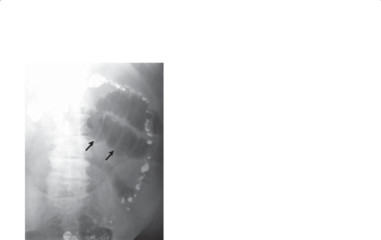
CASE 4.41. Small bowel follow-through. Multiple dilated small bowel loops are present. The valvulae conniventes are thin and straight (type I folds) and closely stacked together.
CASE 4.42. Magnified contrast-enhanced CT enterography. Mildly dilated loops of small bowel with crowded thin folds are present in the pelvis.
Di erential Diagnosis
1.Scleroderma
2.Celiac disease
3.Mechanical obstruction
Diagnosis
Scleroderma
Discussion
Th is case has the typical hidebound appearance of scleroderma involving the small bowel, with dilatation and crowding of the straight and thin
mucosal folds. Delayed transit time, sacculations on the antimesenteric border of the bowel, and even pneumatosis cystoides intestinalis also can be seen radiographically. Pneumatosis cystoides intestinalis may be related to the frequent use of corticosteroids among patients with scleroderma.
Small bowel changes of scleroderma often occur relatively late in the course of disease, usually after the typical skin changes, Raynaud phenomenon, or arthropathy. Pathologically, atrophy of the mucosa and submucosa, submucosal fibrosis, and round cell infiltration are seen. Mesenteric vascular arteritis may be present.
Distinguishing scleroderma from sprue is usually easy because there is no hypersecretion in scleroderma and patients with sprue usually have normal small bowel motility. In mechanical obstruction, the small bowel usually has more peristaltic activity, the folds are displaced from one another, and there is a considerable amount of retained fluid proximal to the obstruction. Esophageal changes also can be observed in patients with scleroderma (cases 1.67 and 1.68).
Disease type: Di use and Segmental Diseases
246 MAYO CLINIC GASTROINTESTINAL IMAGING REVIEW
CASE 4.43
Findings
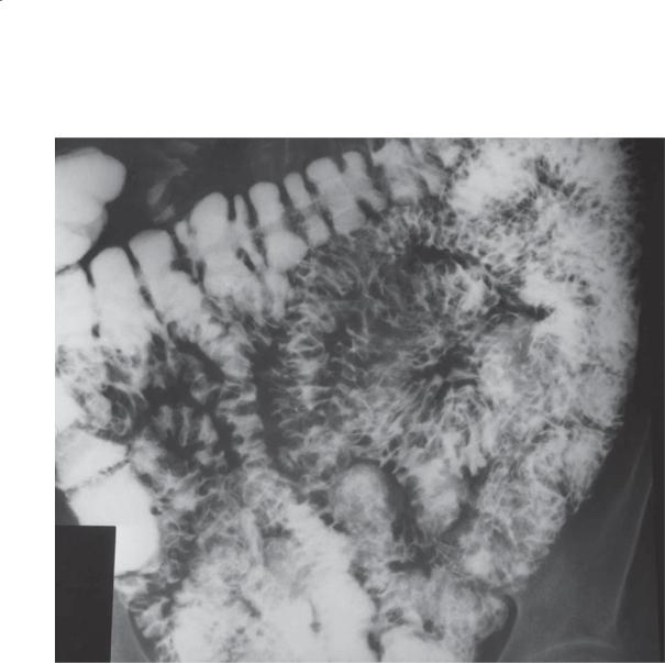
Small bowel follow-through. Mild dilatation of bowel loops with increased number of thin, straight (type I) folds in the ileum. Reversal of the normal jejunal and ileal fold patterns is seen in this case.
Di erential Diagnosis
Celiac disease (sprue)
Diagnosis
Celiac disease (sprue)
Discussion
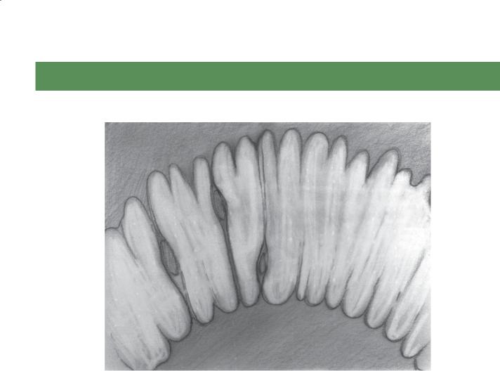
Th is typical example of the reversed fold pattern generally is found in patients with celiac disease. The “jejunization” of the ileum is believed to be an adaptive response to
the loss of absorptive surface in the proximal small bowel caused by villous atrophy. The jejunal folds have been shown to decrease in number (“ilealization” of the jejunum), a feature particularly evident on enteroclysis examinations. Three folds or less per inch in the jejunum on an enteroclysis examination is strong evidence for this disease. Additional cases of celiac disease are included in this chapter (cases 4.90 through 4.98).
Disease type: Di use and Segmental Diseases

|
|
4. SMALL BOWEL 247 |
|
|
|
|
|
|
TABLE 4.2 |
|
|
Di use and Segmental Small Bowel Diseases: |
|
|
|
Type I Folds |
|
CASE |
|
|
|
|
|
Mechanical obstruction |
Dilated loops with transition to decompressed |
4.40 |
|
|
loops of small bowel, usually caused by adhesions |
|
|
|
or hernia |
|
|
|
|
|
|
Paralytic ileus |
Diffusely dilated small bowel loops and colon, |
Not shown |
|
|
often due to surgery or narcotic medications |
|
|
|
|
|
|
Scleroderma |
Thin, straight folds stacked together (hidebound |
4.41 and 4.42 |
|
|
appearance), sacculations on antimesenteric |
|
|
|
border of small bowel or colon, diminished |
|
|
|
peristalsis |
|
|
|
|
|
|
Celiac disease (sprue) |
Fold pattern reversal, hypersecretion, dilatation |
4.43 |
|
|
|
|
|
Disease type: Di use and Segmental Diseases
248 MAYO CLINIC GASTROINTESTINAL IMAGING REVIEW
TYPE II FOLDS: THICK (>3 MM), STRAIGHT FOLDS, CAUSED BY INTRAMURAL EDEMA OR HEMORRHAGE
Cases 4.44–4.48
Segmental |
Di use |
||
1. |
Ischemia |
1. |
Venous congestion |
2. |
Radiation enteritis |
2. |
Hypoproteinemia |
3. |
Intramural hemorrhage |
3. |
Cirrhosis |
4. |
Adjacent inflammatory process |
|
|
Disease type: Di use and Segmental Diseases
4. SMALL BOWEL 249
CASE 4.44
Findings
Supine abdominal radiograph. Two small bowel loops in the left upper abdomen are dilated and contain straight, thickened (≥3 mm) folds (arrows), characteristic of a segmental type II fold pattern. Residual barium is seen in the colon.
Di erential Diagnosis
1.Ischemic bowel
2.Proximal small bowel obstruction
3.Radiation enteritis
Diagnosis
Ischemic bowel
Discussion
Ischemia of the small bowel and colon remains a difficult diagnosis because of the variable and nonspecific clinical findings. Patients may complain of bloating, gas, nausea, or vomiting. Peritoneal signs usually indicate transmural necrosis and possibly perforation. However, this is generally a late and infrequent finding. Gastrointestinal blood loss may be present.
Th e pathologic findings depend on the extent and duration of ischemia. Ischemia may be due to arterial embolization, hypoperfusion, or venous thrombosis. Arterial hypoperfusion is believed to be the most frequent cause, often due to either congestive heart failure or prolonged hypotension in association with mesenteric atherosclerotic disease. Histologic findings of bowel ischemia within the first 24 hours include initial submucosal edema and intramural hemorrhage, followed by transmural ischemia and eventually necrosis. Depending on the severity and depth of bowel wall injury from the ischemic insult, the three possible results of bowel ischemia are complete healing, stricture formation, and perforation.
Radiologic findings depend on the timing of the examination in relation to the vascular insult. Many abdominal radiographs (up to half) may be normal or have findings of only adynamic ileus. Suggestive findings include an isolated, rigid, often dilated, and unchanging small bowel loop with thickened mucosal folds. Additional cases of small bowel ischemia are included in this chapter (cases 4.75 through 4.80 and 4.82 through 4.85).
Disease type: Di use and Segmental Diseases
250 MAYO CLINIC GASTROINTESTINAL IMAGING REVIEW
CASE 4.45
Findings
Small bowel follow-through. Several loops of small bowel in the mid abdomen have straight, thickened folds with separation of the loops, consistent with a segmental type II fold pattern.
Di erential Diagnosis
1.Segmental type II folds (ischemia, hemorrhage, radiation)
2.Diffuse type II folds (venous congestion, hypoproteinemia, cirrhosis)
Diagnosis
Radiation enteritis (diffuse type II folds)
Discussion
A recent history of abdominal radiation therapy is critical for determining the correct cause of the abnormal, straight, thickened folds in this patient.
The small bowel is the portion of the alimentary tract most sensitive to radiation. Usually doses greater than 40 Gy (4,000 rad) are required before radiographic changes occur. Endarteritis obliterans is the underlying pathologic process responsible for the bowel changes. Acute changes can be found several weeks to a few months after treatment. Fold thickening and serration due to edema are frequent. The affected folds may appear fixed and angulated.
Chronic changes can be found 6 months after therapy and may develop many years later. These changes are usually due to bowel ischemia from arteriolar damage. Intestinal loops are often narrowed, separated, and straightened and the bowel wall is thickened. Nodularity and thumbprinting may be observed. These changes may become progressively more severe with stenosis, obstruction, perforation, and fistulization. The majority of patients with chronic radiation changes in the small bowel present with obstruction from adhesion or stenosis rather than with acute ischemia.
Disease type: Di use and Segmental Diseases
CASE 4.46
Findings
Small bowel follow-through. A loop of abnormal small bowel is seen in the pelvis. The lumen
is slightly narrowed with thick, straight folds (segmental type II pattern).
Di erential Diagnosis
1.Ischemia
2.Intramural hemorrhage
3.Adjacent inflammatory process (appendiceal or pelvic abscess)
Diagnosis
Intramural hemorrhage
4. SMALL BOWEL 251
Discussion
Th is patient was receiving anticoagulant therapy and had a spontaneous intramural hemorrhage. The small bowel is the most common site for intramural
hemorrhage, which can be caused by anticoagulation, bleeding, diathesis, or trauma. Spontaneous bleeds such as this one often result in thick, straight folds with a stack-of-coins appearance. Ischemic bowel or reactive small bowel edema due to an adjacent inflammatory process in the pelvis also could result in similar findings on a barium small bowel examination.
Disease type: Di use and Segmental Diseases

252 MAYO CLINIC GASTROINTESTINAL IMAGING REVIEW
CASE 4.47 |
CASE 4.48 |
Findings
CASE 4.47. Small bowel follow-through. The folds of the small bowel are diffusely thickened. Classification of the fold type is difficult, but it is best classified as diffuse type II folds.
CASE 4.48. Contrast-enhanced CT. Diffuse ascites is present in the abdominal cavity. Small bowel fold thickening and increased mesenteric markings are present.
Di erential Diagnosis
1.Hypoproteinemia
2.Venous congestion
3.Cirrhosis
Diagnosis
Hypoproteinemia
Discussion
Th is patient had chronic active hepatitis and secondary hypoalbuminemia. Intestinal edema due to hypoproteinemia (usually the serum albumin level is ≤2 g/dL) can be idiopathic or due to various diseases. Cirrhosis of the liver is the most frequent underlying disease.
Various radiographic changes also can be seen with diffusely thickened folds (type II), including haustral thickening and ascites. Venous congestion due to congestive heart failure could give a similar appearance.
CT findings in patients with hypoalbuminemia include not only edematous changes within the bowel and mesentery but also edema within other body tissues. These edematous changes include soft tissue stranding and a generalized increased density within the subcutaneous and mesenteric fat.
Disease type: Di use and Segmental Diseases

|
|
4. SMALL BOWEL 253 |
|
|
|
|
|
|
TABLE 4.3 |
|
|
Di use and Segmental Small |
Segmental Folds |
CASE |
|
Bowel Diseases: Type II Folds |
|
|
|
|
|
|
|
Ischemia (arterial or venous |
Acute abdomen, pneumatosis and portal |
4.44 |
|
occlusion) |
venous gas should be sought |
|
|
|
|
|
|
Radiation enteritis |
Confined to radiation port |
4.45 |
|
|
|
|
|
Intramural hemorrhage |
History of trauma or anticoagulation |
4.46 |
|
(trauma or bleeding diathesis) |
|
|
|
|
|
|
|
Adjacent inflammatory process |
Appendicitis, diverticulitis, pancreatitis |
Not shown |
|
|
|
|
|
|
Di use Folds |
|
|
|
|
|
|
Venous congestion |
History of congestive heart failure |
Not shown |
|
|
|
|
|
Hypoproteinemia |
History of nephrotic syndrome, cirrhosis |
4.47 and 4.48 |
|
|
|
|
|
Cirrhosis |
Changes of portal venous hypertension |
Not shown |
|
|
should be sought |
|
|
|
|
|
|
Disease type: Di use and Segmental Diseases
254 MAYO CLINIC GASTROINTESTINAL IMAGING REVIEW
TYPE III FOLDS: THICK, NODULAR FOLDS, CAUSED BY INFILTRATIVE
DISEASE OF THE BOWEL WALL
Cases 4.49–4.61
Segmental |
Di use |
||
1. |
Crohn disease |
1. |
Whipple disease |
2. |
Infection |
2. |
Intestinal lymphangiectasia |
3. |
Lymphoma |
3. |
Nodular lymphoid hyperplasia |
4. |
Metastases |
4. |
Polyposis syndromes |
|
|
5. |
Eosinophilic gastroenteritis |
|
|
6. |
Amyloidosis |
|
|
7. |
Mastocytosis |
|
|
8. |
Lymphoma |
|
|
9. |
Metastases |
Disease type: Di use and Segmental Diseases
CASE 4.49
Findings
Small bowel follow-through. Thickened, somewhat nodular (type III) folds are present in the majority of the visualized small bowel loops.
Di erential Diagnosis
Segmental and diffuse type III fold differentials
Diagnosis
Crohn disease
4. SMALL BOWEL 255
Discussion
Crohn disease is a chronic disorder of unknown origin. The disease involves the small bowel in at least 80% of patients. The clinical activity of the disease (abdominal pain, diarrhea, fever, weight loss, anemia) correlates poorly with radiologic changes. The location of the involved small bowel segment does affect prognosis. Crohn disease confined to the distal small bowel
has the best long-term prognosis, whereas ileocolic involvement has the highest incidence of complications (abscess, fistula). The terminal ileum is usually involved (spared in only 5% of all patients). Recurrent disease after resection of a diseased small bowel segment invariably occurs about the anastomosis. Recurrences are usually detectable radiographically within 2 years after operation. Additional cases of Crohn disease are included in this chapter (cases 4.62 through 4.74).
Disease type: Di use and Segmental Diseases
256 MAYO CLINIC GASTROINTESTINAL IMAGING REVIEW
CASE 4.50
A B
Findings
Small bowel follow-through. A and B. The folds of the proximal small bowel are thickened; some fold nodularity or contour irregularity (segmental type III folds) is present.
Di erential Diagnosis
Segmental and diffuse type III fold differentials
Diagnosis
Giardiasis
Discussion
Th ese findings are nonspecific and could be due to any disease causing a type III fold pattern. Localization
of the findings within the proximal small bowel suggests giardiasis. Giardiasis is a disease caused by infection with the protozoan Giardia lamblia.
Ingested trophozoites attach to the duodenal mucosa and reproduce. Cysts are shed and passed in the stool. The host response to the protozoan varies from the asymptomatic carrier state to severe symptoms of diarrhea and malabsorption. Patients with hypogammaglobulinemia or agammaglobulinemia are believed to be more prone to infection, and often these patients have changes of nodular lymphoid
hyperplasia (case 4.56) in the small bowel. The majority of people infected with G lamblia have no clinical or radiographic manifestations of their infection.
Radiographically, the proximal small bowel usually shows inflammatory changes, including fold
thickening, increased secretions, irritability, and spasm or rapid transit. Tiny nodular lesions are frequent
and result from hypertrophied lymphoid follicles. A spruelike pattern may occur in the distal jejunum and ileum, with reversal of the normal fold pattern. These changes revert to normal after treatment.
Disease type: Di use and Segmental Diseases
4. SMALL BOWEL 257
CASE 4.51
Findings |
Discussion |
Small bowel follow-through. Somewhat nodular, |
Whipple disease is caused by the bacillus Tropheryma |
thickened folds (type III) are present throughout the |
whipplei. It is characterized clinically by malabsorption, |
proximal small bowel. |
arthritis or arthralgias, lymphadenopathy, abdominal |
|
tenderness, and increased skin pigmentation. |
Di erential Diagnosis |
Histologically, a periodic acid-Schiff–positive glycoprotein |
Segmental and diffuse type III fold differentials |
is deposited within macrophages in the lamina propria |
|
and lymph nodes of the small bowel. Treatment usually |
Diagnosis |
consists of a long-term course of antibiotics. |
Whipple disease |
Radiographically, fold thickening and nodularity are |
|
common in the proximal small bowel. Hypersecretion, |
|
dilatation, and diffuse small bowel involvement are |
|
usually absent, which helps to differentiate this disease |
|
from sprue. In the immunocompromised patient, |
|
infection with Mycobacterium avium-intracellulare, |
|
Giardia, or Cryptosporidium can produce identical |
|
radiographic findings and should be considered. |
Disease type: Di use and Segmental Diseases
258 MAYO CLINIC GASTROINTESTINAL IMAGING REVIEW
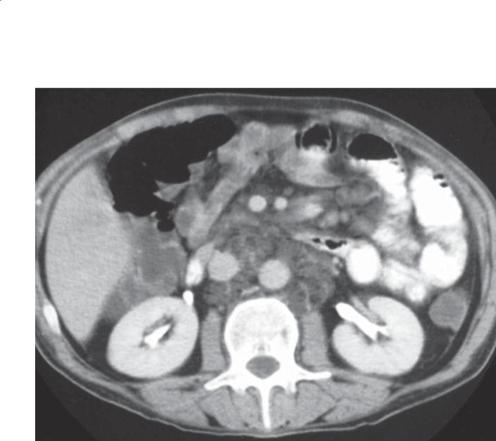
CASE 4.52
Findings |
Discussion |
|
Contrast-enhanced CT. Multiple low-density lymph |
Whipple disease often is associated with abdominal |
|
nodes are present within the retroperitoneum and |
lymphadenopathy, but this finding is rarely appreciated |
|
small bowel mesentery. |
on small bowel examinations. The lymph nodes |
|
|
|
usually have a low attenuation on CT because of the |
Di erential Diagnosis |
deposition of fat and fatty acids within the nodes. |
|
1. |
Whipple disease |
Occasionally, only mesenteric lymphadenopathy is |
2. |
Mycobacterium avium-intracellulare |
present. Sacroiliitis, a component of this systemic |
3. |
Testicular metastases |
illness, also may be detectable on CT of the abdomen. |
4. |
Lymphoma |
The sacroiliac joints may be affected either unilaterally |
5. |
Epidermoid carcinoma |
or bilaterally, and the articular symptoms may precede |
|
|
the gastrointestinal symptoms (usually diarrhea) by |
Diagnosis |
5 years or more. |
|
Whipple disease |
|
|
Disease type: Di use and Segmental Diseases
4. SMALL BOWEL 259
CASE 4.53
Findings
Small bowel follow-through. Nodular fold thickening is present diffusely throughout the visualized dilated loops of small bowel. There is also nodular deformity of the distal stomach.
Di erential Diagnosis
Diffuse type III fold differentials
Diagnosis
Eosinophilic gastroenteritis
Discussion
Although this small bowel pattern is nonspecific, the patient was found to have eosinophilic gastroenteritis. Eosinophilic gastroenteritis is a disease of unknown origin, in which the patient presents with abdominal pain, diarrhea, vomiting,
and occasionally malabsorption. Usually eosinophilia
is present on the peripheral blood smear. Often the clinical course is benign and self-limited, responding to corticosteroid treatments. Some patients have a history of allergy.
Pathologically, eosinophils and chronic inflammatory cells are present in the bowel wall. Localized and diffuse bowel involvement occur. Localized eosinophilic granuloma is usually confined to the stomach. Various clinical syndromes have been attributed to the portion of the bowel wall infiltrated by eosinophils. Predominantly mucosal infiltration results in protein loss and malabsorption, intramural disease presents with obstructive symptoms or diarrhea, and serosal eosinophilia results in ascites.
Th e radiographic findings are similar to those of any other infiltrative small bowel disease. Marked bowel wall infiltration can result in luminal narrowing and rigidity of the affected segment(s). Any portion of the alimentary tract may be affected.
Disease type: Di use and Segmental Diseases
260 MAYO CLINIC GASTROINTESTINAL IMAGING REVIEW
CASE 4.54
A B
Findings
A.Small bowel follow-through. Thickened folds and tiny nodules (type III folds) are present within the proximal small bowel. Dilution of barium as a result of excess intraluminal fluid is seen in the distal small bowel.
B.Lymphangiogram. Multiple dilated and bulbous lymphatic channels are present within the small bowel mesentery.
Di erential Diagnosis
Intestinal lymphangiectasia
Diagnosis
Intestinal lymphangiectasia
Discussion
Intestinal lymphangiectasia is a disorder of abnormal lymph flow, with loss of lymphatic fluid (most importantly protein) into the alimentary tract. Patients often present with hypoalbuminemia, hypoproteinemia, and occasionally malabsorptive
symptoms. Diarrhea, vomiting, and abdominal pain are often present. Lymphangiectasia is often a congenital condition, or it may be acquired later in life from inflammatory or neoplastic lymphatic obstruction.
Pathologically, lymph channels are dilated in the lamina propria and submucosa of the bowel wall with associated enlarged and distorted villi. These lymph channels rupture into the gut lumen and are responsible for the protein loss. Treatment may be difficult, but some patients respond to a low-fat diet with medium-chain triglycerides, which do not
require lymphatic transport for absorption. Lymphatic abnormalities elsewhere in the body are often found.
Disease type: Di use and Segmental Diseases
CASE 4.55
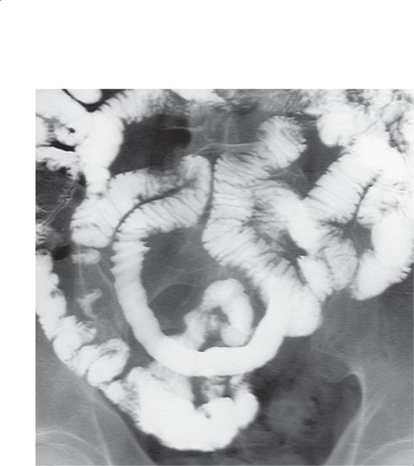
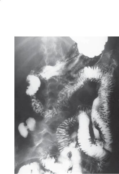
Findings
Small bowel follow-through. Multiple polypoid filling defects are present throughout the small bowel.
Di erential Diagnosis
1.Polyposis syndrome
2.Metastases
3.Lymphoma
4.Lymphangiectasia
Diagnosis
Intestinal lymphangiectasia
4. SMALL BOWEL 261
Discussion
Th e nodular filling defects that may occur in lymphangiectasia can vary considerably in size. Large filling defects (as seen in this case) can be several millimeters in diameter, whereas tiny defects may appear as sandlike lucencies.
Disease type: Di use and Segmental Diseases
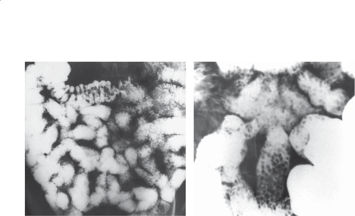
262 MAYO CLINIC GASTROINTESTINAL IMAGING REVIEW
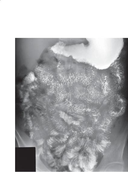
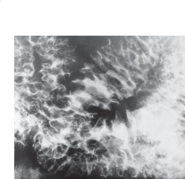
CASE 4.56
A B
Findings
Small bowel follow-through. A and B. Multiple tiny nodular filling defects are present throughout the small bowel (diffuse type III pattern). All the nodules are uniform in size and shape.
Di erential Diagnosis
1.Nodular lymphoid hyperplasia
2.Lymphoma
3.Normal lymphoid nodules
Diagnosis
Nodular lymphoid hyperplasia
Discussion
Nodular lymphoid hyperplasia usually is associated with an immunologic disorder, primarily a deficiency of
immunoglobulin A and immunoglobulin M. Occasionally,
this disease may be present without an immunologic disorder. Malabsorption and an intestinal infection (Giardia lamblia, Strongyloides, or Monilia) are often associated conditions. The incidence of gastric and colonic cancers is increased in all patients with enteropathic immunoglobulin deficiencies, especially children.
Innumerable tiny nodules are seen in the involved portions of the small bowel. The nodules are usually less than 4 mm in diameter and of uniform size. They may be centrally umbilicated and resemble an aphthous ulcer. The main differential consideration is lymphoma; however, lymphomatous nodules are large, vary in size, and may ulcerate (cases 4.57 and 4.58). Normal lymphoid nodules can regularly be seen in patients of any age but are usually found in children and young adults. Normal nodules are uniform in size, nearly always less than 4 mm in diameter, and primarily involve the distal small bowel and proximal colon.
Disease type: Di use and Segmental Diseases
CASE 4.57
A
Findings
A.Small bowel follow-through. Diffuse nodular fold thickening (type III) is present throughout the small bowel. This appearance is nonspecific.
B.Contrast-enhanced CT. Adenopathy (arrows) is present within the small bowel mesentery. A small bowel loop with thickened wall is visible (arrowhead).
Di erential Diagnosis
1.Lymphoma
2.Crohn disease
Diagnosis
Lymphoma
4. SMALL BOWEL 263
B
Discussion
CT is often included in the evaluation of patients with nonspecific complaints. In patients with lymphoma affecting the small bowel, CT can
be helpful in suggesting the diagnosis. Bowel abnormalities vary from thickened folds (as in this case) to circumferential, long segments of bowel wall thickening (cases 4.16, 4.19, and 4.21). Mesenteric adenopathy is displayed well at CT, and, although nonspecific, it is a supportive finding of lymphoma. Other type III fold differential possibilities are unlikely with the focal bowel wall thickening and mesenteric lymphadenopathy. Both lymphoma and Crohn disease can present with these two findings, although the extensive lymphadenopathy favors lymphoma.
Disease type: Di use and Segmental Diseases
264 MAYO CLINIC GASTROINTESTINAL IMAGING REVIEW
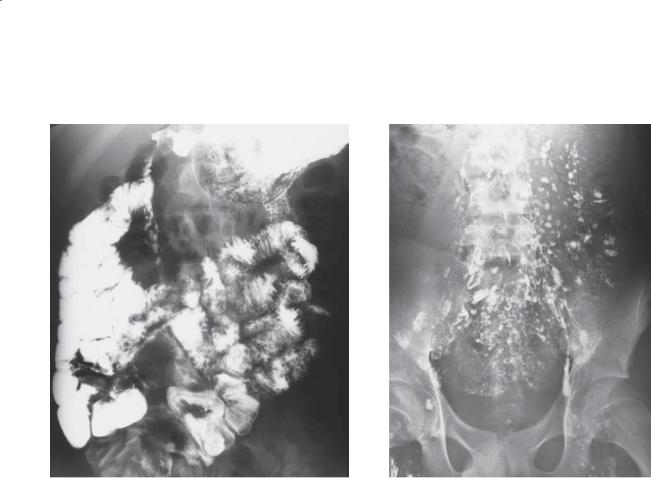
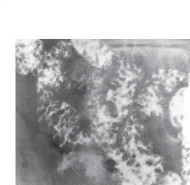
CASE 4.58
Findings
Small bowel follow-through. Diffuse nodular fold thickening (type III) is present throughout these small bowel loops. The smooth surface of the nodules suggests their submucosal location. These nodules are variable in size.
Di erential Diagnosis
Diffuse type III fold differentials
Diagnosis
Lymphoma
Discussion
Multiple nodules of varying size and fold thickening are manifestations of lymphoma diffusely involving the small bowel. In addition to the diffuse form, 10%
to 20% of patients with gastrointestinal lymphoma have multiple focal lesions. These lesions may appear to be of submucosal origin and their surface may
be ulcerated. The polyposis syndromes (cases 2.18, 4.8, 4.9, 5.72 through 5.76) are a differential consideration, but usually they are diagnosed before a small bowel examination is performed. A prior history of malignancy (eg, melanoma) is often known in patients with metastatic tumors of the small bowel (cases 4.29 through 4.33). Metastases are usually not as numerous as the diffuse nodularity in this case. Nodular lymphoid hyperplasia has small (<4 mm) nodules of uniform size (case 4.56). Lymphangiectasia (cases 4.54 and 4.55) usually is diagnosed by early adulthood, whereas non-Hodgkin lymphoma generally occurs during the fifth and sixth decades
of life.
Disease type: Di use and Segmental Diseases
4. SMALL BOWEL 265
CASE 4.59
Findings |
Discussion |
Small bowel follow-through. The small bowel is |
Diffuse hematogenous seeding of the gastrointestinal |
involved with diffusely nodular folds (type III fold |
tract with metastatic tumor can result in nodular |
pattern). |
changes within the bowel wall. A history of melanoma |
|
would make this radiographic appearance nearly |
Di erential Diagnosis |
diagnostic of metastases. Nodular lymphoid |
Diffuse type III fold differentials |
hyperplasia (case 4.56) has smaller nodules (usually |
|
<4 mm diameter) of uniform size. Whipple disease |
Diagnosis |
affects the proximal small bowel most severely. Patients |
Metastatic melanoma |
with Crohn disease usually have segmental areas of |
|
intervening normal small bowel, often with luminal |
|
stenosis and fistulas. Polyps developing in patients with |
|
Peutz-Jeghers syndrome are usually fewer and larger. |
|
Lymphoma and lymphangiectasia could present with |
|
these findings. |
Disease type: Di use and Segmental Diseases
266 MAYO CLINIC GASTROINTESTINAL IMAGING REVIEW
CASE 4.60
Findings
Small bowel follow-through. Multiple thickened and nodular (type III) folds are present throughout the small bowel.
Di erential Diagnosis
Segmental and diffuse type III fold differentials
Diagnosis
Systemic amyloidosis
Discussion
Th e small bowel is that portion of the alimentary tract most often affected with amyloid. Vascular compromise can result in bowel ischemia, infarction, and bleeding. Submucosal amyloid deposition causes polypoid protrusions, fold thickening, and irregular, fine filling defects.
Amyloidosis is caused by deposition of an insoluble fibrillar protein within the extracellular
space of various organs. Deposition within arterial walls is often present, resulting in possible ischemia or infarction of the end organ. Several classifications have been devised for this disease. Generally, systemic and localized forms exist. Systemic amyloidosis is most common and can result from a wide variety of causes; it can be idiopathic, related to a plasma cell dyscrasia (multiple myeloma, light-chain and heavy-chain disease, Waldenström macroglobulinemia), due to chronic infections or inflammatory conditions, or familial. Amyloid can be deposited throughout the gastrointestinal tract. Patients often complain of weight loss, fatigue, and abdominal pain.
Radiographic findings may be normal, even in patients with debilitating gastrointestinal symptoms. Diminished motor activity, thickened or atrophic folds, dilatation, and an obstructive pattern may be seen. Changes identical to those of ulcerative colitis can be seen in the colon.
Disease type: Di use and Segmental Diseases
CASE 4.61
Findings
Small bowel follow-through. Multiple small bowel loops have a featureless, atrophic appearance. Thickened folds are present diffusely.
Di erential Diagnosis
1.Ischemic small bowel
2.Infectious enteritis
3.Celiac disease
4.Acute radiation enteritis
5.Graft -versus-host disease
6.Amyloidosis
Diagnosis
Systemic amyloidosis
4. SMALL BOWEL 267
Discussion
See discussion of case 4.60.
Disease type: Di use and Segmental Diseases

268 MAYO CLINIC GASTROINTESTINAL IMAGING REVIEW
TABLE 4.4
Di use and Segmental Small |
Segmental Folds |
CASE |
Bowel Diseases: Type III Folds |
|
|
|
|
|
Crohn disease |
Nodules associated with ulcers, usually |
4.49 |
|
distal ileum, asymmetric bowel involvement, |
|
|
skip lesions |
|
|
|
|
Infection |
Giardiasis (proximal), Yersinia and |
4.50 |
|
tuberculosis (distal) |
|
|
|
|
Lymphoma |
Nodules of differing sizes |
4.57 and 4.58 |
|
|
|
Metastases |
Multiple nodules with or without ulcers |
4.59 |
|
|
|
Di use Folds
Whipple disease |
Proximal small bowel affected more than |
4.51 and 4.52 |
|
distal small bowel, weight loss, arthralgias, |
|
|
lymphadenopathy |
|
|
|
|
Intestinal lymphangiectasia |
May require lymphangiography for diagnosis |
4.54 and 4.55 |
|
|
|
Nodular lymphoid hyperplasia |
Diffuse <4-mm monotonous nodules |
4.56 |
|
|
|
Polyposis syndromes |
Peutz-Jeghers most common, any type can |
4.8–4.10 |
|
have small bowel polyps |
|
|
|
|
Eosinophilic gastroenteritis |
Peripheral eosinophilia |
4.53 |
|
|
|
Amyloidosis |
Irregular fine filling defects and fold |
4.60 and 4.61 |
|
thickening |
|
|
|
|
Mastocytosis |
Deposits of mass cells in skin and bowel wall, |
Not shown |
|
skeletal sclerosis should be sought |
|
|
|
|
Lymphoma |
Nodules of different sizes, adenopathy |
4.57 and 4.58 |
|
|
|
Metastases |
Multiple nodules, history of primary tumor |
4.59 |
|
|
|
Disease type: Di use and Segmental Diseases
CASE 4.62
Findings
Small bowel follow-through (spot radiograph). Multiple discrete ulcerations are present in the distal small bowel. The central barium collections (ulcer crater) and mounds of edema are characteristic of aphthous ulcers. A compression device with metallic marker is seen.
Di erential Diagnosis
1.Crohn disease
2.Infectious disorders of the ileum Yersiniosis
Amebiasis Tuberculosis
4. SMALL BOWEL 269
Diagnosis
Crohn disease
Discussion
Th ese tiny mucosal ulcers are believed to be the first mucosal lesions of Crohn disease. These lesions may coalesce and form longitudinal and transverse ulcerations that are typical of more advanced disease
(case 4.63). Discrete ulcers are not specific for Crohn disease and can occur in various infectious disorders that affect the terminal ileum.
Disease type: Common Small Bowel Diseases
270 MAYO CLINIC GASTROINTESTINAL IMAGING REVIEW
CASE 4.63
Findings
Small bowel follow-through (spot radiograph). A cobblestone mucosal pattern affects a nonstenotic segment of small bowel. Longitudinal and transverse ulcerations, in conjunction with bowel wall edema, produce a cobblestone mucosal pattern. A metallic marker on a compression device is present.
Di erential Diagnosis
1.Crohn disease
2.Small bowel polyps
3.Infectious enteritis
Diagnosis
Crohn disease
Discussion
Patients with Crohn disease often have the insidious onset of abdominal cramping, diarrhea, weight loss, low-grade fever, anorexia, and anemia. Patients usually are treated conservatively with rest, dietary changes, and antidiarrheal and anti-inflammatory agents. Surgical treatment is usually reserved for complications of the disease that do not respond to medical therapy— fistulas, obstruction, and abscess.
Extraintestinal manifestations of Crohn disease include arthritis (and ankylosing spondylitis), erythema nodosum, pyoderma gangrenosum, and, rarely, primary sclerosing cholangitis. The incidence of cholesterol gallstones and oxalate renal stones can be increased in patients with ileal disease as a result of abnormalities in the enterohepatic bile acid circulation.
Small bowel polyps usually do not focally involve the bowel and are not associated with ulcerations. Infection can simulate Crohn disease and must be excluded. Yersiniosis and tuberculosis can often mimic Crohn disease.
Disease type: Common Small Bowel Diseases
CASE 4.64
Findings
CASE 4.64. Small bowel follow-through. A long segment of narrowed ileum (string sign) is present with areas of bowel wall asymmetry.
CASE 4.65. Small bowel follow-through. An abnormal loop of distal small bowel is present in the right lower quadrant (arrows). The loop is separate from others, and the medial wall is flattened and featureless. Pseudodiverticula are present along the lateral wall. These findings are due to circumferential asymmetric bowel wall involvement.
Di erential Diagnosis
1.Crohn disease
2.Segmental ischemia
3.Small bowel infection
Diagnosis
Crohn disease
4. SMALL BOWEL 271
CASE 4.65
Discussion
Th e string sign of Crohn disease may or may not represent a fixed stricture. This region of narrowing may be inconstant because of marked spasm, and proximal small bowel dilatation may be lacking. Fixed narrowing from transmural fibrosis usually is associated with a short segmental stricture and
dilatation of proximal loops. The terminal ileum often is affected.
Circumferential asymmetry of the bowel lumen can be a helpful radiographic finding in Crohn disease. This finding may be due to skip areas of fibrosis or ulceration with folding and sometimes dilatation of the opposite wall.
Small bowel ischemia can cause featureless strictures; however, multiple skip areas usually are absent. A patient’s age and history also can be helpful. Patients with ischemia often are older and
have an acute onset of abdominal pain and adynamic ileus. Patients with Crohn disease are usually young adults with a chronic history of abdominal pain and diarrhea. Infectious disorders such as tuberculosis, actinomycosis, histoplasmosis, and blastomycosis can present with findings that are indistinguishable from those of Crohn disease. Yersinia and Salmonella can produce superficial erosions and fold thickening, but strictures are uncommon.
Disease type: Common Small Bowel Diseases
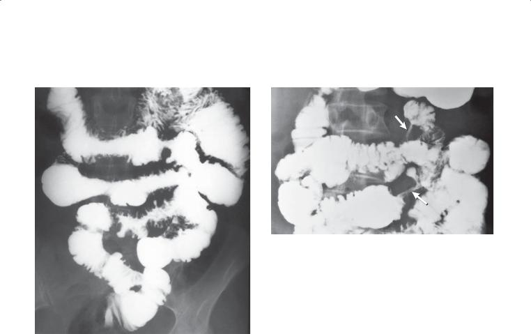
272 MAYO CLINIC GASTROINTESTINAL IMAGING REVIEW
CASE 4.66 |
CASE 4.67 |
Findings
CASE 4.66. Small bowel follow-through. Mucosal fold thickening, ulceration, nodularity, asymmetric bowel wall involvement, regions of narrowing, and separation of small bowel loops are present.
CASE 4.67. Small bowel follow-through. Two regions of high-grade stenosis (arrows) are present in the proximal small bowel.
Di erential Diagnosis
CASE 4.66
1.Crohn disease
2.Acute gastroenteritis
3.Small bowel hemorrhage
CASE 4.67
1.Crohn disease
2.Small bowel tumor
Diagnosis
Crohn disease
Discussion
Advanced changes of Crohn disease are often due to transmural inflammation and fibrosis. Transmural inflammation leads to bowel wall thickening and bowel loop separation (as seen in case 4.66). Fistulas and abscesses also result from transmural disease. Fibrosis nearly always accompanies the healing phase of the disease. Patchy discontinuous regions of fibrosis result in circumferential asymmetry of the bowel wall.
Stenotic Crohn disease can affect both long and short segments of the bowel. Proximal dilatation of the small bowel usually indicates significant obstruction by the lesion. Complete obstruction from Crohn disease is rare. In some patients, particularly those with a solitary short stenosis, it may be impossible to exclude neoplasm radiologically. These patients may require resection of the diseased bowel.
Disease type: Common Small Bowel Diseases
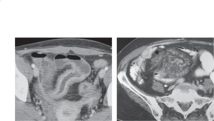
CASE 4.68
Findings
CASE 4.68. Contrast-enhanced CT. Marked homogeneous segmental bowel wall thickening and mucosal hyperenhancement are present in the distal small bowel. Dilatation of the proximal small bowel is visible.
CASE 4.69. Contrast-enhanced CT. A mass composed of fat and soft tissue stranding is present in the right
lower quadrant. A loop of distal small bowel has mildly thickened walls just posterior to the mass.
Di erential Diagnosis
1.Crohn disease
2.Small bowel primary tumor
3.Metastases
Diagnosis
Crohn disease
4. SMALL BOWEL 273
CASE 4.69
Discussion
Bowel wall thickening and mesenteric soft tissue stranding are the commonest CT findings of Crohn disease. The location of the bowel wall involvement in the ileum is often helpful for suggesting the diagnosis.
Th e fibrofatty changes in the mesentery of patients with Crohn disease are referred to as “creeping fat” by surgeons and pathologists. Enlarged and normal-sized lymph nodes are often visible. The mass may be of homogeneous fat density or may contain streaks and poorly defined soft tissue changes within it. Soft tissue changes within the mass are often associated with acute inflammation. This fibrofatty proliferation has been reported to cause ureteral compression and obstruction when the retroperitoneum is affected.
Disease type: Common Small Bowel Diseases
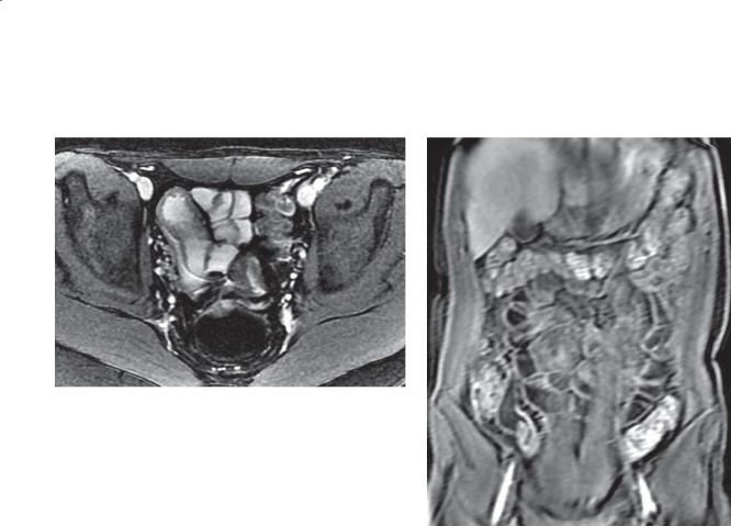
274 MAYO CLINIC GASTROINTESTINAL IMAGING REVIEW
CASE 4.70
A B
Findings
MR enterography. A. The distal small bowel has a thickened wall. B. Contrast-enhanced series shows intense enhancement in the thickened ileal wall.
Di erential Diagnosis
1.Crohn disease
2.Infectious enteritis
3.Nonsteroidal anti-inflammatory drug enteropathy
Diagnosis
Crohn disease
Discussion
MRI can be a useful tool in the management of young people with Crohn disease because of its avoidance of ionizing radiation. Young patients with Crohn disease that will require multiple follow-up studies are most appropriate for MR enterography. The preparation of the bowel is the same as that with CT enterography (3-4 glasses of water-attenuation contrast material, each consumed at 15-minute intervals before scanning) plus dynamic intravenous contrast-enhanced acquisitions.
Disease type: Common Small Bowel Diseases
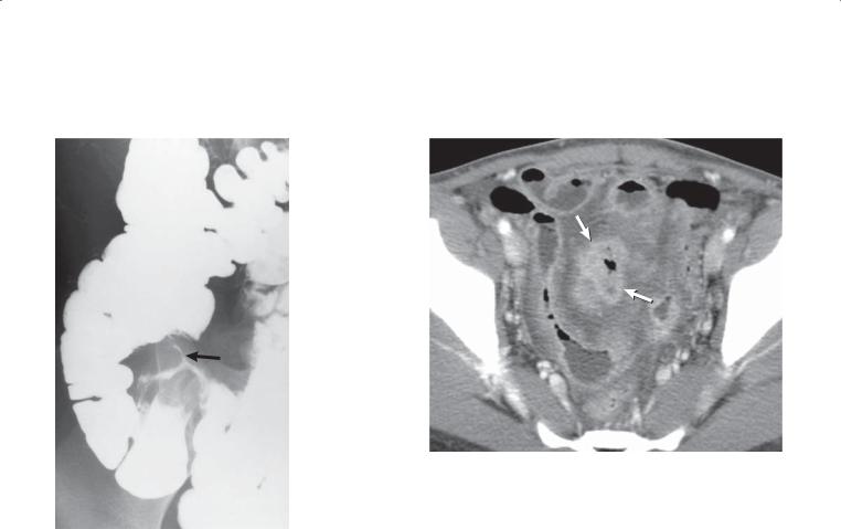
CASE 4.71
Findings
CASE 4.71. Small bowel follow-through. The terminal ileum is narrowed, and a fistulous communication (arrow) exists between the distal ileum and colon.
Inflammatory changes with tethered folds are present along the medial aspect of the cecum.
CASE 4.72. Contrast-enhanced CT. A segmental loop of small bowel is thick-walled, and the lumen is narrowed. A gas-containing cavity is located within
the small bowel mesentery, medial to the affected small bowel loop (arrows). This has the appearance of an interloop abscess.
Di erential Diagnosis
Crohn disease
Diagnosis
Crohn disease
4. SMALL BOWEL 275
CASE 4.72
Discussion
Fistula formation (case 4.71) is a common finding in patients with transmural Crohn disease. Fistulas can communicate with other small bowel loops, colon, genitourinary tract, retroperitoneum, mesentery, and skin. Occasionally they are multiple, and often they are incorporated within an inflammatory mass of bowel and mesentery. Abscess formation (case 4.72) often accompanies fistulization and sinus tracts. CT has the advantage of direct visualization of the entire small bowel, mesentery, and adjacent organs.
Disease type: Common Small Bowel Diseases
276 MAYO CLINIC GASTROINTESTINAL IMAGING REVIEW
CASE 4.73
Findings
Double-contrast barium enema. Freely functioning ileotransverse colostomy. The neoterminal ileum is irregular in contour, and the mucosa appears ulcerated and nodular.
Di erential Diagnosis
1.Recurrent Crohn disease
2.Infectious ileitis
Diagnosis
Recurrent Crohn disease
Discussion
Recurrent Crohn disease is common after operation. Recurrence almost always involves the neoterminal ileum. The features of recurrent disease are the same as those preoperatively. This is a typical case of nonstenotic recurrent Crohn disease.
Disease type: Common Small Bowel Diseases

CASE 4.74
Findings
Contrast-enhanced CT. The terminal ileum has a markedly thickened wall that contains a central ring the attenuation of fat.
Di erential Diagnosis
Inactive Crohn disease
Diagnosis
Inactive Crohn disease
4. SMALL BOWEL 277
Discussion
CT findings of Crohn disease include bowel wall thickening, mesenteric fibrofatty proliferation, abscess, mesenteric inflammation, and lymphadenopathy. In Crohn disease, bowel wall thickening is usually greater than 1 cm and is the commonest CT finding. The wall thickening in active disease may be the density of
soft tissue or contain a central water-density ring. Fat within the bowel wall has been shown to accumulate in the submucosa of patients with either ulcerative colitis or Crohn disease. Active inflammation is not present when this finding is identified. The cause of the fat deposition is unknown, although long-term corticosteroid treatment may be responsible.
Disease type: Common Small Bowel Diseases
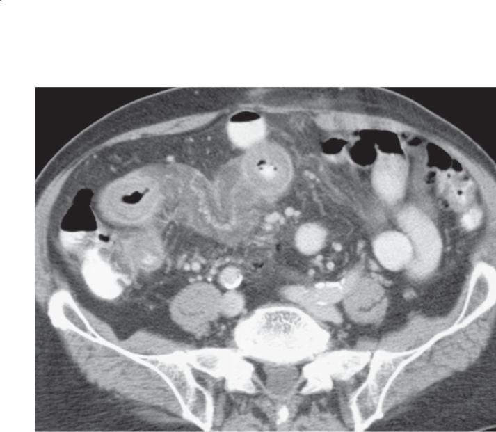
278 MAYO CLINIC GASTROINTESTINAL IMAGING REVIEW
CASE 4.75
Findings
Contrast-enhanced CT. A thick-walled small bowel loop is present in the right side of the abdomen. A portion of the thickened wall is of low attenuation, near that of water, indicating submucosal edema.
Di erential Diagnosis
1.Ischemia
2.Crohn disease
3.Lymphoma
4.Radiation enteritis
5.Angioedema due to angiotensin-converting enzyme inhibitors
Diagnosis
Ischemic small bowel
Discussion
CT findings in patients with bowel infarction include diffuse or focal bowel wall thickening, segmental dilatation, mesenteric edema, ascites, intramural gas, and mesenteric or portal venous gas. Only intramural and venous gases are considered specific for ischemia, but even these findings occasionally are seen in patients without bowel infarction. Edema within the bowel wall is nonspecific and could be due to any inflammatory disease within the bowel or adjacent mesentery or
to hypoalbuminemia. Despite its nonspecific nature, bowel dilatation with a thickened and edematous wall without apparent cause in a patient with abdominal pain should be regarded as highly suggestive
of ischemia.
Disease type: Common Small Bowel Diseases
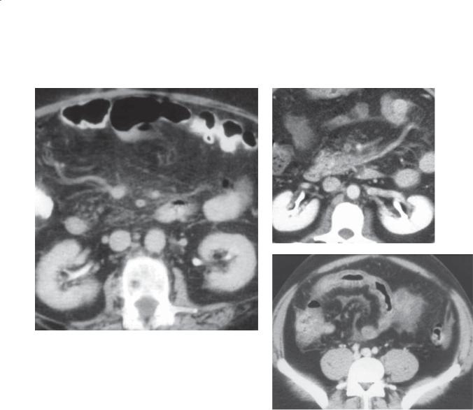
4. SMALL BOWEL 279
CASE 4.76 |
CASE 4.77 |
|
A |
B
Findings
CASE 4.76. Contrast-enhanced CT. A tiny filling defect is present within the superior mesenteric artery. (Courtesy of J. Scott Kriegshauser, MD, Mayo Clinic, Scottsdale, Arizona. Used with permission.)
CASE 4.77. Contrast-enhanced CT. A. A filling defect is present within the superior mesenteric vein. B. Edematous bowel wall thickening involves several small bowel loops and the right colon.
Di erential Diagnosis
Ischemic small bowel
Diagnosis
Ischemic small bowel (superior mesenteric artery embolus in case 4.76, superior mesenteric vein thrombosis in case 4.77)
Discussion
Small bowel ischemia can be due to low flow states (usually from prolonged hypotension), an arterial embolus (case 4.76), or superior mesenteric vein thrombosis (case 4.77). CT often can identify filling defects within the main branches of the superior mesenteric artery or vein.
Disease type: Common Small Bowel Diseases
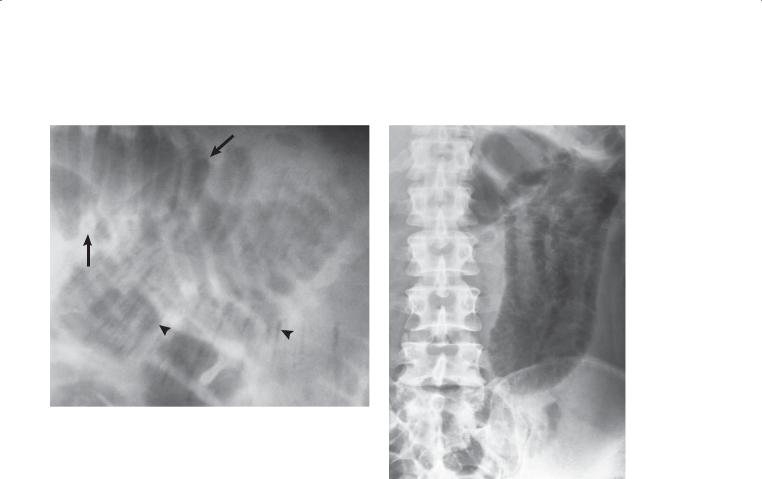
280 MAYO CLINIC GASTROINTESTINAL IMAGING REVIEW
CASE 4.78 |
CASE 4.79 |
Findings
CASE 4.78. Supine abdominal radiograph (close-up). Multiple small bowel folds (arrows) are straight and thickened. In addition, linear intramural pneumatosis (arrowheads) is present.
CASE 4.79. Supine abdominal radiograph. Extensive cystic pneumatosis is present within the jejunum.
Di erential Diagnosis
1.Small bowel ischemia
2.Benign pneumatosis
3.Mechanical small bowel obstruction
Diagnosis
Small bowel ischemia
Discussion
Th e finding of intramural pneumatosis in a patient with ischemic bowel disease suggests severe ischemia. Urgent operation usually is required to prevent perforation and to decrease the high morbidity and mortality associated with such a complication.
Pneumatosis intestinalis due to ischemic or infarcted bowel can have either a cystic or a linear appearance. Large cystic collections are unusual and more commonly associated with nonischemic
disease. Clinical correlation is always necessary when pneumatosis intestinalis is found so as not to delay treatment or overlook the diagnosis of bowel ischemia.
Disease type: Common Small Bowel Diseases
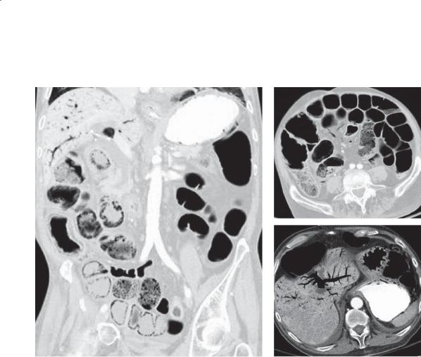
4. SMALL BOWEL 281
CASE 4.80
A B
C
Findings
Contrast-enhanced CT. A. Coronal. Lung windows show multiple small bowel loops containing pneumatosis. B. Axial. Lung windows. The mesenteric veins contain air. C. Axial. Soft tissue windows. Portal venous gas is present within the liver.
Di erential Diagnosis
1.Mesenteric ischemia
2.Benign pneumatosis intestinalis and portal venous gas
Diagnosis
Mesenteric ischemia with secondary pneumatosis intestinalis and portal venous gas
Discussion
Th e appearance of intramural gas on CT can be either cystic or linear-curvilinear (case 4.78 and this case). CT is more sensitive than abdominal plain radiography for detecting pneumatosis intestinalis. In addition, other signs of ischemic bowel can be searched for,
including portal venous gas (cases 4.82 and 4.83), bowel wall thickening (case 4.75), and thrombi or occlusion of the superior mesenteric artery and vein. If a patient with abdominal pain has pneumatosis, it is most important to review the radiologic and clinical findings with the referring physician because benign pneumatosis and pneumatosis due to bowel ischemia can have a similar radiographic appearance. In some patients, lung window settings are helpful for detecting intramural air at CT.
Intramural gas (pneumatosis) is most easily identified within the bowel wall by its posterior, dependent location. It is also usually present within the nondependent wall, but it is often more difficult to distinguish from intraluminal gas. Clinical correlation is necessary to distinguish pneumatosis intestinalis due to intestinal ischemia from other conditions causing pneumatosis, generally referred to as benign pneumatosis. The presence of mesenteric and portal venous gas is usually a sign of advanced ischemia and often is associated with a grave prognosis.
Disease type: Common Small Bowel Diseases
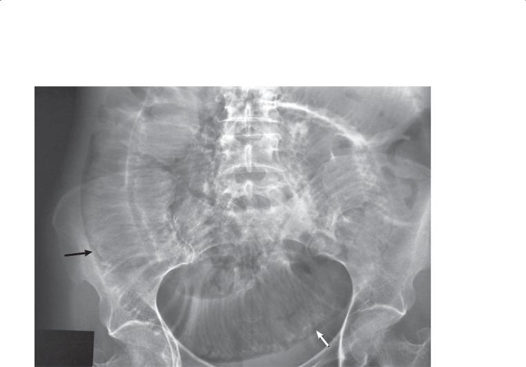
282 MAYO CLINIC GASTROINTESTINAL IMAGING REVIEW
CASE 4.81
Findings
Abdominal radiograph. Extensive intramural gas (arrows) is present within the small bowel.
Di erential Diagnosis
1.Mesenteric ischemia or infarction
2.Benign pneumatosis intestinalis
Diagnosis
Benign pneumatosis intestinalis
Discussion
Th is patient with scleroderma had been hospitalized several times for intestinal pseudo-obstruction.
She recovered uneventfully. The pneumatosis was considered benign, due to her scleroderma and corticosteroid medication.
Pneumatosis intestinalis, or air within the wall of the small bowel, can result from several causes. Commonly associated conditions include corticosteroid medications, scleroderma, chronic
obstructive lung disease, and ischemic bowel. The exact cause is often not known, but many theories suggest that a slow-healing mucosal ulceration develops, followed by dissection of gas into the bowel wall. Alternatively, some authors have suggested
that in patients with obstructive lung disease, air can dissect from the alveoli into the mediastinum, to retroperitoneum, and eventually to small bowel mesentery and bowel wall. Patients are usually asymptomatic and do not require treatment for this condition unless they have ischemic bowel
disease. Patients with ischemic bowel will have acute abdominal signs and may have associated portal venous gas and bowel wall thickening, and usually they require urgent surgical exploration.
Radiologically, a lucent rim that follows the contours of the bowel wall (as in this case) is pathognomonic
of pneumatosis. The air collections may appear cystlike or linear. Occasionally, the intramural air collections escape into the peritoneal cavity and cause asymptomatic pneumoperitoneum.
Disease type: Common Small Bowel Diseases
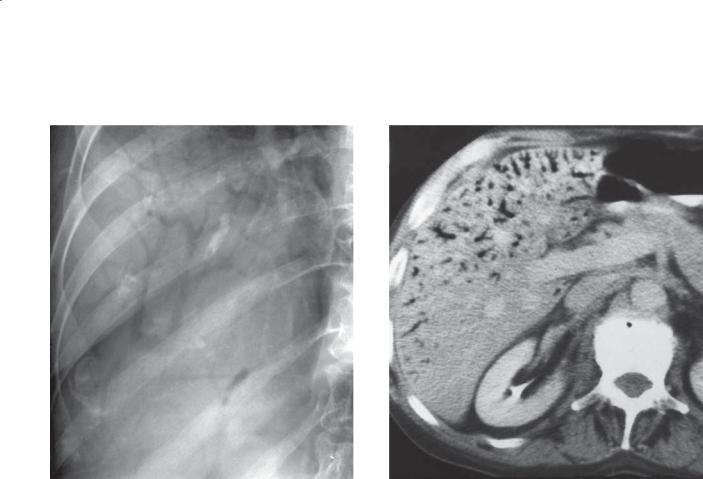
CASE 4.82
Findings
CASE 4.82. Supine abdominal radiograph. Gascontaining tubular structures are visible within the liver. The peripheral distribution of the gas is typical of portal venous gas.
CASE 4.83. Contrast-enhanced CT. Multiple gas-filled portal venous branches are visible throughout the periphery of the liver. The nondependent portal veins contain more gas than those in the dependent, posterior right lobe.
Di erential Diagnosis
1.Portal venous gas, ischemic bowel
2.Portal venous gas, benign cause
Diagnosis
Portal venous gas, ischemic bowel
4. SMALL BOWEL 283
CASE 4.83
Discussion
Portal venous gas is often a late finding in patients with intestinal ischemia. The mucosa is the bowel layer most sensitive to ischemia. As the mucosa breaks down, gas can enter the bowel wall (pneumatosis intestinalis, case 4.80) and eventually enter the mesenteric and portal venous system. Although nonischemic benign causes of portal venous gas have been reported, portal venous gas is unusual, and urgent clinical assessment of the patient is recommended.
Disease type: Common Small Bowel Diseases
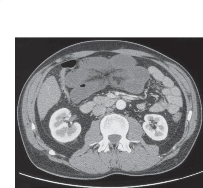
284 MAYO CLINIC GASTROINTESTINAL IMAGING REVIEW
CASE 4.84
Findings
Contrast-enhanced CT. Several dilated small bowel loops have little or no bowel wall enhancement. The bowel loops appear to be grouped together and pulled toward a central point. Soft tissue stranding is present within the mesentery.
Di erential Diagnosis
1.Closed loop small bowel obstruction with secondary ischemia
2.Focal small bowel ischemia (arterial embolus or mesenteric venous thrombosis)
Diagnosis
Closed loop obstruction with secondary ischemia
Discussion
Closed loop obstruction most often is caused by entrapment of a bowel loop(s) by an adhesive band. External and internal hernias also can lead to closed loop obstruction. CT often is helpful for demonstrating the dilated small bowel loop and the site of the compressed adjacent bowel loops. Evidence of vascular compromise by delayed, partial, or absent bowel wall perfusion can be detected at CT. Treatment is urgent operative intervention.
Disease type: Common Small Bowel Diseases
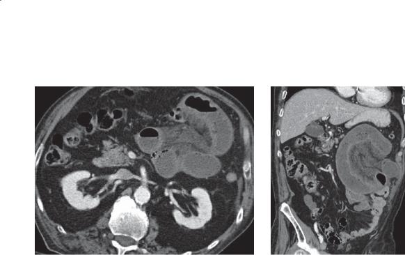
CASE 4.85
A
Findings
Contrast-enhanced CT. A. Axial. Several dilated, thickwalled small bowel loops are present in the left upper quadrant. There is mesenteric soft tissue standing adjacent to the affected small bowel loops. B. Coronal. The abnormal loops are confined to the jejunum, and there is diminished wall enhancement of the dilated small bowel loops compared with the normal loops.
Di erential Diagnosis
1.Closed-loop obstruction with secondary small bowel ischemia
2.Focal ischemia (arterial embolus or mesenteric venous thrombosis)
3.Internal hernia with ischemia
Diagnosis
Closed-loop obstruction with secondary ischemia
4. SMALL BOWEL 285
B
Discussion
A closed-loop obstruction is a type of mechanical small bowel obstruction in which blood supply to the loop can be compromised; this condition can lead to ischemia and necrosis with perforation if untreated. The commonest causes are adhesive bands, internal and external hernias, and small bowel volvulus. Additional findings to support strangulation of the affected loops include bowel wall thickening, vascular engorgement, mesenteric stranding, and differential perfusion of the loop compared with normal bowel (as present in this case).
Disease type: Common Small Bowel Diseases
286 MAYO CLINIC GASTROINTESTINAL IMAGING REVIEW
CASE 4.86
Findings |
Discussion |
Contrast-enhanced CT. Small bowel and mesentery |
Patients with rotational anomalies of the gut lack |
encircle the superior mesenteric artery in a whorl-like |
normal posterior peritoneal attachments. Normal |
pattern. |
peritoneal attachments prevent the small bowel and |
|
colon from moving or twisting into an abnormal |
Di erential Diagnosis |
location or position. In the absence of sufficient |
Midgut volvulus |
fixation, the small bowel can twist about the superior |
|
mesenteric artery, leading to bowel obstruction |
Diagnosis |
and ischemia. Symptoms of obstruction usually |
Midgut volvulus |
develop. Urgent surgical repair often is required to |
|
prevent ischemia or to resect infarcted bowel loops. |
|
Intermittent or partial obstruction can lead to bowel |
|
congestion, edema, or symptoms of malabsorption. |
Disease type: Common Small Bowel Diseases
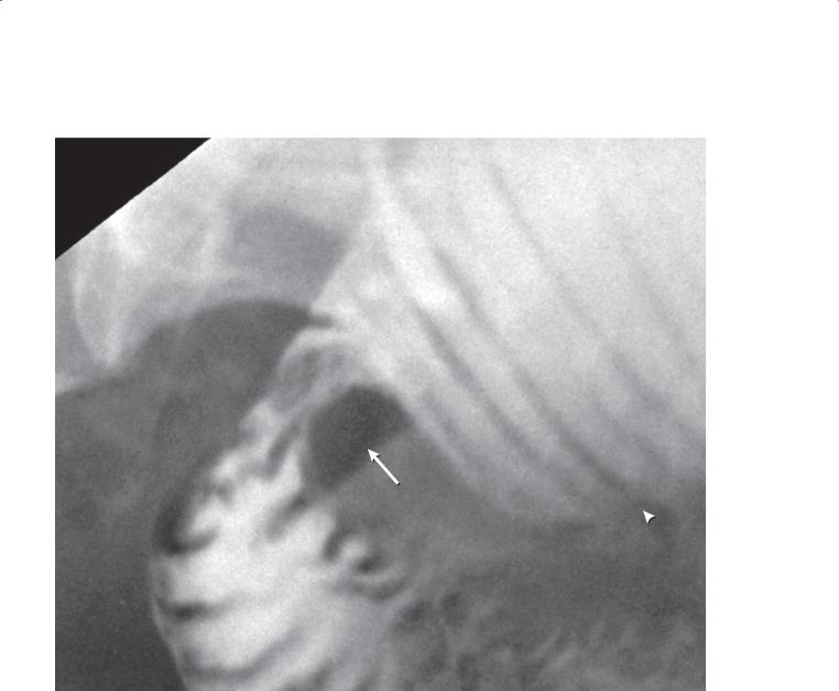
CASE 4.87
Findings
Small bowel follow-through. The proximal small bowel is dilated and abruptly narrows from a smooth rounded defect (arrow) that eccentrically compresses the lumen and causes partial obstruction. Notice the straight thin folds in the dilated loop (arrowhead).
Di erential Diagnosis
1.Mechanical small bowel obstruction due to adhesions
2.Medication-induced focal ulceration and scar
Diagnosis
Mechanical small bowel obstruction due to adhesions
4. SMALL BOWEL 287
Discussion
Defining the site and cause of obstruction is more commonly done at CT than with a small bowel follow-through or enteroclysis examination. This case illustrates the typical appearance at small bowel follow-through of an obstructing adhesion with the smooth, focal, abrupt transition between dilated and nondistended small bowel.
Disease type: Common Small Bowel Diseases
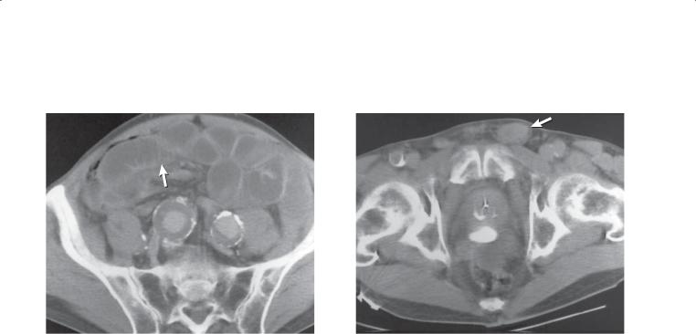
288 MAYO CLINIC GASTROINTESTINAL IMAGING REVIEW
CASE 4.88
A B
Findings
Enhanced abdominal CT. A. Multiple dilated fluidfilled small bowel loops are present in the abdomen. Aneurysmal dilatation of both iliac arteries is seen, as are thin, straight small bowel folds (arrow). B. A loop of small bowel could be traced into the enlarged inguinal canal (arrow).
Di erential Diagnosis
Mechanical small bowel obstruction
Diagnosis
Mechanical small bowel obstruction due to an incarcerated inguinal hernia
Discussion
CT is now widely used as a primary imaging method for evaluating patients with a suspected small bowel obstruction. In nearly three-quarters of the patients, the cause of the obstruction can be identified at CT.
The diagnosis of mechanical intestinal obstruction should be suspected when a discrepancy in the caliber of the proximal and distal bowel is identified. The commonest causes of a mechanical small bowel obstruction are adhesions or hernias.
Disease type: Common Small Bowel Diseases
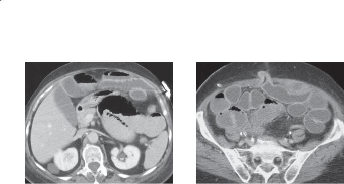
CASE 4.89
A
Findings
Contrast-enhanced CT. A and B. Multiple dilated small bowel loops are present throughout the abdomen. A loop of small bowel enters a ventral abdominal wall hernia. At this site, there is an abrupt transition in the caliber of the small bowel.
Di erential Diagnosis
Mechanical small bowel obstruction, ventral hernia
Diagnosis
Mechanical small bowel obstruction, ventral hernia
4. SMALL BOWEL 289
B
Discussion
Adhesions and hernias are the commonest causes of mechanical small bowel obstruction. In this case, the transition between dilated and collapsed small bowel is visualized as the small bowel loop enters the ventral hernia sac. Operative repair was required.
Disease type: Common Small Bowel Diseases

290 MAYO CLINIC GASTROINTESTINAL IMAGING REVIEW
CASE 4.90 |
CASE 4.91 |
|
A |
B
Disease type: Common Small Bowel Diseases

Findings
CASE 4.90. Small bowel follow-through. The jejunum is devoid of most of its normal mucosal markings. The tubular, featureless appearance of these loops has been referred to as the moulage sign.
CASE 4.91. Contrast-enhanced CT. A. The jejunum lacks the normal fold pattern. Several loops are fluid-filled, thick-walled, and dilated. B. Ileal loops in the pelvis have a prominent fold pattern. Many loops are fluidfilled and dilated.
Di erential Diagnosis
Celiac disease
Diagnosis
Celiac disease
4. SMALL BOWEL 291
Discussion
Excessive intraluminal fluid obscures and prevents adequate mucosal coating of the proximal small bowel and results in the moulage sign at small bowel follow-through. Moulage refers to a molded or casted structure. The jejunum in this patient resembles a tubular cast of small bowel because of its paucity of mucosal folds. The featureless ileum at both small bowel follow-through and CT and the multiple ileal folds at CT demonstrate the reversal of fold pattern characteristic of celiac disease.
Disease type: Common Small Bowel Diseases
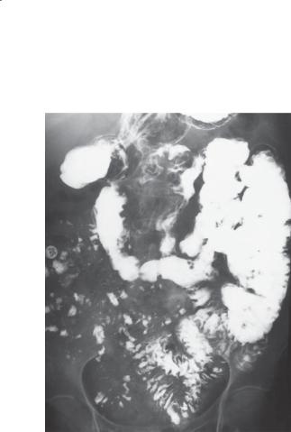
292 MAYO CLINIC GASTROINTESTINAL IMAGING REVIEW
CASE 4.92
Findings
Small bowel follow-through. Flocculation and segmentation of barium indicate the presence of excessive fluid in the distal small bowel. The normal jejunal fold pattern also is absent. These loops appear featureless. A few small bowel loops in the pelvis have thickened folds.
Di erential Diagnosis
1.Celiac disease
2.Zollinger-Ellison syndrome
3.Mechanical small bowel obstruction
4.Acute caustic ingestion
Diagnosis
Celiac disease
Discussion
Th e term sprue is given to 3 diseases with common radiographic and pathologic manifestations. Childhood and adult (nontropical) sprue are probably the same disease, due to a gluten (a water-insoluble protein found in various cereals and grains, including wheat, rye, oats, and barley) sensitivity. The cause
of tropical sprue is unknown, but patients do not respond to dietary gluten restrictions. These patients often respond to folate therapy or antibiotic (usually tetracycline) therapy or both.
Pathologically, villous atrophy is usually seen with elongation of the crypts of Lieberkühn and infiltration of the lamina propria with inflammatory cells. Changes are often more marked in the proximal small bowel.
Th is case demonstrates many of the radiologic features of excess intraluminal fluid (hypersecretion) that commonly are encountered in patients with sprue. Barium may become radiolucent and separated into clumps (segmentation) or tiny pieces (flocculation)
as a result of dilution. This is most common in the distal small bowel. Small bowel folds in patients with sprue may remain normally thin or they may become thickened. Thickened folds usually are due to intramural edema from hypoalbuminemia. The gastrointestinal loss of albumin is the result of the
malabsorption that is typically present in these patients. Patients with Zollinger-Ellison syndrome usually have thickened folds in the proximal small bowel. In patients with a mechanical obstruction, a transition to normalcaliber small bowel usually can be shown.
Disease type: Common Small Bowel Diseases
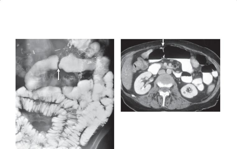
4. SMALL BOWEL 293
CASE 4.93
A B
Findings
A.Small bowel follow-through. The jejunum has a tubular, featureless appearance without the normal jejunal fold pattern. In addition, there is a focal smooth stricture in the proximal jejunum (arrow) with 2 or
3 additional focal strictures in the proximal small bowel which are not well shown on this single image. Extensive postoperative changes also are visible in the abdomen.
B.Contrast-enhanced CT. This corresponding CT image also shows the tubular, featureless appearance of the small bowel with the focal stricture in the proximal jejunum (arrow).
Di erential Diagnosis
1.Celiac disease (sprue) with a benign stricture due to prior ulcerative jejunoileitis
2.Nonsteroidal anti-inflammatory drug–induced enteropathy
Diagnosis
Celiac disease (sprue) with a benign stricture
Discussion
Th is patient has the classic moulage sign in the proximal small bowel associated with celiac disease. Patients with long-standing celiac disease occasionally present with benign focal strictures, probably as a result of prior ulcerative jejunoileitis. Focal strictures and bowel wall thickening can also develop in patients with nonsteroidal anti-inflammatory drug–induced enteropathy; however, the reversal of the fold pattern is not expected.
Disease type: Common Small Bowel Diseases
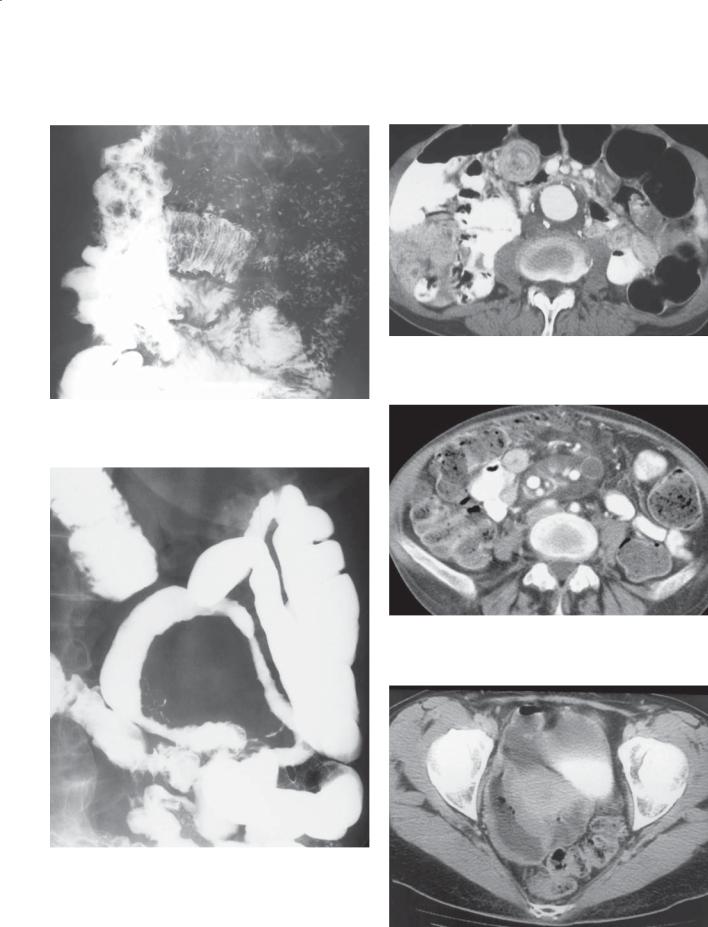
294 MAYO CLINIC GASTROINTESTINAL IMAGING REVIEW
CASES 4.94 |
CASES 4.96 |
CASES 4.97
CASES 4.95
CASES 4.98
Disease type: Common Small Bowel Diseases

Findings
CASE 4.94. Small bowel follow-through. The typical coiled-spring appearance of a jejunal intussusception is present in the upper abdomen.
CASE 4.95. Small bowel follow-through. Several small bowel loops appear featureless, and other loops are narrowed with nodular and thickened folds. The narrowed segments may be ulcerated.
CASE 4.96. Contrast-enhanced CT. A small bowel intussusception is present in the central abdomen.
CASE 4.97. Contrast-enhanced CT. Multiple low-density lymph nodes are present within the mesentery.
CASE 4.98. Contrast-enhanced CT. A dilated, focally thick-walled loop of small bowel is present in the right side of the pelvis.
Di erential Diagnosis
CASES 4.94 AND 4.96
1.Intussusception
2.Lead point mass
3.Celiac disease
CASE 4.95
1.Crohn disease
2.Segmental ischemia
3.Celiac disease with segmental ulceration
CASE 4.97
1.Mycobacterium avium-intracellulare (tuberculosis)
2.Lymphoma (treated)
3.Celiac disease
CASE 4.98
1.Lymphoma
2.Crohn disease
3.Celiac disease with lymphoma
Diagnosis
Complications of celiac disease
Celiac disease with intussusception: cases 4.94 and 4.96 Celiac disease with ulcerating jejunoileitis: case 4.95 Celiac disease with cavitary lymph node syndrome:
case 4.97
Celiac disease with small bowel lymphoma: case 4.98
4. SMALL BOWEL 295
Discussion
Intussusceptions occur often in patients with celiac disease. One report noted this disorder in 20% of patients. Intussusceptions are usually
asymptomatic, transient, and self-reducing. As with any intussusception, careful fluoroscopy should be performed to exclude a small bowel mass as a lead point for the intussusception.
Segmental ulceration in patients with sprue is an unusual complication of the disease. Patients often present with worsening of their malabsorptive symptoms and abdominal pain. Bleeding and perforation can occur as a result of the nonspecific ulcers. The ulcers are morphologically nonspecific, are often multiple, and usually involve the jejunum. They can be difficult to detect radiologically. Spasm, irritability, and persistent areas of narrowing can make it difficult to differentiate these changes from
lymphoma. As a result, the involved loops are usually resected. In some patients, the nonspecific ulcers may precede the development of strictures or recognition of intestinal lymphoma.
Several disorders are associated with celiac disease, including hyposplenism and cavitary lymph node syndrome (case 4.97). Sometimes the enlarged nodes can have lipid-fluid layers within them. Lymphoma and carcinoma are the best known complications. In addition, dermatitis herpetiformis, immunoglobulin A deficiency, and adenopathy can occur.
Disease type: Common Small Bowel Diseases
296 MAYO CLINIC GASTROINTESTINAL IMAGING REVIEW
TABLE 4.5
Common Small Bowel |
|
|
Diseases |
|
CASE |
|
|
|
Crohn disease |
Usually distal ileum involved, aphthous ulcers, short- |
4.62–4.74 |
|
segment strictures, skip lesions, cobblestone pattern, |
|
|
asymmetric bowel wall involvement, fistula, abscess |
|
|
|
|
Ischemia |
Segmental straight, thickened folds, pneumatosis, portal |
4.75–4.85 |
|
venous gas, mesenteric artery or venous thrombosis or |
|
|
emboli should be sought |
|
|
|
|
Small bowel obstruction |
Dilated small bowel with transition point, most commonly |
4.85–4.89 |
|
caused by adhesions or hernias |
|
|
|
|
Celiac disease |
Reversal of jejunal and ileal fold pattern, hypersecretion, |
4.90–4.98 |
|
dilatation, transient intussusceptions. Complications: |
|
ulcerating jejunoileitis, lymphoma, carcinoma, cavitating lymph node syndrome
Disease type: Common Small Bowel Diseases

CASE 4.99
A
Findings
Small bowel follow-through (spot radiograph).
A. Short focal narrowing of a small bowel loop with an irregular luminal contour and abrupt, shouldered margins. B. An eccentric ulceration involves another small bowel loop.
Di erential Diagnosis
1.Metastases
2.Lymphoma
3.Crohn disease
4.Nonspecific ulcerations
5.Nonsteroidal anti-inflammatory drug–induced enteropathy
Diagnosis
Nonspecific ulcerations
4. SMALL BOWEL 297
B
Discussion
In the past, intestinal ulcerations frequently were due to enteric-coated potassium chloride. Because this is no longer used, these types of ulcerations are encountered less often. Similar-appearing ulcers and strictures can occur in patients taking nonsteroidal anti-inflammatory drugs (indomethacin and
ibuprofen). The ulcers may be solitary or multiple and can heal with stenosis or diaphragm-like strictures. The ulcers have a sharp demarcation with normal bowel, without surrounding inflammatory change. Other causes of isolated small bowel ulcers include tuberculosis, Crohn disease, Behçet syndrome, celiac disease, ischemia, trauma, heterotopic gastric mucosa, and arsenic poisoning. Adenocarcinoma is usually a solitary lesion.
Disease type: Miscellaneous
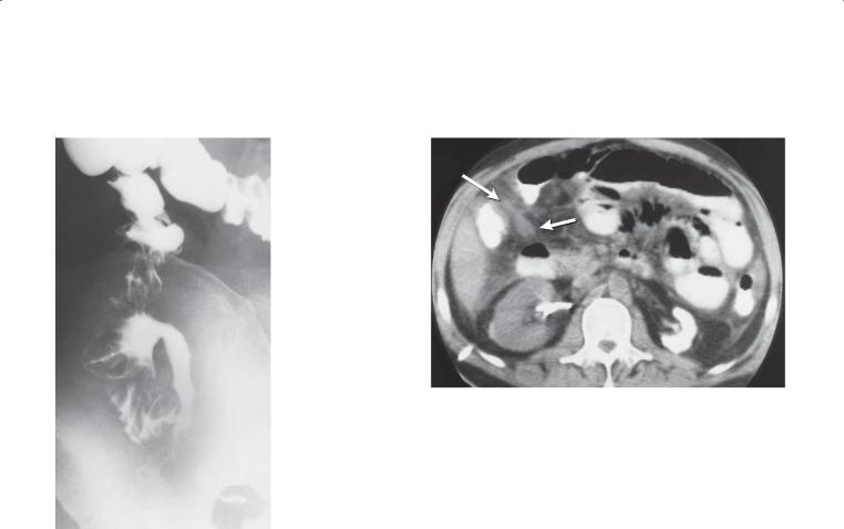
298 MAYO CLINIC GASTROINTESTINAL IMAGING REVIEW
CASE 4.101
Findings |
Discussion |
|
CASE 4.100. Single-contrast barium enema examination. |
Tuberculosis involving the gastrointestinal tract |
|
The terminal ileum is narrowed with fine contour |
remains common in parts of the world. Many patients |
|
irregularities. The cecum is contracted. The ileocecal |
with gastrointestinal tuberculosis have a normal chest |
|
valve is widely patent. |
radiograph. Tuberculosis most often affects the distal |
|
CASE 4.101. Unenhanced CT. The mesenteric fat is |
small bowel and cecum. Pathologically, granulomas in |
|
the bowel wall ulcerate or form a localized mass. Nodal |
||
of increased attenuation (arrows). The anterior |
||
and peritoneal involvement are common. Chronic |
||
pararenal fascia is thickened. The wall of the right |
||
infection leads to fibrosis and luminal narrowing. |
||
colon is thickened. The left kidney is shrunken |
||
Radiographically, findings can mimic those of |
||
and homogeneously hyperdense, consistent |
||
Crohn disease. Ulcerations, luminal narrowing, |
||
with tuberculous nephropathy (putty kidney). A |
||
multiple segmental regions of involvement, bowel wall |
||
nephrostomy tube is present within the right kidney. |
||
thickening, fistulas, and a localized mass are typical. |
||
|
||
Di erential Diagnosis |
The cecum may be shrunken and deformed (acutely |
|
as a result of spasm and chronically from fibrosis). The |
||
1. Crohn disease |
||
ileocecal valve may be patulous and incompetent. |
||
2. Mycobacterial infection |
||
Tuberculous peritonitis is relatively rare. Ascites, |
||
3. Amebiasis |
||
mesenteric soft tissue stranding, and mesenteric |
||
4. Peritoneal metastases |
||
adenopathy are usual radiographic findings. |
||
|
||
Diagnosis |
Adenopathy involves predominantly the upper |
|
abdomen. Low-density centers within the nodes |
||
Mycobacterial tuberculosis |
||
occasionally are identified. |
||
|
Disease type: Miscellaneous
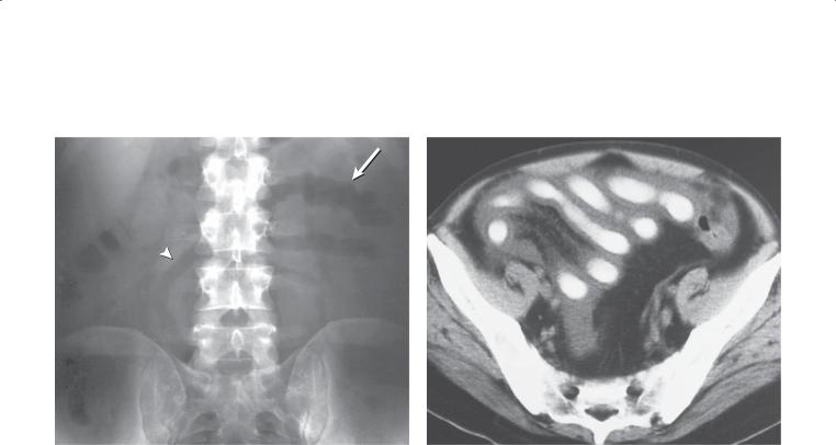
CASE 4.102
Findings
CASE 4.102. Plain abdominal radiograph. Several abnormal small bowel loops are present on this plain film radiograph. The mucosal folds are markedly thickened (arrow) or nearly absent (arrowhead). Separation of small bowel loops suggests bowel-wall thickening.
CASE 4.103. Contrast-enhanced CT. Diffuse wall thickening is seen throughout the small bowel. Normal mucosal folds were not visible. The small bowel has a ribbon-bowel appearance.
Di erential Diagnosis
1.Ischemic small bowel
2.Infectious enteritis
3.Celiac disease
4.Acute radiation enteritis
5.Graft -versus-host disease
6.Amyloidosis
Diagnosis
Graft-versus-host disease
4. SMALL BOWEL 299
CASE 4.103
Discussion
Graft-versus-host disease occurs commonly in patients receiving a bone marrow transplant. New antirejection drugs have reduced this problem such that it develops in only 25% to 35% of patients. The skin, liver, and gastrointestinal tract are affected most often. Severe mucosal inflammation, destruction, and atrophy occur, resulting in a profuse secretory diarrhea. Graft-versus- host disease can affect the stomach, small bowel, and colon; the small bowel usually is affected most severely.
Radiographic changes include fold thickening, effacement, and featureless (atrophic) loops that have been referred to as ribbon bowel. A feature unique to patients who have had bone marrow transplantation is prolonged coating of the affected bowel segments
with barium for several days after the examination. The cause of this finding is unknown. Knowledge that the patient has had bone marrow transplantation makes this diagnosis straightforward.
Disease type: Miscellaneous
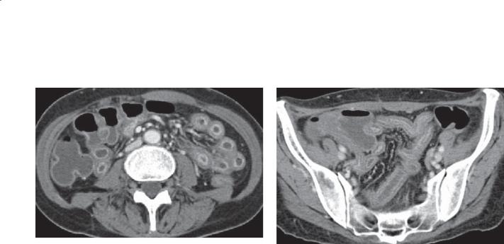
300 MAYO CLINIC GASTROINTESTINAL IMAGING REVIEW
CASE 4.104
A B
Findings
Contrast-enhanced CT. A. Diffuse small bowel wall thickening. The lumen is narrowed, and there is mucosal hyperenhancement. B. Diffuse small bowel thickening, mucosal hyperenhancement, waterattenuation changes within the bowel wall (submucosal edema), and mesenteric vascular engorgement.
Di erential Diagnosis
1.Crohn disease
2.Ulcerative jejunoileitis of celiac disease
3.Graft -versus-host disease
4.Nonsteroidal anti-inflammatory drug–enteropathy
Diagnosis
Graft-versus-host disease
Discussion
Th e findings of diffuse inflammatory changes throughout the small bowel are nonspecific. A more precise diagnosis requires correlation with the patient’s medical history. Crohn disease is the most common, although the findings here are diffuse rather than the expected segmental involvement. Ulcerative disease in sprue is possible but rare. Graft-versus-host disease requires a history of a bone marrow transplant. Nonsteroidal anti-inflammatory drug–enteropahy usually presents with multiple areas of stenoses, most common in the ileum, but diffuse disease has been reported.
Disease type: Miscellaneous
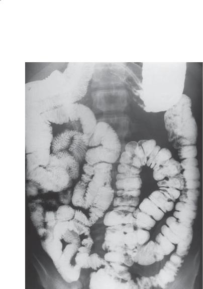
CASE 4.105
Findings
Small bowel follow-through. All of the small bowel occupies the right side of the abdomen. The colon resides on the left side of the abdomen.
Di erential Diagnosis
Intestinal nonrotation
Diagnosis
Intestinal nonrotation
4. SMALL BOWEL 301
Discussion
Normally, the intestines undergo a rotation of 270° during fetal life. Rotation involves two major segments of intestine: the duodenal-jejunal region, which migrates to its normal position early in fetal life, and the cecocolic segments, which enter the abdominal cavity and rotate into their normal positions later. Abnormalities of rotation can interrupt this sequence of events at any stage of development, resulting in a nonrotation (small bowel resides in the right portion of the abdomen, colon on the left), incomplete rotation (normal small bowel rotation but the cecum is located in the left portion of the abdomen, in the epigastrium, or high on the right side), and incomplete mesenteric fixation (mobile cecum). Midgut volvulus or small bowel obstruction from mesenteric bands is an important complication of malrotation (case 4.86). Congenital diaphragmatic hernias and omphaloceles are also sequelae of nonrotation.
Disease type: Miscellaneous

302 MAYO CLINIC GASTROINTESTINAL IMAGING REVIEW
CASE 4.106
Findings
Small bowel follow-through. Multiple large saclike structures project from multiple small bowel loops.
Di erential Diagnosis
Intestinal diverticulosis
Diagnosis
Intestinal diverticulosis
Discussion
Small bowel diverticula are often found incidentally during a small bowel examination. The majority are single or few in number, and most affect the proximal small bowel, especially the duodenum. Tiny diverticula also frequently can be seen arising from the terminal ileum. In our experience, most diverticula are incidental findings and rarely cause clinical symptoms. Occasionally, significant stasis
occurs in one or more diverticula, resulting in bacterial overgrowth, vitamin B12 deficiency, and macrocytic anemia. Malabsorption can be problematic as a result of bile acid deconjugation by the abundant bacteria and subsequent fat malabsorption. Bacterial exotoxins also can damage the intestinal lining and impair absorption. Bleeding, perforation, diverticulitis, enterolith formation, and pneumoperitoneum are other rarely reported complications.
Disease type: Miscellaneous

4. SMALL BOWEL 303
CASE 4.107
B
Findings
Contrast-enhanced CT. A. Axial. Well-defined mass adjacent to a loop of jejunum that contains fluid and air. There is surrounding mesenteric soft tissue
stranding. B. Coronal. The adjacent jejunum appears to have a narrowed lumen and wall thickening.
Di erential Diagnosis
1.Perforated neoplasm
2.Foreign body perforation (toothpick injury)
3.Jejunal diverticulitis
4.Postoperative abscess
Diagnosis
Jejunal diverticulitis
Discussion
Jejunal diverticulitis is a rare complication of jejunal diverticulosis. Most diverticula within the small bowel are asymptomatic. If they do cause problems, bacterial overgrowth is the most common cause. Jejunal diverticulitis is usually seen in elderly patients and can result in perforation, abscess formation (as in this case), bleeding, or obstruction. CT is the examination of choice for detecting this condition. Treatment
is usually by operative resection of the affected bowel segment.
Disease type: Miscellaneous
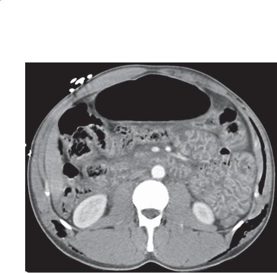
304 MAYO CLINIC GASTROINTESTINAL IMAGING REVIEW
CASE 4.108
Findings
Contrast-enhanced CT. The small bowel is hyperattenuating, and the inferior vena cava is collapsed. (Courtesy of Cookie Menias, MD, Washington University Medical Center, St. Louis, Missouri. Used with permission.)
Di erential Diagnosis
1.Hypoperfusion complex (shock bowel)
2.Angioedema
3.Infl ammatory bowel disease
4.Radiation enteritis
5.Infectious enteritis
Diagnosis
Hypoperfusion complex
Discussion
Hypoperfusion complex (shock bowel) is usually due to blunt trauma and is more common in children
than adults. The findings of intense enhancement of the bowel wall, kidneys, adrenals, and aorta and a small or collapsed inferior vena cava are typical of
this condition. The diagnosis of shock bowel indicates tenuous hemodynamic stability and the urgent need for fluid resuscitation. The condition is associated with a high mortality rate (85%). The abdomen should be assessed for a source of bleeding.
Angioedema usually affects only a few small bowel segments, and collapse of the inferior vena cava would not be expected. Other inflammatory and infectious conditions would be expected to cause thickened small bowel loops with mucosal hyperenhancement. Radiation enteritis could acutely have a similar enhancement pattern of the bowel. Chronically, radiation changes would be expected to have normal or reduced bowel wall enhancement and wall thickening (resembling ischemia).
Disease type: Miscellaneous
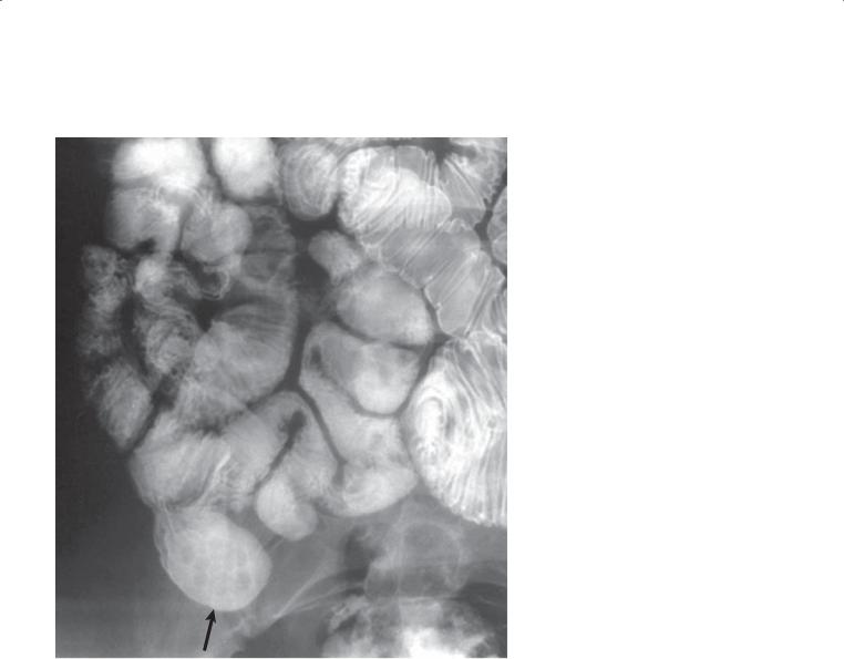
4. SMALL BOWEL 305
CASE 4.109
Findings
Enteroclysis. A diverticulum (arrow) arising from the distal small bowel is present in the right lower portion of the abdomen.
Di erential Diagnosis
1.Meckel diverticulum
2.Postoperative deformity
Diagnosis
Meckel diverticulum
Discussion
Meckel diverticulum is an outpouching of the small bowel representing the remnant of the omphalomesenteric duct (a connection between the yolk sac and the midgut). Meckel diverticula usually
are located within the distal 3 feet of the small bowel, and approximately two-thirds contain heterotopic gastric mucosa. Symptoms can develop at any age, but most patients seek medical attention before
age 3 years. Signs and symptoms usually include melena and abdominal pain. Ulceration, perforation, intussusception, or enterolith formation have all been reported to occur in Meckel diverticulum.
Radiographic evaluation may include an abdominal plain film. Calcified enteroliths and free air or a mechanical small bowel obstruction may be found. Barium examination of the small bowel may reveal the diverticulum; however, this entity is identified less frequently than the expected incidence (3%) for the general population. Some investigators believe an enteroclysis small bowel examination is more sensitive for detecting this entity.
Disease type: Miscellaneous
306 MAYO CLINIC GASTROINTESTINAL IMAGING REVIEW
CASE 4.110
A B
Findings
Contrast-enhanced CT. A. A small bowel structure within the right abdomen has a mildly thickened wall and contains at least 2 calculi. The surrounding mesenteric fat is normal in appearance. B. Bone windows enable optimal visualization of the calculi.
Di erential Diagnosis
1.Meckel diverticulum containing multiple enteroliths
2.Ectopic gallstones
3.Medication within the small bowel lumen
Diagnosis
Meckel diverticulum containing enteroliths
Discussion
Th e connection of this diverticulum to the small bowel lumen is not shown in these images, but it could be traced on contiguous slices. Enteroliths develop
in areas of stasis, most often within a diverticulum. Although enteroliths can reside in ordinary diverticula, the location of these within the right lower quadrant should prompt a consideration of a Meckel diverticulum. Enteroliths can cause obstruction of the diverticulum, which can lead to perforation, inflammation, or infection. Ectopic gallstones are almost never identified unless they are causing obstruction of the small bowel lumen (gallstone ileus).
Medication would not have central low attenuation, but it would be expected to have solid high attenuation.
Disease type: Miscellaneous
CASE 4.111
1 minute |
10 minutes |
Findings
Technetium pertechnetate radionuclide examination. A region of abnormally increased tracer (arrows) is visible in the left lower portion of the abdomen.
Di erential Diagnosis
1.Duplication cyst
2.Ectopic island of gastric mucosa
3.Meckel diverticulum
Diagnosis
Meckel diverticulum
4. SMALL BOWEL 307
30 minutes
Discussion
Th is finding is consistent with a Meckel diverticulum containing ectopic gastric mucosa; however, usually tracer is identified in the right lower quadrant in this condition. Technetium pertechnetate scans are useful for identifying a Meckel diverticulum because the tracer is taken up and secreted by gastric mucosa. Some controversy exists as to which cell type specifically accumulates the tracer. Because many Meckel diverticula contain ectopic gastric mucosa, they can be identified with this examination. Organs that normally take up pertechnetate include stomach, salivary glands, and urinary tract (because a small amount of tracer
is cleared by the kidneys). Uptake also can occur in other organs with gastric mucosa, including Barrett esophagus, duplication cysts, and ectopic islands of gastric mucosa in the small bowel.
Disease type: Miscellaneous
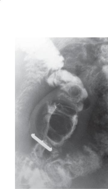
308 MAYO CLINIC GASTROINTESTINAL IMAGING REVIEW
CASE 4.112
Findings
Small bowel follow-through (spot radiograph). An oblong, sausage-shaped filling defect is present within the lumen of the distal small bowel. A compression device with a metallic marker is present.
Di erential Diagnosis
1.Large polyp
2.Gastrointestinal stromal tumor
3.Foreign body
4.Inverted Meckel diverticulum
Diagnosis
Inverted Meckel diverticulum
Discussion
Complications that can occur from Meckel diverticulum include ulceration, bleeding, perforation, obstruction, intussusception, and enterolith formation. Occasional inversion of a diverticulum can present
as an intraluminal filling defect and simulate a polypoid tumor.
Disease type: Miscellaneous
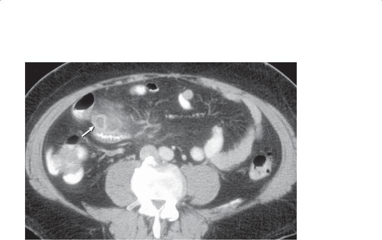
CASE 4.113
Findings
Contrast-enhanced CT. A blind-ending tubular structure arises from the ileum. The wall of this structure is thickened (arrow), and there is
considerable soft tissue stranding in the adjacent fat.
Di erential Diagnosis
1.Meckel diverticulitis
2.Crohn disease
3.Mesenteric panniculitis
Diagnosis
Meckel diverticulitis
4. SMALL BOWEL 309
Discussion
Th e presence of a blind-ending tubular structure arising from the ileum is typical of a Meckel diverticulum. The wall thickening and mesenteric soft tissue stranding indicate acute inflammation of this structure. The ileum is not thickened, thereby excluding Crohn disease. Panniculitis can be excluded because the process is not centered in the mesentery.
Disease type: Miscellaneous
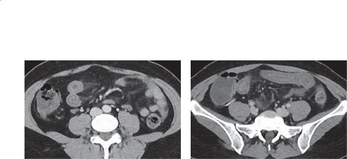
310 MAYO CLINIC GASTROINTESTINAL IMAGING REVIEW
CASE 4.114
A B
Findings
Contrast-enhanced CT. A and B. Bowel wall thickening within a segment of mid to distal small bowel. The attenuation of the bowel wall is near-water, likely indicating edema.
Di erential Diagnosis
1.Ischemia
2.Infl ammatory bowel disease
3.Angiotensin-converting enzyme inhibitor–induced angioedema
4.Vasculitis
Diagnosis
Angiotensin-converting enzyme inhibitor–induced angioedema
Discussion
Angioedema occurring in patients taking angiotensinconverting enzyme inhibitors is well recognized. Patients can present with edema involving the face, tongue, and upper respiratory tract. Involvement of the abdominal viscera, usually the small bowel, has been reported. Patients usually present with acute or recurrent abdominal pain and nausea. The onset of the symptoms can occur days to years after beginning use of the medication. The radiographic findings
are nonspecific (as in this case), with edematous thickening involving a segment of small bowel. Treatment is to stop use of the medication and observe for improvement (usually within 24–48 hours).
Disease type: Miscellaneous
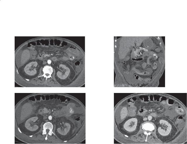
4. SMALL BOWEL 311
CASE 4.115
A C
B D
Findings
A–D. Contrast-enhanced CT images of the abdomen. A–C. Extravasation of intravenous contrast material within the small bowel lumen during the arterial phase of contrast enhancement. D. Enlargement of the extravasation and a change in its configuration during the portal phase of contrast enhancement.
Di erential Diagnosis
1.Acute gastrointestinal bleed
2.Ingested high-attenuation oral medication
3.Small bowel hemangioma with phleboliths in the bowel wall
Diagnosis
Acute gastrointestinal bleed. Nonspecific ulceration was found at operation
Discussion
It can be difficult to identify the site and cause of gastrointestinal bleed in some patients. Generally, when a patient presents to the emergency department and is actively bleeding, it is helpful to commence
CT scanning as soon as feasible. The rationale is that bleeding is often intermittent, and scanning should optimally be performed during the bleeding episode for detection. In our practice, a source of acute bleeding is detected with CT in about 44% of patients.
Th e CT technique for detecting gastrointestinal blood loss often starts with precontrast scanning. This is done to allow differentiation of high-attenuation medication, calcification, or food in the gastrointestinal tract from extravasated vascular contrast material. Usually an arterial and 1 or 2 delayed phases are also acquired to assess for growth of the extravasation (indicating active bleeding) and changed configuration of the contrast puddle (differentiation from solid high-attenuation material vs blood that changes shape). A tagged red blood cell study would also be an appropriate imaging test to select.
Detection of the cause and source of chronic gastrointestinal blood loss is more difficult at CT; the study shows positive results in only 22% of cases. For chronic gastrointestinal blood loss, capsule
endoscopy should probably be considered as a first-line imaging test.
Disease type: Miscellaneous

312 MAYO CLINIC GASTROINTESTINAL IMAGING REVIEW
TABLE 4.6
Miscellaneous Small |
|
|
Bowel Conditions |
|
CASE |
|
|
|
Nonspecific ulcerations |
Potassium chloride, nonsteroidal and anti- |
4.99 and 4.115 |
|
inflammatory drugs. Ulcers can be solitary or |
|
|
multiple. Can heal with bandlike narrowing |
|
|
|
|
Tuberculosis |
Resembles Crohn disease, peritoneal nodules, |
4.100 and 4.101 |
|
masses, adenopathy may be low-attenuation |
|
|
|
|
Graft-versus-host disease |
Ribbon bowel (thickened, featureless loops). Usually |
4.102–4.104 |
|
occurs after bone marrow transplantation |
|
|
|
|
Nonrotation |
Abnormal duodenal-jejunal flexure or colon |
4.105 |
|
location or both. Predisposes to midgut volvulus |
|
|
and cecal ileus |
|
|
|
|
Diverticulosis |
Usually asymptomatic, bacterial overgrowth and |
4.106 and 4.107 |
|
malabsorption occasionally |
|
|
|
|
Shock bowel |
Intense enhancement of bowel, kidneys, adrenals, |
4.108 |
|
aorta; collapsed inferior vena cava |
|
|
|
|
Meckel diverticulum |
Within 3 feet of distal ileum, ectopic gastric mucosa |
4.109–4.113 |
|
bleeds, infection, inversion possible |
|
|
|
|
Angioedema |
Focally thickened bowel wall (water attenuation); |
4.114 |
|
consider angiotensin-converting enzyme inhibitors |
|
|
for cause |
|
|
|
|
Gastrointestinal bleeding |
Intraluminal contrast that changes size and shape |
4.115 |
|
on subsequent images; look for underlying lesion |
|
|
|
|
Disease type: Miscellaneous

4. SMALL BOWEL 313
TABLE 4.7
Diff erential Diagnoses
Solitary Filling Defects
BENIGN TUMORS
Lipoma
Hemangioma
Adenoma
Hamartoma
MALIGNANT TUMORS
Carcinoid
Lymphoma
Adenocarcinoma
Malignant GIST
Metastasis
NONNEOPLASTIC
Duplication cyst
Gallstone ileus
Ascariasis
Type I Folds (Thin, Straight, Dilated Lumen)
Mechanical obstruction
Paralytic ileus
Scleroderma
Celiac disease (sprue)
Type II Folds (Thick, Straight)
SEGMENTAL
Ischemia
Intramural hemorrhage
Radiation enteritis
Adjacent inflammatory process
DIFFUSE
Venous congestion
Hypoproteinemia
Cirrhosis
(Table continued on next page)

314 MAYO CLINIC GASTROINTESTINAL IMAGING REVIEW
Type III Folds (Thick, Nodular)
SEGMENTAL
Crohn disease
Infection
Lymphoma
Metastases
DIFFUSE
Whipple disease
Intestinal lymphangiectasia
Nodular lymphoid hyperplasia
Polyposis syndromes
Eosinophilic gastroenteritis
Amyloidosis
Mastocytosis
Lymphoma
Metastases
Excessive Fluid in Lumen
Proximal to mechanical obstruction
Celiac disease (sprue)
Zollinger-Ellison syndrome
Atrophic (Featureless) Folds
Graft-versus-host disease
Ischemic small bowel (chronic)
Celiac disease (sprue)
Radiation enteritis (chronic)
Amyloidosis
Infectious enteritis
Luminal Narrowing
Adhesions or bands
Crohn disease
Adenocarcinoma
Lymphoma
Metastases
Intramural hemorrhage
Tuberculosis
Ischemia
Radiation enteritis
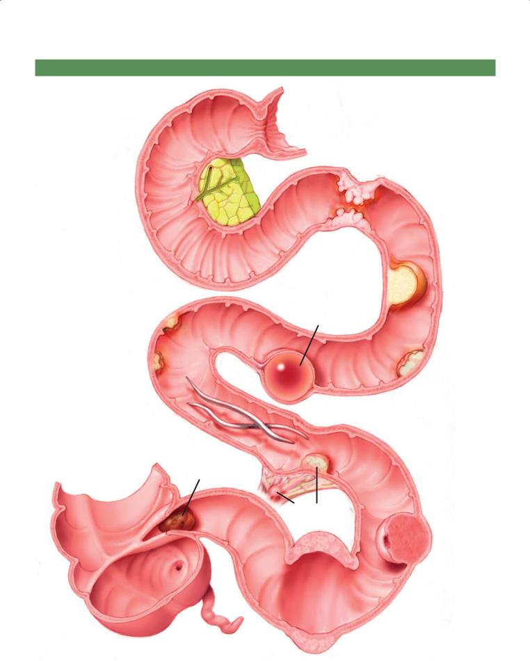
4. SMALL BOWEL 315
SMALL BOWEL FILLING DEFECTS AND MASSES
Annular adenocarcinoma
Lipoma (fat density)
Enteric duplication cyst (fluid density)
Metastases
Adenoma
Ascariasis
Gallstone ileus
Carcinoid tumor (with mesenteric metastasis)
Gastrointestinal
stromal tumor
© MAYO 2013
Lymphoma (aneurysmal dilatation)
Figure 4.116 (Used with permission of Mayo Foundation for Medical Education and Research.)
316 MAYO CLINIC GASTROINTESTINAL IMAGING REVIEW
QUESTIONS
Multiple Choice (choose the best answer)
4.1.Which of the following is the most likely diagnosis for the findings in the figure?
(Courtesy of Cookie Menias, MD, Washington University Medical Center, St. Louis, Missouri. Used with permission.)
a.Closed-loop obstruction
b.Shock bowel
c.Crohn disease
d.Ischemia
e.Whipple disease

|
4. SMALL BOWEL 317 |
4.2. Which of the following is most often associated with 4.3. |
For the case described in Question 4.2, what symptoms |
the condition shown in the figure? |
would be expected? |
|
a. Diarrhea |
|
b. Melena |
|
c. Fever |
|
d. Cold intolerance |
|
e. Arthralgias |
a.Esophagitis
b.Response to antibiotic therapy
c.Small bowel obstruction
d.Gluten intolerance
e.Lymphadenopathy
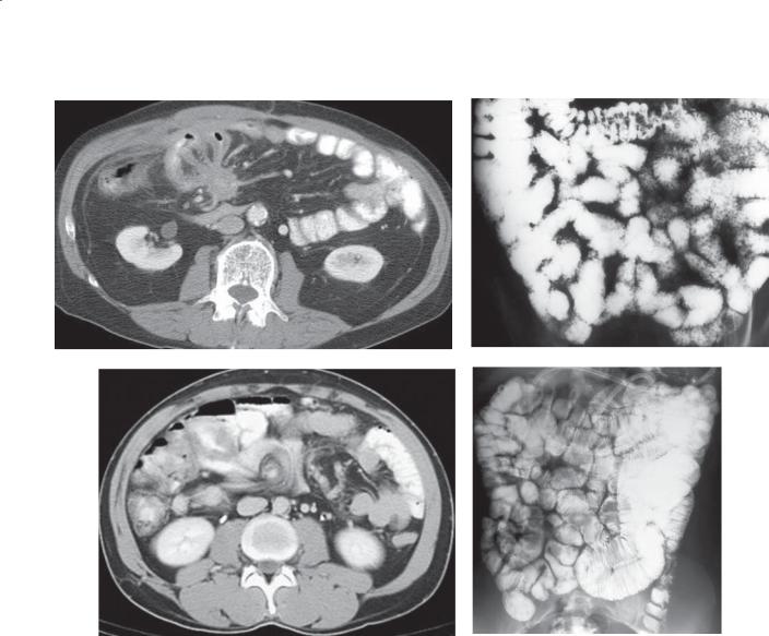
318 MAYO CLINIC GASTROINTESTINAL IMAGING REVIEW
A B
C D
Match images A through G with the responses listed below.
4.4. |
_____ Usually associated with immunodeficiency |
4.6. |
_____ Associated with tricuspid insufficiency |
4.5. |
_____ Source of gastrointestinal bleeding |
4.7. |
_____ Associated with a hypercoagulable state |
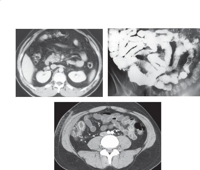
4. SMALL BOWEL 319
E F
G
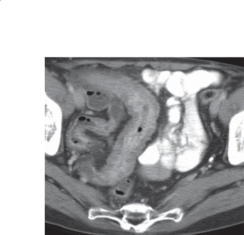
320 MAYO CLINIC GASTROINTESTINAL IMAGING REVIEW
4.8.Which of the following is most likely associated with the disease shown in the figure?
a.Primary sclerosing cholangitis
b.Hepatic abscess
c.Enterocolonic fistula
d.Pulmonary granulomas
e.Neutropenia

4. SMALL BOWEL 321
ANSWERS
4.1.Answer b.
Th e findings include hyperenhancing and thickened small bowel folds. The aorta is also of very high attenuation, even though the phase of enhancement is portal venous (inferior vena cava contains contrast material and the right kidney parenchyma is enhancing). There is a small amount of fluid about the liver. These findings are characteristic of shock bowel. In some patients with severe shock, the inferior vena cava may also be collapsed.
A closed-loop obstruction would present with several loops of dilated bowel and compressed loops at the site of entrapment. There may be evidence of hypoperfusion if vascular impairment has occurred.
In Crohn disease, usual findings are bowel wall thickening and mucosal hyperenhancement in acute disease. Diffuse involvement is possible but atypical because the terminal ileum is most often segmentally affected.
Acute ischemia presents with thickened folds, hypoattenuation of the bowel wall, and possibly hemorrhage within the mesentery. Ischemia can be focal or diffuse depending on the underlying cause.
Whipple disease presents with thickened and nodular folds, usually in the proximal small bowel. The bowel wall enhances normally, and there may be adenopathy within the retroperitoneum or mesentery.
4.2.Answer a.
Th e findings in this case include thin small bowel folds that are abnormally stacked too close together. This hidebound appearance of the small bowel is characteristic of scleroderma. Patients with scleroderma can have aperistalsis of the smooth muscle portion of the esophagus and a patulous gastroesophageal junction. These features lead to esophagitis and often complications from reflux esophagitis.
Scleroderma does not respond to antibiotic therapy. Whipple disease is now known to be caused by a bacterium that responds to antibiotic therapy. Whipple disease would be expected to have thickened or nodular small bowel folds.
Although the small bowel is dilated, a transition zone to suggest a mechanical small bowel obstruction is not apparent on this radiograph.
Gluten intolerance is a finding of celiac disease (sprue). Patients often have a reversal of the normal small bowel pattern (not present here), dilation, and hypersecretion. The fold pattern in sprue is either normal or thickened.
Lymphadenopathy is not usually associated with scleroderma, but it would be a finding in patients with Whipple disease.
4.3.Answer d.
Patients with scleroderma will have typical skin changes and often Raynaud phenomenon leading to cold intolerance of their extremities. The CREST syndrome (calcinosis, Raynaud phenomenon, esophageal dysfunction, sclerodactyly, and telangiectasias) has been reported in patients with scleroderma.
Melena would indicate a source of gastrointestinal bleeding, which is not common in these patients. Fever and arthralgias are not commonly associated with scleroderma.
4.4.Answer b.
Figure b shows lymphoid nodular hyperplasia. Diffuse tiny nodules are present throughout the small bowel. The nodules are too small and uniform for lymphoma, Whipple disease, or Crohn disease and too numerous for lymphangiectasia. This condition is usually associated with an immunoglobulin deficiency.
4.5.Answer d.
Figure d shows Meckel diverticulum. The lesions can be found incidentally but are known to harbor ectopic gastric mucosa. The acid secreted can lead to a mucosal ulceration and gastrointestinal bleeding. Small bowel follow-through examinations may not always detect these if the neck of the diverticulum is small. Technetium-99m pertechnetate scanning can be helpful to detect the aberrant gastric mucosa. Answer f could also be correct, as ulceration secondary to lymphoma can also lead to gastrointestinal bleeding.
4.6.Answer a.
Figure a shows carcinoid tumor. These tumors usually arise in the distal ileum and metastasize to mesenteric lymph nodes. The serotonin secreted by these tumors causes a desmoplastic response in the regional mesentery that causes thickening and retraction of the mesenteric leaves (resulting in the starburst appearance). In patients with metastatic carcinoid to the liver, tricuspid insufficiency can develop because of the fibrotic response of the tricuspid valve to serotonin that is directly excreted into the hepatic veins.
4.7.Answer e.
Figure e shows mesenteric vein thrombosis. The superior mesenteric vein is enlarged and contains a central thrombus. This condition can lead to bowel

322 MAYO CLINIC GASTROINTESTINAL IMAGING REVIEW
ischemia and is often associated with a hypercoagulable clotting disorder.
Figure c shows a midgut volvulus. Notice the twisting or whorled appearance of the mesenteric vessels.
Figure f shows a lymphoma within the ileum. Note that the lumen at the level of the neoplasm is enlarged greater than the lumen of the adjacent small bowel (aneurysmal dilatation).
Figure g shows Crohn disease with edematous thickening of a distal small bowel loop with adjacent soft tissue stranding and fluid. Note the mucosal hyperenhancement characteristic of an active ileitis.
4.8.Answer c.
Th e figure shows marked ileal wall thickening, typical of Crohn disease. The 3 main complications in Crohn disease are fistula formation, abscess, and obstruction. Primary sclerosing cholangitis is associated with ulcerative colitis. Hepatic abscess (pyogenic) is most
commonly associated with diverticulitis, appendicitis, and cholecystitis. Pulmonary granulomas could be associated with tuberculosis; however, this disease is much less common than Crohn disease in North America. Neutropenia is associated with neutropenic colitis; however, small bowel findings are usually absent.