


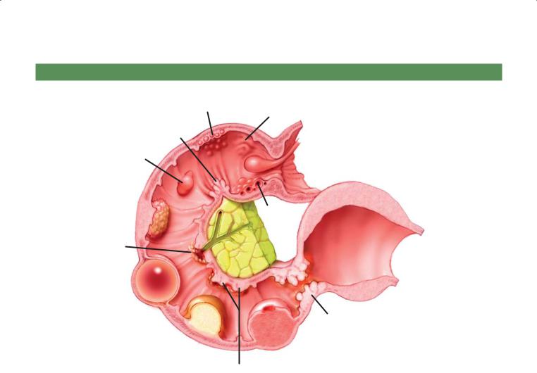
C H A P T E R 3
DUODENUM
Cases 3.1–3.18 Inflammatory and Ulcerative Diseases
Cases 3.19–3.42 Masses and Filling Defects
Malignant Tumors
Benign Tumors
Nonneoplastic
Cases 3.43–3.52 Miscellaneous

This page intentionally left blank
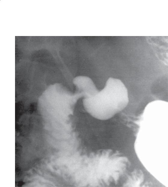
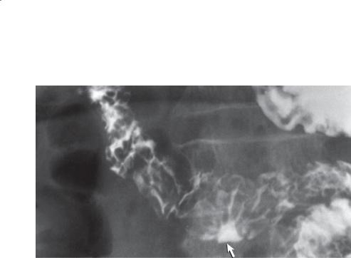
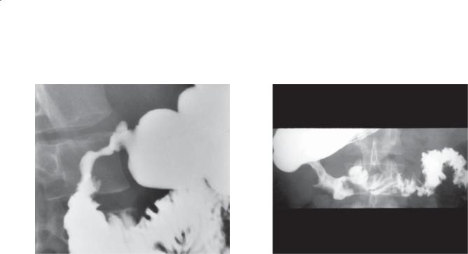
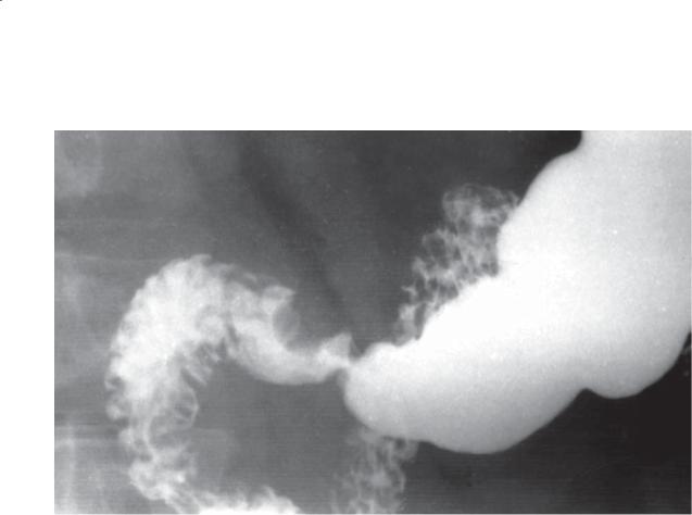
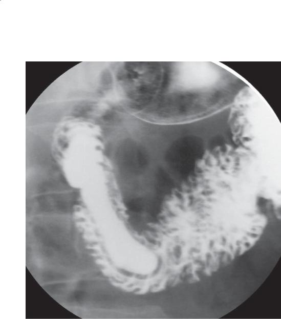
CASE 3.1
Findings
Single-contrast UGI. Multiple thick and nodular folds are present in the first and second portions of the duodenum. A diverticulum (arrows) is seen lateral to the descending duodenum.
Di erential Diagnosis
1.Duodenitis
2.Brunner gland hyperplasia
3.Crohn disease
Diagnosis
Duodenitis
3. DUODENUM 159
Discussion
Th e most frequent cause of thickened duodenal folds is peptic ulcer disease due to Helicobacter pylori infection. The folds may be enlarged as a result of edema, or Brunner gland hyperplasia (case 3.38) may simulate fold thickening. Usually, multiple enlarged, discrete nodules that form a cobblestone appearance are present in patients with Brunner gland hyperplasia. Treatment is with antibiotics and acid blockers. Patients with Crohn disease (cases 3.15, 3.16, and 3.17), giardiasis (case 4.50), sprue (case 3.18), Whipple disease (case 4.51), and lymphoma (case 4.57) also can present with thickened duodenal folds.
Disease type: Inflammatory and Ulcerative Diseases
160 MAYO CLINIC GASTROINTESTINAL IMAGING REVIEW
CASE 3.2
Findings
Single-contrast UGI. A collection of barium is present within the pyloric canal. A metallic compression device is present adjacent to the pylorus.
Di erential Diagnosis
1.Pyloric channel ulcer
2.Pyloric torus defect
Diagnosis
Pyloric channel ulcer
Discussion
Ulcers can occur within the pyloric canal. In some cases, they are difficult to visualize because of secondary gastric outlet obstruction. Ulcers can be differentiated from the pyloric torus defect by the unchanging shape of an ulcer. The mid-channel widening of a pyloric torus is often diamond or triangular in shape and changes as the pyloric muscle contracts and relaxes.
Disease type: Inflammatory and Ulcerative Diseases
3. DUODENUM 161
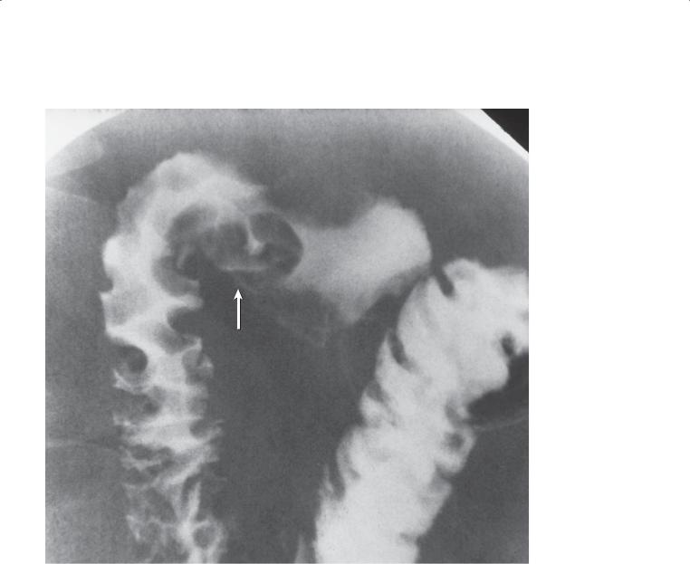
CASE 3.3
Findings
Single-contrast UGI. The duodenum is deformed along its inferior margin. A small ulcer niche (arrow) with a surrounding mound of edema is present.
Di erential Diagnosis
1.Duodenal ulcer
2.Duodenal scarring
Diagnosis
Duodenal ulcer
Discussion
Duodenal ulcers are common problems and are encountered 2 to 3 times more often than gastric
ulcers. Correlation between clinical symptoms and radiographic findings is often imprecise. Some patients with relatively large duodenal ulcers remain asymptomatic.
Duodenal ulcers are associated with increased secretion of acid and with a high incidence of Helicobacter pylori infection. Patients without H pylori infection often are using nonsteroidal anti-inflammatory drugs. Other contributing causes include stress, smoking, alcohol, caffeine, and corticosteroid medication.
Th e key radiologic finding is a persistent collection of barium in the duodenal bulb that does not change shape with compression or peristalsis. Ulcers can vary in shape from pinpoint erosions to linear niches to the commonest round crater.
Disease type: Inflammatory and Ulcerative Diseases
162 MAYO CLINIC GASTROINTESTINAL IMAGING REVIEW
CASE 3.4
Findings
Double-contrast UGI. A persistent, round ulcer crater is present in the duodenal bulb. Folds radiate up to the crater. This is the classic appearance of a duodenal ulcer.
Di erential Diagnosis
Duodenal ulcer
Diagnosis
Duodenal ulcer
Discussion
Th is radiograph was obtained with the patient in the supine position. Because barium collected within the crater, its location is within the posterior duodenal wall. An anteriorly located ulcer crater could appear as a ring shadow in this position, because barium would empty from the crater, leaving the ulcer edges bariumcoated. Anteriorly located ulcers can be difficult
to detect with only the double-contrast technique.
Compression of the duodenal bulb is an important part of every UGI. With compression, most anterior wall ulcers can be identified.
Duodenal ulcers are identified less frequently than in the past because of improved medical therapy. Duodenal ulcers alone are rarely malignant. These ulcers tend to follow the same morphologic patterns as benign gastric ulcers. Most are located along the anterior surface of the duodenum within the bulb. Acute ulcers without fibrosis can heal without scarring. Chronic ulcers with submucosal fibrosis usually
result in residual duodenal deformity. Complications of duodenal ulcers are similar to those of benign gastric ulcers: bleeding, perforation, obstruction, and penetration. Perforation is more common among duodenal ulcers (case 3.6). The ulcer can penetrate into adjacent structures, such as pancreas, omentum, biliary tract, colon, liver, and mesocolon. Multiple duodenal ulcers can be identified in 10% to 15% of patients. The presence of multiple postbulbar ulcerations should suggest Zollinger-Ellison syndrome (case 3.11).
Disease type: Inflammatory and Ulcerative Diseases
CASE 3.5
Findings
Single-contrast UGI. A large ulcer crater resides at the junction of the first and second portions of the duodenum. A symmetric mound of edema causes a
filling defect around the ulcer base. An ulcer this size is sometimes referred to as a giant ulcer crater.
Di erential Diagnosis
1.Duodenal deformity (peptic disease)
2.Duodenal ulcer
Diagnosis
Giant duodenal ulcer
3. DUODENUM 163
Discussion
Giant ulcers have been described as being larger than 2 to 2.5 cm in diameter or as replacing most of the duodenal bulb. These ulcers may be so large as to mimic a normal duodenal configuration. Recognition depends on observing the fixed shape and size of
the ulcer, which contains no normal duodenal folds. Narrowing of the duodenum proximal and distal to the ulcer is common. The complication and mortality rates are higher with this type than with usual duodenal ulcers.
Disease type: Inflammatory and Ulcerative Diseases
164 MAYO CLINIC GASTROINTESTINAL IMAGING REVIEW
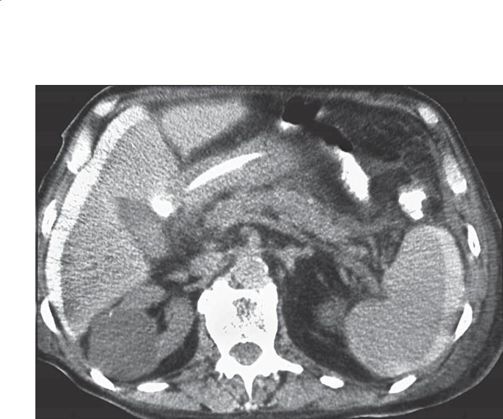
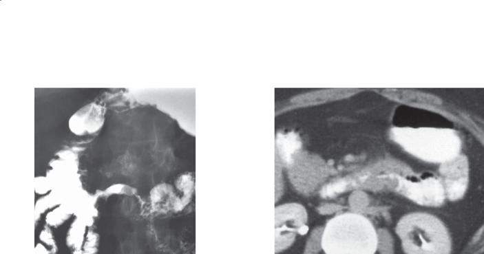
CASE 3.6
Findings
Unenhanced abdominal CT. High-density contrast material is seen in an extraluminal location (lateral to the duodenum). High-density ascites is visible about the liver and spleen. Benign cyst is present in the right kidney.
Di erential Diagnosis
Perforated duodenal bulb ulcer with extravasation of oral contrast material into the peritoneal space
Diagnosis
Perforated duodenal bulb ulcer with extravasation of oral contrast material into the peritoneal space
Discussion
Th e commonest cause of a perforated viscus in a patient who has not undergone trauma or a surgical procedure is a duodenal ulcer. Most duodenal ulcers perforate anteriorly with leakage of air (pneumoperitoneum) or contrast material (as in this case) or both. The presence of high-density contrast material adjacent to the duodenum increases the probability of a perforated duodenal ulcer.
Disease type: Inflammatory and Ulcerative Diseases
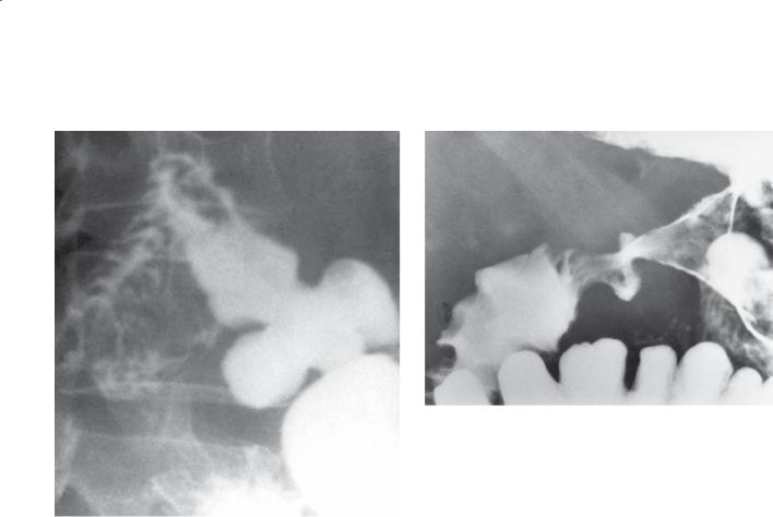
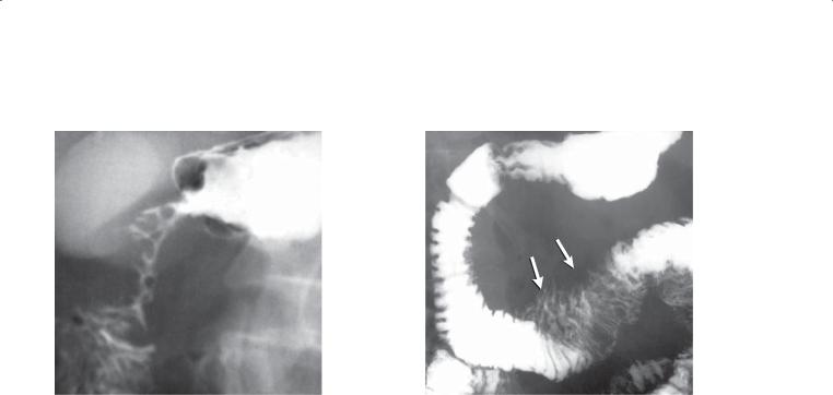
CASE 3.7
Findings
CASE 3.7. Single-contrast UGI. The duodenal bulb is deformed and resembles a cloverleaf. This is the classic appearance of duodenal bulb scarring from a peptic ulcer.
CASE 3.8. Double-contrast UGI. Two collections of barium protrude from the pyloric channel. Deformity of the distal gastric antrum and duodenal bulb is present.
Di erential Diagnosis
Duodenal bulb scarring from prior ulcer disease
Diagnosis
Duodenal bulb scarring from prior ulcer disease
3. DUODENUM 165
CASE 3.8
Discussion
Scarring of the duodenal bulb from previous peptic ulcer disease is a common finding. A centrally located duodenal bulb ulcer characteristically produces this cloverleaf deformity (as seen in case 3.7). The challenge to the radiologist is to exclude an active ulcer in
the scarred bulb. Sometimes this is impossible and endoscopy should be recommended to examine the mucosa directly. Active ulcers are fixed abnormalities within the duodenum that do not change shape with peristalsis. Scarred duodenal bulbs change shape as the duodenum fills and empties. Compression should be routinely performed to exclude concomitant ulcers within the deformed bulb.
Disease type: Inflammatory and Ulcerative Diseases
166 MAYO CLINIC GASTROINTESTINAL IMAGING REVIEW
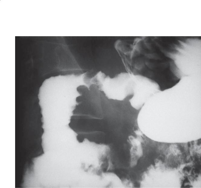
CASE 3.9
Findings
Single-contrast UGI. Two pyloric channels are present on this radiograph taken with the patient in the prone position. One channel (arrow) connects the greater curvature of the gastric antrum with the apex of the duodenal bulb.
Di erential Diagnosis
1.Congenital double pylorus
2.Fistulous channel from peptic ulcer disease
Diagnosis
Fistulous channel from peptic ulcer disease
Discussion
Th e majority of cases of multichannel pylorus are due to a persistent fistula from peptic ulcer disease or Crohn disease. Rarely, this finding may represent a congenital abnormality. No treatment may be necessary if no active ulcer is present.
Disease type: Inflammatory and Ulcerative Diseases
3. DUODENUM 167
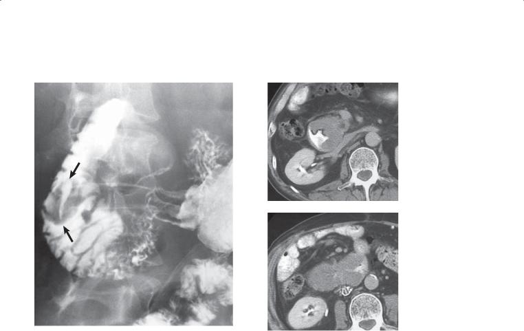
CASE 3.10
Findings
Single-contrast UGI. A round ulcer crater lies along the medial aspect of the second portion of the duodenum in association with duodenal narrowing, concentric edema about the crater, and fold thickening.
Di erential Diagnosis
Postbulbar duodenal ulcer
Diagnosis
Postbulbar duodenal ulcer
Discussion
Duodenal ulcers are most common in the duodenal bulb. Postbulbar ulcers are unusual; when they develop they are usually proximal to the ampulla, often
along the medial wall of the descending duodenum. About 1 in 20 peptic ulcers is located beyond the bulb. Ulcerations should be considered malignant, or evidence for a gastrin-secreting tumor (ZollingerEllison syndrome) should be sought if an ulcer is identified distal to the ampulla of Vater (case 3.11). Healing of these ulcers can result in a ring stricture of the duodenum. This can be difficult to diagnose, often necessitating distention of the duodenum with barium or effervescent gas. Associated ulcer niches also can be easily overlooked and should be suspected if a focal area of narrowing is found in the postbulbar duodenum. CT may be useful to assess for complications of peptic ulcer disease: abscess,
fistula, and pancreatitis. CT has no primary role in the diagnosis of peptic ulcers.
Disease type: Inflammatory and Ulcerative Diseases
168 MAYO CLINIC GASTROINTESTINAL IMAGING REVIEW
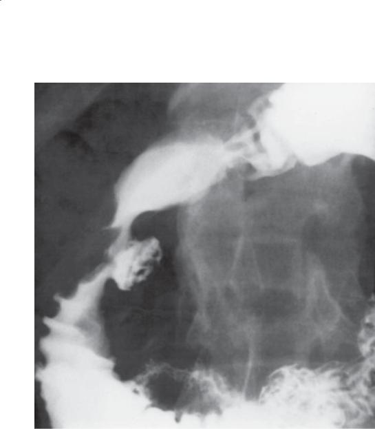
CASE 3.11
Findings
Single-contrast UGI. An ulceration (arrow) is present within the distal duodenum. The ulcer has benign features radiographically, with no associated mass and folds that radiate to the edge of the crater.
Di erential Diagnosis
1.Postbulbar ulcer due to Zollinger-Ellison syndrome
2.Metastases
3.Primary adenocarcinoma
Diagnosis
Postbulbar ulcer due to Zollinger-Ellison syndrome
Discussion
Th is patient was found to have Zollinger-Ellison syndrome. Peptic ulcers in patients with ZollingerEllison syndrome most commonly occur in the pylorus and proximal duodenal region. Ulcers occurring
distal to the ampulla are usually malignant, except in patients with Zollinger-Ellison syndrome. Normally, acidic gastric secretions are neutralized by the alkaline pancreatic secretions in the postampullary duodenum. Multiple ulcers occur in about 10% of patients
with Zollinger-Ellison syndrome. The stomach and duodenum may appear hypotonic with dilatation of the duodenum (“megaduodenum”). Thickened duodenal folds and Brunner gland hyperplasia (case 3.38) also may be present.
Disease type: Inflammatory and Ulcerative Diseases
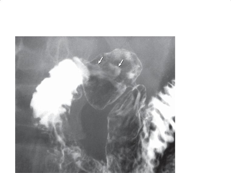
CASE 3.12
Findings
CASE 3.12. Single-contrast UGI. The proximal duodenum is markedly narrowed. The mucosa through this region is intact, and the narrowing ends abruptly.
CASE 3.13. Single-contrast UGI. The duodenal C loop is mildly widened. Tethered folds (arrows) are present along the cranial aspect of the transverse duodenum.
Di erential Diagnosis
1.Duodenitis
2.Cholecystitis
3.Pancreatitis
4.Annular pancreas
5.Pancreatic carcinoma
Diagnosis
Pancreatitis
3. DUODENUM 169
CASE 3.13
Discussion
Occasionally, patients with pancreatitis have a region of focal stenosis in the proximal duodenum. This narrowing is believed to be due to pancreatitis-induced edema and spasm. A persistent stricture can develop. The stenosis may mimic a carcinoma because of
the abrupt edges and focal nature of the narrowing. Identifying intact mucosa through the stenosis is a key finding in distinguishing it from a tumor. A history of pancreatitis is also helpful information. Inflammation from cholecystitis or duodenitis could have a presentation similar to that in case 3.12. Annular pancreas should not have associated fold thickening unless superimposed pancreatitis is present.
Tethered folds of the bowel (as seen in case 3.13) usually develop from either an extraluminal inflammatory or neoplastic process. Normally the folds of the bowel are oriented perpendicular to the lumen and meet the edge of the bowel wall with a square, flat appearance. Tethered folds usually appear to be pulled from the normal bowel wall toward a central area extrinsic to the bowel. The folds are usually pointed and elongated.
Disease type: Inflammatory and Ulcerative Diseases
170 MAYO CLINIC GASTROINTESTINAL IMAGING REVIEW
CASE 3.14
Findings
Double-contrast UGI. A mottled collection of gas is present within the pancreatic bed (the region between the stomach and the duodenum). There is a region
of focal narrowing in the proximal duodenum. A drainage catheter is seen in the left upper quadrant.
Di erential Diagnosis
Acute pancreatitis with abscess
Diagnosis
Acute pancreatitis with abscess
Discussion
Th e location and appearance of the extraluminal gas are pathognomonic of a pancreatic abscess.
Pancreatic abscess is a dreaded complication of severe acute pancreatitis. High morbidity and mortality
are associated with this disease. Pancreatic necrosis is present in many patients with abscess; however, infection of a peripancreatic fluid collection(s) is possible without pancreatic parenchymal necrosis. If a pancreatic abscess is suspected, CT is the
best initial examination. This condition often is diagnosed from plain radiographs in patients with nonspecific abdominal complaints or incidentally when radiographs are obtained for documenting tube position. The key radiographic finding of a pancreatic abscess is a variable amount of extraluminal gas within the pancreatic bed. The gas collection may appear either mottled or homogeneous.
Disease type: Inflammatory and Ulcerative Diseases
CASE 3.15
Findings
Double-contrast UGI. Multiple flat ulcerations (arrows) are present within the duodenal bulb and descending duodenum.
Di erential Diagnosis
1.Peptic ulcer disease
2.Crohn disease
3.Viral (herpes and cytomegalovirus) ulceration
4.Chemical-induced ulcers (medication, alcohol)
5.Zollinger-Ellison syndrome
Diagnosis
Crohn disease
3. DUODENUM 171
Discussion
Crohn disease affecting the upper gastrointestinal tract is most common within the proximal duodenum. Aphthous ulcers are probably the earliest radiologic lesion detectable. The ulcers in Crohn disease can vary in appearance from pinpoint ulcers with surrounding halos of edema (aphthous ulcers) to small shallow ulcerations (as seen in this case).
Duodenal erosions are nonspecific lesions. They can be caused by various medications (aspirin and nonsteroidal anti-inflammatory agents), drugs (alcohol), and infectious agents (herpesvirus and cytomegalovirus). The erosions found in patients with Crohn disease are often identical to those due to other conditions. The clinical setting is an important consideration. Usually patients with duodenal Crohn disease also have radiologic findings in the more distal small bowel.
Disease type: Inflammatory and Ulcerative Diseases
172 MAYO CLINIC GASTROINTESTINAL IMAGING REVIEW
CASE 3.16 |
CASE 3.17 |
Findings
CASE 3.16. Single-contrast UGI. Marked deformity of the pylorus and duodenum is present with asymmetric bowel wall involvement and sacculations.
CASE 3.17. Single-contrast UGI. Duodenal narrowing, luminal irregularity, and sacculations (due to asymmetric bowel wall involvement) are visible throughout most of the transverse duodenum.
Di erential Diagnosis
1.Scarring from prior peptic ulcer disease
2.Crohn disease
3.Strictures from prior pancreatitis
Diagnosis
Crohn disease
Discussion
Chronic changes of Crohn disease involving the duodenum mimic the findings of Crohn disease found elsewhere in the small bowel. These changes vary from slight luminal narrowing and mucosal thickening to marked narrowing and irregularity causing gastric obstruction. Similar changes can affect the gastric antrum. A differential consideration in this case is scarring from previous peptic ulcer disease. Conceivably, prior severe pancreatitis can leave residual duodenal strictures. If the diagnosis of Crohn disease is not definite, it is often worthwhile extending the examination to include the remainder of the small bowel, because often another characteristic lesion(s) is found (cases 4.66 through 4.72).
Disease type: Inflammatory and Ulcerative Diseases
3. DUODENUM 173
CASE 3.18
Findings
Single-contrast UGI. The duodenum is mildly dilated, and thickened folds are present in its first and second portions.
Di erential Diagnosis
1.Duodenitis
2.Adult celiac disease
Diagnosis
Adult celiac disease
Discussion
Changes of sprue are usually most marked in the jejunum and ileum (cases 4.92, 4.93, and 4.96), but
changes in the duodenal mucosa also can be identified both radiographically and endoscopically in a majority of patients. Both villous atrophy and inflammatory changes are usually found histologically. Celiac disease probably impairs the protective effects of the duodenal mucosa against peptic damage.
Th e radiographic findings of sprue in the duodenum include thickened and often nodular duodenal folds. Duodenal erosions and luminal dilatation may be seen. Brunner gland hyperplasia is often present. Small (1-4 mm), angular filling defects have been reported, resulting in a mosaic or “bubbly” appearance of the duodenal bulb. Patients with long-standing disease may have an atrophic, featureless duodenal mucosal pattern.
Disease type: Inflammatory and Ulcerative Diseases

174 MAYO CLINIC GASTROINTESTINAL IMAGING REVIEW
TABLE 3.1
Inflammatory and |
|
|
Ulcerative Diseases of |
|
|
the Duodenum |
|
CASE |
|
|
|
Peptic disease |
Most frequent cause of thickened duodenal folds. |
3.1–3.10 |
|
Often results in duodenal bulb ulcers |
|
|
|
|
Zollinger-Ellison syndrome |
Multiple and postbulbar ulcers. Thick gastric folds. |
3.11 |
|
Hypersecretion |
|
|
|
|
Pancreatitis |
Narrowing and tethered folds along duodenal C loop |
3.12–3.14 |
|
|
|
Crohn disease |
Usually erosions in proximal duodenum. Most |
3.15–3.17 |
|
patients also have ileal disease |
|
|
|
|
Adult celiac disease |
Thickened nodular duodenal folds. Classic findings |
3.18 |
|
are usually seen in jejunum and ileum. Chronic |
|
|
changes include featureless mucosal pattern |
|
|
|
|
Disease type: Inflammatory and Ulcerative Diseases
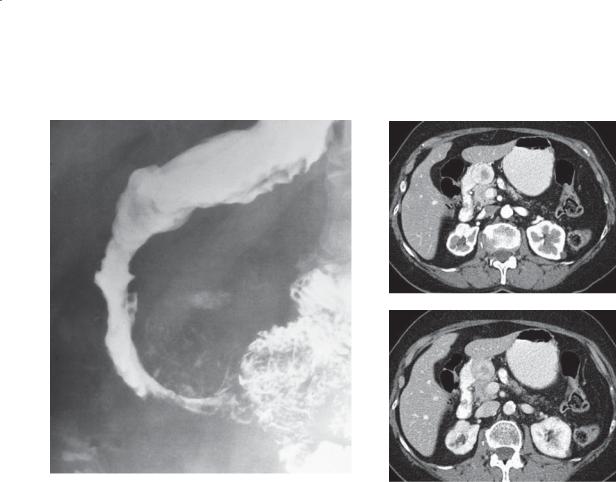
CASE 3.19
Findings
CASE 3.19. Single-contrast UGI. Focal, eccentric narrowing is present within the third portion of the duodenum. The intraluminal characteristics of the lesion are featureless and devoid of any normal mucosal markings (ulcerated surface).
CASE 3.20. Contrast-enhanced abdominal CT. Short, segmental bowel-wall thickening is present within the fourth portion of the duodenum. (Adapted from Dudiak KM, Johnson CD, Stephens DH. Primary tumors of the small intestine: CT evaluation. AJR Am J Roentgenol. 1989 May;152(5):995-8. Used with permission.)
Di erential Diagnosis
1.Adenocarcinoma
2.Lymphoma
Diagnosis
Duodenal adenocarcinoma
3. DUODENUM 175
CASE 3.20
Discussion
Adenocarcinoma was removed surgically. Adenocarcinoma is the primary malignant tumor that most frequently affects the duodenum. It is most commonly distal to the ampulla. Short segmental regions of narrowing (apple core) with abrupt edges or polypoid masses are most common (case 3.19). Lymphoma in the duodenum (cases 3.23 and 3.24) is unusual but can present as a bulky solitary mass.
CT commonly is used to evaluate patients for nonspecific complaints and possible obstruction. Small bowel masses can be detected and staged at CT. Adenocarcinomas characteristically cause short, segmental wall thickening of the duodenum. Luminal narrowing is often present.
Disease type: Masses and Filling Defects
176 MAYO CLINIC GASTROINTESTINAL IMAGING REVIEW
CASE 3.21 |
CASE 3.22 |
Findings
CASE 3.21. Double-contrast UGI. A lobulated filling defect occupies the medial wall of the second portion of the duodenum.
CASE 3.22. Contrast-enhanced CT. A 2.7-cm, solid, enhancing mass is within the duodenum at the level of the ampulla.
Di erential Diagnosis
1.Ampullary neoplasm
2.Edematous papilla (recent stone passage or impacted stone)
3.Duodenal polyp
4.Metastasis
Diagnosis
Ampullary adenocarcinoma (case 3.21) Tubulovillous adenoma with high-grade dysplasia (case 3.22)
Discussion
Carcinomas involving the ampulla of Vater are nearly always adenocarcinomas arising from the duodenum, distal common bile duct, or distal pancreatic duct.
They may present as a polypoid mass or an annular constricting lesion. Biliary tree obstruction is common. Malignant lesions usually have an irregular surface contour. Duodenal (periampullary) adenomas and adenocarcinomas may present as a manifestation of familial adenomatous polyposis syndrome (cases 2.18 and 3.33).
Differential considerations include edematous papilla, usually due to an impacted distal common bile stone or a large duodenal polyp. Edematous changes nearly always present with the enlarged papilla having a smooth surface.
ERCP is usually indicated if an ampullary lesion is suspected. The tumor is often directly visible, and a biopsy specimen can be obtained. Surgical treatment is usually pancreatoduodenectomy (Whipple procedure).
Disease type: Masses and Filling Defects
3. DUODENUM 177
CASE 3.23 |
CASE 3.24 |
|
A |
B
Findings
CASE 3.23. Single-contrast UGI. A well-defined mass (arrows) with smooth borders is present within the second portion of the duodenum. The mass contains a linear ulcer crater.
CASE 3.24. Contrast-enhanced CT. A and B. A long segment of circumferential bowel-wall thickening involves the second portion of the duodenum. A large ulceration is present within the lesion (best seen in A).
Di erential Diagnosis
1.Metastases
2.Lymphoma
3.Adenocarcinoma
4.Gastrointestinal stromal tumor
Diagnosis
Lymphoma
Discussion
Any of the primary duodenal tumors and metastases to the duodenum could have the appearance of case 3.23 on the UGI, including adenocarcinoma (cases 3.19 and 3.20), gastrointestinal stromal tumors (case 3.25), and lymphoma. Lymphoma was removed surgically. Notice the well-defined minor and major papillae located medial to the tumor in case 3.23.
Lymphoma involving the duodenum is unusual. It may present as a solitary mass, as one of many lesions within the alimentary tract, or as an ulcerated mass. Occasionally, the ulcer crater is larger than the associated unaffected bowel lumen (aneurysmal ulceration). Characteristic findings of lymphoma on CT include a long segment of marked bowel-wall thickening (as seen in case 3.24). Regional or diffuse adenopathy may be associated with the mass.
Disease type: Masses and Filling Defects
178 MAYO CLINIC GASTROINTESTINAL IMAGING REVIEW
CASE 3.25 |
CASE 3.26 |
|
A |
B
Findings
CASE 3.25. Single-contrast UGI. An extramucosal mass narrows and deforms the duodenum at the junction of its second and third portions. The smooth surface of the lesion indicates its extramucosal location. Calcification is present within the mass.
CASE 3.26. Contrast-enhanced CT. A. Arterial phase. B. Portal venous phase. A 2.6-cm well-circumscribed mass is adjacent to the medial wall of the descending duodenum. The mass enhances briskly during the arterial phase of contrast enhancement, with visible washout during the portal venous phase on contrast enhancement.
Di erential Diagnosis
1.Acute and chronic pancreatitis
2.Gastrointestinal stromal tumor
3.Crohn disease
Diagnosis
Malignant gastrointestinal stromal tumor
Discussion
Malignant gastrointestinal stromal tumors are often impossible to differentiate radiologically from those that are benign. They are intramural tumors with
a smooth surface. Tumoral calcification (as seen in case 3.25) is usually specific for smooth muscle tumors. Large lesions and ulcerative lesions are more likely malignant. Hyperenhancement during the arterial phase of contrast enhancement is characteristic at CT. Bulky lesions with areas of central necrosis are typically found on CT. Often the tumor appears larger at CT than anticipated from the barium study. A history
and symptoms of pancreatitis are helpful to consider this possibility. Although Crohn disease can alter the duodenal contour, a focal mass is rarely present in this location.
Disease type: Masses and Filling Defects
3. DUODENUM 179
CASE 3.27
A B
C
Findings |
Discussion |
|
Contrast-enhanced CT. A through C. Several small |
Gastrinomas are classified as a type of islet cell |
|
enhancing nodules are present within the proximal |
neoplasm. The majority of gastrinomas are malignant |
|
duodenum. |
and can lead to Zollinger-Ellison syndrome as a result |
|
|
|
of the overproduction of gastrin; secondary effects are |
Di erential Diagnosis |
hyperacidity with gastroduodenal inflammation and |
|
1. |
Multiple gastrinomas |
ulceration. Gastrinomas are often extrapancreatic in |
2. |
Enhancing duodenal polyps |
location, often confined to the the proximal duodenum |
3. |
Metastases |
(gastrinoma triangle) and surrounding tissues (lymph |
|
|
nodes and bile ducts). Arterial-phase imaging is |
Diagnosis |
important for detection of these hyperenhancing, |
|
Multiple duodenal gastrinomas |
1- to 20-mm tumor nodules. |
|
Disease type: Masses and Filling Defects
180 MAYO CLINIC GASTROINTESTINAL IMAGING REVIEW
CASE 3.28 |
CASE 3.29 |
Findings
CASE 3.28. Single-contrast UGI. A large polypoid mass fills most of the duodenal bulb. A diverticulum is present along the medial aspect of the second portion of the duodenum.
CASE 3.29. Single-contrast UGI. An ulcerated, annular constricting mass is present within the second portion of the duodenum.
Di erential Diagnosis
1.Duodenal adenocarcinoma
2.Metastases
3.Lymphoma
Diagnosis
Duodenal metastases
Discussion
CASE 3.28. The mass was metastatic renal adenocarcinoma. Renal adenocarcinomas can directly invade or metastasize hematogenously to the alimentary tract. Either route of spread can lead to bulky polypoid filling defects, as seen in this case.
CASE 3.29. This mass was a metastatic large cell carcinoma from a primary lesion in the lung. The tumor is radiographically indistinguishable from a primary duodenal adenocarcinoma (cases 3.19 and 3.20). Statistically, a metastasis to the duodenum is more likely than is a primary adenocarcinoma.
Disease type: Masses and Filling Defects

3. DUODENUM 181
CASE 3.30
Findings
Single-contrast UGI. The second portion of the duodenum is narrowed and distorted, with ulceration along its medial border. (Adapted from Stephens DH. Pancreatic adenocarcinoma: radiologic diagnosis and staging. In: Freeny PC, Sevenson GW, editors. Margulis and Burhenne’s alimentary tract radiology.
Vol 2. 5th ed. St. Louis [MO]: Mosby-Year Book; c1994. p. 1107-26. Used with permission.)
Di erential Diagnosis
Pancreatic adenocarcinoma
Diagnosis
Pancreatic adenocarcinoma
Discussion
Th is is a typical example of the reverse 3, or epsilon, sign, described by Frostberg. Barium examination is no longer the method of choice for examination of the pancreas. Direct pancreatic imaging with CT, MRI, or sonography is preferable. Many intraluminal contrast examinations in patients with pancreatic cancer are negative. Radiographic findings vary depending on the size of the tumor or its location within the pancreas. Tumors in the pancreatic head can compress, invade, and ulcerate the medial border of the duodenal sweep and inferior aspect of the gastric antrum. Masses in the pancreatic body and tail can cause mass effect
and tethering of the gastric body and fundus. Tumor also can extend along the normal tissue planes and ligaments of the transverse colon and duodenojejunal flexure. Pancreatic cancer is also described in
cases 8.27 through 8.38.
Disease type: Masses and Filling Defects
182 MAYO CLINIC GASTROINTESTINAL IMAGING REVIEW
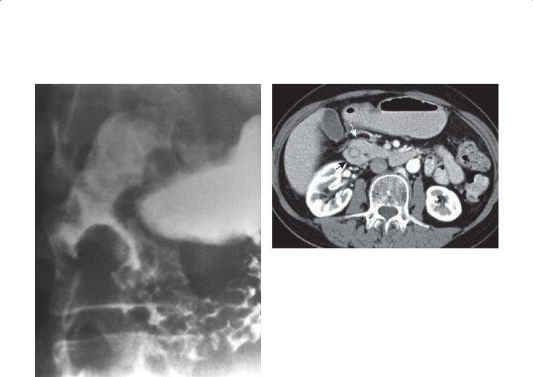
CASE 3.31
A B
Findings
A.Single-contrast UGI. A well-defined mass is located in the region of the ampulla.
B.Contrast-enhanced CT. A soft tissue mass causes focal wall thickening of the medial wall of the second portion of the duodenum.
Di erential Diagnosis
1.Ampullary neoplasm
2.Duodenal polyp
3.Edematous major papilla
Diagnosis
Villous adenoma (duodenal polyp)
Discussion
A villous adenoma was removed at operation. Benign tumors of the duodenum are unusual. Villous adenomas often have a raspberryor cauliflower-like surface, although the surface of the tumor in this case does not appear villous. These tumors can be
suggested radiographically when barium fills the many tiny surface interstices. The incidence of malignant transformation within villous adenomas is higher than in usual tubular adenomatous polyps (cases 3.21 and 3.22).
Polyps can be detected at CT with optimal technique. CT is also helpful for determining the extraluminal extent of disease. Local tumor
invasion, regional lymphadenopathy, and liver and lung metastases can be assessed preoperatively with CT. Follow-up of these findings with endoscopy is indicated.
Disease type: Masses and Filling Defects
CASE 3.32
A
Findings
Contrast-enhanced MRI. A. T1-weighted axial arterial phase. B. T2-weighted coronal image.
A 1.3-cm filling defect is within the duodenal bulb. The defect is smooth-surfaced and enhances briskly after intravenous gadolinium injection.
Di erential Diagnosis
1.Benign duodenal polyp
2.Metastasis
3.Brunner gland hyperplasia or duodenitis
Diagnosis
Benign duodenal polyp
3. DUODENUM 183
B
Discussion
Most polyps in the gastrointestinal tract enhance after the administration of intravenous contrast material. Radiographically, it is not possible to distinguish benign from malignant polyps of this size and shape. Endoscopic resection is often required for definitive diagnosis.
Disease type: Masses and Filling Defects
184 MAYO CLINIC GASTROINTESTINAL IMAGING REVIEW
CASE 3.33
Findings
Double-contrast UGI. Multiple tiny filling defects are present within the duodenal bulb. Larger and more numerous polyps are also present in the stomach in this patient with Gardner syndrome.
Di erential Diagnosis
Fundic gland polyposis syndrome (familial adenomatous polyposis syndrome)
Diagnosis
Fundic gland polyposis syndrome (familial adenomatous polyposis syndrome)
Discussion
Duodenal polyps are rare; when present, they are usually adenomas, gastrointestinal stromal tumors, or lipomas. Multiple polyps in the duodenum can be present in patients with a polyposis syndrome. Gastric polyps in familial polyposis (and Gardner syndrome) are most commonly hyperplastic, although adenomas also can be found. Duodenal polyps are usually adenomatous. Because patients with familial polyposis have an increased incidence of ampullary carcinoma, the duodenum should
be assessed carefully in all patients with familial polyposis. Hamartomas are usually present in PeutzJeghers syndrome.
Other causes of multiple filling defects in the duodenal bulb include heterotopic gastric mucosa (case 3.37), benign lymphoid hyperplasia, and Brunner gland hyperplasia (case 3.38). Heterotopic gastric mucosa usually appears as multiple tiny, angular filling defects. Nodular lymphoid hyperplasia consists of tiny, round, uniform filling defects that usually diffusely involve the entire small bowel (case 4.56). Brunner gland hyperplasia is often larger and involves fewer filling defects that vary in size.
Disease type: Masses and Filling Defects
CASE 3.34
Findings
Single-contrast UGI. Two polypoid filling defects are present within the duodenal bulb. At fluoroscopy, these were observed to traverse the pylorus and return to the stomach.
Di erential Diagnosis
1.Prolapsing gastric polyps
2.Brunner gland adenomas
3.Foreign bodies in duodenum
Diagnosis
Prolapsing gastric polyps
3. DUODENUM 185
Discussion
Th ese lesions were found to be hyperplastic gastric polyps and were removed with endoscopic
polypectomy. Gastric polyps are more common than duodenal polyps. In fact, most duodenal bulb polyps are actually gastric polyps that have prolapsed through the pylorus into the duodenum. Fluoroscopic observation can be helpful for watching the polyps move between stomach and duodenum and for identifying the pedicle. Duodenal polyps are attached and do not prolapse into the stomach. Foreign bodies (usually food) will move, during fluoroscopic observation, into the descending duodenum and beyond.
Disease type: Masses and Filling Defects
186 MAYO CLINIC GASTROINTESTINAL IMAGING REVIEW
CASE 3.35 |
CASE 3.36 |
Findings
CASE 3.35. Double-contrast UGI. A well-circumscribed mass is located at the junction of the second and third portions of the duodenum. Its smooth surface suggests a submucosal origin of the tumor. The sausage shape of the mass suggests it is soft and pliable.
CASE 3.36. Contrast-enhanced CT. A mass with the density of fat is present within the duodenum. The mass is well circumscribed.
Di erential Diagnosis
Duodenal lipoma
Diagnosis
Duodenal lipoma
Discussion
Th is radiographic appearance is typical for a lipoma. No further diagnostic tests should be necessary.
Lipomas are submucosal tumors that nearly always have a smooth surface and are compressible at fluoroscopy. These tumors may change shape during a
peristaltic contraction. They may grow to be large. The fat attenuation at CT is pathognomonic.
Disease type: Masses and Filling Defects

3. DUODENUM 187
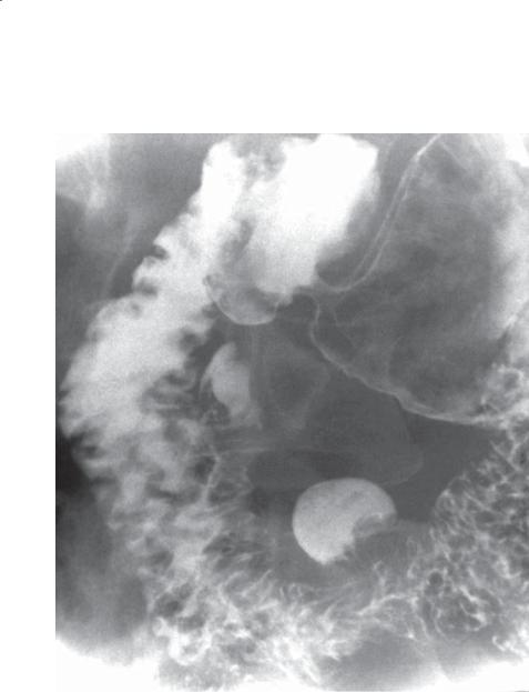
CASE 3.37
Findings
Double-contrast UGI. Multiple tiny filling defects of variable size (arrows) are present within the duodenal bulb.
Di erential Diagnosis
1.Brunner gland hyperplasia
2.Lymphoid pattern
3.Heterotopic gastric mucosa
4.Eff ervescent granules
Diagnosis
Heterotopic gastric mucosa
Discussion
Th e normal duodenal mucosa can vary in appearance, but it is often either featureless or a fine nodular pattern. Heterotopic gastric mucosa is a congenital, benign, and asymptomatic condition consisting of islands of gastric mucosa within the
duodenal bulb. This type of mucosa may have a protective effect against peptic ulcer disease. Radiographically, raised nodules, often with an angular configuration, that measure one to
several millimeters in diameter are seen about the pylorus either diffusely or in clusters. Differential considerations include effervescent granules, Brunner gland hyperplasia, and nodular lymphoid hyperplasia. Brunner glands are usually larger, less numerous, and more uniform in size (case 3.38). Lymphoid nodules (case 4.56) are small and uniform in size, usually evenly distributed throughout the duodenum. Effervescent granules change size and shape on different views as they dissolve. Gastric metaplasia of duodenal epithelium also can occur, analogous to Barrett metaplasia in the esophagus. This condition is commonly associated with peptic ulcer disease. The mucosal changes in metaplasia cannot be appreciated radiologically because the lesions are flat.
Disease type: Masses and Filling Defects
188 MAYO CLINIC GASTROINTESTINAL IMAGING REVIEW
CASE 3.38
Findings
Single-contrast UGI. Multiple filling defects of various sizes are present within the duodenal bulb. A metallic marker embedded on a radiolucent compression paddle is projected over the bulb.
Di erential Diagnosis
1.Duodenitis
2.Brunner gland hyperplasia
3.Nodular lymphoid hyperplasia
4.Heterotopic gastric mucosa
5.Crohn disease
Diagnosis
Brunner gland hyperplasia
Discussion
Brunner glands are normal glandular elements located predominantly in the proximal portion of the duodenum. Glandular secretions are rich in mucus and bicarbonate, substances that protect the duodenum against acid. Hyperplasia of these glands occurs mainly in response to peptic ulcer disease. Radiographically, nodular filling defects are seen
in the duodenal bulb and the second portion of the duodenum.
Differential considerations include thickened folds from duodenitis (case 3.1), which often can be obliterated with compression. Crohn disease often involves a more extensive portion of the duodenum and often is associated with changes in the distal stomach and small bowel (cases 3.15, 3.16, and 3.17). Nodular lymphoid hyperplasia usually presents with smaller, uniform-size, and diffusely and evenly distributed nodular filling defects (case 4.56). Heterotopic gastric mucosa is usually smaller and confined to the duodenal bulb (case 3.37).
Disease type: Masses and Filling Defects
CASE 3.39
Findings
Single-contrast UGI. An extramucosal (smoothsurfaced) mass indents the superior aspect of the duodenal bulb.
Di erential Diagnosis
1.Lipoma
2.Enteric duplication cyst
3.Ectopic pancreatic rest
4.Gastrointestinal stromal tumor
Diagnosis
Enteric duplication cyst
3. DUODENUM 189
Discussion
Intestinal duplication occurs as a result of abnormal recanalization of the intestinal tract. Duplications can occur anywhere in the alimentary tract, but they usually are found in the esophagus or ileum. With cysts in the small bowel, patients usually present with symptoms of obstruction during late infancy or early childhood. Peptic ulceration and bleeding can occur when ectopic gastric mucosa is present.
Barium studies usually show an extrinsic mass causing narrowing and obstruction of the affected bowel loop. Rarely, communication with the adjacent bowel is seen. Compressibility and pliable shape with palpation and peristalsis are helpful findings that can suggest the diagnosis of either duplication cyst or lipoma. CT often is helpful in a suspected duplication cyst by identifying a well-defined cylindrical or spherical cystic mass the density of water adjacent to the bowel (cases 1.49 and 1.50). Treatment is operative resection in symptomatic patients.
Disease type: Masses and Filling Defects
190 MAYO CLINIC GASTROINTESTINAL IMAGING REVIEW
CASE 3.40
Findings
Single-contrast UGI. A polypoid filling defect (arrow) is present near the junction of the first and second portions of the duodenum.
Di erential Diagnosis
1.Duodenal flexural pseudopolyp
2.Polyp
3.Ectopic pancreatic rest
4.Duodenal ulcer
Diagnosis
Flexural pseudopolyp
Discussion
Th is is the typical appearance of a flexural pseudopolyp. Redundant mucosa is often present at the junction of the first and second portions of the duodenum. This extra mucosa in combination with some buckling medially as the duodenum curves can resemble a polyp (case 3.34), a small ulcer (case 3.3), or ectopic pancreatic tissue (case 2.39). The changing size and configuration of a pseudopolyp can be visualized at fluoroscopy during duodenal filling and emptying.
Disease type: Masses and Filling Defects
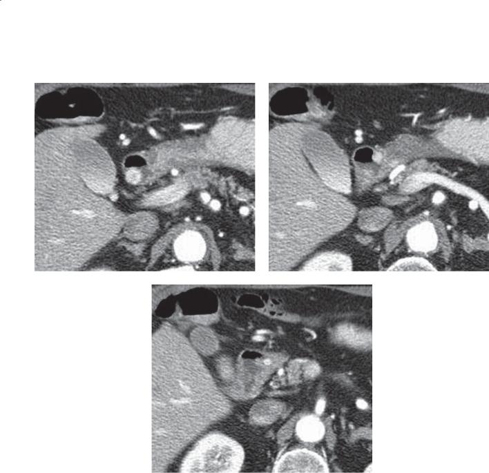
CASE 3.41
Findings
CASE 3.41. Single-contrast UGI. A well-defined filling defect with associated narrowing of the descending duodenum is present.
CASE 3.42. Contrast-enhanced CT. The duodenum is located within the pancreatic head. Pancreatic tissue is seen to encircle the descending duodenum (arrows).
Di erential Diagnosis
1.Pancreatitis
2.Duodenal neoplasm (primary or metastatic)
3.Annular pancreas
Diagnosis
Annular pancreas
3. DUODENUM 191
CASE 3.42
Discussion
Annular pancreas is a congenital abnormality in which the dorsal and ventral pancreatic tissues encircle and narrow the duodenum at or above the level of the ampulla. Complete duodenal obstruction often occurs in the neonatal period. In patients with duodenal obstruction, the classic “double bubble”
is present with gas in the distended stomach and proximal duodenum. Less severe narrowing may be asymptomatic or lead to symptoms that are delayed until adulthood. Endoscopic retrograde cholangiopancreatography or magnetic resonance cholangiopancreatography is diagnostic of this
condition when the ventral pancreatic duct encircles the duodenum.
Disease type: Masses and Filling Defects

192 MAYO CLINIC GASTROINTESTINAL IMAGING REVIEW
TABLE 3.2
Duodenal Masses and Filling |
|
CASE |
Defects |
|
|
|
|
|
Malignant tumors |
|
|
Adenocarcinoma |
Most common primary malignant tumor of duodenum. |
3.19 and 3.20 |
|
“Apple-core” or polypoid mass |
|
Ampullary carcinoma |
Polypoid mass at ampulla or annular constricting lesion. |
3.21 and 3.22 |
|
Usually causes biliary and pancreatic duct obstruction |
|
Lymphoma |
Often long-segment bowel-wall thickening. Aneurysmal |
3.23 and 3.24 |
|
ulceration and associated adenopathy should be sought |
|
Malignant gastrointestinal |
Smooth-surfaced submucosal tumor. Can calcify |
3.25 and 3.26 |
stromal tumor |
and ulcerate |
|
Duodenal gastrinoma(s) |
Small enhancing masses usually in duodenal wall |
3.27 |
Metastases |
Direct extension (pancreatic cancer) or hematogenous |
3.28–3.30 |
|
spread (eg, melanoma, lung carcinoma) |
|
|
|
|
Benign tumors |
|
|
Villous adenoma |
Cauliflower-like surface. Higher incidence of malignant |
3.22 and 3.31 |
|
transformation than in usual tubular adenomatous polyps |
|
Polyps |
Small sessile or pedunculated lesions |
3.32–3.34 |
Gastrointestinal stromal tumor |
Submucosal tumor. Can ulcerate |
3.25 and 3.26 |
Lipoma |
Smooth submucosal tumor. Fat density on CT |
3.35 and 3.36 |
|
|
|
Nonneoplastic |
|
|
Heterotopic gastric mucosa |
Often nodules with angular configuration clustered about |
3.37 |
|
pylorus |
|
Brunner gland hyperplasia |
Nodular filling defects of varying size in proximal |
3.38 |
|
duodenum. Results from gland hyperplasia in response to |
|
|
peptic ulcer disease |
|
Enteric duplication cyst |
Abnormal recanalization of intestinal tract. Fluid density on |
3.39 |
|
CT. Extrinsic mass |
|
Flexural pseudopolyp |
Redundant mucosa at junction of first and second portions |
3.40 |
|
of duodenum medially |
|
Annular pancreas |
Dorsal and ventral pancreatic tissue encircle and narrow |
3.41 and 3.42 |
|
second portion of duodenum |
|
|
|
|
Disease type: Masses and Filling Defects
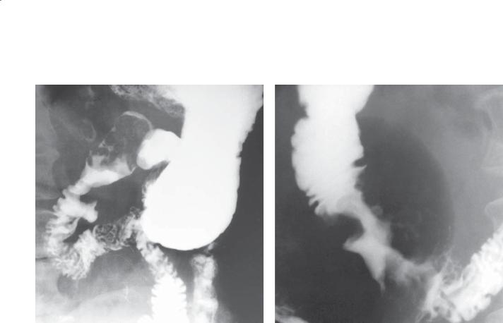
CASE 3.43
Findings
Double-contrast UGI. Two outpouchings arise from the second and third portions of the duodenum.
Di erential Diagnosis
Duodenal diverticula
Diagnosis
Duodenal diverticula
3. DUODENUM 193
Discussion
Diverticula can occur at any location in the duodenum, but they most commonly arise from the medial
wall of its second portion. Usually these are of little clinical significance; however, rarely hemorrhage and perforation have been reported. Diverticula often can be differentiated from ulcers by observing duodenal folds entering the diverticula and the diverticula changing shape and size.
Disease type: Miscellaneous
194 MAYO CLINIC GASTROINTESTINAL IMAGING REVIEW
CASE 3.44
Findings |
Discussion |
Double-contrast UGI. A barium-filled, polypoid, |
An intraluminal duodenal diverticulum is believed |
intraluminal mass is present within the descending |
to develop as a result of an intraluminal duodenal |
duodenum. |
diaphragm. The diaphragm usually arises at or near |
|
the papilla of Vater. The diaphragm balloons within |
Di erential Diagnosis |
the duodenum, eventually developing into a sac. |
Intraluminal duodenal diverticulum |
Most patients present with intermittent obstructive |
|
symptoms and abdominal pain. An obstructive |
Diagnosis |
diverticulum near the papilla of Vater may be |
Intraluminal duodenal diverticulum |
associated with pancreatitis. |
|
Th e radiologic appearance of intraluminal duodenal |
|
diverticula has been likened to that of a wind |
|
sock. Usually a radiolucent band, representing the |
|
diverticular wall, surrounds the diverticulum. If the |
|
diverticulum is not filled with barium, it resembles a |
|
pedunculated polyp (case 3.34). |
Disease type: Miscellaneous
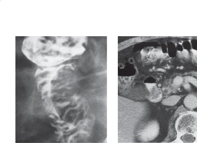
CASE 3.45
Findings
CASE 3.45. Single-contrast UGI. The proximal transverse duodenum is dilated to the level of the spine. The bowel abruptly narrows and appears to be pinched closed by a linear extrinsic mass. The underlying duodenal mucosa is intact.
CASE 3.46. Contrast-enhanced CT. The proximal duodenum is dilated and tapers to a region of highgrade narrowing at the level of the superior mesenteric artery. No mass is present at the level of the duodenal stenosis.
Di erential Diagnosis
1.Superior mesenteric artery syndrome
2.Duodenal neoplasm
3.Abdominal aortic aneurysm
Diagnosis
Superior mesenteric artery syndrome
3. DUODENUM 195
CASE 3.46
Discussion
Findings are characteristic of the superior mesenteric artery syndrome. Narrowing of the third portion of the duodenum can occur as a result of compression by the anteriorly located superior mesenteric artery and posteriorly positioned spine and aorta. This finding often can be observed in asymptomatic persons, but in certain patients a critical narrowing occurs and results in partial duodenal obstruction. Patients who lose weight rapidly or very asthenic persons are most often affected. Patients immobilized for long periods (body casts, hyperextension fixation for spinal injuries, extensive burns) are also at risk.
Radiologically, the first and second portions of the duodenum are dilated to the level of the extramucosal linear obstruction (superior mesenteric artery).
Vigorous peristalsis of the proximal duodenum often is observed fluoroscopically as these segments attempt to propel duodenal contents beyond the obstruction. Turning the patient to the left decubitus or prone position may reduce the compression on the duodenum, and contrast material is observed to pass easily into the distal duodenum.
Other conditions causing dilatation of the proximal small bowel include primary or metastatic duodenal tumors (cases 3.28 and 3.29), pancreatitis (cases 3.12 and 3.13), and abdominal aortic aneurysms.
Disease type: Miscellaneous
196 MAYO CLINIC GASTROINTESTINAL IMAGING REVIEW
CASE 3.47
A B
Findings
Contrast-enhanced CT. A. An end-to-side abdominal aortic graft is in place. B. The normally visible fat plane between the aorta/graft and duodenum is absent. Fluid and gas (arrow) are present within the left limb of the graft.
Di erential Diagnosis
1.Aortoenteric fistula
2.Aortic graft infection
Diagnosis
Aortoenteric fistula
Discussion
Th is patient was experiencing gastrointestinal bleeding. Findings are compatible with an aortoenteric fistula. At operation, a defect was present in the fourth portion of the duodenum. The graft was bile-stained and leaking blood into the duodenum.
An aortoenteric fistula is usually a complication of reconstructive aortic surgery after the placement of a prosthetic graft. Perigraft infection is nearly always present, and fistulization is a potentially catastrophic complication of the infection. The clinical signs and symptoms of this disorder can be subtle; gastrointestinal bleeding is the most obvious manifestation. The commonest CT findings of aortoenteric fistula include an ectopic gas collection and focal bowel-wall thickening. The fat plane is
usually lost between the aorta and the duodenum. The finding of perigraft soft tissue, fluid, or ectopic gas is worrisome for perigraft infection. CT is most helpful for excluding this diagnosis by identification of an intact fat plane between the duodenum and the aortic graft. Arteriography is often necessary to confirm the diagnosis.
Disease type: Miscellaneous
CASE 3.48
Findings
CASE 3.48. Contrast-enhanced CT. Extraluminal gas is present in the subhepatic space, adjacent and anterior to the duodenum. Patient had sustained abdominal trauma in a motor vehicle accident.
CASE 3.49. Extravasated orally administered contrast material (arrow) is tracking from the region of the retroperitoneal duodenum into the right anterior pararenal space. Hazy soft tissue stranding is also present in the right flank. This patient had sustained a blow to the abdomen in a motor vehicle accident.
Di erential Diagnosis
1.Perforated duodenal ulcer
2.Posttraumatic duodenal rupture
3.Gas-producing retroperitoneal infection
Diagnosis
Posttraumatic duodenal rupture
3. DUODENUM 197
CASE 3.49
Discussion
Th e location of the extraluminal air and the history of abdominal trauma are consistent with duodenal perforation, which was surgically confirmed and repaired.
Th e diagnosis of duodenal perforation often is not apparent clinically. If the diagnosis is delayed beyond 24 hours, reports suggest that mortality may reach 65%. Repair within 24 hours has only a 5% mortality. For traumatic duodenal perforation, the most important finding on plain radiography is
retroperitoneal air. Other nonspecific findings include psoas obliteration, segmental ileus near the duodenum, and scoliosis. Plain radiographic findings of duodenal perforation are present in only one-third of patients. Free intraperitoneal air is unusual in traumatic duodenal perforations, and it is more common in patients with a perforated peptic ulcer. CT can directly visualize the duodenum and adjacent tissues. CT findings of a traumatic duodenal perforation include extravasated gas or orally administered contrast material, fluid collection in the anterior pararenal space, and considerable soft tissue stranding in the retroperitoneum. Retroperitoneal gas and fluid also can occur from traumatic injuries to the thorax, bladder, or pancreas.
Disease type: Miscellaneous

198 MAYO CLINIC GASTROINTESTINAL IMAGING REVIEW
CASE 3.50 |
CASE 3.51 |
Findings
CASE 3.50. Single-contrast UGI. A localized mass (arrow) indents and narrows the duodenum at the junction of its second and third portions. This child was brought to the emergency department after falling from playground equipment onto the abdomen.
CASE 3.51. Contrast-enhanced CT. A large, mixed attenuation mass is present in the location of the transverse duodenum. Duodenal folds are thickened and splayed by the mass. This patient was brought to the emergency room after a motor vehicle accident.
Di erential Diagnosis
1.Duodenal hematoma
2.Retroperitoneal neoplasm or abscess
3.Pancreatitis
4.Gastrointestinal stromal tumor
5.Metastases
Diagnosis
Duodenal hematoma
Discussion
A duodenal hematoma is most likely, considering the history of trauma. Hemorrhage can affect any portion of the gastrointestinal tract, but the small bowel is most frequently affected. Anticoagulants, trauma, and bleeding diatheses are the usual causes. Patients who sustain blunt abdominal trauma usually have masslike changes involving the duodenum because of its lack of mobility and retroperitoneal fixation. Clearing of the hematoma usually can be appreciated within 1 week, with complete resolution in 2 to 3 weeks. Gastric outlet obstruction often occurs due to the obstructing hematoma.
Radiographic findings include a segmental region of fold thickening where the folds are straight and have been likened to a stack of coins. Smooth or scalloped narrowing of the duodenum with effacement of the folds also can be seen, as in case 3.50. It is important to assess for a duodenal leak (extravasated contrast material or extraluminal gas), an important complication of a severe injury (cases 3.47 and 3.49).
Differential considerations on the barium examination include any process causing localized fold thickening. Generally, the diagnosis is obvious if recent trauma has occurred.
Disease type: Miscellaneous
CASE 3.52
A
Findings
Single-contrast UGI. A and B. An angular-shaped collection of barium is seen in an elongated pyloric canal. The triangular collection was observed to change shape at fluoroscopy.
Di erential Diagnosis
1.Pyloric channel ulcer
2.Pylorus torus defect
3.Adult hypertrophic pyloric stenosis
4.Annular carcinoma
Diagnosis
Pyloric torus defect with hypertrophic pyloric stenosis
3. DUODENUM 199
B
Discussion
Th e normal pylorus is composed of two prominent muscle bundles that converge toward the lesser curvature, known as the torus. In some patients, these muscular bundles diverge far enough to allow mucosa to prolapse between them. This prolapse creates an annulus-shaped niche within the pyloric canal that can simulate a pyloric channel crater. A pyloric torus niche is transient and changes shape and possibly disappears with fluoroscopic observation or serial radiography.
It has been reported in 14% of normal patients and is also prominent in patients with hypertrophic pyloric stenosis.
Adult hypertrophic stenosis is rare. Patients frequently complain of nausea, postprandial upset, intermittent vomiting, and heartburn. Radiologically, the pyloric canal is narrowed and elongated. The torus defect is often identified and the pyloric muscle mass indents the base of the duodenal bulb. Nearly half of all patients have concomitant gastric ulceration.
Differential considerations include a pyloric ulcer, carcinoma, and antral gastritis. A fixed pyloric channel should be viewed with suspicion, and endoscopy with biopsy should be recommended to exclude neoplasm.
Disease type: Miscellaneous

200 MAYO CLINIC GASTROINTESTINAL IMAGING REVIEW
TABLE 3.3
Miscellaneous Duodenal Conditions |
|
CASE |
Diverticula |
Often periampullary, but can occur anywhere. Folds |
3.43 |
|
radiate into outpouching |
|
|
Intraluminal diverticula have windsock deformity |
3.44 |
|
due to prior web |
|
|
|
|
Superior mesenteric artery syndrome |
Dilation of duodenum to superior mesenteric artery. |
3.45 and 3.46 |
|
Usually active peristalsis proximally. Asthenic patients |
|
|
|
|
Aortoenteric fistula |
Exclude with fat between aorta and duodenum on all |
3.47 |
|
CT slices. Usually due to graft infection |
|
|
|
|
Rupture |
Blunt trauma to abdomen is commonest cause. |
3.48 and 3.49 |
|
Retroperitoneal air, soft tissue stranding, oral |
|
|
contrast leakage |
|
|
|
|
Hematoma |
Blunt trauma commonest. Soft tissue mass (often |
3.50 and 3.51 |
|
mixed attenuation at CT) involving duodenum |
|
|
|
|
Pylorus torus |
Triangular collection of contrast centered in pylorus. |
3.52 |
|
Changes shape with observation |
|
|
|
|
Disease type: Miscellaneous

3. DUODENUM 201
|
TABLE 3.4 |
|
Di erential Diagnoses |
|
|
Thickened Folds |
Narrowing |
|
BENIGN |
BENIGN |
|
Peptic disease |
Peptic ulcer disease |
|
Zollinger-Ellison syndrome |
Pancreatitis |
|
Brunner gland hyperplasia |
Crohn disease |
|
Pancreatitis |
Hematoma |
|
Crohn disease |
Superior mesenteric artery syndrome |
|
Adult celiac disease |
Annular pancreas |
|
Whipple disease |
MALIGNANT |
|
Hematoma |
||
Adenocarcinoma |
||
Cystic fibrosis |
||
Lymphoma |
||
|
||
MALIGNANT |
Malignant gastrointestinal stromal tumor |
|
Lymphoma |
Metastases |
|
Filling Defects |
|
|
BENIGN TUMORS |
|
|
Polyp |
|
|
Gastrointestinal stromal tumor |
|
|
Lipoma |
|
|
Villous adenoma |
|
|
MALIGNANT TUMORS |
|
|
Adenocarcinoma |
|
|
Lymphoma |
|
|
Malignant gastrointestinal stromal tumor |
|
|
Metastases |
|
|
Ampullary carcinoma |
|
|
Gastrinoma |
|
|
NONNEOPLASTIC |
|
|
Flexural pseudotumor |
|
|
Hamartoma |
|
|
Intraluminal duodenal diverticulum |
|
|
Brunner gland hyperplasia |
|
|
Enteric duplication cyst |
|
|
Heterotopic gastric mucosa |
|
|
Annular pancreas |
|
202 MAYO CLINIC GASTROINTESTINAL IMAGING REVIEW
DUODENAL FILLING DEFECTS
|
Lymphoid nodular |
Heterotopic |
|
|
|
hyperplasia |
|
|
|
|
|
gastric mucosa |
|
|
|
|
|
|
|
Flexural |
|
|
|
|
pseudopolyp |
|
|
|
|
Duodenal |
|
|
Prolapsing |
|
|
|
gastric polyp |
Lymphoma |
|
polyp |
|
|
||
|
|
|
|
(aneurysmal dilatation) |
Villous |
|
Brunner |
|
|
adenoma |
|
|
gland |
|
|
|
hyperplasia |
|
|
Ampullary |
|
|
|
D.F. |
carcinoma |
|
|
|
|
|
|
|
© MAYO |
|
|
|
|
|
2013 |
Enteric
duplication cyst Annular adenocarcinoma
Lipoma
GIST
Metastases
GIST, gastrointestinal stromal tumor
Figure 3.53 (Used with permission of Mayo Foundation for Medical Education and Research.)

|
3. DUODENUM 203 |
|
|
|
QUESTIONS |
Multiple Choice (choose the best answer) |
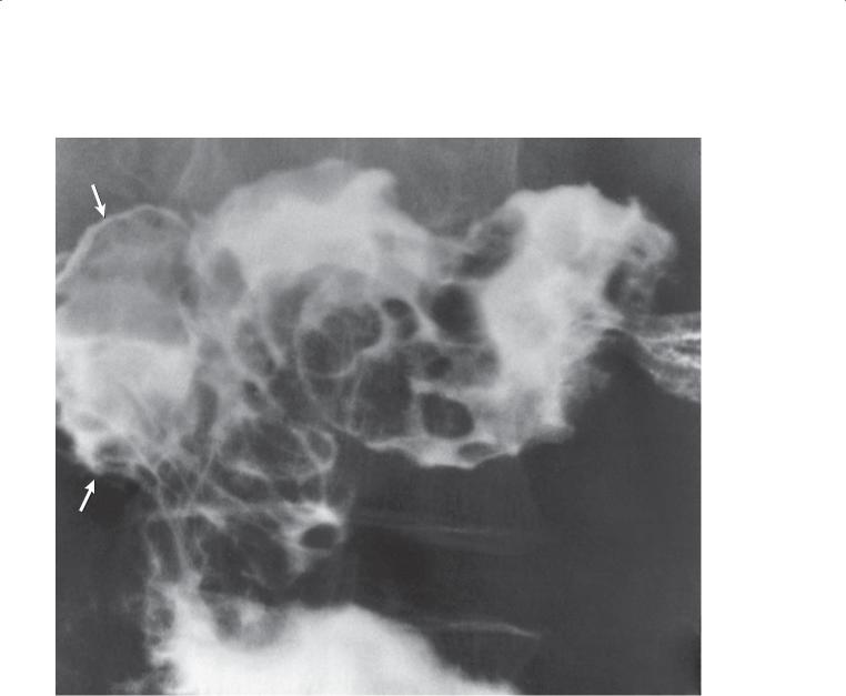
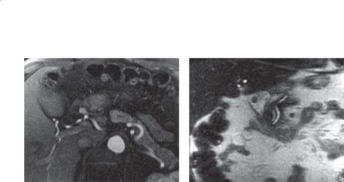
3.3. Whatsymptomswouldbeexpectedwiththecondition |
shown in Question 3.2?
3.1. Which of the following is the most likely diagnosis |
a. Nausea |
|
for the findings in the figure? |
||
b. Epigastric burning |
||
|
||
|
c. Hematemesis |
|
|
d. Melena |
|
|
e. Fever |
a.Carcinoid tumor
b.Ectopic pancreatic rest
c.Prolapsed gastric mucosa
d.Brunner gland hyperplasia
e.Heterotopic gastric mucosa
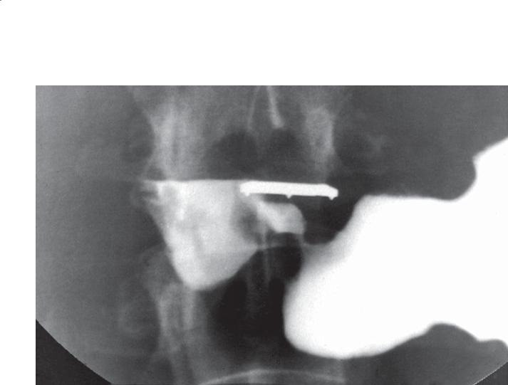
3.2.Which of the following is most often associated with the condition shown in the figure?
a.Increased level of carcinoembryonic antigen
b.Increased level of immunoglobulin G4
c.Chance fracture
d.Increased level of serum gastrin
e.Pancreatic necrosis
204 MAYO CLINIC GASTROINTESTINAL IMAGING REVIEW
A B
C
D
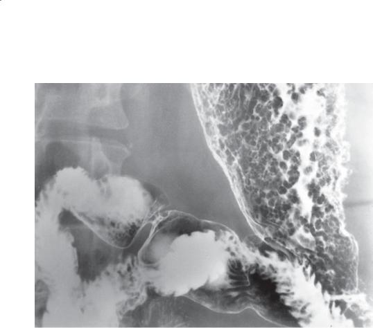
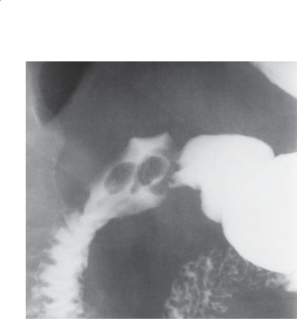
Match images A through F with the responses listed below.
3.4. |
_____ Usually due to infection |
3.6. |
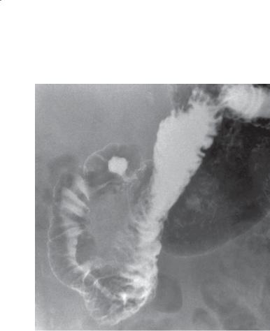
_____ Causes obstruction |
3.5. |
_____ May be associated with pneumoperitoneum |
3.7. |
_____ A normal variant |

3. DUODENUM 205
E F
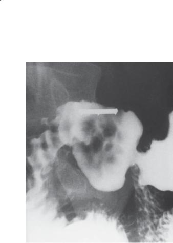
3.8.Which of the following is the most likely diagnosis for the findings in this figure?
a.Primary carcinoma
b.Crohn disease
c.Zollinger-Ellison syndrome
d.Pancreatic cancer
e.Duodenitis
206 MAYO CLINIC GASTROINTESTINAL IMAGING REVIEW
ANSWERS
3.1.Answer d.
Multiple well-defined filling defects in the proximal duodenum are most likely due to Brunner gland hyperplasia, usually a manifestation of duodenitis. Brunner glands secrete mucin and an alkaline fluid to protect the duodenum. In hyperacid states, these glands can hypertrophy and present as multiple fixed filling defects.
A carcinoid tumor can be found in the duodenum, but it is usually a solitary submucosal finding.
An ectopic pancreatic rest can be seen in the distal stomach or proximal duodenum as a solitary filling defect, typically with a central umbilication (the site of the duct) that collects barium.
Prolapsed gastric mucosa moves in and out of the bulb with peristaltic waves.
Heterotopic gastric mucosa presents as multiple small, finely nodular mucosal filling defects, often with angular edges. These are usually smaller and more numerous than Brunner gland hyperplasia.
3.2.Answer c.
Th is is a case of a duodenal hematoma, most commonly associated with blunt trauma to the abdomen with compression of the duodenum against the spine. A seatbelt injury in an automobile accident is a common clinical scenario for this finding. A chance fracture of the spine is also commonly associated with a seat-belt injury.
An increased level of carcinoembryonic antigen is associated with many types of carcinomas, such as a pancreatic carcinoma. There is no evidence for a pancreatic mass in this case.
An increased level of immunoglobulin G4 is associated with autoimmune pancreatitis, usually presenting with diffuse pancreatic enlargement. No evidence of this is present in this case.
An increased level of serum gastrin can occur in patients with a gastrinoma and in patients with peptic ulcer disease.
Pancreatic necrosis is a finding of severe pancreatitis. This finding is centered on the transverse duodenum rather than the pancreas.
3.3.Answer a.
Duodenal hematomas can obstruct the duodenum and result in retained fluid and debris within the stomach and proximal duodenum. Nausea and vomiting are the most common symptoms expected with gastrointestinal obstruction.
Epigastric burning is a symptom associated with ulcer disease of the stomach and duodenum.
Gastric or duodenal bleeding could lead to either hematemesis or melena; however, the hemorrhage in this condition is submucosal, usually without a mucosal leak. Severe duodenal trauma could lead to duodenal perforation with extraluminal air and fluid (not seen in this case).
Fever would be expected with an infection, which is not present in this case.
3.4.Answer a.
Figure a shows an aortoenteric fistula. There is loss of the normal fat plane between the aorta and the duodenum. The aorta contains a graft. Aortoenteric fistula is usually due to infection at the proximal limb of an aortic stent graft. In many cases, a fluid collection containing air is seen between the aorta and transverse duodenum. In nearly all cases, the normal fat plane between the aorta and duodenum is obliterated. The condition can cause catastrophic bleeding into the gastrointestinal tract and requires emergency repair.
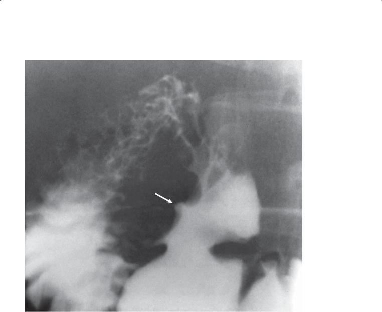
3.5.Answer c.
Figure c shows a duodenal ulcer. There is narrowing in the descending duodenum about a persistent collection of barium (ulcer crater). Peptic ulcers (gastric or duodenal) are the commonest cause of non-iatrogenic pneumoperitoneum.
3.6.Answer d.
Figure d shows an annular pancreas. The lowattenuation duodenal lumen is seen within the pancreatic parenchyma. This is the classic finding of an annular pancreas. In children, this condition is identified because of persistent nausea and vomiting due to obstruction of the duodenum by the annular pancreatic tissue. In symptomatic patients, operative repair is usually required to relieve the obstruction. In adults, the condition can be discovered incidentally or can be the cause of duodenal obstruction.
3.7.Answer f.
Figure f shows a flexural pseudotumor. There is a filling defect at the junction of the first and second portions of the duodenum along its medial wall. This finding is typical of redundant tissue at the angle of the duodenal flexure. The redundant tissue can cause a filling defect and should not be mistaken for a mass.
Figurebshowsanintraluminalduodenaldiverticulum. Over time, a web of duodenal tissue can elongate into

a sac with the duodenal lumen. Contrast material will fill this sac, unlike a filling defect, which will displace barium. If the condition causes symptoms, it can be surgically removed. Usually it is an incidental finding.
Figure e shows malrotation, whereby the duodenum is never seen to cross the midline. Proximal small bowel is seen within the right upper quadrant. This condition may be asymptomatic, but it can also be associated with midgut volvulus. Ladd bands develop in this condition, and they can act as a fulcrum around which the small bowel will twist.
3. DUODENUM 207
3.8.Answer a.
Primary adenocarcinomas are usually located within the duodenum and are short, annular, constrictive lesions (as seen in this case). Crohn disease can involve the duodenum, but there is usually ileal disease present to suggest the correct diagnosis. Zollinger-Ellison syndrome can cause postbulbar ulcerations but not focal masses, and associated findings in the stomach and proximal duodenum would be expected. Pancreatic cancers can involve the duodenum, but there is nearly always a contiguous pancreatic mass. Duodenitis involves the duodenum proximal to the ampulla.
This page intentionally left blank