


MAYO CLINIC
Gastrointestinal
Imaging Review
Second Edition

Mayo Clinic Scientific Press
Mayo Clinic Atlas of Regional Anesthesia and Ultrasound-Guided Nerve Blockade
Edited by James R. Hebl, MD, and Robert L. Lennon, DO
Mayo Clinic Preventive Medicine and Public Health Board Review
Edited by Prathibha Varkey, MBBS, MPH, MHPE
Mayo Clinic Challenging Images for Pulmonary Board Review
Edward C. Rosenow III, MD
Mayo Clinic Gastroenterology and Hepatology Board Review, Fourth Edition Edited by Stephen C. Hauser, MD
Mayo Clinic Infectious Diseases Board Review
Edited by Zelalem Temesgen, MD
Mayo Clinic Antimicrobial Handbook: Quick Guide, Second Edition Edited by John W. Wilson, MD, and Lynn L. Estes, PharmD
Just Enough Physiology
James R. Munis, MD, PhD
Mayo Clinic Cardiology: Concise Textbook, Fourth Edition Edited by Joseph G. Murphy, MD, and Margaret A. Lloyd, MD
Mayo Clinic Internal Medicine Board Review, Tenth Edition
Edited by Robert D. Ficalora, MD
Mayo Clinic Internal Medicine Board Review: Questions and Answers
Edited by Robert D. Ficalora, MD
Mayo Clinic Electrophysiology Manual
Edited by Samuel J. Asirvatham, MD

MAYO CLINIC
Gastrointestinal
Imaging Review
Second Edition
C. Daniel Johnson, MD
Chair, Department of Radiology
Mayo Clinic, Scottsdale, Arizona
Professor of Radiology
College of Medicine, Mayo Clinic
MAYO CLINIC SCIENTIFIC PRESS |
OXFORD UNIVERSITY PRESS |

Th e triple-shield Mayo logo and the words MAYO, MAYO CLINIC, and MAYO CLINIC SCIENTIFIC PRESS are marks of Mayo Foundation for Medical Education and Research.
3
Oxford University Press is a department of the University of Oxford.
It furthers the University’s objective of excellence in research, scholarship, and education by publishing worldwide.
Oxford New York |
|
|
|
|
|
|
||
Auckland |
Cape Town |
Dar es Salaam Hong Kong |
Karachi |
|||||
Kuala Lumpur |
Madrid |
Melbourne |
Mexico City |
Nairobi |
||||
New Delhi |
Shanghai |
Taipei Toronto |
|
|
|
|||
With offices in |
|
|
|
|
|
|
|
|
Argentina |
Austria Brazil |
Chile |
Czech Republic |
France Greece |
||||
Guatemala |
Hungary |
Italy |
Japan |
Poland |
Portugal |
Singapore |
||
South Korea |
Switzerland Thailand |
Turkey |
Ukraine |
Vietnam |
||||
Oxford is a registered trademark of Oxford University Press in the UK and certain other countries.
Published in the United States of America by Oxford University Press
198 Madison Avenue, New York, NY 10016
© Mayo Foundation for Medical Education and Research 2014
All rights reserved. No part of this publication may be reproduced, stored in a retrieval system, or transmitted, in any form or by any means, electronic, mechanical, photocopying, recording, or otherwise, without the prior permission of Mayo Foundation for Medical Education and Research. Inquiries should be addressed to Scientific Publications, Plummer 10, Mayo Clinic, 200 First St SW, Rochester, MN 55905.
Catalog record is available from the Library of Congress
ISBN: 978–0–19–986215–3
Mayo Foundation does not endorse any particular products or services, and the reference to any products or services in this book is for informational purposes only and should not be taken as an endorsement by the authors or Mayo
Foundation. Care has been taken to confirm the accuracy of the information presented and to describe generally accepted practices. However, the authors, editors, and publisher are not responsible for errors or omissions or for any consequences from application of the information in this book and make no warranty, express or implied, with respect to the contents of the publication. This book should not be relied on apart from the advice of a qualified health care provider.
Th e authors, editors, and publisher have exerted efforts to ensure that drug selection and dosage set forth in this text are in accordance with current recommendations and practice at the time of publication. However, in view of ongoing research, changes in government regulations, and the constant flow of information relating to drug therapy and drug reactions, readers are urged to check the package insert for each drug for any change in indications and dosage and for added wordings and precautions. This is particularly important when the recommended agent is a new or infrequently employed drug.
Some drugs and medical devices presented in this publication have US Food and Drug Administration (FDA) clearance for limited use in restricted research settings. It is the responsibility of the health care providers to ascertain the FDA status of each drug or device planned for use in their clinical practice.
9 8 7 6 5 4 3 2 1
Printed in China on acid-free paper

To Th erese, my wife, the love of my life
This page intentionally left blank

ABOUT THE COVER
Th e cover for the book was adapted from the cover of the first edition. For the first edition, a magnificent Kandinsky painting was the inspiration and foundation for an illustration depicting each portion of the gastrointestinal tract. The paintings of Kandinsky were seen as analogous to radiographic images—abstract to many viewers and understood only with knowledge of anatomy, radiography, and pathology. To demonstrate that this edition is an updated and more modern version of the first, I selected a next-generation artistic style, that of Pablo Picasso, as the underlying structure. The beauty of the interrelated gastrointestinal organs has been preserved, but with an increasingly modern flair.
Jim Rownd, the artist for the cover of the first edition, again designed and created this second edition cover. The individual gastrointestinal components are placed on the title pages of their respective chapters. I am most grateful to Jim for his artistic guidance and talents. The freedom given to me to create the book of my choice is also greatly appreciated. I hope you enjoy it.
C. Daniel Johnson, MD
vii
This page intentionally left blank

PREFACE
Th e purpose of this second edition is to update and supplement the first edition of
Mayo Clinic Gastrointestinal Imaging Review. The goal of the book is to provide a radiologic atlas of common abnormalities that affect the gastrointestinal tract that can be detected with imaging examinations. Emphasis is placed on images that are large enough to study the findings with a minimal amount of text. The book is not intended to be an inclusive source of gastrointestinal diseases and their many manifestations. Rather, the most important disease processes affecting the gastrointestinal tract and their commonest presentations are included. Only a few selected pediatric cases are included to provide a comparison with adult disease processes. Readers are directed to pediatric textbooks for a more comprehensive review of these disorders. The content of this book is directed primarily to radiology residents learning the fundamentals of gastrointestinal imaging and for board preparation. In addition, radiologists preparing for recertification examinations may find the book helpful as an efficient review.
Many of the cases in this second edition were in the first edition, although outdated images have been replaced when possible and new disease entities and imaging approaches have been added. For example, many magnetic resonance images have been added. The format for cases remains standardized to a description of the radiologic findings, pertinent differential diagnoses, the actual diagnosis, and a brief discussion. Students are urged to review the case first without reading the text (answers). Numerous summary tables and illustrations synthesize information and provide key points and case references for review. New features of the book are the questions at the end of each chapter and a final chapter of examination-type questions. These have been added to help students prepare for written examinations and to assess their learning as they progress through the book. Selected readings and references are not included—an acknowledgment that readers of this book are likely looking for an efficient single-source review and that references are easily accessible online. There are multiple other textbooks on gastrointestinal imaging with comprehensive references that the reader should access for supplementation of their knowledge in this field.
C. Daniel Johnson, MD
ix
This page intentionally left blank

ACKNOWLEDGMENTS
I am deeply indebted to the many members of the Department of Radiology at Mayo Clinic who provided cases for the book. I have had many mentors and assistants who made this project possible. My training in radiology was highly influenced by distinguished radiologists, including Harley C. Carlson, David H. Stephens, Robert L. MacCarty, Reed P. Rice, William M. Thompson, and Igor Laufer. I am grateful to all of them. Grant Schmidt, the coauthor of the first edition, provided much assistance in making the first edition successful. Although he didn’t have time to coauthor this second edition, I am grateful to him for the many good ideas that he had for the initial textbook that remain part of this edition and for a long list of interesting cases that contributed to needed improvements for the book. My administrative assistant, Lisa Kay, was very helpful with printing, sorting, and organizing the revisions. The Section of Scientific Publications and Media Support Services at Mayo Clinic worked tirelessly to provide the product that I envisioned. I am grateful for the expert editorial assistance provided by the staff in Scientific Publications: LeAnn M. Stee (editor), Kenna Atherton (manager), Jane Craig (editorial assistant), and John Hedlund (copy editor/proofreader). The radiologic images were meticulously reviewed and prepared by Deborah Veerkamp, the drawings were originally created by David A. Factor, MS, and beautifully updated with color by Joanna King, MSMI, and the cover art was created by Jim Rownd.
As always in my career, my wife, Therese Johnson, has encouraged me and supported me in this effort. Always unselfish with her time and always willing to pick up the slack when I was not available at home, she continues to help me achieve when I would not have otherwise.
I hope readers find the book an efficient way to learn the myriad and fascinating ways of the gastrointestinal tract through imaging.
C. Daniel Johnson, MD
xi
This page intentionally left blank

CONTENTS
1Esophagus
77 Stomach
157 Duodenum
209 Small Bowel
323 Colon
463 Liver
561 Bile Ducts and Gallbladder
655 Pancreas
753 Spleen
793 Peritoneum and Mesentery
857 Final Examination
879 Abbreviations Used in This Book
881 Case Index
893 Subject Index
xiii
This page intentionally left blank
C H A P T E R 1
ESOPHAGUS
Cases 1.1–1.21 |
Inflammatory and Ulcerative Diseases |
Cases 1.22–1.30 |
Strictures |
Cases 1.31–1.52 |
Masses and Filling Defects |
|
Benign Tumors |
|
Malignant Tumors |
|
Nonneoplastic |
Cases 1.53–1.58 |
Diverticula |
Cases 1.59–1.72 |
Miscellaneous |
This page intentionally left blank
1. ESOPHAGUS 3
CASE 1.1
Findings |
Discussion |
Double-contrast esophagram. Thickened and |
Reflux esophagitis is a common indication for and |
somewhat nodular folds are seen in the distal |
finding at UGI. If fold thickening is the only finding on |
esophagus. |
a triphasic esophagram, mild esophagitis commonly is |
|
found endoscopically. Folds are considered abnormal if |
Di erential Diagnosis |
they exceed 2 to 3 mm in diameter. |
1. Refl ux esophagitis |
Development of reflux esophagitis depends on |
2. Esophageal varices |
several factors, including the acidity of the refluxed |
|
contents, the efficacy of esophageal clearance, and |
Diagnosis |
the frequency of reflux. These factors are particularly |
Mild reflux esophagitis |
important in patients with Zollinger-Ellison syndrome |
|
and those with scleroderma, groups commonly affected |
|
with reflux esophagitis. Varices (cases 1.46 and 1.47) |
|
change shape with fluoroscopic observation and often |
|
have a serpentine and nodular configuration. |
Disease type: Inflammatory and Ulcerative Diseases

4MAYO CLINIC GASTROINTESTINAL IMAGING REVIEW
CASE 1.2
Findings
Double-contrast esophagram. Linear erosions (arrow) and punctate superficial erosions (arrowhead) are present within mildly nodular mucosal background. The esophagus is shortened, and a small hiatal hernia is present.
Di erential Diagnosis
1.Refl ux esophagitis
2.Herpes esophagitis
3.Candida esophagitis
Diagnosis
Moderate reflux esophagitis
Discussion
Moderate esophagitis usually is characterized by the presence of superficial erosions that may be either punctate or linear. Fold thickening and nodularity also often are findings on the mucosal-relief phase images. In patients with long-standing disease with esophageal intramural fibrosis, the esophagus shortens and pulls the stomach into the thorax. This type of “short esophagus” hiatal hernia is present in this case. Herpes esophagitis (cases 1.17 and 1.18) is usually associated with multiple discrete superficial ulcers that may be located anywhere in the esophagus. Candida esophagitis (cases 1.15 and 1.16) most often has multiple plaquelike filling defects throughout the esophagus.
Disease type: Inflammatory and Ulcerative Diseases
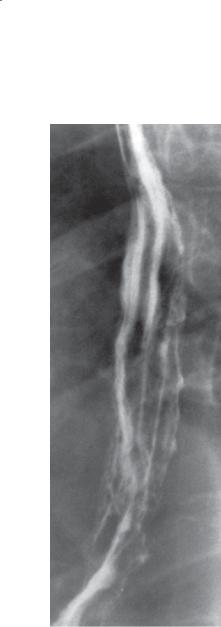
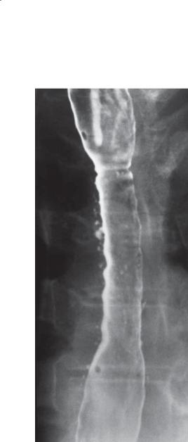
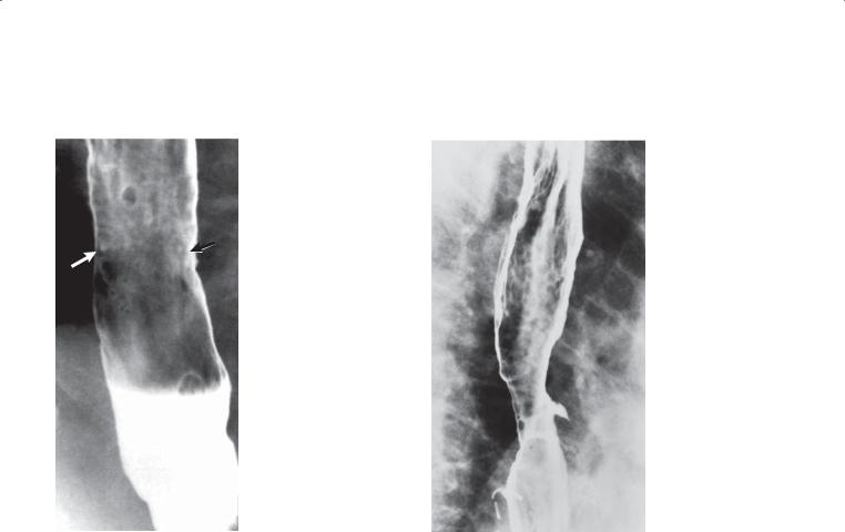
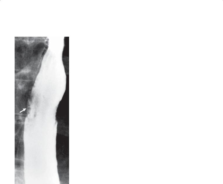
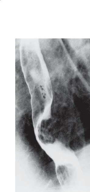
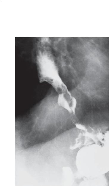
CASE 1.3
Findings
CASE 1.3. Double-contrast esophagram. A large, flat ulcer is present in the distal esophagus (arrow) with associated diminished distensibility of the lower esophageal segment. There is also a long linear ulcer (arrowheads) with a surrounding halo of edema.
CASE 1.4. Double-contrast esophagram. Mucosal nodularity, a deep ulcer, and luminal narrowing are present. Transverse folds due to chronic scarring and buckling of the mucosa also are present (arrows). Sharp spiculations are seen just superior to the deep ulcer; these are due to transverse folds seen in profile (arrowheads). Asymmetric scarring causes the distal deformity and narrowing.
Di erential Diagnosis
1.Transverse folds and severe reflux esophagitis
2.Feline esophagus
3.Carcinoma of the esophagus
Diagnosis
Severe reflux esophagitis
1. ESOPHAGUS 5
CASE 1.4
Discussion
Transverse folds develop as a result of a prior linear ulceration with scar formation within the longitudinal muscle layers of the distal esophagus. They usually do not span the entire esophageal lumen.
Severe esophagitis usually is characterized by the presence of an ulcer crater. Typically, patients with severe esophagitis have superficial erosions and fold thickening in addition to the characteristic ulcer.
Transverse folds from esophagitis should be distinguished from the folds in feline esophagus (case 1.5) because transverse folds are fixed, fewer in number, coarser, and shorter. Changes of both active and chronic disease commonly coexist (as seen in these cases).
Disease type: Inflammatory and Ulcerative Diseases
6MAYO CLINIC GASTROINTESTINAL IMAGING REVIEW
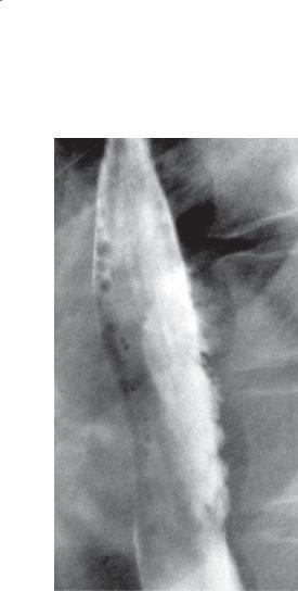
CASE 1.5
A
Findings
Double-contrast esophagram. A. Multiple thin, transverse folds are present in lower esophagus. B. Exposure taken a few seconds later is normal, without evidence of previously seen folds.
Di erential Diagnosis
1.Feline esophagus
2.Transverse folds of chronic reflux esophagitis
Diagnosis
Feline esophagus
Discussion
Feline esophagus is recognized by the presence of multiple transverse folds present transiently as the
B
esophagus begins to collapse. The folds are fine and delicate, numerous, and symmetric. They usually cross the entire esophageal lumen. They develop from contractions of the longitudinally oriented muscularis mucosae. Their transience distinguishes them from the fixed, larger folds occasionally found in reflux esophagitis (case 1.4). Transverse folds of esophagitis usually do not cross the esophageal lumen. They also should be distinguished from the broad transverse bands seen in nonpropulsive tertiary contractions (case 1.59).
Th e name is derived from the similar-appearing esophagraphic findings in cats. Usually these findings are associated with active changes of reflux esophagitis. Some investigators have suggested that they are more common in patients with esophagitis.
Disease type: Inflammatory and Ulcerative Diseases
CASE 1.6
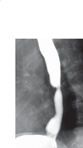
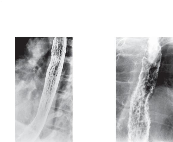
Findings
CASE 1.6. Double-contrast esophagram. Luminal irregularity and narrowing are present in the distal esophagus with associated asymmetric sacculations.
CASE 1.7. Single-contrast esophagram. A short esophagus hiatal hernia has irregularity and outpouchings about the lower esophageal segment.
Di erential Diagnosis
1.Active refl ux esophagitis
2.Chronic reflux esophagitis
3.Esophageal carcinoma
Diagnosis
Chronic reflux esophagitis
1. ESOPHAGUS 7
CASE 1.7
Discussion
Chronic and active changes of reflux esophagitis often coexist. It may be impossible to exclude active ulceration or carcinoma in patients with marked deformity. Endoscopy should be recommended for patients with such problematic findings. Changes of chronic reflux esophagitis can result in scarring and considerable deformity of the esophagus.
Disease type: Inflammatory and Ulcerative Diseases
8MAYO CLINIC GASTROINTESTINAL IMAGING REVIEW
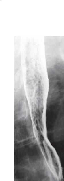
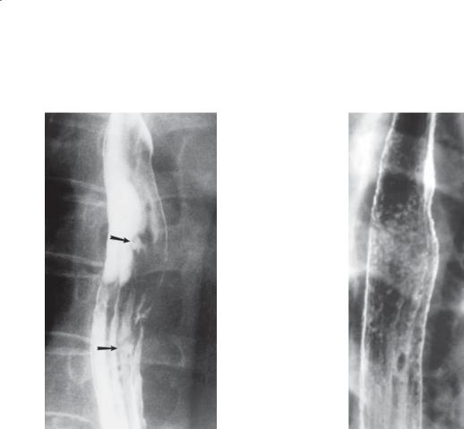
CASE 1.8
Findings
Double-contrast esophagram. Multiple tiny outpouchings are seen within the mid esophagus around a localized stricture.
Di erential Diagnosis
Intramural esophageal pseudodiverticulosis
Diagnosis
Intramural esophageal pseudodiverticulosis
Discussion
Intramural esophageal pseudodiverticulosis is seen as multiple tiny outpouchings that either diffusely or segmentally affect the esophagus. Because the tiny
necks may not fill completely, there is an apparent lack of communication with the esophageal lumen. Pathologically, these outpouchings are dilated submucosal glands, most commonly due to chronic reflux esophagitis. Elderly patients are most frequently affected and complain of progressive dysphagia due to the often-associated esophageal strictures. Up to 90%
have an associated smooth stricture in the mid or upper esophagus. Dilation of the stricture usually cures the symptoms. The radiographic appearance is analogous to Rokitansky-Aschoff sinuses in the gallbladder and
is virtually pathognomonic of this condition. Candida organisms are frequently cultured from the esophagus in patients with this condition, but they are believed to be a secondary invader rather than a causative factor.
Disease type: Inflammatory and Ulcerative Diseases
CASE 1.9
Findings
Single-contrast esophagram. A short stricture is present in the mid esophagus, with some luminal irregularity at the level of the stricture. A short esophagus-type hiatal hernia also is visible.
Di erential Diagnosis
1.Barrett esophagus stricture
2.Refl ux stricture
3.Medication-induced esophagitis or stricture
4.Caustic ingestion-induced stricture
Diagnosis
Barrett esophagus stricture
1. ESOPHAGUS 9
Discussion
A focal esophageal stricture above the gastroesophageal junction is suggestive of Barrett esophagus. Barrett esophagus is histologically described as metaplasia and replacement of the normal squamous epithelium with gastric-type adenomatous mucosa. Chronic reflux esophagitis is nearly always the underlying cause of the disease. Some authorities estimate that nearly
10% of patients with reflux esophagitis have some adenomatous transformation in the esophagus.
Th is case is a typical example of Barrett esophagus. The short-esophagus hiatal hernia indicates longstanding esophagitis. Usual reflux-induced strictures (case 1.20) are located immediately above the gastroesophageal junction. Because adenomatous tissue is acid-resistant, strictures in Barrett esophagus develop near the squamous-adenomatous transition zone. Barrett strictures commonly are seen in the upper and mid esophagus. Medication-induced esophagitis (case 1.13) usually occurs at areas of anatomical narrowing— thoracic inlet, near aortic arch, or left mainstem bronchus. Caustic strictures (case 1.25) are usually long and associated with a typical history of caustic ingestion.
Disease type: Inflammatory and Ulcerative Diseases
10 MAYO CLINIC GASTROINTESTINAL IMAGING REVIEW
CASE 1.10 |
CASE 1.11 |
Findings
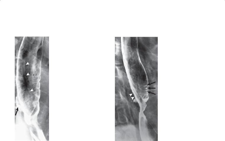
CASE 1.10. Double-contrast esophagram. A featureless distal esophagus is present. Above the featureless zone are findings of active reflux esophagitis (mucosal granularity and superficial erosions [arrows]).
CASE 1.11. Double-contrast esophagram. Mucosal reticularity is present in the esophagus. There is a short esophagus-type hiatal hernia. A surgically induced deformity mimics an ulcer crater. A benign-appearing stricture also is present.
Di erential Diagnosis
1.Barrett esophagus with active reflux esophagitis
2.Refl ux esophagitis
3.Candida esophagitis
Diagnosis
Barrett esophagus
Discussion
It is important to recognize Barrett esophagus because it can develop in 5% to 20% of patients with chronic reflux esophagitis and the adenomatous tissue is
at increased risk for malignant transformation. Reports suggest that the risk for development of adenocarcinoma is less than 1% per year. Barrett esophagus is a major risk factor for development of esophageal cancer.
Reticular changes of Barrett esophagus are often adjacent to an esophageal stricture. The nodularity and crevices may be smaller and more delicate than in case 1.11. The plaquelike filling defects in patients with candidiasis (cases 1.15 and 1.16) usually involve the entire esophagus and are not associated with other changes of reflux esophagitis. Upper endoscopy is the
most accurate method for detecting Barrett esophagus, but radiologists should be aware of key findings when they are present.
Disease type: Inflammatory and Ulcerative Diseases
CASE 1.12
Findings
Double-contrast esophagram. Multiple nodular filling defects are throughout the esophagus.
Di erential Diagnosis
1.Refl ux esophagitis
2.Glycogen acanthosis
3.Candida esophagitis
Diagnosis
Glycogen acanthosis
1. ESOPHAGUS 11
Discussion
Glycogen acanthosis is an occasional incidental finding in older patients, caused by increased cytoplasmic glycogen within the squamous epithelial cells of the esophagus. Radiographically, nodular filling defects are seen, ranging in size from 1 to 15 mm. Rarely, nodules coalesce to form larger plaques. Often, the margins of these lesions are hazy and fade peripherally.
Reflux esophagitis is the main differential consideration. Candida esophagitis also may have a nodular mucosal appearance. However, patients
with Candida esophagitis almost always present with odynophagia, unlike patients with glycogen acanthosis, who are almost always asymptomatic.
Disease type: Inflammatory and Ulcerative Diseases
12 MAYO CLINIC GASTROINTESTINAL IMAGING REVIEW
CASE 1.13
Findings
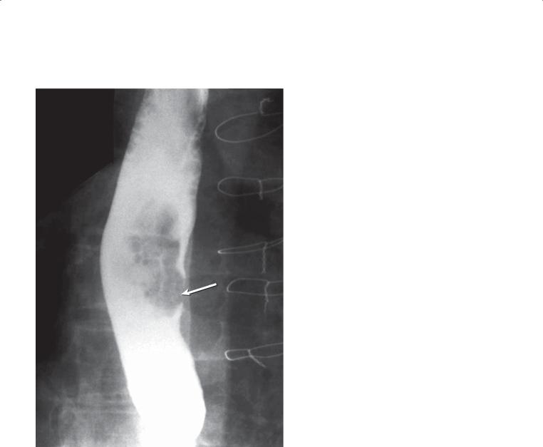
Single-contrast esophagram. An irregular, linearly oriented ulceration with a surrounding soft tissue mass (arrow) is present in the mid esophagus. There is also a region of reduced distensibility on the opposite wall.
Di erential Diagnosis
1.Infectious esophagitis (cytomegalovirus, human immunodeficiency virus)
2.Early esophageal carcinoma
3.Medication-induced esophagitis
Diagnosis
Medication-induced esophagitis
Discussion
Localized inflammation due to medication lodged in the esophagus can lead to focal edema, ulceration, and stricture formation. Pills can lodge anywhere within the esophagus, but usually do so at the arch level or within the distal esophagus. Patients usually complain of odynophagia, dysphagia, and retrosternal pain.
Radiographic findings are usually single or multiple shallow ulcerations. Associated fold thickening also may be seen. Double-contrast examination of the esophagus is sensitive for detecting these small, subtle ulcers. Endoscopy is required to exclude other differential possibilities unless the patient’s history is typical.
Disease type: Inflammatory and Ulcerative Diseases
1. ESOPHAGUS 13
CASE 1.14
Findings
Double-contrast esophagram. Segmental region of deep ulceration affects a noncircumferential portion of mid esophagus.
Di erential Diagnosis
1.Infectious esophagitis
2.Crohn esophagitis
3.Acute caustic ingestion
4.Esophageal carcinoma
Diagnosis
Crohn esophagitis
Discussion
Crohn disease rarely affects the esophagus; when it does, disease is almost always present in the
small bowel and colon. Most patients seek medical attention because of disease symptoms elsewhere in the alimentary tract or an esophageal stricture that is causing dysphagia.
Th e usual radiographic finding is aphthous ulcers (discrete ulcers surrounded by a mound of edema), which usually require double-contrast radiography for detection. Confluent ulcerations and deep ulcers (as seen in this case) can be found in more severe disease. Transmural disease may lead to esophageal perforation and fistula formation. Progressive scarring can lead to stricture formation.
Th e lack of surrounding mass makes cancer unlikely. Caustic ingestion nearly always involves a larger segment of the esophagus (and ingestion history). The ulceration is larger than that usually found with cytomegalovirus (case 1.19) or human immunodeficiency virus.
Disease type: Inflammatory and Ulcerative Diseases
14 MAYO CLINIC GASTROINTESTINAL IMAGING REVIEW
CASE 1.15 |
CASE 1.16 |
Findings
CASE 1.15. Double-contrast esophagram. Multiple plaquelike filling defects are seen throughout the esophagus.
CASE 1.16. Double-contrast esophagram. Shaggy, irregular luminal contours due to extensive plaquelike filling defects are present throughout the esophagus.
Di erential Diagnosis
1.Candidiasis
2.Refl ux esophagitis
3.Glycogenic acanthosis
4.Herpes esophagitis
Diagnosis
Candidiasis
Discussion
Th ere is a spectrum of radiographic findings associated with candidiasis. Patients may present early with findings only of dysmotility (atonicity and tertiary contractions). Discrete plaquelike lesions are the
most common finding, ranging from a few to diffuse esophageal involvement. Nodularity, granularity, and fold thickening may occur as a result of mucosal inflammation and edema. More severe disease is manifested as a shaggy, irregular luminal surface. Rarely, large filling defects occur as a result of giant plaques and fungus balls.
Reflux esophagitis usually is confined to the lower esophagus and rarely is associated with well-defined plaques. Patients with glycogenic acanthosis are usually asymptomatic and older but could present with similar radiographic findings. Herpes esophagitis occasionally can present with multiple plaquelike filling defects, but discrete ulcerations are more common.
Disease type: Inflammatory and Ulcerative Diseases
CASE 1.17
Findings
CASE 1.17. Double-contrast esophagram. Multiple discrete ulcerations (arrows) are present on a normal esophageal background. (Adapted from Levine MS, Laufer I. Esophagus. In: Levine MS, Rubesin SE, Laufer I, editors. Double contrast gastrointestinal radiology. 3rd ed. Philadelphia [PA]: WB Saunders Company; c2000. p. 90–125. Used with permission.)
CASE 1.18. Double-contrast esophagram. Multiple discrete ulcerations are clustered within the mid esophagus. (Courtesy of Marc S. Levine, MD, Hospital of the University of Pennsylvania, Philadelphia, Pennsylvania. Used with permission.)
Di erential Diagnosis
1.Herpes esophagitis
2.Candidiasis
3.Refl ux esophagitis
4.Cytomegalovirus or varicella esophagitis
Diagnosis
Herpes esophagitis
1. ESOPHAGUS 15
CASE 1.18
Discussion
Radiographically, discrete ulcerations on an otherwise normal esophageal mucosal background are characteristic findings in herpes esophagitis. Multiple plaquelike filling defects, with or without ulcerations, also can be seen.
Differential considerations include candidiasis, which usually presents with plaquelike filling defects (cases 1.15 and 1.16). Other members of the herpesvirus group, including cytomegalovirus and
varicella zoster, can present with identical radiographic findings. Cytomegalovirus infection often presents with a large, solitary, discrete ulcer (case 1.19). Ulcers from reflux esophagitis (cases 1.3 and 1.4) are usually confined to the distal esophagus and associated with hiatal hernia, reflux, and lower esophageal stricture.
Disease type: Inflammatory and Ulcerative Diseases
16 MAYO CLINIC GASTROINTESTINAL IMAGING REVIEW
CASE 1.19
Findings
Double-contrast esophagram. A large, flat ulceration (arrow) is present in the distal esophagus. (Courtesy of Kyunghee C. Cho, MD, NYU Langone Medical Center, New York, New York. Used with permission.)
Di erential Diagnosis
1.Cytomegalovirus esophagitis
2.Human immunodeficiency virus esophagitis
3.Herpes esophagitis
4.Crohn esophagitis
Diagnosis
Cytomegalovirus esophagitis
Discussion
Cytomegalovirus infection is a disorder arising from the herpesvirus family. Most patients with this opportunistic infection have AIDS. The small bowel and colon are most frequently affected. Diagnosis is
usually made histologically by examining endoscopic brushings and biopsy specimens. Intranuclear inclusions are seen within endothelial cells or fibroblasts.
Radiographically, cytomegalovirus esophagitis may be indistinguishable from herpes esophagitis (cases 1.17 and 1.18) or human immunodeficiency virus (HIV) infection with multiple discrete ulcerations. Advanced disease may be associated with extensive ulceration. Giant and flat ulcers suggest cytomegalovirus or HIV infection rather than herpes esophagitis. Cytomegalovirus esophagitis should be considered in any patient with AIDS and esophageal ulcerations.
Disease type: Inflammatory and Ulcerative Diseases
CASE 1.20
Findings
Single-contrast esophagram. A moderate-sized hiatal hernia is present with esophageal shortening and narrowing beginning at the gastroesophageal junction. The margins of the stricture taper gradually.
Di erential Diagnosis
1.Chronic reflux-induced stricture
2.Caustic ingestion-induced stricture
Diagnosis
Chronic reflux esophagitis: focal stricture
1. ESOPHAGUS 17
Discussion
Stricture formation is the most frequent radiographic manifestation of chronic reflux esophagitis. The classic stricture begins immediately above the gastroesophageal junction and extends proximally
a variable distance. Usually the margins are smooth and taper gradually. A mild stricture may present as a subtle region of incomplete esophageal distention. Severe strictures that narrow the lumen to less than
1 cm are nearly always symptomatic—dysphagia is the most frequent complaint. Treatment of symptomatic strictures is usually by either balloon or bougie dilation. An antireflux operation is reserved for patients who do not respond to more conservative therapy. In many cases, fibrosis causes esophageal shortening with the development of a short esophagustype hiatal hernia. Caustic strictures (case 1.25) are usually longer, often involving a longer length of the esophagus.
Disease type: Inflammatory and Ulcerative Diseases
18 MAYO CLINIC GASTROINTESTINAL IMAGING REVIEW
CASE 1.21
Findings
Single-contrast esophagram. A long distal esophageal stricture, with an associated hiatal hernia, is present.
Di erential Diagnosis
1.Caustic ingestion-induced stricture
2.Prolonged nasogastric tube stricture
3.Radiation-induced stricture
4.Blistering skin disorders
5.Chronic reflux-induced stricture
Diagnosis
Chronic reflux esophagitis: long stricture
Discussion
Th is benign-appearing stricture is due to chronic reflux esophagitis. The smooth, tapered margins usually indicate a benign cause. Long strictures such as this are unusual for peptic esophagitis-induced strictures. Other considerations may have to be excluded after review of the patient’s history.
Disease type: Inflammatory and Ulcerative Diseases

|
1. ESOPHAGUS 19 |
|
|
|
|
|
TABLE 1.1 |
|
Peptic and Inflammatory |
|
CASE |
Esophagitis |
|
|
|
|
|
Active reflux esophagitis |
|
|
Mild reflux esophagitis |
Thickened folds only |
1.1 |
Moderate reflux esophagitis |
Thickened folds and tiny linear ulcerations |
1.2 |
Severe reflux esophagitis |
Thickened folds and moderate to large ulcer |
1.3 and 1.4 |
Feline esophagitis |
Delicate, transverse folds, transient |
1.5 |
|
|
|
Chronic reflux esophagitis |
Smooth, tapering stricture immediately above |
1.6–1.8, |
(with or without active disease) |
gastroesophageal junction. Scarring with deformity, |
1.20, 1.21 |
|
transverse folds, intramural pseudodiverticulosis |
|
|
|
|
Barrett esophagus |
Benign stricture at an abnormally high location, |
1.9–1.11 |
|
transition zone with active esophagitis above normal |
|
|
distal segment, or reticulated mucosal pattern |
|
|
|
|
Medication-induced esophagitis |
Single or multiple shallow ulcers, usually at level of |
1.13 |
|
arch or distal esophagus |
|
|
|
|
Crohn esophagitis |
Aphthous ulcers. Confluent ulcers in more |
1.14 |
|
severe disease |
|
|
|
|
Infectious Esophagitis |
|
|
|
|
|
Candidiasis |
Multiple plaquelike filling defects |
1.15 and 1.16 |
|
Advanced disease has shaggy appearance |
|
|
|
|
Herpes esophagitis |
Multiple discrete ulcers, normal mucosa |
1.17 and 1.18 |
|
between lesions |
|
|
|
|
Cytomegalovirus and human |
Large, flat, solitary ulcer. Radiographically |
1.19 |
immunodeficiency virus |
indistinguishable |
|
esophagitis |
|
|
|
|
|
Miscellaneous |
|
|
|
|
|
Glycogen acanthosis |
Multiple filling defects in elderly, asymptomatic patient |
1.12 |
|
|
|
Disease type: Inflammatory and Ulcerative Diseases

20 MAYO CLINIC GASTROINTESTINAL IMAGING REVIEW
CASE 1.22
Findings
Single-contrast esophagram. Dilatation of the esophagus with a beaklike deformity near the gastroesophageal junction. Aperistalsis of the smooth muscle portion (distal two-thirds) was observed.
Di erential Diagnosis
1.Achalasia
2.Scleroderma
3.Refl ux-induced stricture
4.Chagas disease
5.Carcinoma of gastroesophageal junction (pseudoachalasia)
Diagnosis
Achalasia
Discussion
Achalasia is a motor disorder of the esophagus characterized by aperistalsis of the distal two-thirds (smooth muscle portion) of the esophagus and failure of the lower esophageal sphincter to relax. Patients usually complain of dysphagia, recumbent regurgitation, and episodes of aspiration pneumonia.
Radiographically, the esophagus is often dilated, and a standing column of contrast material is seen (the height of the column is proportional to the severity of lower esophageal obstruction). Periodic relaxation of the lower esophageal sphincter with continued drinking is a critical observation for distinguishing primary achalasia from pseudoachalasia—carcinoma of the gastroesophageal junction (a fixed, nonrelaxing obstruction) (case 1.24). Manometry is the standard for diagnosing achalasia.
Transesophageal balloon dilation and surgical incision of the lower esophageal muscle fibers (Heller myotomy) are the primary treatment options.
Patients with scleroderma have a patulous gastroesophageal junction, esophagitis, and a lower esophageal stricture. In reflux esophagitis, peristalsis remains normal. Chagas disease may be indistinguishable from achalasia.
Disease type: Strictures
CASE 1.23
Findings
Single-contrast UGI. The esophagus is dilated to the gastroesophageal junction. The lower esophageal segment has smooth, tapered narrowing and intermittent, transient opening. In addition, multiple nonpropulsive contractions are present in the lower esophageal segment. Normal stripping wave is not observed.
Di erential Diagnosis
1.Achalasia
2.Carcinoma gastroesophageal junction (pseudoachalasia)
3.Refl ux-induced stricture
4.Scleroderma
5.Chagas disease
Diagnosis
Vigorous achalasia
1. ESOPHAGUS 21
Discussion
Vigorous achalasia is considered a less severe or early form of achalasia with motor abnormalities similar to those of achalasia seen fluoroscopically, in addition to repetitive, simultaneous, nonpropulsive contractions. Patients may have chest pain and typical dysphagia.
Esophageal dilatation may be only minimal. Complications from achalasia include carcinoma
and Candida esophagitis. Carcinomas are squamous cell type, developing after 20 years of achalasia. The upper and mid esophagus are common locations for these cancers. Cancer surveillance should begin after a disease duration of 10 to 15 years. Candidiasis (cases 1.15 and 1.16) develops from chronic stasis.
Endoscopic treatment with botulinum toxin does not provide long-term benefit. The value of endoscopic myotomy, esophageal stents, and robotic-assisted myotomy is not currently established.
Disease type: Strictures
22 MAYO CLINIC GASTROINTESTINAL IMAGING REVIEW
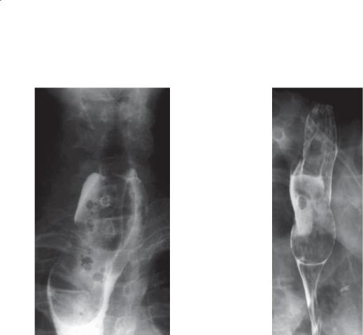
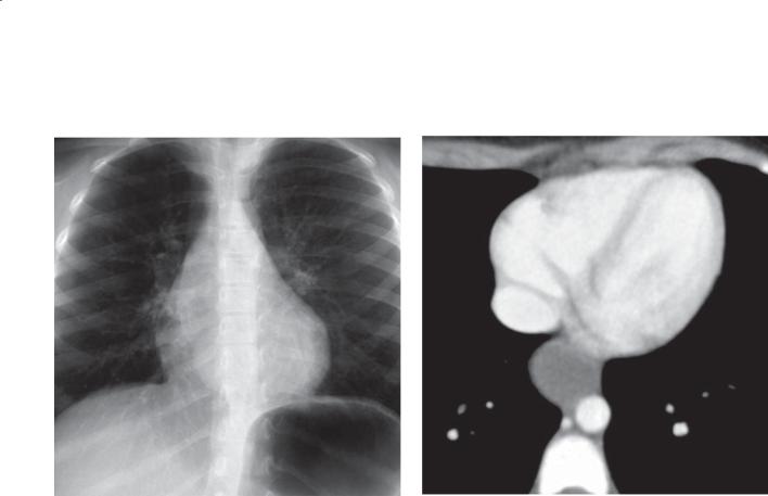
CASE 1.24
A B
Findings
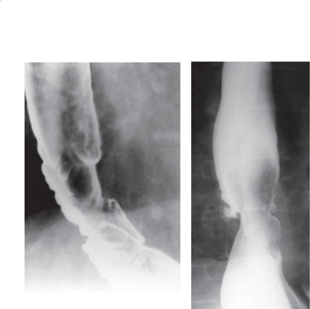
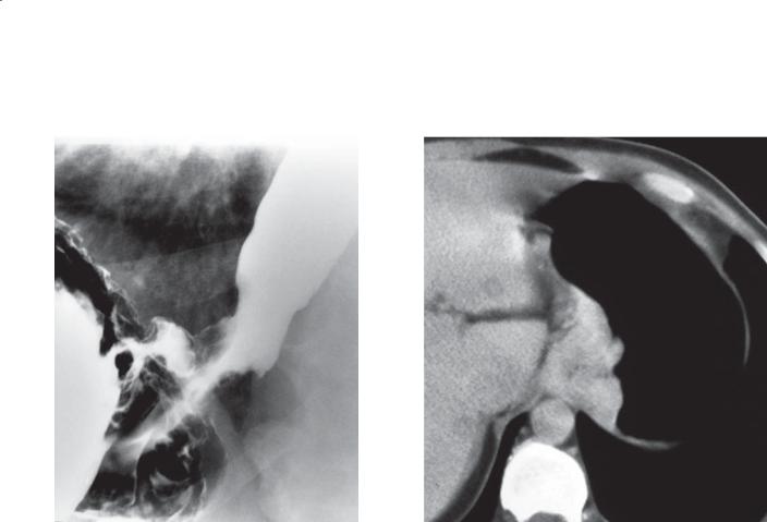
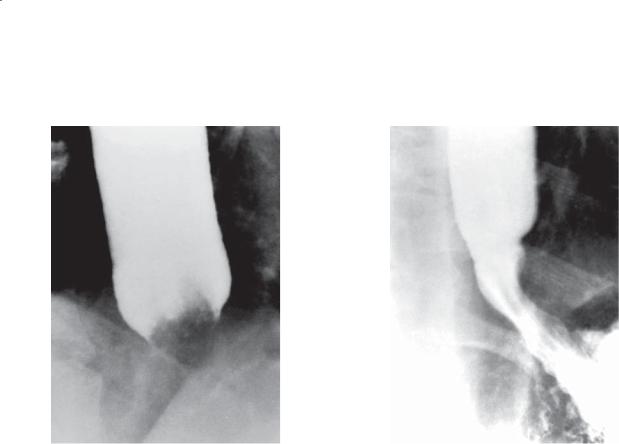
A.Single-contrast esophagram. Fixed, smooth narrowing is present in distal esophagus. Proximal esophagus is dilated. At fluoroscopy, narrowing remained unchanged, even after esophagus was half filled with contrast material.
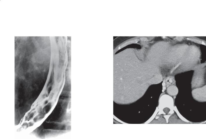
B.Abdominal CT with oral contrast. A soft tissue mass (arrows) causing gastric wall thickening is present at the level of the gastroesophageal junction. The mass extends into the distal esophagus.
Di erential Diagnosis
1.Carcinoma of gastroesophageal junction (pseudoachalasia)
2.Achalasia
Diagnosis
Carcinoma of the gastroesophageal junction (pseudoachalasia)
Discussion
Tumors such as this have predominantly submucosal growth and often arise from a gastric primary lesion. The radiographic findings can mimic those of achalasia. The terms pseudoachalasia and secondary achalasia have been used for such cases. On careful fluoroscopic observation of the cancerous esophagogastric junction during swallowing, a fixed, rigid stricture is seen
that never distends during esophageal emptying. The esophagogastric junction in achalasia (case 1.22) opens transiently and distends when the hydrostatic barium column in the esophagus exceeds lower esophageal sphincter muscle tone. Peristalsis of the distal esophagus is absent in both conditions. The exact mechanism for this motor abnormality in patients with pseudoachalasia is uncertain.
CT may be helpful when findings are equivocal, because a soft tissue mass may be visible in patients with tumor.
Disease type: Strictures
1. ESOPHAGUS 23
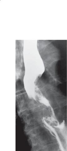
CASE 1.25
Findings
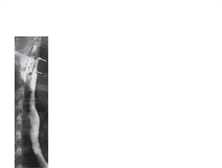
Single-contrast esophagram. A long, narrow stricture involves the mid and distal esophagus.
Di erential Diagnosis
1.Caustic stricture
2.Stricture due to bullous skin disease
3.Radiation stricture
4.Prolonged nasogastric tube stricture
5.Chronic reflux stricture
Diagnosis
Caustic (lye-induced) stricture
Discussion
Caustic damage to the esophagus can occur from several agents, including alkali (lye), acid, phenols,
ammonium chloride, and silver nitrate. The extent of damage depends on the concentration of the agent, the amount ingested, and the duration of mucosal contact. Radiographic examinations are helpful for documenting the severity and extent of stricture formation.
Radiographic findings of acute caustic esophagitis include dysmotility, irregular margins, edema, ulcerations, plaques of sloughing mucosa, and occasionally an intramural collection of contrast material. Esophageal strictures usually develop 1 to 3 months after injury. The strictures may be focal and confined to the upper esophagus, or the esophagus may be diffusely involved with asymmetric regions of narrowing and sacculations. Patients are also at increased risk (1%-4%) of esophageal cancer after
20 years.
Disease type: Strictures
24 MAYO CLINIC GASTROINTESTINAL IMAGING REVIEW
CASE 1.26
A B
Findings
Single-contrast esophagram. Frontal (A) and lateral (B) images show persistent multifocal strictures in the cervical and upper thoracic esophagus.
Di erential Diagnosis
1.Caustic ingestion strictures
2.Strictures due to medication-induced esophagitis
3.Strictures due to bullous disease
Diagnosis
Cicatricial pemphigoid strictures (benign mucous membrane pemphigoid)
Discussion
Th is patient had a known history of cicatricial pemphigoid with characteristic blistering lesions of the oral mucosa and face. Bullous diseases of the esophagus include cicatricial pemphigoid and epidermolysis bullosa dystrophica. Cicatricial
pemphigoid is a rare (1:20,000), chronic, autoimmune blistering disorder involving the mucous membranes of the mouth, eyes, nose, esophagus, larynx, urethra, and anus. Peak incidence is between 60 and 80 years of age. Transient skin lesions (small blisters and erosions) occur in more than 20% of patients and usually are located on the head and neck. Biopsy often is required for diagnosis. Patients generally are treated with corticosteroids, the results of which are variable. Chronic changes of the disease in the esophagus often result in strictures. Scarring due to other causes of esophagitis also could present as multifocal strictures. Caustic (lye) ingestion (case 1.25) usually results in a long esophageal stricture, but multifocal strictures also could occur with variable areas of mucosal damage and healing.
Disease type: Strictures
CASE 1.27
Findings
Double-contrast esophagram. A long, smooth stricture is present in the mid esophagus.
Di erential Diagnosis
1.Radiation stricture
2.Caustic stricture
3.Nasogastric tube peptic stricture
4.Zollinger-Ellison reflux stricture
Diagnosis
Radiation stricture
1. ESOPHAGUS 25
Discussion
Th is patient had radiation therapy 1 year previously for lung cancer. Severe esophagitis and strictures can occur with high-dose (generally >50 Gy) radiation therapy to the chest. Acute radiation esophagitis usually occurs 1 to 4 weeks after the start of radiation therapy, whereas strictures often develop 4 to 8 months after its completion. Long-segment esophageal strictures also can be caused by caustic ingestion (case 1.25), peptic stricture due to acid reflux with long-term nasogastric tube placement, or reflux stricture in Zollinger-Ellison syndrome. However, these strictures almost always extend to the gastroesophageal junction. A radiation stricture should be a primary diagnostic consideration for a long, smooth esophageal stricture that spares the distal esophagus.
Disease type: Strictures
26 MAYO CLINIC GASTROINTESTINAL IMAGING REVIEW
CASE 1.28 |
CASE 1.29 |
Findings
CASE 1.28. Double-contrast esophagram. Esophagus is diffusely narrowed.
CASE 1.29. Single-contrast esophagram. Multiple ringlike narrowings within the upper thoracic esophagus are associated with luminal narrowing.
Di erential Diagnosis
1.Lye stricture
2.Radiation-induced stricture
3.Prolonged use of nasogastric tube
4.Refl ux esophagitis
5.Eosinophilic esophagitis
6.Blistering skin disorder
Diagnosis
Eosinophilic esophagitis
Discussion
Eosinophilic esophagitis is a chronic immune-mediated disorder characterized by dysphagia and eosinophilia within the esophageal mucosa at biopsy. Some
patients have esophageal dysmotility that contributes considerably to their dysphagia. Some patients have a history of a food impaction, allergies, or peripheral eosinophilia; however, none of these factors are required for diagnosis. Topical corticosteroid therapy
has been reported to be successful for these symptoms. Radiographically, the spectrum of findings includes
a normal esophagus (with symptoms attributed to dysmotility). A diffusely small-caliber esophagus (case 1.28), a short stricture, strictures with ringlike indentations (case 1.29), and a granular mucosa are findings at esophagography.
A clinical history is helpful for narrowing the differential list. Biopsy confirmation is usually necessary for definitive diagnosis.
Disease type: Strictures
CASE 1.30
A
Findings
A.Single-contrast esophagram. Extrinsic compression and narrowing involve the mid esophagus.
B.Contrast-enhanced CT. Bulky mediastinal and hilar lymphadenopathy is present.
Di erential Diagnosis
1.Mediastinal adenopathy
2.Esophageal carcinoma
Diagnosis
Extrinsic esophageal compression by lymphomatous mediastinal adenopathy
1. ESOPHAGUS 27
B
Discussion
Lymphomatous involvement of the esophagus is unusual. Usually patients have known generalized lymphoma before esophageal disease is discovered. This disease can affect the esophagus by mass effect or by direct spread from enlarged, contiguous lymph nodes. Lymphomatous involvement of the stomach can spread directly to the esophagus, or a lesion can develop in the esophagus simultaneously with disease elsewhere in the body. Benign inflammatory causes of mediastinal adenopathy also can result in extrinsic narrowing of the esophagus.
Disease type: Strictures

28 MAYO CLINIC GASTROINTESTINAL IMAGING REVIEW
TABLE 1.2
Esophageal Strictures |
|
CASE |
|
|
|
Peptic stricture |
Immediately above gastroesophageal junction, often above |
1.20 and 1.21 |
|
esophageal hiatal hernia. Smooth, tapered margins |
|
|
|
|
Barrett esophagus |
Usually seen in mid esophagus near squamous-adenomatous |
1.9 |
stricture |
transition zone |
|
|
|
|
Carcinoma |
Irregular luminal contour with abrupt, shouldered margins |
1.37 |
|
|
|
Achalasia |
2-cm smooth stricture in region of gastroesophageal junction. |
1.22 and 1.23 |
|
Transiently relaxes with column of barium in esophagus while |
|
|
patient is standing |
|
|
|
|
Pseudoachalasia |
Carcinoma of the gastroesophageal junction. Fixed obstruction, |
1.24 |
|
never relaxes with full column of esophageal barium |
|
|
|
|
Caustic stricture |
Usually long and very narrow. Childhood injury, prolonged |
1.25 |
|
nasogastric tube common |
|
|
|
|
Blistering skin disorders |
Strictures, webs, cicatricial deformity. Epidermolysis bullosa |
1.26 |
|
dystrophica or benign mucous membrane pemphigoid |
|
|
|
|
Radiation stricture |
Long, narrow, smooth stricture. History of radiation therapy |
1.27 |
|
|
|
Eosinophilic esophagitis |
Diffusely narrowed lumen, benign stricture, stricture with |
1.28 and 1.29 |
|
multiple rings, dysmotility |
|
|
|
|
Lymphoma |
Extrinsic compression by mediastinal adenopathy |
1.30 |
|
|
|
Disease type: Strictures
1. ESOPHAGUS 29
CASE 1.31
A B
1
2
3 
 4
4
5 |
3 |
6
5
6
7
8
Findings
Double-contrast esophagram. Normal anatomic structures on the lateral (A) and frontal (B) views.
1.Soft palate
2.Tongue base
3.Epiglottis
4.Posterior wall pharynx
5.Vallecula
6.Pyriform sinuses
7.Esophagus
8.Trachea
Diagnosis
Normal oropharynx
Discussion
Performing swallowing studies requires knowledge of normal oropharyngeal anatomy. During a normal swallow, the soft palate elevates and seals the pharynx from the nasal cavity, preventing nasopharyngeal regurgitation. The pharynx elevates and contracts, propelling food toward the esophagus as the epiglottis tilts posteriorly to seal the laryngeal vestibule. Contrast material entering the laryngeal vestibule
without crossing the vocal cords is termed penetration. Contrast material that enters the trachea (beyond
the vocal cords) is termed aspiration. Different food consistencies (thin liquid, thick liquid, nectar,
honey, paste, solid) can be evaluated in patients with penetration or aspiration, because thicker foods may be swallowed more safely. Various maneuvers can also be attempted to assess for safe passage of liquids and solids. These include tucking the chin, turning the head, and supraglottic swallow.
Disease type: Masses and Filling Defects
30 MAYO CLINIC GASTROINTESTINAL IMAGING REVIEW
CASE 1.32
A B
Findings
Double-contrast esophagram. A. Abnormal soft tissue mass is present in region of tongue base
(1). The epiglottis (2) is tilted posteriorly. B. Incomplete filling of the right vallecula.
Di erential Diagnosis
Pharyngeal tumor arising from base of tongue
Diagnosis
Squamous cell carcinoma of base of tongue
Discussion
Pharyngeal tumors can be difficult to detect without magnified spot radiographs. This relatively large tumor was difficult to appreciate at fluoroscopy, but it is seen well on the lateral and anteroposterior radiographs. Patients with suspicious lesions should be referred for laryngoscopy and biopsy, if indicated.
Disease type: Masses and Filling Defects
CASE 1.33
A
Findings
A.Double-contrast esophagram. There is a smooth impression on the esophageal contour by a wellcircumscribed mass. The lateral margins of the mass (arrows) extend to the right of the spine.
B.Contrast-enhanced CT of the chest. A wellcircumscribed, exophytic, enhancing tumor (arrow) is seen along the right side of the distal esophagus.
Di erential Diagnosis
1.Leiomyoma
2.Gastrointestinal stromal tumor (GIST)
3.Fibroma, neuroma, neurofibroma, lipoma, hemangioma
4.Duplication cyst
5.Lymphoma
Diagnosis
Leiomyoma
1. ESOPHAGUS 31
B
Discussion
Th e location of the mass in relationship to the esophagus, its smooth surface, and the obtuse angle between the mass and the esophageal lumen suggest a submucosal mass. Leiomyomas are the most frequently occurring submucosal neoplasm of the esophagus, accounting for more than half of all benign esophageal tumors. They are more common in the mid esophagus and distal esophagus—segments where smooth muscle is most abundant. Approximately 3% to 4% of tumors are multiple.
Differential considerations include GISTs and other types of intramural tumors such as fibromas,
neuromas, neurofibromas, lipomas, and hemangiomas. GISTs are mesenchymal neoplasms that do not arise from smooth muscle cells, but they can have an identical radiologic appearance. Enteric duplication cysts or a lymphomatous mass could also have a similar appearance.
A focal and enhancing mass makes a neoplasm most likely at CT. Duplication cysts often show water attenuation and do not enhance with intravenous contrast material.
Disease type: Masses and Filling Defects
32 MAYO CLINIC GASTROINTESTINAL IMAGING REVIEW
CASE 1.34
Findings
Single-contrast esophagram. A lobulated filling defect is present in the distal esophagus.
Di erential Diagnosis
1.Adenoma
2.Papilloma
3.Infl ammatory esophagogastric polyp
Diagnosis
Adenoma
Discussion
Adenomas of the esophagus are rare lesions arising from adenomatous tissue in the distal esophagus,
usually within Barrett esophagus. Malignant degeneration has been reported in these lesions; therefore, endoscopic removal is recommended. Large polyps are more likely to be malignant than small polyps.
Esophageal papillomas (fibrovascular excrescences that are covered with squamous epithelium) can have a similar radiographic appearance and are probably more common than previously believed. These polyps are usually removed endoscopically, but no malignant transformation has been reported in humans. Inflammatory esophagogastric polyps (case 1.35) also could be confused with adenomas; however, these polypoid protuberances are enlarged gastric folds that protrude into the lower esophagus.
Disease type: Masses and Filling Defects
CASE 1.35
Findings
Double-contrast esophagram. An enlarged polypoid fold extends from the stomach into the esophageal lumen.
Di erential Diagnosis
1.Infl ammatory esophagogastric polyp
2.Adenoma
3.Papilloma
Diagnosis
Inflammatory esophagogastric polyp
1. ESOPHAGUS 33
Discussion
Th e inflammatory esophagogastric polyp is an enlarged gastric fold that projects into the lower esophagus. Inflammation is due to reflux esophagitis in affected patients. The polyp has no malignant potential. Usual radiographic features are a clubbed, bulbous fold that arises from the fundus of the stomach and projects into the lower esophagus. Occasionally, it may be difficult to differentiate an inflammatory esophagogastric polyp from a papilloma or an adenoma. In these cases, endoscopy is recommended to obtain a definitive diagnosis.
Disease type: Masses and Filling Defects
34 MAYO CLINIC GASTROINTESTINAL IMAGING REVIEW
CASE 1.36
A B
Findings
Double-contrast esophagram. A and B. A large, smooth, intraluminal mass arising from a thin stalk is present in the cervical esophagus. The esophagus is dilated around the intraluminal mass.
Di erential Diagnosis
1.Fibrovascular polyp
2.Adenomatous polyp
3.Spindle cell carcinoma (carcinosarcoma)
4.Intraluminal gastrointestinal stromal tumor
Diagnosis
Fibrovascular polyp
Discussion
Fibrovascular polyps are benign, pedunculated, intraluminal lesions composed of various mesenchymal elements. They are covered by normal squamous esophageal mucosa and usually occur in the
cervical esophagus. In the past, fibrovascular polyps were named according to the predominant histologic component (fibroepithelial polyps, pedunculated lipomas, fibrous lipomas, fibromyxomas, fibrolipomas). Fibrovascular polyps are typically longer than 7 cm and can exceed 20 cm in length. Occasionally, they present dramatically with regurgitation of the polyp into the pharynx. In these cases, they can result in respiratory compromise or even death. Fibrovascular polyps are not at risk for malignant transformation. Treatment
of these lesions is surgical or endoscopic excision. Fibrovascular polyps often have a considerable lipomatous component and have fatty density on CT.
Adenomatous polyps (case 1.34) rarely attain this size. Spindle cell carcinomas (case 1.45) can arise from a stalk, but the site of attachment is usually in the mid or distal esophagus. Gastrointestinal stromal tumor is also a diagnostic consideration, but it rarely has this large an endoluminal component and does not arise from a stalk.
Disease type: Masses and Filling Defects
1. ESOPHAGUS 35
CASE 1.37
Findings
Single-contrast esophagram. A large, centrally ulcerated mass with an irregular luminal contour and an abrupt inferior edge is present in the mid esophagus.
Di erential Diagnosis
1.Primary esophageal carcinoma
2.Metastases
3.Lymphoma
Diagnosis
Ulcerative esophageal carcinoma (squamous cell)
Discussion
Features of a malignant mass include an irregular contour of the barium-filled lumen, abrupt (shouldered) edges, and an ulcer that does not project beyond the expected location of the normal esophageal mucosa. This case illustrates these features
of a malignancy. Esophageal carcinoma is a highly lethal disease that usually presents with chest pain or progressive dysphagia. Symptomatic tumors are often large and of advanced stage. Small tumors are
usually discovered incidentally. Important risk factors in the development of esophageal cancer include tobacco and alcohol consumption, sex (threefold to fourfold increase in males), race (twice as common in nonwhites), lye ingestion, achalasia, prior head and neck cancer, nontropical sprue, tylosis, PlummerVinson syndrome, and exposure to tannins and nitrosamines. Tobacco and alcohol consumption are the most important risk factors in the United States.
Squamous cell carcinomas and adenocarcinomas are the most common histologic types of esophageal cancers. Squamous cell tumors do not usually cross the esophagogastric junction. Squamous cell carcinoma
is the most common esophageal cancer worldwide (90%-95%), but it accounts for less than half of esophageal cancers in the United States.
Disease type: Masses and Filling Defects
36 MAYO CLINIC GASTROINTESTINAL IMAGING REVIEW
CASE 1.38
Findings
Double-contrast esophagram. A large polypoid filling defect with irregular borders and abruptly shouldering edges is present within the distal esophagus.
Di erential Diagnosis
1.Esophageal carcinoma
2.Spindle cell carcinoma (carcinosarcoma)
3.Lymphoma
Diagnosis
Polypoid esophageal adenocarcinoma
Discussion
An esophageal adenocarcinoma, spindle cell carcinoma (carcinosarcoma) (case 1.45), or lymphoma (case 1.44) should be suspected when a bulky polypoid tumor is identified. Squamous cell carcinomas usually present as an annular constricting mass. Adenocarcinomas nearly always arise within Barrett esophagus and are therefore usually located in the distal esophagus. Esophageal adenocarcinomas are increasing in frequency because of an increase in the number of patients with Barrett esophagus, and they now account for 50% to 80% of all esophageal cancers in the United States.
Disease type: Masses and Filling Defects

CASE 1.39
Findings
Single-contrast esophagram. A large mass is present in the distal esophagus with associated thickened and nodular folds. An abrupt edge forms the superior margin of the tumor.
Di erential Diagnosis
1.Primary esophageal carcinoma
2.Varices
Diagnosis
Varicoid esophageal adenocarcinoma
1. ESOPHAGUS 37
Discussion
Most esophageal tumors do not have a varicoid appearance. This type of adenocarcinoma has a substantial proportion of tumor extending within the submucosa, causing the distorted fold appearance. Tumors are fixed and rigid, unchanging with swallowing. Usually a peristaltic wave will not pass through such a diseased esophageal segment. Varices change shape, especially with passage of a peristaltic stripping wave (cases 1.46 and 1.47).
Disease type: Masses and Filling Defects
38 MAYO CLINIC GASTROINTESTINAL IMAGING REVIEW
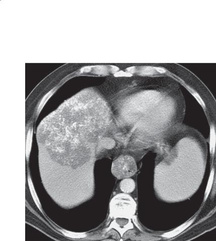
CASE 1.40
Findings
Contrast-enhanced CT. Marked distal esophageal wall thickening and calcification are present. A large, finely calcified hepatic mass is also visible.
Di erential Diagnosis
1.Primary esophageal carcinoma
2.Lymphoma
3.Peptic esophagitis
4.Infectious esophagitis
Diagnosis
Esophageal carcinoma with hepatic metastases
Discussion
Th e normal esophageal wall is less than 3 mm thick. Wall thickening by itself is a nonspecific finding; both benign and malignant processes can have
this appearance. Often, endoscopy is necessary to determine the cause of the wall thickening. Detection of paraesophageal invasion can be difficult, especially if the patient has a paucity of mediastinal fat or the fat planes are obliterated by operative or radiation therapy. Intravenously administered contrast material may help display tissue boundaries better
in some patients with questionable findings. The presence of enlarged nodes (1.0–1.5 cm) suggests metastases; however, the absence of adenopathy does not exclude disseminated disease. Metastases to normal-sized lymph nodes is common and not reliably detected on CT. In addition, enlarged nodes can be due to benign inflammatory disease. Acute inflammatory changes within the esophagus can sometimes be distinguished from tumor infiltration by visualization of water attenuation within the thickened wall.
Disease type: Masses and Filling Defects
CASE 1.41
Findings
CASE 1.41. Double-contrast barium esophagram. Distal esophagus has irregular luminal narrowing and nodularity.
CASE 1.42. Contrast-enhanced CT. A circumferential soft tissue mass encases the esophagogastric junction.
Di erential Diagnosis
Primary esophageal carcinoma
Diagnosis
Infiltrative adenocarcinoma of the gastroesophageal junction
1. ESOPHAGUS 39
CASE 1.42
Discussion
Esophageal adenocarcinomas nearly all arise from Barrett esophagus or from a fundal carcinoma extending into the esophagus. The spread of adenocarcinoma resembles that of squamous cell carcinoma, except there is a higher likelihood
of involvement of the gastric cardia or fundus. Radiographic features that suggest an adenocarcinoma rather than a squamous carcinoma include distal location, gastric invasion, polypoid morphologic appearance, and evidence for chronic reflux esophagitis. CT can be used to identify mediastinal invasion and subdiaphragmatic extent of tumor. If extensive metastases are identified, the patient often receives chemotherapy and radiation therapy rather than surgery. CT often understages tumors at the gastroesophageal junction.
Disease type: Masses and Filling Defects
40 MAYO CLINIC GASTROINTESTINAL IMAGING REVIEW
CASE 1.43
Findings
Double-contrast esophagram. There is a polypoid, lobulated filling defect in the distal esophagus, and an associated extrinsic mass displaces the esophagus posteriorly.
Di erential Diagnosis
1.Esophageal carcinoma
2.Metastases
Diagnosis
Lung cancer with metastatic invasion into the esophagus
Discussion
CT (not shown) showed extensive mediastinal lymphadenopathy causing the changes on the esophagram. Metastases to the esophagus usually arise from cancers of the stomach, lung, breast, or ovary. Most of these tumors metastasize to
mediastinal lymph nodes, and with growth they can displace or directly invade the esophagus. In some patients, the abnormality may resemble a primary esophageal cancer or present as a long, narrow stricture. Displacement of the lumen by a nodal mass is common. The mid esophagus is most commonly affected because mediastinal lymph nodes are most often affected. Hematogenous metastases to the esophagus are unusual.
Disease type: Masses and Filling Defects
1. ESOPHAGUS 41
CASE 1.44
Findings |
Discussion |
|
Esophagram. A large lobulated and ulcerated mass |
Th e appearance of lymphoma can closely |
|
constricts the distal esophagus. |
resemble that of primary carcinoma. The most |
|
|
|
common intrinsic manifestation of lymphoma is a |
Di erential Diagnosis |
polypoid, ulcerative, or infiltrative mass—usually |
|
1. |
Esophageal carcinoma |
indistinguishable from primary esophageal cancer. |
2. |
Lymphoma |
Less frequently, submucosal infiltration of lymphoma |
3. |
Varices |
results in varicoid folds, discrete small submucosal |
|
|
masses, or diffuse nodular changes. Varices are usually |
Diagnosis |
more serpentine, and they change shape during |
|
Esophageal lymphoma |
fluoroscopic observation (especially from a peristaltic |
|
|
|
stripping wave). |
Disease type: Masses and Filling Defects
42 MAYO CLINIC GASTROINTESTINAL IMAGING REVIEW
CASE 1.45
Findings
Single-contrast esophagram. A large, bulky filling defect is seen within the distal esophagus. A pedicle (arrow) is seen attaching the tumor to the left side of the esophagus.
Di erential Diagnosis
1.Pedunculated gastrointestinal stromal tumor
2.Fibrovascular polyp
3.Spindle cell carcinoma
4.Adenocarcinoma
Diagnosis
Spindle cell carcinoma (carcinosarcoma)
Discussion
Spindle cell carcinomas are rare malignancies that contain histologic elements of both carcinoma and sarcoma. In the past, these tumors have been called carcinosarcomas. These tumors usually appear as bulky, polypoid, intraluminal tumors in the mid or distal esophagus. The mass may expand the esophageal lumen, but obstruction is rare.
Disease type: Masses and Filling Defects
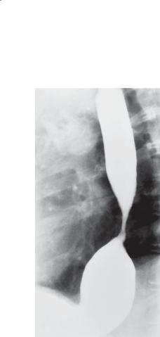
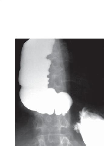
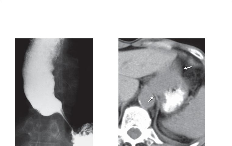
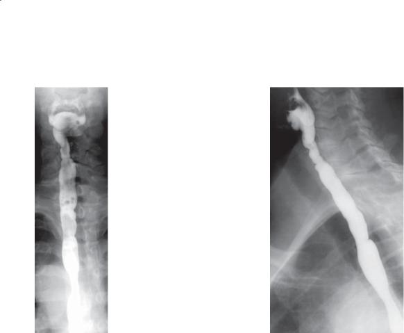
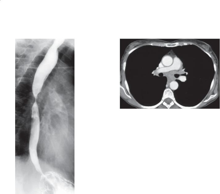
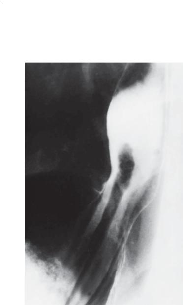
CASE 1.46
Findings
CASE 1.46. Double-contrast esophagram. Multiple serpentine filling defects are seen in the distal esophagus. These defects change in size and shape.
CASE 1.47. Contrast-enhanced CT. Several esophageal varices are present within the distal periesophageal tissues.
Di erential Diagnosis
1.Esophageal varices
2.Varicoid esophageal carcinoma
Diagnosis
“Uphill” esophageal varices
1. ESOPHAGUS 43
CASE 1.47
Discussion
Esophageal varices are dilated submucosal veins that develop as a result of portal venous hypertension. Hepatic cirrhosis is the most frequent cause of portal venous hypertension. Increased portal venous pressure reverses normal venous blood flow such that flow occurs “uphill” from the portal vein, to the left gastric (coronary) vein, to the periesophageal venous plexus, and, finally, to the azygous and hemiazygous collaterals that empty into the superior vena cava. Varices almost never cause dysphagia, but they can bleed and cause life-threatening hemorrhage.
Th e radiographic diagnosis of esophageal varices is most sensitive when the partially collapsed esophagus is examined. Varices can be obliterated by the filled, distended esophagus as well as the collapsed esophagus immediately after a stripping peristaltic wave. Varices appear as linear, often serpentine, filling defects with a scalloped esophageal contour. Uphill varices are more prominent in the distal esophagus. The changing nature of varices with esophageal distention, peristalsis, and respiratory effort is helpful in differentiating varices from varicoid carcinoma (case 1.39) that appears fixed, is noncompliant, and often has shouldered margins.
Disease type: Masses and Filling Defects
44 MAYO CLINIC GASTROINTESTINAL IMAGING REVIEW
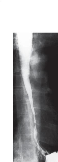
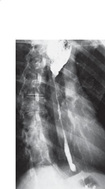
CASE 1.48
Findings
Single-contrast esophagram. Tubular thickened folds (arrows) are present in the upper thoracic esophagus.
Di erential Diagnosis
1.Esophageal varices
2.Varicoid esophageal carcinoma
Diagnosis
“Downhill” esophageal varices
Discussion
“Downhill” varices develop as a result of obstruction of the superior vena cava, most commonly due to
bronchogenic carcinoma, lymphoma, or fibrosing mediastinitis. Collaterals of the supreme intercostal vein, bronchial veins, and inferior thyroidal veins and other periesophageal collaterals enlarge and may be visible on an esophagram in the upper one-third of the esophagus. Blood flows “down” (caudally) the azygous-hemiazygous system and reenters the systemic circulation by way of the left gastric and portal veins. Unlike uphill varices, which often cause gastrointestinal bleeding, downhill varices usually are asymptomatic. In fact, in clinical practice, these varices are rare. In the absence of
the superior vena cava syndrome, other diagnostic considerations should be entertained, such as varicoid carcinoma (case 1.39).
Disease type: Masses and Filling Defects
CASE 1.49
Findings
CASE 1.49. Chest radiograph. There is a wellcircumscribed mass behind the heart.
CASE 1.50. Contrast-enhanced CT. A paraesophageal mass with the attenuation of water is present in the lower mediastinum.
Di erential Diagnosis
Bronchopulmonary foregut malformation
Diagnosis
Esophageal (enteric) duplication cyst
1. ESOPHAGUS 45
CASE 1.50
Discussion
Esophageal duplication cysts are a type of foregut cyst, as are bronchogenic cysts and neurenteric cysts. Pathologically, esophageal duplication cysts are lined with squamous epithelium and have a smooth muscle wall. Bronchogenic cysts have respiratory epithelium, and neurenteric cysts have associated vertebral
body anomalies. Symptoms from these cysts can be caused by compression on the adjacent esophagus or tracheobronchial tree or by infection of the cyst. If acid is secreted by the lining mucosa, peptic ulceration, perforation, and bleeding occur rarely.
Esophageal duplication cysts can be located anywhere in the posterior mediastinum. Contrast esophagraphy usually shows an extramucosal (smooth, well-demarcated) mass, which is often impossible
to differentiate from a leiomyoma (case 1.33), gastrointestinal stromal tumor, or other mass arising from the esophageal wall. CT allows differentiation of an enteric cyst (attenuation of water) from a solidenhancing mass.
Disease type: Masses and Filling Defects
46 MAYO CLINIC GASTROINTESTINAL IMAGING REVIEW
CASE 1.51
Findings
Lateral radiograph of the neck. There is a bone fragment (arrow) in the region of the esophagus at the level of the C4 interspace.
Di erential Diagnosis
Foreign body
Diagnosis
Esophageal foreign body
Discussion
Th is patient complained of food sticking and pain while eating chicken. A chicken bone was removed endoscopically.
Bony foreign bodies such as chicken or fish bones often lodge in the upper esophagus, whereas meat impactions (case 1.52) most commonly occur at the gastroesophageal junction. Impacted bones often are best visualized on a lateral radiograph of the cervical region or at CT. Contrast material may obscure a small fragment.
Disease type: Masses and Filling Defects
1. ESOPHAGUS 47
CASE 1.52
A
Findings
A.Single-contrast esophagram. An intraluminal filling defect is present in the distal esophagus just above the gastroesophageal junction.
B.Subsequent esophagram. The filling defect is no longer present, and a lower esophageal ring is seen.
Di erential Diagnosis
1.Foreign body
2.Esophageal carcinoma
3.Esophageal adenoma
Diagnosis
Esophageal foreign body
Discussion
Th is patient had a typical history of swallowing a large piece of meat and immediately experiencing odynophagia. Glucagon (1 mg) was administered
intravenously after image A was obtained, and the meat bolus passed into the stomach.
B
Impacted food typically lodges above the gastroesophageal junction. Several techniques can be used to remove the food. Intravenously administered glucagon decreases pressure of the
lower esophageal sphincter and may facilitate passage of the food bolus into the stomach. Generally, if the bolus reaches the stomach, it will pass through the remainder of the alimentary tract without difficulty. Endoscopic retrieval is the traditional means of treatment. Baskets and balloon catheters also have been used successfully to extract these foreign bodies. Effervescent granules and meat tenderizer have been used successfully by some, but they are not used in our practice because of the possible risk of perforation with these methods. If an impaction has persisted for more than 24 hours, the risk of perforation increases because of possible transmural ischemia. Special care must be taken in these cases. It is important to examine the esophagus after the food bolus has passed or been removed in order to exclude an underlying lesion.
Disease type: Masses and Filling Defects

48 MAYO CLINIC GASTROINTESTINAL IMAGING REVIEW
TABLE 1.3
Esophageal Filling Defects |
|
CASE |
|
|
|
|
|
Benign tumors |
|
|
|
Leiomyoma |
Smooth surface. 90° angle with esophageal lumen. Most |
1.33 |
|
|
common submucosal tumor. Gastrointestinal stromal tumor |
|
|
|
has identical appearance |
|
|
Adenoma |
Resembles a polyp. Usually <1.5 cm in diameter |
1.34 |
|
Always contiguous with gastric fold. Due to esophagitis |
1.35 |
||
Inflammatory polyp |
|||
Large polyp in the cervical esophagus on a thin stalk |
1.36 |
||
Fibrovascular polyp |
|||
|
|
||
|
|
|
|
Malignant tumors |
|
|
|
Carcinoma |
Polypoid, ulcerative, or annular. Squamous cell carcinoma, |
1.32, 1.37–1.42 |
|
|
proximal; adenocarcinoma, distal. Most adenocarcinomas are |
|
|
|
from Barrett esophagus, usually polypoid |
|
|
Metastases |
Can appear mucosal or extramucosal |
1.43 |
|
Primary or secondary involvement of esophagus. Associated |
1.44 |
||
Lymphoma |
|||
lymphadenopathy should be sought |
|
||
|
|
||
Spindle cell carcinoma |
Polypoid tumor in mid or distal esophagus. May expand the |
1.45 |
|
esophageal lumen |
|
||
(carcinosarcoma) |
Submucosal, bulky. Can present as intraluminal mass |
Not shown |
|
Malignant gastrointestinal |
|||
|
|
||
stromal tumor |
|
|
|
|
|
|
|
Nonneoplastic |
|
|
|
Varices |
Serpentine shape, shape changes. Also can be present in the |
1.46–1.48 |
|
|
stomach |
|
|
Duplication cyst |
Smooth surface. Oblique angle with esophageal contour, |
1.49 and 1.50 |
|
|
lumen displacement away from mass. Water attenuation at CT |
|
|
|
|
|
|
Foreign body |
Bones lodge in upper esophagus; meat impactions occur |
1.51 and 1.52 |
|
|
above gastroesophageal junction |
|
|
|
|
|
Disease type: Masses and Filling Defects
CASE 1.53
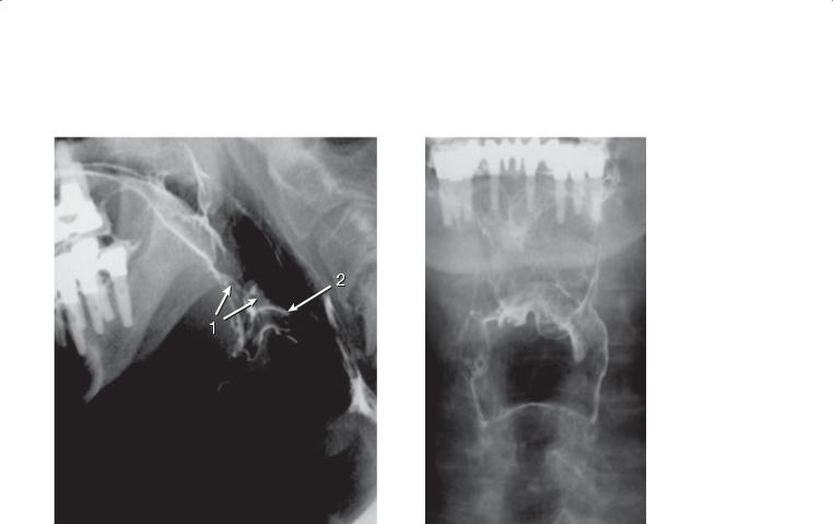
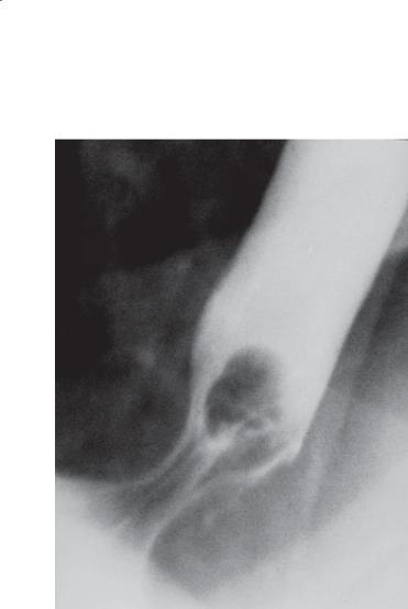
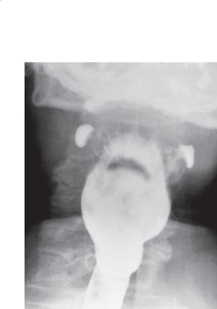
Findings
Single-contrast esophagram. Small outpouchings arise from both sides of the hypopharynx.
Di erential Diagnosis
Lateral pharyngeal pouches
Diagnosis
Lateral pharyngeal pouches
1. ESOPHAGUS 49
Discussion
Lateral pharyngeal pouches arise from an unsupported “weak area” of the thyrohyoid membrane that does not contain a muscular covering. Pouches may be best shown during the pharyngeal phase of swallowing
or by asking a patient to blow through closed lips (modified Valsalva maneuver). These pouches are common in asymptomatic patients and can be large in glassblowers, wind instrument players, and the elderly.
Disease type: Diverticula
50 MAYO CLINIC GASTROINTESTINAL IMAGING REVIEW
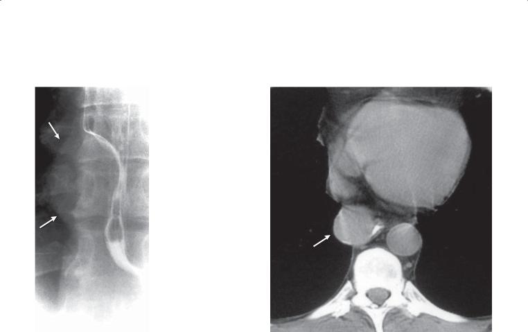
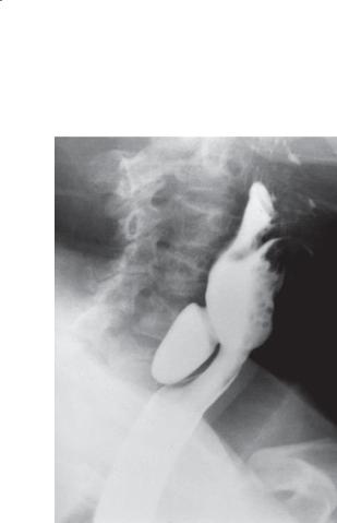
CASE 1.54
Findings
Double-contrast esophagram. A diverticulum arises from the posterior wall of the cervical esophagus.
Di erential Diagnosis
1.Zenker diverticulum
2.Pseudo-Zenker diverticulum
Diagnosis
Zenker diverticulum
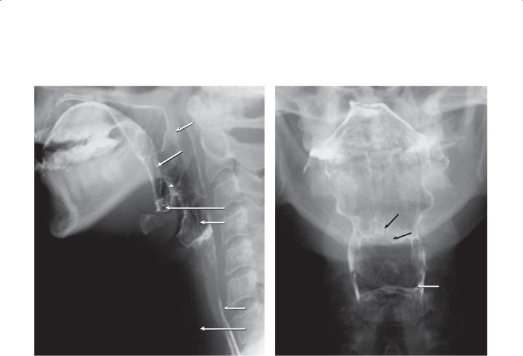
Discussion
Zenker diverticulum is a protrusion of the esophageal mucosa through an anatomically weak area in the posterior wall of the cervical esophagus. The site of weakness lies between the oblique and horizontal fibers of the esophageal wall, known as a Killian dehiscence or triangle. The cricopharyngeus muscle is invariably prominent just caudal to the diverticulum.
Zenker diverticula result from abnormally increased pressure generated in the hypopharynx. This high pressure is due to failure of the cricopharyngeus muscle to relax after pharyngeal contraction during swallowing. Many patients with gastroesophageal reflux have a prominent cricopharyngeus muscle.
Retention of food and fluid within the diverticulum can be uncomfortable, cause halitosis, or even lead to aspiration when the patient is recumbent. Cricopharyngeal myotomy and either surgical diverticulopexy or diverticulectomy may be required.
A pseudo-Zenker diverticulum results from barium being trapped between a pharyngeal contraction wave and a prominent cricopharyngeus muscle.
This can result in a transient, small, saclike bulge off the posterior pharyngeal wall superior to the cricopharyngeus muscle.
Disease type: Diverticula
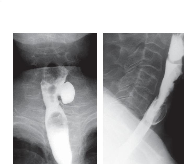
1. ESOPHAGUS 51
CASE 1.55
A B
Findings
A.Single-contrast anteroposterior esophagram.
B.Lateral esophagram. A rounded, barium-filled diverticulum protrudes off lateral wall of upper cervical esophagus.
Di erential Diagnosis
1.Killian-Jamieson diverticulum
2.Zenker diverticulum
Diagnosis
Killian-Jamieson diverticulum
Discussion
A Killian-Jamieson diverticulum protrudes through an area of weakness below the attachment of the cricopharyngeus muscle on the cricoid cartilage and lateral to the suspensory ligaments of the esophagus inserting on the cricoid cartilage, known as the Killian-Jamieson space. It presents radiographically as a diverticulum off the anterolateral wall of the proximal cervical esophagus just below the level
of the cricopharyngeus muscle. Killian-Jamieson diverticula are more often bilateral than unilateral. A unilateral diverticulum can sometimes be confused with a Zenker diverticulum. However, on a lateral view, a Killian-Jamieson diverticulum should protrude anteriorly in relation to the cervical esophagus, unlike a Zenker diverticulum, which protrudes posteriorly.
Disease type: Diverticula
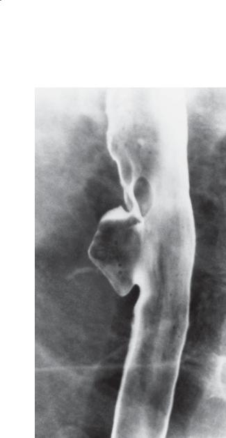
52 MAYO CLINIC GASTROINTESTINAL IMAGING REVIEW
CASE 1.56
Findings |
Discussion |
Double-contrast esophagram. A wide-based |
Th e triangular shape of this diverticulum is often found |
outpouching arises from the mid esophagus. |
among traction diverticula as a result of fibrosis in the |
|
periesophageal tissues. In the past, tuberculosis with |
Di erential Diagnosis |
inflammatory mediastinal lymph nodes was implicated |
1. Traction diverticulum |
as an important cause of traction diverticula. Today, most |
2. Pulsion diverticulum |
esophageal diverticula are pulsion in origin, developing as |
|
a result of esophageal motor disorders. Pulsion diverticula |
Diagnosis |
usually are rounded and fail to empty (they contain no |
Mid-esophageal traction diverticulum |
muscle in their walls) with a peristaltic contraction. |
|
Most mid-esophageal diverticula are asymptomatic. |
|
A large diverticulum can compress the esophagus and |
|
lead to dysphagia. If a diverticulum overflows into the |
|
esophagus, aspiration can occur. Inflammation and |
|
infection can lead to rare complications of perforation |
|
or fistulization. |
Disease type: Diverticula

1. ESOPHAGUS 53
CASE 1.57 |
CASE 1.58 |
Findings
CASE 1.57. Single-contrast esophagram. A large diverticulum is seen in the distal esophagus just above the gastroesophageal junction. Esophageal contractions also are visible.
CASE 1.58. Contrast-enhanced CT. A large, thinwalled sac communicates with the esophagus. Orally administered contrast material and food are seen within the diverticulum.
Di erential Diagnosis
Pulsion diverticulum
Diagnosis
Epiphrenic pulsion diverticulum
Discussion
Diverticula arising in the distal esophagus just above the gastroesophageal junction are referred to as epiphrenic diverticula. These outpouchings are nearly always pulsion in origin. Achalasia or other motor abnormalities of the esophagus often are associated with this condition. Both of these patients had achalasia. The visible contractions were nonpropulsive and characteristic of vigorous achalasia.
Disease type: Diverticula

54 MAYO CLINIC GASTROINTESTINAL IMAGING REVIEW
TABLE 1.4
Esophageal Diverticula |
|
CASE |
Pharyngeal |
Usually referred to as pouches; due to weakness in region of tonsillar |
1.53 |
|
fossa and thyrohyoid membrane. Usually asymptomatic |
|
|
|
|
Zenker |
Posterior, immediately above prominent cricopharyngeus muscle |
1.54 |
|
|
|
Killian-Jamieson |
Arise from lateral esophagus just below cricopharyngeus muscle |
1.55 |
|
|
|
Mid-esophageal |
In the past, due to traction from granulomatous disease |
1.56 |
|
(tuberculosis); today, nearly always pulsion diverticula due to |
|
|
motor abnormalities |
|
|
|
|
Epiphrenic |
Distal esophageal segment due to motor abnormality of esophagus |
1.57 and 1.58 |
|
|
|
Pseudodiverticulosis |
Dilated submucosal glands cause multiple small outpouchings. |
1.8 |
|
Usually due to chronic reflux esophagitis. Candida is often cultured |
|
|
but is not the causative factor |
|
|
|
|
Disease type: Diverticula

CASE 1.59
Findings
Single-contrast esophagram. Multiple nonpropulsive contractions are present in the lower esophagus.
Di erential Diagnosis
1.Tertiary contractions
2.Vigorous achalasia
Diagnosis
Nonspecific motor incoordination, tertiary esophageal contractions
1. ESOPHAGUS 55
Discussion
Esophageal contractions can be categorized as three separate types: primary, secondary, or tertiary. Primary esophageal peristalsis is initiated by a swallow and propagates a smooth, continuous contraction the length of the esophagus. Secondary esophageal peristalsis appears fluoroscopically identical to a primary wave, except it is not initiated by a swallow but rather by a bolus within the esophagus or by intraesophageal distention. Tertiary esophageal contractions are nonpropulsive contractions that may be single or multiple, and they do not result in clearing of esophageal contents. These contractions increase
in frequency with age. Tertiary contractions also can be associated with many other conditions. Incomplete relaxation of the lower esophageal sphincter also can occur in some patients. The single esophagraphic image shown here could be seen in vigorous achalasia (case 1.23). However, more esophageal dilatation might be expected, and, in this case, barium was seen to flow freely into the stomach across a normal gastroesophageal junction during real-time imaging.
Disease type: Miscellaneous
56 MAYO CLINIC GASTROINTESTINAL IMAGING REVIEW
CASE 1.60
Findings
Single-contrast esophagram. Numerous nonpropulsive contractions are present in the lower esophagus. This appearance has been likened to a corkscrew.
Di erential Diagnosis
1.Diff use esophageal spasm
2.Vigorous achalasia
3.Nonspecific motor incoordination
Diagnosis
Diffuse esophageal spasm (nutcracker esophagus)
Discussion
Diffuse esophageal spasm is characterized by dysphagia or chest pain, and at least 30% of swallows are associated with vigorous, repetitive, nonpropulsive contractions. Careful fluoroscopy eventually shows
a normal peristaltic sequence. Lower esophageal sphincter function may be normal; however, nearly
a third of patients have impaired relaxation or increased resting pressures. Various terms have been used to describe the radiographic appearance of the nonpropulsive contractions, including corkscrew, curling, rosary bead, and shish kebab esophagus.
Th e term nutcracker esophagus refers to a disorder characterized by the manometric findings of highamplitude (>180 mm Hg) contractions in conjunction with chest discomfort. Radiographically, peristalsis is normal. Some authorities believe that this disorder is a precursor to diffuse esophageal spasm.
Th e principal differential consideration is vigorous achalasia (case 1.23). Achalasia can be excluded by
a normal stripping wave in response to swallowing. Some patients are symptomatic, and others are not but have identical radiographic findings (nonspecific motor incoordination). It is important to exclude other causes of chest pain, especially coronary artery disease, before attributing it to the esophageal abnormalities.
Disease type: Miscellaneous
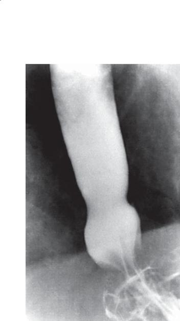
1. ESOPHAGUS 57
CASE 1.61
Findings
Single-contrast esophagram. A ring (circumferential impression) is seen within the distal esophagus.
At fluoroscopy, the ring was noted to be transient.
Di erential Diagnosis
1.Muscular ring (A ring)
2.Mucosal ring (B ring)
Diagnosis
Muscular ring (A ring)
Discussion
A muscular ring appears as a broad, smooth indentation in the lower esophagus, just superior to
the esophageal vestibule. It is caused by a muscular thickening and can be observed to change shape and disappear at fluoroscopy. A prominent muscular ring is encountered more often in patients with a hiatal hernia or gastroesophageal reflux and in some esophageal motor disorders. It is not known whether these associations are significant. The esophageal vestibule roughly corresponds in location to the manometrically defined lower esophageal sphincter and should not
be confused with a hiatal hernia. At fluoroscopy, a peristaltic wave can be seen to pass through this region. Often a mucosal ring (B ring) also is visible during the examination. The B ring is a thin, fixed ring that does not change appearance and marks the location of the esophagogastric junction.
Disease type: Miscellaneous
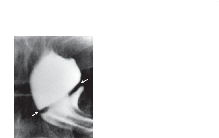
58 MAYO CLINIC GASTROINTESTINAL IMAGING REVIEW
CASE 1.62
Findings
Single-contrast esophagram. There is a thin, smooth, circumferential filling defect at the gastroesophageal junction (arrows) with a small associated hiatal hernia.
Di erential Diagnosis
1.Schatzki ring
2.Chronic reflux stricture
Diagnosis
Schatzki ring
Discussion
A Schatzki ring is a symptomatic, thin constricting ring at the level of the gastroesophageal junction. The pathogenesis of this condition is unknown. Most
patients complain of dysphagia with solid foods. Large pieces of meat are often most troublesome. Treatment varies from instructions on chewing more carefully to endoscopic mechanical disruption of the ring with a bougie or pneumatic dilation.
Radiographically, a thin, weblike constriction is present at the gastroesophageal junction. A hiatal hernia is often present. Some patients experience dysphagia if the luminal diameter is between 10 and 15 mm at the level of the ring. Virtually all patients are symptomatic if the ring narrows to 10 mm or less.
Th e term lower esophageal ring denotes a visible B ring that is asymptomatic. A Schatzki ring has a very characteristic appearance, and other conditions are rarely mistaken for it. It was originally defined as a B ring with luminal narrowing of 13 mm or less. A chronic reflux stricture should be longer and of different morphologic appearance.
Disease type: Miscellaneous
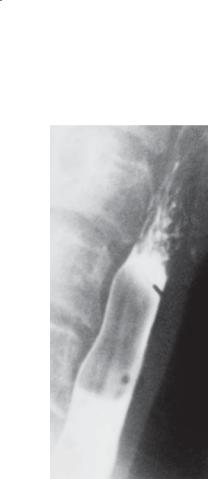
1. ESOPHAGUS 59
CASE 1.63
Findings
Double-contrast esophagram. A thin, smooth, shelflike filling defect is present along the anterior wall of the cervical esophagus.
Di erential Diagnosis
1.Esophageal web
2.Prominent anterior venous plexus
Diagnosis
Esophageal web
Discussion
Esophageal webs are thin folds composed of mucosa and submucosa which result in a 1- to 2-mm–wide shelflike filling defect along the anterior wall of the lower hypopharynx, pharyngoesophageal segment, or proximal cervical esophagus. Occasionally, webs may be multiple and more masslike in configuration.
Esophageal webs are usually asymptomatic. However, dysphagia can develop when the web results in marked luminal narrowing. Clinically significant narrowing may present as a jet phenomenon, in which a thin column of barium jets through the center
of a circumferential web. Many investigators have attempted to associate cervical esophageal webs with other conditions. The association between esophageal webs and iron deficiency anemia (Plummer-Vinson syndrome) remains controversial. Reports also have suggested an association between webs and upper esophageal or pharyngeal carcinoma. The great majority of webs remain an isolated finding, often incidentally discovered without an associated disorder.
Distinction of an esophageal web from the anteriorly located venous plexus may be difficult. Careful study of the venous plexus will show variability in its size, whereas a web remains fixed and unchanging from swallow to swallow.
Disease type: Miscellaneous
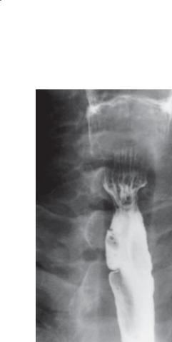
60 MAYO CLINIC GASTROINTESTINAL IMAGING REVIEW
CASE 1.64
Findings
Double-contrast esophagram. A pair of shallow indentations approximately 2 cm apart are seen along the lateral wall of the cervical esophagus.
Di erential Diagnosis
1.Ectopic gastric mucosa
2.Blistering skin disorder
Diagnosis
Ectopic gastric mucosa
Discussion
Ectopic gastric mucosa is thought to be a congenital abnormality, resulting from residue of columnar epithelium not replaced by stratified squamous epithelium during normal embryogenesis. Ectopic gastric mucosa usually is located in the cervical esophagus, which is the last place in the esophagus to undergo replacement with stratified squamous epithelium. Radiographic evidence of ectopic gastric mucosa in the upper esophagus can be found in
up to 3% of patients. The patches of ectopic gastric mucosa are usually less than 2 cm in diameter. Recognition of the characteristic radiographic features of this abnormality makes endoscopy and follow-up unnecessary. However, ectopic gastric mucosa occasionally results in dysphagia and appears polypoid or irregular in shape. An atypical appearance should be evaluated endoscopically. A blistering skin disorder could present with similar findings.
Disease type: Miscellaneous
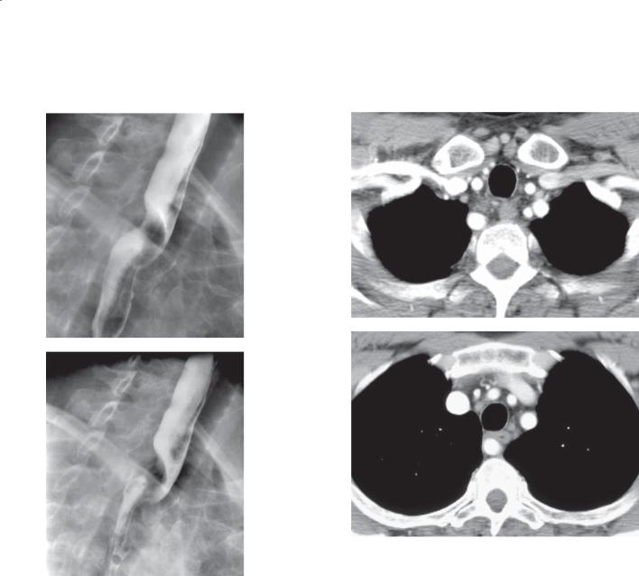
1. ESOPHAGUS 61
CASE 1.65 |
CASE 1.66 |
A |
A |
B
B
Findings
Double-contrast esophagrams. A and
B. There is a prominent oblique, smooth-surfaced, tubular-shaped filling defect on the posterior aspect of the esophagus.
CASE 1.66. Contrast-enhanced CTs. A and B. An aberrant right subclavian artery is present coursing posterior to the esophagus.
Di erential Diagnosis
Aberrant right subclavian artery
Diagnosis
Aberrant right subclavian artery
Discussion
An aberrant right subclavian artery is the most common aortic arch anomaly seen as an impression on the esophagus. The aberrant right subclavian artery arises just distal to the normal left subclavian artery and traverses obliquely to the right, posterior to the esophagus. The impression is extramucosal and is so typical of the abnormality that no further studies are needed. The abnormality is rarely symptomatic and needs no treatment.
Disease type: Miscellaneous
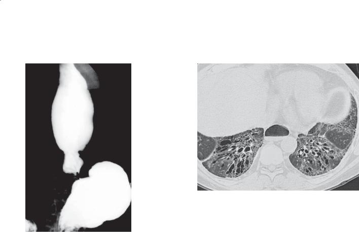
62 MAYO CLINIC GASTROINTESTINAL IMAGING REVIEW
CASE 1.67 |
CASE 1.68 |
Findings
CASE 1.67. Single-contrast esophagram. There is dilatation of the esophagus with a small esophageal hiatal hernia. Real-time imaging showed absent peristalsis in the distal two-thirds of the esophagus and free gastroesophageal reflux of barium in the recumbent position. Slight irregularity is present at the gastroesophageal junction.
CASE 1.68. Unenhanced high-resolution CT. Peripheral interstitial fibrotic changes are present in the lung bases with associated ground-glass infiltrates and traction bronchiectasis. Pulmonary findings are consistent with nonspecific interstitial pneumonitis. Dilatation of the distal esophagus also can be seen with an air-fluid level.
Di erential Diagnosis
1.Scleroderma
2.Achalasia
3.Pseudoachalasia
Diagnosis
Scleroderma
Discussion
Scleroderma (progressive systemic sclerosis) involves the esophagus in 80% of cases, and the radiographic changes in the esophagus often precede development of the characteristic skin changes. Pathologically, degeneration and atrophy of the smooth muscle and fibrosis are present within the distal two-thirds of the esophagus.
Radiographic changes relate to decreased peristalsis in the distal two-thirds of the esophagus, with an incompetent lower esophageal sphincter. Gastroesophageal reflux and changes of peptic esophagitis often are observed. Chronically, a lower esophageal stricture is often present.
Diminished esophageal clearance results in stasis and the possibility of secondary esophageal candidiasis and aspiration pneumonitis. Chronic reflux esophagitis can be complicated by Barrett esophagus.
Th e lungs are involved pathologically in nearly 90% of patients with scleroderma. However, radiographic evidence of pulmonary involvement is found in only 25% of patients. The appearance is indistinguishable from other causes of interstitial pulmonary fibrosis. Findings of scleroderma in the small bowel include diminished peristalsis, dilatation, sacculations, and closely spaced valvulae conniventes.
Disease type: Miscellaneous

CASE 1.69
Findings
Double-contrast esophagram. A portion of the gastric cardia protrudes through the esophageal hiatus into the thorax. Gastric rugae are seen within the hernia sac. A lower esophageal mucosal ring (arrow) (B ring) demarcates the gastroesophageal junction.
Di erential Diagnosis
1.Sliding hiatal hernia
2.Schatzki ring
Diagnosis
Sliding hiatal hernia
1. ESOPHAGUS 63
Discussion
Identification of a B ring at least 2 cm above the diaphragm allows hiatal hernia to be diagnosed with confidence. The significance of esophageal hiatal hernias is controversial. Usual symptoms are from gastroesophageal reflux: heartburn, chest pain, and water brash. Occasionally, large hernias can be associated with aspiration, respiratory distress, and compromised lung excursion. A hiatal hernia as a single radiographic finding is a poor predictor of gastroesophageal reflux or reflux esophagitis. Most patients with significant reflux esophagitis, however, have a hiatal hernia.
Th e main differential consideration in this case is a Schatzki ring (case 1.62). A lower esophageal ring has little clinical relevance unless there is significant compromise of the esophageal lumen. Some patients experience dysphagia if the luminal diameter is between 10 and 15 mm, whereas virtually all patients are symptomatic if the diameter is 10 mm or less. Pneumatic dilation of the mucosal ring is often successful in treating symptomatic patients.
Disease type: Miscellaneous
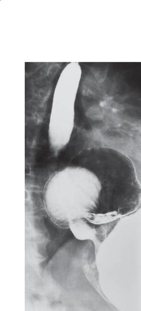
64 MAYO CLINIC GASTROINTESTINAL IMAGING REVIEW
CASE 1.70
Findings
Double-contrast esophagram. The gastroesophageal junction is normally positioned, and a portion of the stomach herniates into the thorax by way of the esophageal diaphragmatic hiatus.
Di erential Diagnosis
1.Paraesophageal hernia
2.Sliding esophageal hiatal hernia
Diagnosis
Paraesophageal hernia
Discussion
Paraesophageal hernias are more prone to complication than sliding esophageal hiatal hernias. Complications
include gastritis and bleeding in the hernia (due to edematous and hemorrhagic rugal folds from venous and lymphatic obstruction), gastric ulcers at the level of the diaphragmatic hiatus, and strangulation of the hernia sac.
Radiographic diagnosis is dependent on identifying a normally positioned esophageal junction with
the stomach passing through the esophageal hiatus anteriorly. A sliding esophageal hiatal hernia
can at times mimic a paraesophageal hernia, but in these cases the esophageal junction is always abnormally located within the thoracic cavity.
Most paraesophageal hernias undergo surgical repair because a high mortality is associated with
strangulation—a complication that may occur in up to 30% of patients.
Disease type: Miscellaneous
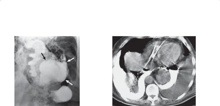
1. ESOPHAGUS 65
CASE 1.71 |
CASE 1.72 |
Findings
CASE 1.71. Single-contrast esophagram with watersoluble contrast. A large collection of extravasated contrast (arrows) is present in the mediastinum.
CASE 1.72. CT with oral contrast. Extravasated oral contrast is seen in the pleural spaces bilaterally. Air also is present within the mediastinum and pleural spaces.
Di erential Diagnosis
Esophageal perforation
Diagnosis
Esophageal perforation
Discussion
In case 1.71, the patient had a known history of achalasia, and pain developed after dilation of the lower esophageal sphincter. In case 1.72, the patient also had pain after endoscopy. Surgical repair was necessary in both cases.
Esophageal perforation usually is due to iatrogenic procedures (endoscopic perforations cause 75%–80% of all esophageal leaks) or spontaneous perforation (Boerhaave syndrome) from violent retching or
vomiting. Most endoscopic perforations involve the cervical esophagus near the cricopharyngeus muscle. Spontaneous rupture nearly always occurs just above the gastroesophageal junction on the left. Early detection of these perforations is critical to
patient survival. Small cervical esophageal leaks often can be treated conservatively, but large leaks must
be surgically repaired to prevent development of a retropharyngeal abscess. Untreated thoracic esophageal perforations have at least a 70% mortality rate (due to mediastinitis) if the leak is undiscovered.
Esophageal perforations can be suspected from the chest radiograph by detection of pneumomediastinum, widening of the mediastinum, hydropneumothorax, pleural effusion, and subcutaneous emphysema. Perforations suspected on the basis of the chest radiograph or on clinical grounds should be confirmed by an esophagram with water-soluble contrast material. Small leaks may be overlooked with watersoluble contrast agents, and negative studies should be followed by barium esophagraphy.
CT is excellent for evaluating patients with suspected or known esophageal perforation. Small amounts of extravasated air (pneumomediastinum) or contrast material and its location within the chest can be identified readily.
Disease type: Miscellaneous
66 MAYO CLINIC GASTROINTESTINAL IMAGING REVIEW
TABLE 1.5
Miscellaneous Esophageal |
|
CASE |
Conditions |
|
|
|
|
|
Nonspecific motor |
Nonpropulsive contractions. Stripping wave present with |
1.59 |
incoordination |
observation. Common in the elderly |
|
|
|
|
Esophageal spasm |
Tertiary contractions with pain |
1.60 |
|
|
|
Muscular ring |
Transient, nonobstructing |
1.61 |
|
|
|
Schatzki ring |
Fixed, narrows lumen 13 mm. B ring, esophagogastric junction |
1.62 |
|
|
|
Esophageal web |
Membranous, usually cervical esophagus. Association with |
1.63 |
|
anemia is controversial |
|
|
|
|
Ectopic gastric mucosa |
Lateral esophageal indentations |
1.64 |
|
|
|
Aberrant right subclavian |
Oblique, smooth filling defect in upper posterior thoracic |
1.65 and 1.66 |
artery |
esophagus |
|
|
|
|
Scleroderma |
Aperistalsis of distal two-thirds. Patulous gastroesophageal |
1.67 and 1.68 |
|
junction, reflux. Reflux esophagitis, lung fibrosis |
|
|
|
|
Hiatal hernia |
Gastroesophageal junction in thorax. Sliding (common) |
1.69 |
|
or short (due to chronic reflux esophagitis) |
|
|
|
|
Paraesophageal hernia |
Gastroesophageal junction normally positioned. High incidence |
1.70 |
|
of incarceration and strangulation |
|
|
|
|
Esophageal perforation |
Contained within mediastinum or pleural communication. |
1.71 and 1.72 |
|
Iatrogenic—usually cervical location; vomiting (Boerhaave |
|
|
syndrome)—usually distal esophageal tear |
|
|
|
|
Disease type: Miscellaneous

1. ESOPHAGUS 67
TABLE 1.6
Di erential Diagnoses
Esophagitis
Peptic (reflux) esophagitis Barrett esophagus Medication-induced esophagitis Crohn esophagitis
Infectious esophagitis Candidiasis Herpes esophagitis
Cytomegalovirus esophagitis
Human immunodeficiency virus esophagitis
Strictures
BENIGN
Peptic (reflux) stricture
Barrett stricture
Caustic stricture
Radiation stricture
Achalasia
Blistering skin disorders
Eosinophilic esophagitis
MALIGNANT
Annular carcinoma Pseudoachalasia
Lymphoma (extrinsic compression by adenopathy)
Filling Defects
BENIGN TUMORS
Leiomyoma
Gastrointestinal stromal tumor Adenoma
Inflammatory esophagogastric polyp Fibrovascular polyp
MALIGNANT TUMORS
Carcinoma
Metastases
Lymphoma Leiomyosarcoma
Malignant gastrointestinal stromal tumor Spindle cell carcinoma
NONNEOPLASTIC FILLING DEFECTS
Varices
Esophageal duplication cyst
Ectopic gastric mucosa
Foreign body
Diverticula
Pharyngeal
Zenker
Killian-Jamieson
Mid-esophageal
Epiphrenic
Pseudodiverticulosis
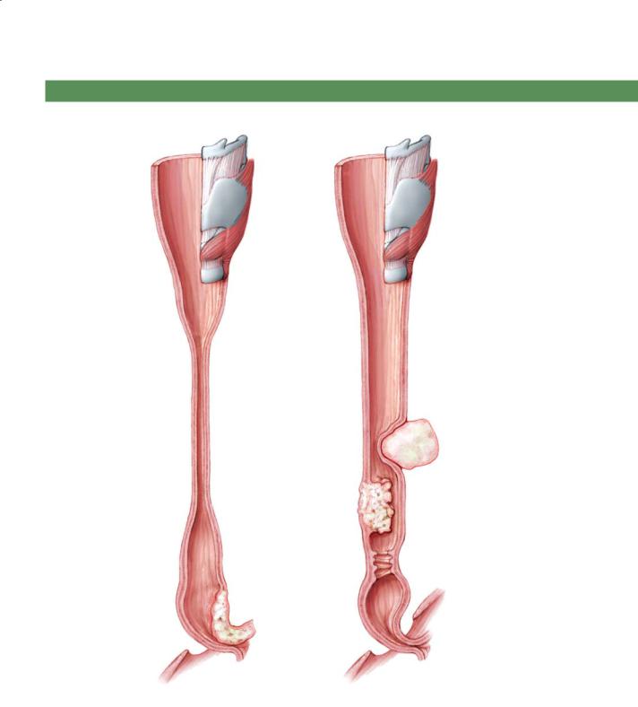
68 MAYO CLINIC GASTROINTESTINAL IMAGING REVIEW
ESOPHAGEAL STRICTURES
Long stricture |
|
|
Caustic stricture |
|
|
Radiation stricture |
Focal mid-esophageal |
|
Reflux stricture |
stricture |
|
Extrinsic mass or |
||
from nasogastric tube |
||
Eosinophilic |
adenopathy |
|
|
||
esophagitis |
|
|
|
Carcinoma |
|
Pseudoachalasia |
Chronic reflux stricture |
|
(submucosal carcinoma |
||
(with transverse folds) |
||
involving gastro- |
||
|
||
esophageal junction) |
|
|
|
Achalasia |
D.F.
© MAYO 2013
Figure 1.73 (Used with permission of Mayo Foundation for Medical Education and Research.)
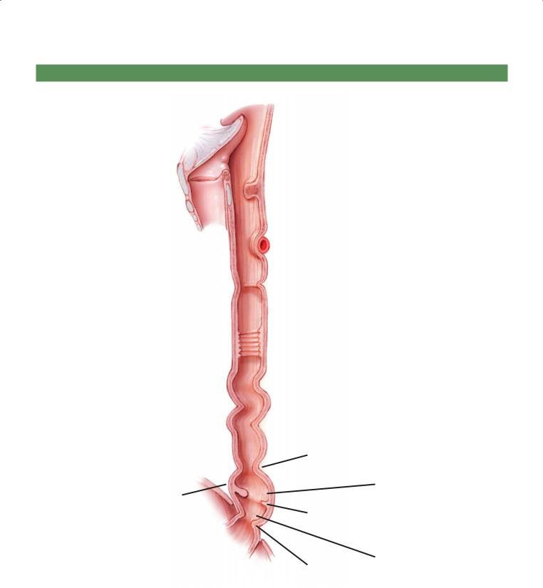
1. ESOPHAGUS 69
ESOPHAGEAL NARROWINGS
D.F.
© MAYO 2013
Schatzki ring
(narrowed B ring)
Cricopharyngeus muscle (bar)
Aberrant right subclavian artery
Esophageal web
Feline esophagus
Eosinophilic esophagitis
Nonspecific motor incoordination
(corkscrew)
A ring
(muscular ring)
Vestibule
B ring
(mucosal ring)
Hiatal hernia
C ring
(diaphragmatic
impression)
Figure 1.74 (Used with permission of Mayo Foundation for Medical Education and Research.)
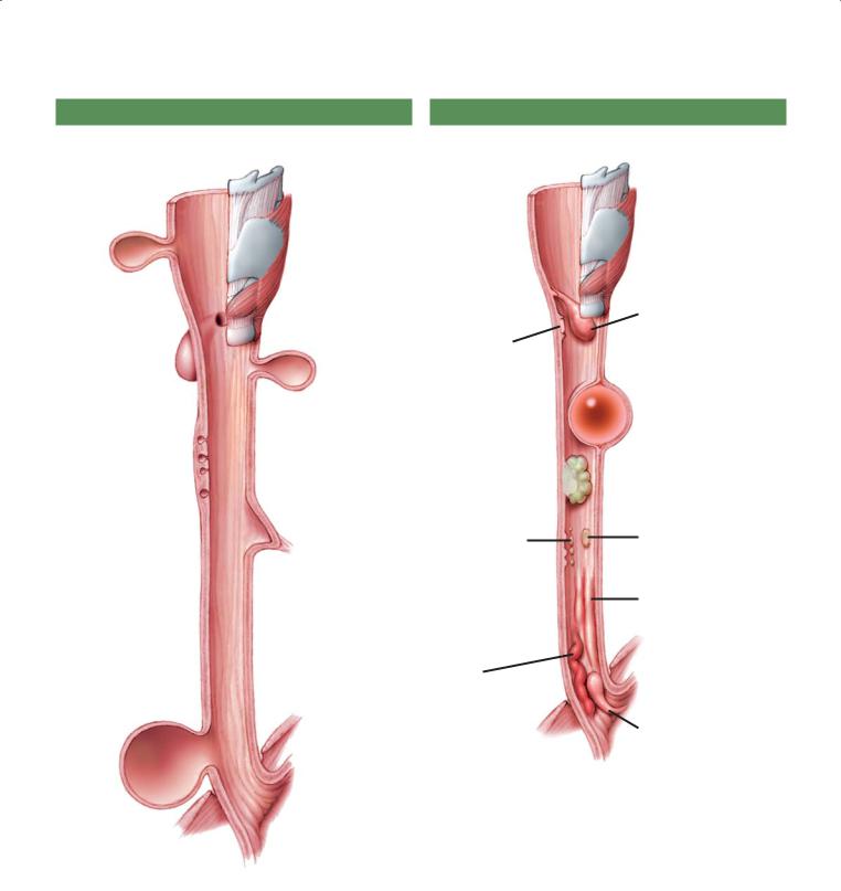
70 MAYO CLINIC GASTROINTESTINAL IMAGING REVIEW
ESOPHAGEAL DIVERTICULA |
ESOPHAGEAL FILLING DEFECTS |
Lateral pharyngeal pouch
Killian-Jamieson diverticulum
Zenker diverticulum
Intramural pseudodiverticulosis
Mid-esophageal diverticulum
Epiphrenic diverticulum
D.F.
© MAYO 2013
Figure 1.75 (Used with permission of Mayo Foundation for Medical Education and Research.)
Ectopic gastric mucosa
Polypoid mass
Carcinoma, lymphoma, metastases, carcinosarcoma
Plaquelike filling defects
Candidiasis Glycogen acanthosis Reflux esophagitis
Varices
Serpentine filling defects
D.F.
© MAYO 2013
Fibrovascular polyp
Smooth mass with acute angles
Leiomyoma, gastrointestinal stromal tumor (GIST), or esophageal duplication cyst
Adenoma
Thickened folds
Reflux esophagitis
Esophagogastric polyp
Figure 1.76 (Used with permission of Mayo Foundation for Medical Education and Research.)
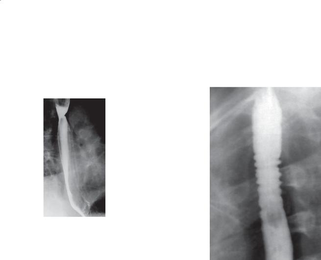
|
|
1. ESOPHAGUS 71 |
|
|
|
|
QUESTIONS |
|
Multiple Choice (choose the best answer) |
|
1.3. Which of the following is the most likely diagnosis |
1.1. Which of the following is most likely to be associated |
for the findings in the figure? |
|
|
||
with the condition shown in the figure?
a.Occult gastrointestinal blood loss
b.Lymphadenopathy
c.Heartburn
d.Halitosis
e.Dysphagia
a. Nonspecific motor incoordination
1.2. For the case shown in Question 1.1, what test should
b. Eosinophilic esophagitis
be recommended next?
c. Medication-induced esophagitis
a. Chest CT
d. Radiation esophagitis
b. Abdominal CT
e. Refl ux esophagitis
c.Endoscopy
d.Transthoracic needle aspiration/biopsy
e.Fecal occult blood testing
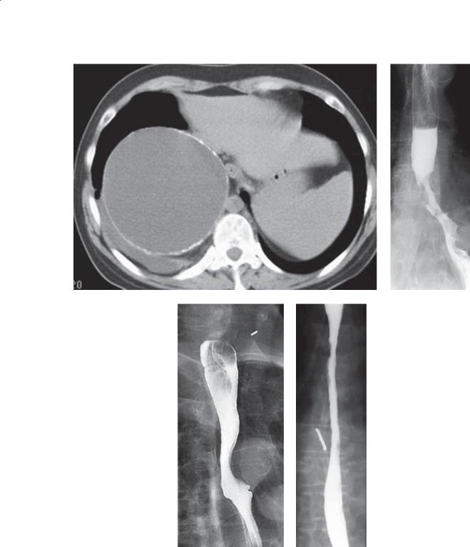
72 MAYO CLINIC GASTROINTESTINAL IMAGING REVIEW
A B
C D
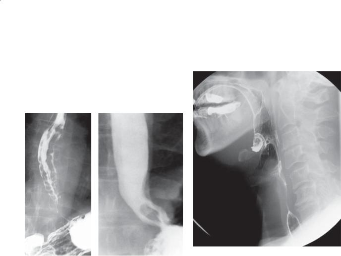
Match images A through F with the responses listed below.
1.4.______ Cirrhosis
1.5.______ Barrett esophagus
1.6.______ Regurgitation
1.7.______ Lymphoma
E F
1. ESOPHAGUS 73
Match the letters on the anatomic structure shown in the figure
with the appropriate label listed below.
E
A
C
B
II
D
F
J
G H
1.8.______ Vallecula
1.9.______ Region of the vocal cords
1.10.______ Pyriform sinus

74 MAYO CLINIC GASTROINTESTINAL IMAGING REVIEW
ANSWERS
1.1.Answer c.
Th e patient has a benign-appearing stricture in the upper esophagus. Differential possibilities include Barrett esophagus or medication-induced esophagitis with a secondary stricture. This patient had Barrett esophagus, a condition of mucosal metaplasia from squamous mucosa to adenomatous (glandular) mucosa. Barrett esophagus is due to long-standing reflux esophagitis, usually associated with chronic heartburn.
Occult blood loss would likely be due to a neoplasm or perhaps varices in the esophagus. There is no evidence of either in this case.
Lymphadenopathy can narrow the esophageal lumen, but there is no evidence of an extrinisic mass on the esophagus. Lymphadenopathy would not be expected to cause such a focal narrowing.
Halitosis can be associated with a Zenker diverticulum, but the radiographic findings do not support this. Potentially abundant gastroesophageal reflux could lead to halitosis, but this answer is much less likely.
Dysphagia usually occurs once the esophageal lumen narrows to less than 1 cm in diameter. Although the luminal diameter is compromised, it remains wide enough that dysphagia is unlikely.
1.2.Answer c.
For patients in whom Barrett esophagus is suspected, endoscopy is recommended to confirm the diagnosis and to exclude complicating dysplasia or carcinoma by biopsy.
1.3.Answer b.
Irregularity of the upper esophagus or stricture, or both, is typical of esosinophilic esophagitis. This patient has both luminal irregularity and narrowing of the esophageal lumen.
Nonspecific motor incoordination refers to nonpropulsive contractions of the esophagus, most commonly affecting the smooth muscle portion of the esophagus (lower two-thirds), where the bolus of contrast does not pass smoothly into the stomach. Occasionally there can be persistent contractions that have the appearance of a shish-kebab or rosary-bead esophagus—with alternating narrowing and dilatation.
Medication-induced |
esophagitis |
is characterized |
by focal changes of |
esophagitis |
(granular mucosa, |
erosions) usually in the upper esophagus, at the level of the thoracic inlet or bronchial impression. In this case, the findings are located too cephalad and are not typical of acute changes from esophagitis.
Radiation esophagitis is usually a long segment of narrowed, featureless esophagus in the chronic state, in the regions of a known radiation port.
Reflux esophagitis is usually first and worst at the gastroesophageal junction, extending into the proximal esophagus (unless the patients have a Barrett esophagus). Fold thickening, mucosal granularity, erosions, and deeper ulcers are most commonly encountered.
1.4.Answer e.
Figure e shows varices. Serpentine filling defects within the lower esophagus are typical of esophageal varices. Portal venous hypertension that leads to the development of esophageal and gastric varices can develop in patients with cirrhosis.
1.5.Answer b.
Figure b shows esophageal cancer. Irregular, abrupt narrowing of the esophageal lumen with ulceration of the mucosa is typical of an annular esophageal cancer. Most cancers within the lower half of the esophagus develop from Barrett esophagus.
1.6.Answer c.
Figure c shows a large, elongated, polypoid filling defect in the upper esophagus, which is typical of a fibrovascular polyp. Because these lesions have a long stalk, they can present clinically with regurgitation of the polyp into the mouth.
1.7.Answer d.
Figure d shows a long stricture of the esophagus; this could be due to radiation treatment, prior caustic ingestion, or a blistering skin disease. Radiation injury to the bowel usually results in a smooth, featureless stricture. Patients with mediastinal lymphoma may receive radiation therapy.
1.8.Answer b.
1.9.Answer f.
1.10.Answer d.

1. ESOPHAGUS 75
Key for all structures shown on the figure for Questions 1.8 through 1.10:
A. |
Base of the tongue |
F. |
Region of the vocal cords |
B. |
Vallecula |
G. |
Trachea |
C. |
Epiglottis |
H. |
Esophagus |
D. |
Pyriform sinus |
I. |
Hyoid bone |
E. |
Soft palate |
J. |
Cricoid cartilage |
This page intentionally left blank