

30 |
3 Imaging Differential Diagnosis of Pediatric CNS Tumors |
3.3.2.5Diffuse Intrinsic Pontine Gliomas (DIPG)
Despite all treatment and scientific efforts DIPGs have remained the entity with the worst prognosis in children [70]. The median survival is below 1 year and the number of children surviving 3 years is very small. For this reason it is desirable to obtain tumor material to enhance further knowledge. This possibly may open new ways of treatment in the future [71]. For this reason the current strategy of only imaging diagnosis will probably be replaced in the future by regular biopsies although the histological diagnosis is usually well predictable by only MRI and clinical symptoms. The WHO grades of DIPG may vary from II to IV and cannot be differentiated by MRI. However, the course of disease, prognosis, and treatment does not depend on the individual WHO grades. For the imaging diagnosis of a DIPG, it is most important to define the main tumor site to be clearly centered in the pons (Fig. 3.18a, b). Diffuse high-grade gliomas can be found as well in the medulla
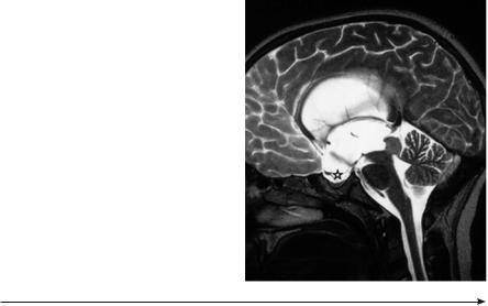
Fig. 3.17 The massive but not acute triventricular hydrocephalus in an older child is caused by a typical tectal glioma (no histological verification). The chronic hydrocephalus can be diagnosed due to a massive enlargement of the sella with vanished clinoid processes (asterisk)
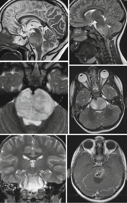
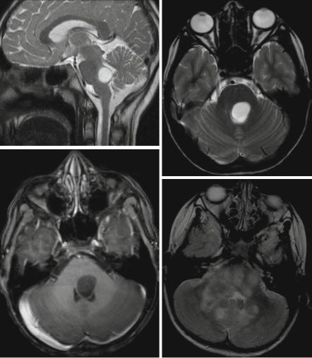
Fig. 3.18 (a–j) (a) on the sagittal T2-weighted MRI, a DIPG confirmed by biopsy resulting in a small defect with hemosiderin deposition in the edge is clearly centered in the pons. (b) On the sagittal T2-weighted MRI the histologically confirmed anaplastic ependymoma is growing from outside into the pons and is not an intrinsic lesion. A DIPG is covering more than half of the cross sectional area on this axial T2-weighted image (c). Sharp borders are rather frequently seen on T2-weighted MRI (d). The intrinsic growth of this glioma is demonstrated by a splitting of the pontine formatio reticularis on the coronal T2-weighted sequence (e). The suspicion of a pilocytic astrocytoma was raised by the dorsal cyst and confirmed by biopsy. The enhancement pattern on this postcontrast T1-sequence (f) is quite characteristic for a pilocytic astrocytoma. A focal pontine glioma in two different patients covers less than half of the cross-sectional area of the pons on T2-weighted images (g, h). Although the tumor is not enhancing (same patient as in h) on a postcontrast T1-weighted image (i), there seems to be a dorsal cyst. This feature is very much in favor of a pilocytic astrocytoma. In a child with NF-1 (j) as well high T2-signal areas in the cerebellum (NF-1 associated lesions) and an engulfment and signal increase in the pons is seen
3.3 Glial Tumors |
31 |
a |
b |
c
d
e |
f |
32 |
3 Imaging Differential Diagnosis of Pediatric CNS Tumors |
g |
h |
i
j
Fig. 3.18 (continued)
oblongata, but in these children a biopsy is indispensible to achieve a histological diagnosis with certainty. Usually a DIPG covers more that half of the pons [72] (Fig. 3.18c). Contrast behavior is variable. The borders may be very sharp on T2-weighted images (Fig. 3.18d). Usually as a sign of infiltrative growth the fibers of the formatio reticularis are seen as engulfed and split by the high T2-signal of the infiltrating tumor (Fig. 3.18e). Real cysts are not found and should warrant a histological clarification of the lesion because they are frequently found in pilocytic astrocytomas, which can grow in the pons as in all parts of the CNS (Fig. 3.18f). Also focal gliomas of the pons covering less than half of the cross sectional area should be closely watched or histologically clarified (Fig. 3.18h, i). Extension in