

4.1 Imaging Techniques |
61 |
h |
i |
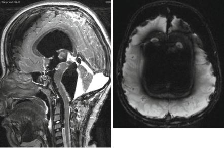
Fig. 4.2 (continued)
can only be identified on the basis of their T2/FLAIR or PD characteristics. Therefore, the comparability of MRI to the preoperative time point is of utmost importance. Also a change of magnetic field strength is problematic and should be avoided for the preand postoperative comparison and also for further follow-up.
4.1.3Meningeal Dissemination
All CNS tumors can disseminate in the CSF. However, for embryonal tumors like MB, AT/RT, and ependymomas especially during follow-up, germ cell tumors, and young children with LGGs of the chiasmatic region, there is a known propensity for higher percentage of CSF seeding. In such cases, it is very useful not only to perform a cranial MRI but also to add a spinal MRI after the cranial examination has been finished. For a correct staging, the visualization of the entire dural space intracranially and in the spinal canal is necessary. MRI is the only noninvasive method for the evaluation of a leptomeningeal dissemination of tumors or a primary spinal cord tumor.
Enhancement can affect the leptomeninx and the pachymeninx. While pachymeningeal enhancement is frequently a consequence of pressure changes in the CSF space, e.g., after surgery, in case of an implanted shunting system or after lumbar punctures leptomeningeal enhancement either represents a neoplastic or inflammatory affection. Nodular enhancement of the leptomeninges is pathognomonic for nodular meningeal dissemination and only rarely can be explained by inflammation like sarcoidosis. In laminar enhancement a lumbar tab can exclude meningitis as underlying reason.

62 |
4 Imaging Guidelines for Pediatric Brain Tumor Patients |
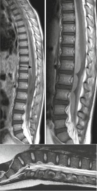
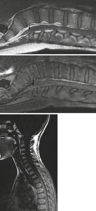
In a considerable number of children with MBs or other tumors prone to a leptomeningeal seeding, the staging examination of the spinal canal is performed after surgery. In the immediate postsurgical time period a physiological phenomenon may occur, which is called nonspecific subdural spinal enhancement. This enhancement closely resembles the MR images known from idiopathic CSF hypotension syndrome [122, 123], where the pachymeninges and not leptomeninges are affected and can easily be differentiated from the MRI characteristics of leptomeningeal dissemination [124]. Subdural enhancement must not be mixed up with leptomeningeal disease. It seems to be a dynamic process because we observed a progressive increase in T1-signal after contrast enhancement in a few patients during repetitive sequences performed during the same MR examination (Fig. 4.3a, b). Patients are always without specific symptoms. In case of extensive subdural enhancement, a leptomeningeal dissemination cannot be excluded (Fig. 4.3b) with sufficient reliability and therefore the spinal MRI has to be repeated after about 1–2 weeks. Within this time period a reduction or complete spontaneous resolution of nonspecific enhancement is to be expected. Occasionally after surgery a fluid level of nonenhancing spinal intradural blood mainly in the sacral region can be seen and resolves spontaneously as well (Fig. 4.3c–e).
For the visualization of possible spinal leptomeningeal dissemination exclusively T1 after contrast medium application is important. Rarely T2-weighted MRI is useful for the identification of small leptomeningeal nodules or in case of nonenhancing leptomeningeal dissemination. For nonenhancing dissemination, thin T2 sequences like CISS sequences seem to be promising and should be considered for the routine spinal protocol. However, as the T1-sequence after contrast is usually the last of the standard spinal MR sequences, the clinical practice in the reference evaluation of children with brain tumors has shown that these important postcontrast T1-images are very frequently deteriorated by movement artifacts in no longer compliant awake patients or by a flattening of sedation towards the expected end of the MR examination in sedated patients. We advice to focus the spinal MRI on the T1-series after contrast enhancement. The T2-weighted MRI may be added thereafter. If a fatty filum terminale is suspected, an additional T1 with fat suppression can clarify the situation. Small deposits of leptomeningeal disease may be mistaken for physiologic vessels of the spinal cord. Vessels are easily depicted if axial slices of all areas showing possible vessel enhancement on the sagittal slices are performed additionally.
Fat suppression is a useful method for the evaluation of bone tumors in the spine. However, it is not necessary for the detection and staging of spinal leptomeningeal dissemination and additionally caries the risk of artifacts rendering the images ine-
Fig. 4.3 (a–f) T1-weighted TSE series early after contrast medium application (a) shows faint subdural enhancement and a thick leptomeningeal laminar dissemination (arrow) on the spinal cord. This dissemination is masked on the repeat postcontrast series (b) 20 min after (a). A small fluid-fluid-level can be seen as horizontally confined structure in prone position (arrow) on T2 (c), T1-weighted MRI before (d) and after contrast (e). The signal is compatible with very fresh blood and this lesion vanishes within a few days. The use of fat suppression techniques for the evaluation of a spinal leptomeningeal dissemination is not necessary and not rarely impaired by artifacts mainly in the transition from the chest to the neck (f)
4.1 Imaging Techniques |
63 |
a |
b |
c
64 |
4 Imaging Guidelines for Pediatric Brain Tumor Patients |
Fig. 4.3 (continued)
d
e
f