

3.5 Germ Cell Tumors |
37 |
e |
f |
Fig. 3.20 (continued)
myxopapillary ependymomas tend to develop not only recurrences but also metastases (Fig. 3.21e, f) and a complete imaging of the CSF space in defined intervals is recommended [85, 86].
3.5Germ Cell Tumors
Germinomas are the most frequent germ cell tumors. The predominant localization is the pineal region. Second most frequently they are found in the suprasellar area [87]. The typical age at diagnosis is in the second and third decade and males predominate. Germ cell tumors and germinomas have a high cell density and nucleus/plasma-rela- tion and are therefore similar to gray matter on T2 [61] with a restricted diffusion [88] (Fig. 3.22a, b). More than 95 % of our tumors showed mostly intense contrast enhancement (Fig. 3.22c). Intense enhancement is also seen in their metastases in the CSF or along the subependymal lining of the ventricles (Fig. 3.22d). A quite distinct picture is found if germinomas grow in the basal ganglia. The clinical picture is dominated by a slowly progressing hemiparesis. The affected side of the basal ganglia rather shows atrophic changes than a mass lesion combined with a highly cellular lesion as derivable from hyperdense CT-density values and isoor hypointense T2-signals sometimes showing contrast uptake [89] (Fig. 3.22e–h). The prognosis for a reduction of symptoms after treatment is worse in this group. Bifocal tumor growth meaning synchronous tumors in the pineal and suprasellar area is not infrequent (10–25 % in our
38 |
3 Imaging Differential Diagnosis of Pediatric CNS Tumors |
database) and not considered as disseminated [90] (Fig. 3.22c). Outside the pineal, suprasellar, and basal ganglia region, they rarely originate from any part of the brain and spinal cord and are then indistinguishable from other brain tumors. We saw one germinoma in the diencephalon, one in the brain stem and two in the spinal cord in about 230 patients. Histology can only be clarified by biopsy or resection in these unusual cases. Germinomas are usually quite homogeneous tumors and are
a |
b |
d
c
Fig. 3.21 Supratentorial ependymomas are tumors with a high cell density with an intermediate to low T2-signal and usually growing outside in ventricles (a, b). Larger cysts (different child in (c) then in (a, b)) are not rare (c). During follow-up a CSF-dissemination in a different patient may occur as seen by a inhomogeneous nodule in the left lateral ventricle (arrow on d). A myxopapillary ependymoma of the filum terminale (e) was completely resected. After some time small disseminated nodules (arrows) recurred without a local recurrence of the tumor (f)

3.5 Germ Cell Tumors |
39 |
e |
f |
Fig. 3.21 (continued)
nonsecreting tumors. Secreting germ cell tumors are defined by a diagnosis of increased levels of human chorion gonadotropin (hCG) and alpha1-fetoprotein (AFP) in the serum and/or CSF. Secreting tumors are usually composed of several histological varieties frequently also including germinoma parts. They bear a more unfavorable prognosis and treatment differs from pure germinomas [91]. Therefore in case of a suspected germ cell tumor, the examination of CSF and blood serum plays a very important role and only after this clarification biopsy or surgery should be considered. In secreting germ cell tumors treatment may be started after a completed staging for CSF dissemination without a histological clarification. In nonsecreting tumors, only bifocal tumors are considered as germinomas and may also be treated without histology. In all other tumors, a histological clarification preferably by biopsy is necessary. Biopsy is preferred to attempted complete resection in these frequently well treatment
40 |
3 Imaging Differential Diagnosis of Pediatric CNS Tumors |
a |
b |
c |
d |
|
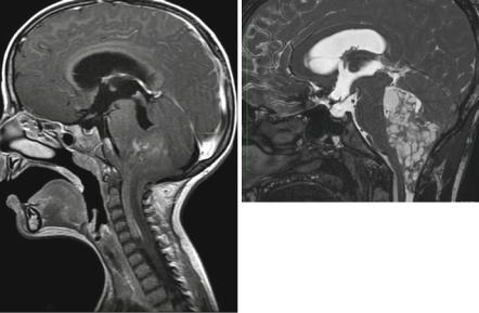
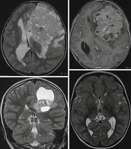
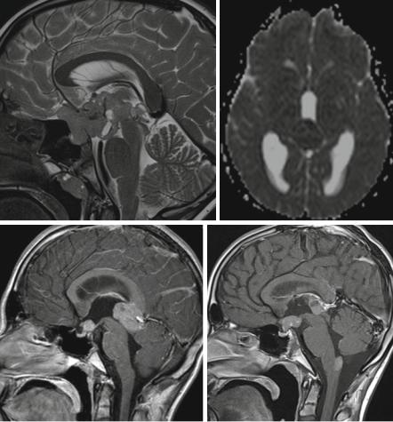
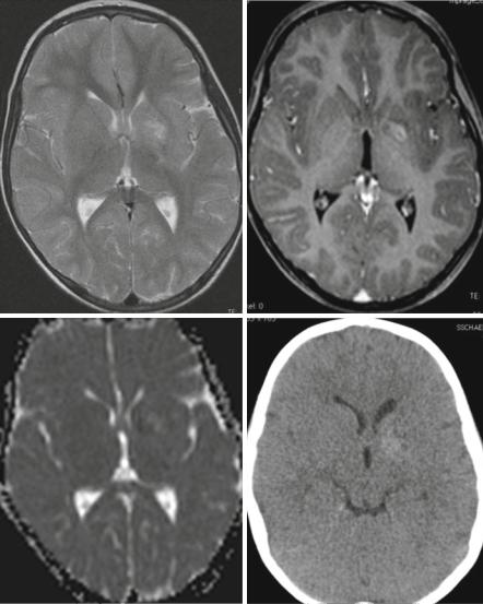
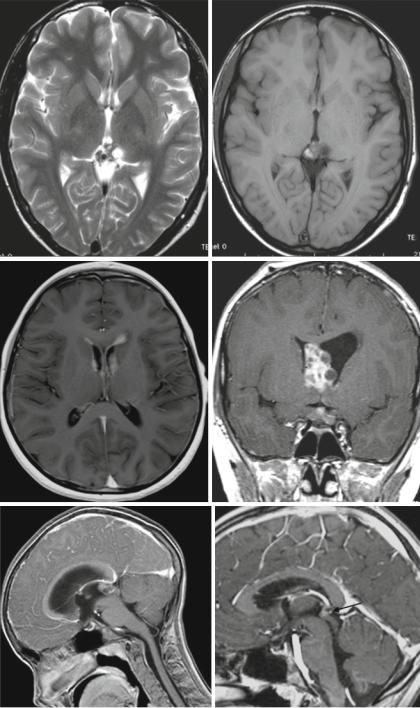
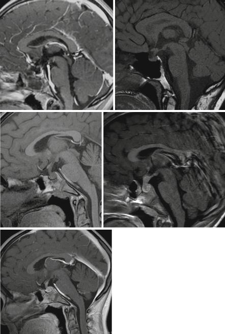
Fig. 3.22 MRIs from different patients: The T2-signal of a bifocal germ cell tumor (a) is comparable to gray matter. High cell density leads to a dark ADC (b) of a tumor in the pineal region. A bifocal localization in the suprasellar and pineal region (a and c) is not considered as a disseminated tumor and nearly all germ cell tumors enhance (c). Metastases also enhance (in d at the level of the obex). A basal ganglia germ cell tumor (e–h) can be suspected if atrophic changes in the basal ganglia are combined with hyperdense CT values (h), a dark ADC (g) of the tumor and some contrast enhancement (f). Irregular and inhomogeneous T2- (i) and T1-signals (j) are more frequent in nongerminomatous germ cell tumors as shown in this secreting tumor. A frequent route of dissemination in germ cell tumors is along the borders here shown in the lateral ventricles (k) or even the optic nerve sheaths (l) in another child. These extensions even if in continuity with the primary tumor are considered as dissemination in the SIOP-CNS-GCT study. Response assessment in this study is sometime not possible without doubt because the affected areas like the pituitary stalk and the pineal gland (arrow) may vary in normal size between individuals. We advice a short term control to prove additional shrinkage and therefore rendering the first examination as very good partial response (n) but not complete response (o). (m, MRI before treatment (n), after the first part of treatment (o), after further 4 weeks of treatment). Loss of pituitary bright spot on nonenhanced T1-weighted sagittal slices (SL 2–3 mm) in two different patients (p, q) with “unexplained” diabetes insipidus and thickened pituitary stalk. Both patients showed growth of a suprasellar germ cell tumor on follow-up. Brighter structure after contrast enhancement (r, s) anterior to an intraand suprasellar germinoma in two different patients probably representing the anterior pituitary (arrow)
3.5 Germ Cell Tumors |
41 |
e |
f |
g |
h |
Fig. 3.22 (continued)
42 |
3 Imaging Differential Diagnosis of Pediatric CNS Tumors |
i |
j |
k |
l |
m |
n |
Fig. 3.22 (continued)
3.5 Germ Cell Tumors |
43 |
o |
p |
q |
r |
s
Fig. 3.22 (continued)