

24 |
3 Imaging Differential Diagnosis of Pediatric CNS Tumors |
a |
b |
c |
d |
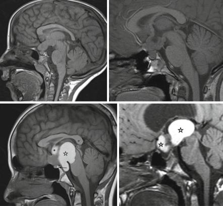
Fig. 3.13 Diagnostic significance of the bright pituitary spot on unenhanced T1-weighted MRI. In LGGs (a) virtually always the physiological hyperintensity of the posterior pituitary lobe (arrow) is present, while this bright spot is very frequently missing in suprasellar germ cell tumors (b). Note the compressed pituitary gland (arrow) below the tumor extending into the third ventricle. In craniopharyngiomas (c, d) the behavior of the bright spot is not predictable and depends on the site and size of the tumor. In (c) it is present despite a large tumor with colloid filled cysts (asterisk). In (d) it is absent. Note the flat and very small pituitary gland
3.3.2Gliomas of Higher Grades (HGG)
Comparable to gliomas of higher grades of malignancies in adults, HGG in children tend to show more indistinct margins, larger perifocal edema, and areas of higher cell density or necrosis. However, a differentiation between entities or grades is not possible with a reasonable certainty on the basis of imaging alone in an individual patient. So the suspicion of a high-grade glioma may be raised but a definite diagnosis is usually not possible by MRI (or CT) (Fig. 3.14a–d).
3.3 Glial Tumors |
25 |
a b
c |
d |
Fig. 3.14 In high-grade gliomas the prediction of tumor type and WHO grade is much less precise. General but rather nonprecise predictors of a high WHO-grade are: inhomogeneous tumor with larger perifocal edema and indistinct margins (a, T2-weighted axial MRI). Blood product deposition like met-hemoglobin on T1-weighted MRI before contrast (b). Irregular, increasing enhancement after contrast medium application (c). Areas with a higher cell density than in low-grade gliomas (d: ADC)
26 |
3 Imaging Differential Diagnosis of Pediatric CNS Tumors |
3.3.2.1Gliomatosis Cerebri (omitted acc. to the new WHO-classification)
According to the WHO classification 2007, gliomatosis cerebri is defined as a diffuse growth of a glioma grade II–IV within more than two cerebral lobes [62] (Fig. 3.15a). The central parts of the brain like basal ganglia or thalami may be involved or not and growth into the infratentorial compartment or even the spinal cord may be present or not (Fig. 3.15b). As a consequence this definition only applies to supratentorial tumors. Usually the mass effect is not predominant and contrast enhancement like in DIPG may signify higher grades but is not essential for the diagnosis (Fig. 3.15c) because also on biopsy vascular hyperplasia is usually missing [63]. Frequently, inflammation like encephalitis is the initial suspicion because many patients develop a seizure as the first symptom. These tumors are never completely resectable and prognosis is therefore grave like in other diffuse gliomas depending on age, Karnofsky performance status, and WHO grade of the gliomatosis. Comparable to gliomatosis cerebri, the prognosis of multifocal gliomas (Fig. 3.15d–f) is grave because multifocal diffuse lesions are usually also not resectable. Contrary to embryonal tumors like MBs, gliomas may grow in separate parts of the brain parenchyma either synchronously or during progression [64, 65].
3.3.2.2Brain Stem Gliomas
Depending on their localization, the WHO grade and prognosis of brain stem gliomas differ considerably. The following description of individual entities is ordered according to the tumor site.
3.3.2.3Cerebral Peduncles
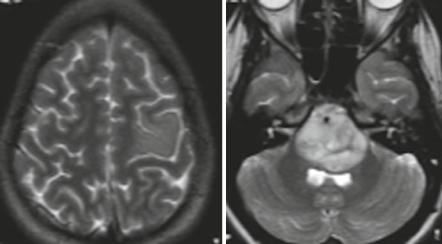
The most frequent gliomas in this localization in children are low-grade gliomas, predominantly pilocytic astrocytomas [66–68] (Fig. 3.16a–d). They are usually sharply demarcated and may extend as well into the basal ganglia as downwards into the pons. It is of utmost importance to respect the main mass of the tumor and its localization in the brain stem to discriminate these gliomas from the typical diffuse intrinsic pontine gliomas. Symptomatic tumors in the cerebral peduncles are usually decompressed by neurosurgery and therefore a histological clarification is the regular consequence.
3.3.2.4Tectal Plate Gliomas
Tectal plate gliomas belong frequently to the group of presumably low-grade gliomas [68], although a histological clarification is usually not performed in the majority of uncomplicated cases. They become symptomatic not by regional disturbances
3.3 Glial Tumors |
27 |
a b
d
c
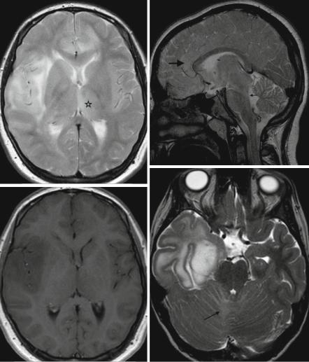
Fig. 3.15 (a–f) Gliomatosis cerebri was defined as tumor in more than two cerebral lobes with or without involvement of the central parts of the cerebrum. The T2-weighted MRI (a) shows a glioma in the right frontal and temporal lob with extension into the left frontal lobe and affection of the left thalamus (asterisk). On the sagittal T2-weighted image (b) of another patient, gliomatosis is visible in enlarged frontal (large arrow) and parietal gyri along the corpus callosum. Via central cerebral structures the dorsal midbrain (small arrow) and tegmentum pontis are involved as well. On T1 after contrast (c) (same patient as in a), no enhancement is visible. No connection of T2-hypersignal is seen on (d) between the right temporal tumor and the T2-hyperintense signal predominantly in the right cerebellum (arrow). In a patient with a typical DIPG (f), a second glioma is seen on the T2-weighted image. It involves the left precentral gyrus (e)
28 3 Imaging Differential Diagnosis of Pediatric CNS Tumors
e f
Fig. 3.15 (continued)
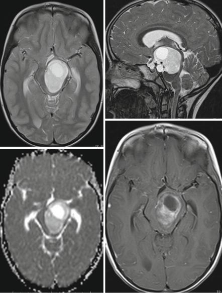
of the tectal plate itself but rather by the development of a triventricular hydrocephalus even in very small tumors due to their strategic localization close to the aqueduct. The patients are usually adapted to the hydrocephalus for many years. Consequently they are frequently diagnosed with a tectal glioma only by chance or by a decompensation of the increased CSF pressure equilibrium (Fig. 3.17). Therefore very young children or those with tectal plate symptoms are not found among children bearing typical tectal plate gliomas. In children with young age and local symptoms you should plan a rather short-term control (4–6 weeks) if histology is not clarified right away. We saw 2 PNETs in very young children mimicking tectal plate gliomas on imaging at the time of first MRI. However, extremely rapid growth and local symptoms like gaze abnormalities unmasked these tumors as highly malignant lesions later confirmed by histology. Of course all kinds of tumors can exceptionally be found in this area.
The typical tectal gliomas are usually smaller lesions with a high and mostly homogeneous T2-signal. Due to the usually massive ventricular dilatation, small tumors sometimes cannot be detected before a relief of hydrocephalus and tectal plate decompression was achieved. Contrast enhancement is more frequently missing. A relief of hydrocephalus by a third-ventriculostomy or a shunt is usually the only intervention needed for typical tectal gliomas. However, follow-up imaging is necessary to avoid a misleading imaging diagnosis and to identify the demand for treatment. Larger tumors, tumors extending into the thalamus, and those with contrast enhancement may need additional treatment like chemotherapy or irradiation after correction of increased CSF pressure [69].
3.3 Glial Tumors |
29 |
a |
b |
d
c
Fig. 3.16 A pilocytic astrocytoma in the left cerebral peduncle exhibits a sharp border on the axial T2-weighted MRI (a). On sagittal T2-weighted images (b) the main mass is confined to the preduncle only reaching the upper pons. Cellularity is low demonstrated by a high signal on ADC (c). The tumor enhances irregularly and intensively (d)