

3.4 Ependymomas |
33 |
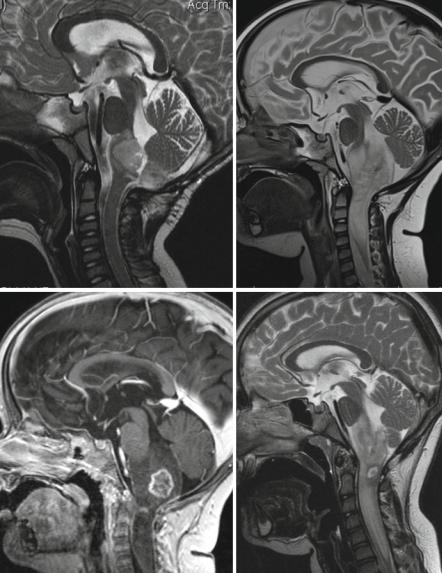
inferior and more frequently superior direction or into the cerebellum is seen either at diagnosis or during follow-up. A perifocal edema cannot be distinguished from the tumor itself and the entire T2/FLAIR hyperintensity is considered tumor. Asymmetric growth is not unusual. If enhancement is present at the time of diagnosis it does not contradict the diagnosis of a DIPG. However, it has been described as a sign of a worse prognosis as well in our patient group as also in other larger treatment studies [73, 74]. Leptomeningeal dissemination is possible but rare compared to embryonal tumors at diagnosis and increases during follow-up [75]. As in all diffuse gliomas, further intracerebral tumors distant from the pons may be seen either synchronously or during follow-up [76] (Fig. 3.15e, f). During or soon after treatment the tumor may shrink and the high T2-signal may approximate to normal. A new reactive contrast enhancement within a DIPG is often found on the postradiation MRI and must not be interpreted as a sign of tumor progression during this phase of treatment.
In NF1, gliomas of the pons can imitate a DIPG on MRI (Fig. 3.18j). However, the clinical course in NF1 patients is much more favorable and a specific treatment is not initiated due to the fact that a tumor in the pons is present but rather depends on the course of disease and the patient’s clinical complaints.
3.3.2.6Gliomas of the Medulla Oblongata
While gliomas of the pons are not at all resectable, their dorsal exophytic counterpart in the medulla oblongata sometimes extending downwards into the spinal cord (Fig. 3.19a, b) is frequently considered to be decompressable by the experienced neurosurgeon. This is the initial method of choice for treating a dorsal exophytic, symptomatic glioma of the medulla oblongata and a good way to confirm the histology of a low-grade glioma [77, 78]. Most frequent histologies are pilocytic astrocytomas, gangliogliomas (Fig. 3.19c, d), and diffuse astrocytomas grade II in our patients. Contrast accumulation is variable.
3.4Ependymomas
Ependymomas show different genetic alterations and behavior according to their localization [79]. In the posterior fossa they are the main differential diagnosis to MBs. They usually grow in close spatial relation to the ependymal lining in the posterior fossa either within the mainly lower parts of the ventricle or near the ependymal protrusions into the cerebellopontine angle (Fig. 3.20a, b). As they are tumors with a dense cellularity, the T2-signal is low in the solid parts of the tumor but usually not as low as in MB and free water diffusion may be impeded. The internal structure frequently shows rim enhancing cystic formations (necroses) with an elevated protein content. Inhomogeneity is predominant (Fig. 3.20a, b). Occasionally ependymomas are homogeneous on T2 or do not take up contrast [80] (Fig. 3.20c, d)
34 |
3 Imaging Differential Diagnosis of Pediatric CNS Tumors |
a |
b |
c |
d |
|
Fig. 3.19 In the medulla oblongata, diffuse tumors with a definite dorsal exophytic component are very frequently low-grade gliomas. They may involve only the medulla oblongata (as seen in a) or extend into the cervical cord (b–e) and are then called cervicomedullary tumors. Pilocytic astrocytomas (b, c) or diffuse astrocytomas WHO grade II are not well distinguishable from gangliogliomas (d, e). Contrast enhancement in gangliogliomas (e) is more frequently irregular and looking “infiltrative”
3.4 Ependymomas |
35 |
Fig. 3.19 (continued) |
e |
|
although enhancement is intense and striking in most ependymomas. The most characteristic feature is the so-called plastic growth pattern [81], which leads to an early sprouting out of the posterior fossa mainly into the spinal canal and the encasement of prepontine vessels and cranial nerve roots (Fig. 3.20e). Involvement of the foramen of Magendie is much more frequent (Fig. 3.20f) than in MB [80]. The encasing growth pattern is the main obstacle for a complete resection and a good outcome. Complete resection is an important precondition for cure in ependymomas.
In the supratentorial compartment, most ependymomas do not grow in the ventricles (Fig. 3.21a, b) but in the vicinity of the outer ventricular border within the brain parenchyma [82, 83]. Intraventricular growth is rarely found and was associated with lack of RELA fusion transcript genes in our patients. Especially supratentorially large cysts are frequent (Fig. 3.21c).
As well infraas also supratentorial tumors rarely show leptomeningeal dissemination already at the time of diagnosis. During follow-up the incidence of dissemination in cranial ependymomas increases significantly and even isolated spinal deposits or isolated intraventricular nodules may be found (Fig. 3.21d). Therefore a complete screening of the CSF space may not be as important at diagnosis as it is during follow-up [84]. Nevertheless, the standard staging examination in ependymomas includes a complete spinal MRI. Spinal ependymomas in children are mainly myxopapillary WHO grade I tumors of the filum terminale. They behave differently than the WHO grade II and III tumors affecting the brain. However,
36 |
3 Imaging Differential Diagnosis of Pediatric CNS Tumors |
a |
b |
c
d
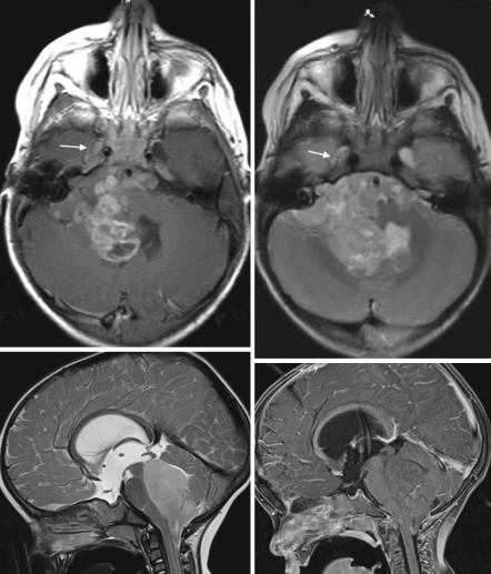
Fig. 3.20 Ependymomas in the posterior fossa frequently show irregular but rather intense contrast enhancement and an inhomogeneous internal structure with cyst-like components like this tumor arising from the right cerebellopontine angle on axial T1-weighted postcontrast (a) and T2-weighted (b) images. The “plastic” growth pattern led to a surrounding of the pontine structures and an infiltration into the internal auditory canal and even Meckels’ cave on the right hand side (arrow). Also homogeneous tumors without significant enhancement (c, d) may be seen. An early extension into the spinal canal is frequent (e) and the foramen of Magendie is frequently involved as seen on a sagittal CISS image (f)