- •Foreword
- •Acknowledgements
- •Contents
- •1.1 Postoperative Residual Tumor
- •1.2 Metastases
- •3.1 Explanatory Note
- •3.2 Embryonal Tumors
- •3.2.1 Medulloblastoma
- •3.2.1.5 Typical Localization of the MB Variants
- •3.2.3 Atypical Teratoid/Rhabdoid Tumor (AT/RT)
- •3.3 Glial Tumors
- •3.3.1 Astrocytomas
- •3.3.1.1 Visual Pathway Gliomas
- •3.3.1.2 Differential Diagnosis of Suprasellar and Visual Pathway Lesions
- •3.3.2 Gliomas of Higher Grades (HGG)
- •3.3.2.2 Brain Stem Gliomas
- •3.3.2.3 Cerebral Peduncles
- •3.3.2.4 Tectal Plate Gliomas
- •3.3.2.5 Diffuse Intrinsic Pontine Gliomas (DIPG)
- •3.3.2.6 Gliomas of the Medulla Oblongata
- •3.4 Ependymomas
- •3.5 Germ Cell Tumors
- •3.6 Craniopharyngiomas
- •3.7 Choroid Plexus Tumors
- •4.1 Imaging Techniques
- •4.1.2 Early Postoperative Imaging
- •4.1.3 Meningeal Dissemination
- •4.1.4.1 Differential Diagnosis Between Recurrence or Treatment Related Changes
- •References
- •Index

14 |
3 Imaging Differential Diagnosis of Pediatric CNS Tumors |
Plexus carcinomas can very well resemble AT/RTs on imaging. Interestingly common features in immunohistochemistry between these two entities have been described [25].
3.2.2Central Nervous System Primitive Neuroectodermal Tumors (CNS PNET changed to ETMR in the new WHO classification)
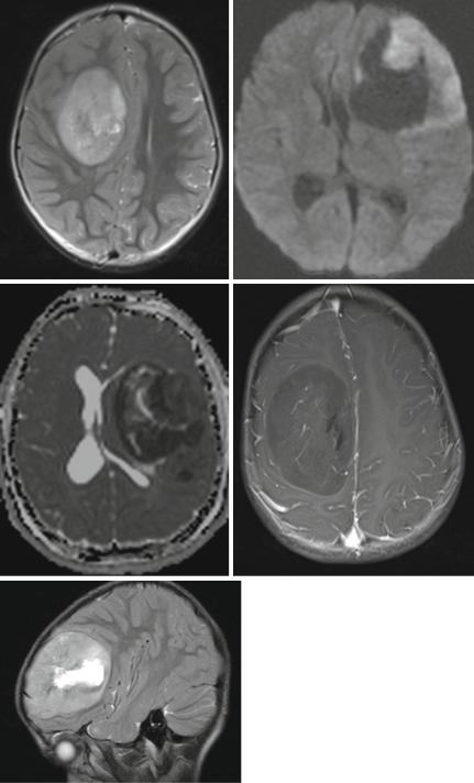
CNS PNETs arise in the cerebral hemispheres, the brain stem, and the spinal cord [26] of children and mainly young adults. A variant of this clinically very aggressive entity according to a specific differentiation are cerebral neuroblastomas or ganglioneuroblastomas, medulloepitheliomas, and ependymoblastomas. On imaging we see highly cellular tumors with a low T2 and restricted diffusion (Fig. 3.7a–c). Contrast enhancement is variable but more frequently sparse than intense or complete (Fig. 3.7d). It is often missing completely. Perifocal edema is mostly minor or absent (Fig. 3.7e), and in contradiction to a fast growth and malignant behavior the tumor seems to be surrounded by a capsule and usually is on imaging well delineated from the surrounding brain [27]. Like MB also CNS PNET are prone to tumor dissemination in the leptomeninges prompting an MRI evaluation of the complete cranial and spinal CSF space.
3.2.3Atypical Teratoid/Rhabdoid Tumor (AT/RT)
AT/RTs are highly malignant tumors affecting young children. Meanwhile they are considered the most frequent malignant brain tumors within the first 6 months of age [28, 29]. They are closely linked to rhabdoid tumors of the kidneys as well on histology as also genetic findings and may be found in familial rhabdoid tumor predisposition syndrome [30, 31]. AT/RTs are restricted to the CNS and supraor infratentorial localization is varying with age. Older children or rarely adults more frequently develop supratentorial tumors [32]. AT/RTs may also arise in the spinal cord [33, 34] and are then not distinguishable from other cord tumors.
In the brain it is rather the combination of multiple features than one single characteristic on imaging that can help to find the likely diagnosis. Like all tumors with increased cell density, the solid parts are very hypointense on T2-weighted MRI and
Fig. 3.7 T2-weighted axial (a) and sagittal (e) MRI, diffusion-weighted MRI (b) and ADC (c) and T1-weighted image after contrast enhancement (d) of different children with a cPNET. It is characteristic that cPNETs are sharply delineated without perifocal edema. The high cell density is reflected by a restricted diffusion and thus a dark ADC. Contrast enhancement is frequently little or missing

3.2 Embryonal Tumors |
15 |
a |
b |
c |
d |
e
