


3.7 Choroid Plexus Tumors |
51 |
Fig. 3.25 Development of hypothalamic obesity demonstrated by the thickness of the subcutaneous fat in the dorsal neck in a patient after subfrontal resection of a craniopharyngioma. The early postoperative MRI (a) shows surgical changes in the access route in a slim girl. After only 3 months the thickness of the subcutaneous fat has at least doubled (b). Note the large defect after extensive resection. Anatomic illustration of the anterior hypothalamus (in front of the mammillary bodies) and the posterior hypothalamus (including the mammillary bodies and behind) in a patient with a small tectal glioma (c). The floor of the third ventricle is clearly seen as a thin line (arrow) and the normal mammillary bodies are marked with a white cross. Note also a small, triangular intermediate lobe cyst in the pituitary. This must not be diagnosed as a pathologic structure but as a harmless incidental finding. Hypothalamic obesity is not exclusive for craniopharyngiomas but rather a general consequence of damage to the hypothalamus. After complete resection (d: preoperative MRI) of a secreting germ cell tumor the subcutaneous fat has considerably increased after only 8 months (e). Note the large defect in the floor of the third ventricle and the tiny mammillary bodies behind the defect
3.7Choroid Plexus Tumors
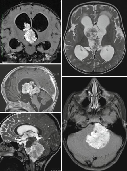
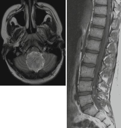
Choroid plexus tumors are rare overall (0.3–0.6 % of all brain tumors) but represent 2–4 % of tumors in children and 10–20 % of tumors in the first year of life [111]. Three grades of malignancy are found with plexus papillomas representing WHO grade I (Fig. 3.26a–f), atypical plexus papillomas WHO grade II (Fig. 3.26g, h), and choroid plexus carcinomas WHO grade III (Fig. 3.26i, j). Papillomas are about 5 times more frequent than carcinomas. A correct differentiation between the three possible entities is not always possible although the typical papilloma is a circumscribed, sharply delimited, cauliflower-like tumor attached to the ventricular plexus with intense contrast enhancement. A ventricular dilatation at diagnosis may be due to hyperproduction of CSF or a blockage of the CSF pathways. Infiltration of the ventricular wall or the brain parenchyma is unusual. Carcinomas are typically larger and inhomogeneous tumors, which infiltrate into the brain and lead to a perifocal edema. Leptomeningeal metastases do not exclude the diagnosis of a papilloma, although carcinomas disseminate more frequently (Fig. 3.26k, l). Not infrequent in plexus carcinomas is a contrast enhancement pattern resembling AT/RTs. Interestingly also on neuropathology AT/RTs are a possible differential diagnosis to plexus carcinomas [112, 113].
52 |
3 Imaging Differential Diagnosis of Pediatric CNS Tumors |
a |
b |
c
d
e
Fig. 3.26 (a–m) A papilloma of the plexus in the third ventricle in a young child shows the typical cauliflower aspect and a hydrocephalus (a–c). In an adolescent (d–f) a papilloma in the fourth ventricle is localized in the left cerebellopontine angle and could be mistaken for a WNT-MB due to its location. However, there is no restricted diffusion (f) with a bright ADC rendering a highly cellular tumor like an MB unlikely. The atypical PPL in a young child (g, h) cannot be distinguished from the typical case on (a–c). In a plexus carcinoma (i–k) there is obvious infiltration of the temporal lobe with edema, an indistinct border and an irregular structure. Note also the nodular leptomeningeal metastasis in the fourth ventricle (j). There were also other metastases not shown on the pictures. In a young adult with a typical, completely resected PPL (l) spinal leptomeningeal metastases (m) persist completely unchanged on follow-up for several years despite treatment with chemotherapy some years ago

3.7 Choroid Plexus Tumors |
53 |
f |
g |
h |
i |
j |
k |
Fig. 3.26 (continued)
54 |
3 Imaging Differential Diagnosis of Pediatric CNS Tumors |
l |
m |
Fig. 3.26 (continued)