

Глава из книги Случаи УЗДиагностики
.pdfA N S W E R S
C A S E 3 8
Distal Ureteral Stone
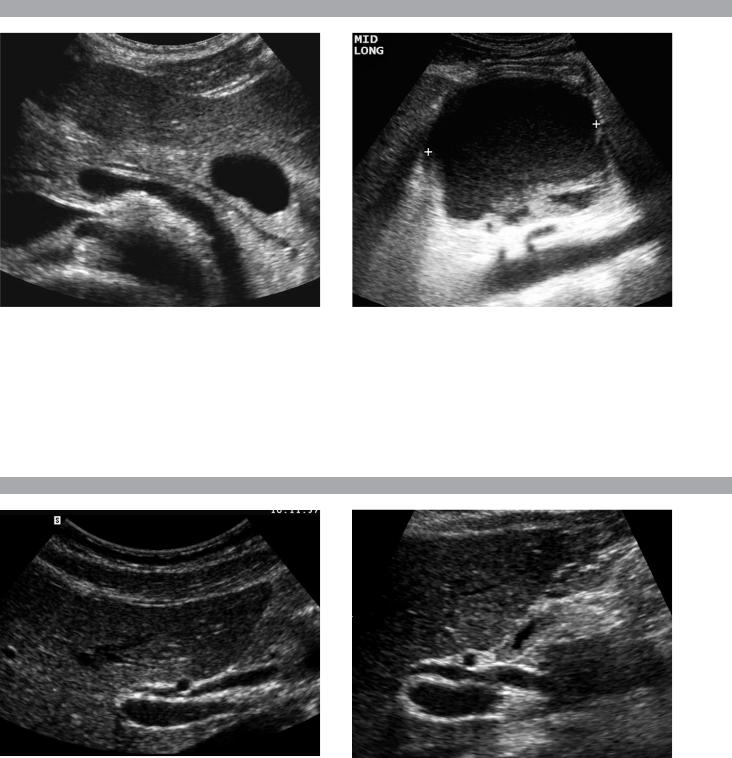
1.The transverse view shows an echogenic shadowing structure in the left pelvis in the expected location of the distal ureter. The longitudinal view confirms that it is located in the ureter.
2.Ultrasound is generally not used as a primary means of evaluating patients with suspected ureteral stones. The exception is when the patient is pregnant and radiation needs to be avoided.
3.The majority of ureteral stones impact in the distal ureter. Therefore, views through the urinary bladder give the highest yield for stone detection.
4.Stones in the distal ureter are seen with similar success in men and in women using transabdominal scanning through the bladder. In women, transvaginal scanning can be used in difficult cases.
Reference
Ohnishi K, Watanabe H, Ohe H, Saitoh M: Ultrasound
findings in urolithiasis in the lower ureter. Ultrasound
Med Biol 1986;12:577–579.
Cross-Reference
Ultrasound: THE REQUISITES, 2nd ed, pp 133–136.
Comment
Patients with renal colic can be imaged in a number of ways. Currently, noncontrast CT is the recommended first test since it avoids the small risk of reactions to contrast media and provides information about structures other than the kidneys, ureters, and bladder. Ultrasound has a very limited role in the initial workup of patients with suspected renal colic. This is not to say that ultrasound is not capable of establishing the correct diagnosis in the majority of patients. It is simply easier and more effective to start with other tests. One legitimate role of ultrasound is in the pregnant patient in whom radiation needs to be avoided.
Most, but certainly not all, patients who are passing a kidney stone will have at least mild hydronephrosis. They may also have a small amount of perinephric fluid, usually best seen around the upper or lower pole. In the proper clinical setting, either of these findings is very specific for ureteral stones. Identification of the ureteral calculus is easiest when it is in the proximal ureter near the ureteropelvic junction (using the kidney as a window) and in the distal ureter near the ureterovesicle junction (using the fluid-filled bladder as a window). The most common place for stones to impact is in the distal ureter, so it is very important to carefully evaluate the distal ureters. In addition to a transabdominal approach using the bladder as a window, the distal
ureters in women can be imaged quite well from a transvaginal approach. In men, a transrectal approach can be used, although it is not as successful as the transvaginal approach in women.
C A S E 3 9
Lower Extremity Deep Vein Thrombosis
1.Ultrasound is very good at diagnosing deep vein thrombosis (DVT) in the femoral–popliteal system.
2.Color Doppler plays a minor role in diagnosing DVT. It is usually evaluated completely with gray-scale imaging.
3.Lower extremity DVT is usually unilateral.
4.The diagnosis of DVT is made based on lack of venous compressibility. Detection of intraluminal echoes is not a reliable way to diagnose DVT, and lack of intraluminal echoes is not a reliable way to exclude DVT.
Reference
Fraser JD, Anderson DR: Deep venous thrombosis: Recent advances and optimal investigation with US. Radiology 1999;211:9–24.
Cross-Reference
Ultrasound: THE REQUISITES, 2nd ed, pp 293–295.
Comment
Ultrasound has become the procedure of choice in the evaluation of suspected lower extremity DVT. In symptomatic patients, the sensitivity and specificity exceed 95% and 98%, respectively, in the femoral–popliteal system. Results in asymptomatic high-risk patients (predominantly post hip and knee surgery) and in the calf are more variable.
Normally, the deep veins should be completely compressible. The diagnosis of DVT is made when the veins fail to compress completely. Many normal veins will have low-level internal echoes that are artifactual, and it is not uncommon for an intraluminal clot to be hypoechoic or anechoic. Therefore, analysis of echogenicity is not a primary focus of lower extremity venous examinations.
In patients with marked obesity or with severe edema, identification of the femoral and popliteal veins may be very difficult. In these situations, color Doppler may help to localize the vessels. Augmentation of proximal venous flow by compression of the calf or plantarflexion can accentuate the veins and further assist when color Doppler is required.
42
C A S E 4 0
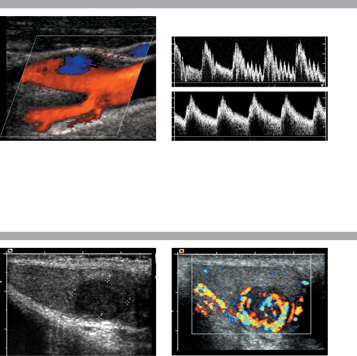
Longitudinal color Doppler view of the carotid bifurcation and pulsed Doppler waveforms from the internal and external carotid arteries.
1.Identify the internal and the external carotid arteries on the color Doppler image.
2.Which vessel is typically located anterior and medial?
3.Which waveform is from the internal and which is from the external carotid artery?
4.On longitudinal views of the bifurcation, is the internal carotid artery located superficial or deep to the external carotid?
C A S E 4 1
Longitudinal gray-scale and color Doppler images of the testis.
1.Describe the abnormality.
2.What nonneoplastic conditions can appear as a focal hypoechoic mass in the testis?
3.Are testicular tumors most likely benign or malignant?
4.Does associated enlargement and hypervascularity of the epididymis favor a neoplastic or nonneoplastic condition?
43

A N S W E R S
C A S E 4 0
Normal Carotid Bifurcation
1.Branches arise from the deep vessel, so that vessel is the external carotid artery. The larger vessel without branches is the internal carotid artery.
2.The external carotid artery is anterior and medial.
3.The top waveform shows a low resistance pattern typical of the internal carotid. The bottom waveform shows pulsations in the last two cardiac cycles due to manual tapping on the superficial temporal artery. This identifies the external carotid.
4.When the probe is positioned in the anterior and medial neck, the external carotid will be superficial. When the probe is positioned posterior and lateral, the internal carotid will be superficial.
Reference
Cardoso T, Middleton WD: Duplex sonography and color Doppler of carotid artery disease. Semin Interv Radiol 1990;7:1–8.
Cross-Reference
Ultrasound: THE REQUISITES, 2nd ed, pp 259–262.
Comment
Proper interpretation of carotid artery Doppler examinations requires reliable differentiation of the internal and external carotid arteries. Several features distinguish these vessels. Location, size, and branches are all useful. Realize, however, that although branches are always present on the external carotid artery, they are not always detectable on color Doppler. Therefore, branches are useful only when they are seen.
Doppler waveform analysis is also a valuable means of distinguishing the internal and external carotids. Since the internal carotid supplies a solid organ with a low vascular resistance, its waveform has a low resistance profile with broad systolic peaks, gradual systolic deceleration into early diastole, and well-maintained diastolic flow throughout the cardiac cycle. The external carotid supplies primarily muscle, bone, and cutaneous tissue, which has a high resistance to blood flow. Therefore, the external carotid waveform has narrower systolic peaks, more abrupt transition between systole and diastole, and less end diastolic flow. Another feature of the waveform that can be useful is transmission of fluctuations into the external carotid when the superficial temporal artery is tapped. This is called the temporal tap maneuver. Although the effects tend to be greatest and most frequent in the external carotid, occasionally some changes can be seen in the common and in the internal carotid.
C A S E 4 1
Testicular Seminoma
1.The images show a homogeneous, hypoechoic, hypervascular mass. The appearance is typical for a seminoma.
2.Infarcts, focal atrophy, focal orchitis, hematoma, abscess, sarcoid, and contusions.
3.They are much more likely to be malignant.
4.Involvement of the epididymis favors epididymoorchitis.
Reference
Horstman WG, Melson GL, Middleton WD, Andriole
GA: Color Doppler ultrasonography of testicular
tumors. Radiology 1992;185:733–737.
Cross-Reference
Ultrasound: THE REQUISITES, 2nd ed, pp 160–162.
Comment
Primary testis tumors are the most common malignancy in young adult males. Germ cell tumors account for the vast majority of testis tumors. Seminoma is the most common germ cell tumor, accounting for 40% to 50% of these malignancies. Seminomas frequently occur in a pure form as well as in mixed germ cell tumors. They tend to occur in a slightly older age group than the other germ cell tumors. Although 25% of patients with pure seminomas have metastases at the time of presentation, the 5-year survival rate is excellent. Most testis tumors manifest as a painless, palpable mass. Approximately 10% will manifest with testicular pain and 10% with symptoms due to metastatic disease (such as back pain due to retroperitoneal adenopathy).
On sonography, detection of an intratesticular lesion other than a simple cyst should always raise the possibility of a testicular tumor. Pure seminomas are typically homogeneous, hypoechoic, solid masses. When they are large, they tend to become more heterogeneous. Cystic components and calcification are distinctly uncommon in pure seminomas.
With modern color Doppler, most testicular tumors larger than 1 cm (and many that are less than 1 cm) are seen to have detectable internal blood flow. Detecting internal flow can be helpful in making a diagnosis since many benign intratesticular lesions that can potentially be confused with tumors, such as hematomas, abscesses, and infarcts, do not have internal flow. In addition, many nonneoplastic lesions seen on ultrasound are not palpable, whereas the majority of testicular tumors are palpable.
44
C A S E 4 2
Longitudinal views of the right and left upper quadrants in different patients with the same condition.
1.What are the major complications of the condition shown in this case?
2.What other associated abnormalities are there?
3.Would your diagnosis be the same if there were no family history of this condition?
4.Is there any effective therapy?
45

A N S W E R S
C A S E 4 2
Autosomal Dominant Polycystic Kidney
Disease
1.The most devastating complications of polycystic kidney disease (PKD) are renal failure and hypertension. Patients are also at increased risk for cyst hemorrhage, renal infection, and stone formation.
2.Findings besides renal cysts include cysts in the liver (and rarely in the pancreas) and berry aneurysms.
3.Yes. Up to 50% of cases of PKD are due to spontaneous mutations and have no family history.
4.There is no proven therapy. Laparoscopic cyst decortication can help relieve symptoms and may improve renal function.
References
Levine E, Hartman DS, Meilstrup JW, et al: Current concepts and controversies in imaging of renal cystic diseases. Urol Clin North Am 1997;24:523–544.
Ravine D, Gibson RN, Walker RG, et al: Evaluation of ultrasonographic criteria for autosomal dominant polycystic kidney disease 1. Lancet 1994;343:824.
Cross-Reference
Ultrasound: THE REQUISITES, 2nd ed, pp 113–114.
Comment
Autosomal dominant PKD is an inherited disorder that has 100% penetrance but is expressed to varying degrees. Patients generally present in the fourth or fifth decade with symptoms related to the mass effect of enlarged kidneys, hypertension, hematuria, or urinary tract infections. Renal failure develops on average in the sixth decade of life. Up to 10% of cases of end-stage renal disease in North America and Europe are due to PKD. If untreated, patients survive approximately 10 years from the onset of symptoms.
Two genetic defects cause PKD. Type 1 accounts for approximately 90% of families and is due to a defect on the short arm of chromosome 16. Type 2 is caused by a defect on the long arm of chromosome 4 and accounts for 10% of families. The major clinical difference in these two types is the lower age of onset of end-stage renal disease in type 1 than in type 2.
Although the kidneys are affected to a greater degree than any other organ, cysts can also develop in the liver (in up to 50% of patients), the pancreas (in up to 7% of patients), and the spleen (in less than 5% of patients). Cysts in these organs rarely cause clinical symptoms. In particular, the liver can be almost replaced by multiple cysts and still function normally. Approximately 10% of patients have intracranial aneurysms.
On sonography, the diagnosis is made by detecting multiple, bilateral renal cysts located in the renal cortex. In older patients, the kidneys are almost always enlarged, sometimes to the point that they cannot be effectively measured with sonography. Hemorrhage into renal cysts is very common and may result in low-level internal echoes, fluid-blood levels, or solid nodular internal structures. Cyst wall calcification and renal stones are also common.
In affected families, it is common to screen children to determine whether they inherited the disease. Criteria that have been established include the presence of at least two cysts in one kidney or one cyst in each kidney in an at-risk person younger than 30 years of age, the presence of at least two cysts in each kidney in an at-risk person between 30 and 59 years of age, and at least four cysts in each kidney for persons at risk aged 60 years or older. Most patients have many cysts present bilaterally, and the diagnosis is not in doubt. Ultrasound has a high sensitivity in making the diagnosis in patients with type 1 disease and in patients with type 2 disease who are older than 30 years of age. Ultrasound is less sensitive in patients with type 2 disease who are younger than 30 years of age. When ultrasound results are indeterminate or confusing, DNA linkage analysis can be performed if it is available.
The differential diagnosis for multiple renal cysts includes acquired cystic disease of dialysis, von Hipple– Lindau disease, tuberous sclerosis, and lymphoma. The kidneys are almost always small in acquired cystic disease. Von Hipple–Lindau disease is associated with central nervous system tumors, retinal angiomas, and pheochromocytomas. Solid renal masses due to renal cell cancer are also common in von Hipple–Lindau disease. Tuberous sclerosis has central nervous system and dermal abnormalities that are lacking with PKD. Lymphoma can rarely produce multifocal renal masses that simulate cysts, but these patients almost always have a history of lymphoma and there is usually evidence of lymphoma elsewhere in the body.
46
C A S E 4 3
Longitudinal and transverse views of the gallbladder in a patient with fever.
1.What are the abnormalities in the images shown above?
2.What is the differential diagnosis?
3.What is the preferred way of establishing this diagnosis?
4.What is the treatment of choice?
C A S E 4 4
Views of the testis in two patients.
1.What is the significance of the shadowing shown in the first image?
2.In patients such as these, what else should be scanned in addition to the scrotum?
3.Is scrotal MRI useful in patients with sonographic findings such as these?
4.What conditions predispose to this abnormality?
47

A N S W E R S
C A S E 4 3
Acute Cholecystitis
1.The gallbladder is enlarged (12 × 7 cm), has a thick wall (5 mm), and has an intraluminal stone.
2.The most likely diagnosis is acute cholecystitis. Many things can cause wall thickening, including edema-forming states, portal hypertension, hepatitis, adenomyomatosis, and gallbladder cancer. However, the association of gallbladder enlargement, stones, and wall thickening in a febrile patient is strongly predictive of acute cholecystitis.
3.Ultrasound is the best initial test in patients with suspected acute cholecystitis. Scintigraphy is also very helpful in a problem-solving mode when the sonogram is confusing or indefinite.
4.Surgery. In some cases, surgery will be postponed until the patient has been treated with antibiotics and the acute inflammatory changes have resolved.
Reference
Middleton WD: Right upper quadrant pain. In Bluth EI, Benson C, Arger P, et al (eds): The Practice of Ultrasonography. New York, Thieme, 1999, pp 3–16.
Cross-Reference
Ultrasound: THE REQUISITES, 2nd ed, pp 36–39.
Comment
Ultrasound is the procedure of choice in the evaluation of patients with suspected acute cholecystitis. In the majority of patients, by documenting a gallstone-free gallbladder, ultrasound can exclude the diagnosis rapidly and effectively. This is important since most patients with suspected acute cholecystitis do not actually have that problem. Ultrasound has also been shown to be as effective at establishing the diagnosis of acute cholecystitis as any other noninvasive technique.
The signs of acute cholecystitis are gallstones, wall thickening (>3 mm), gallbladder enlargement (> 4 cm transverse diameter), pericholecystic fluid, an impacted stone in the gallbladder neck or cystic duct, and a positive sonographic Murphy’s sign. The combination of stones and either wall thickening or a positive sonographic Murphy’s sign has a very high positive predictive value for acute cholecystitis.
Cholescintigraphy can also be used in patients with suspected acute cholecystitis. Accuracy is similar to that of ultrasound. However, there are several limitations to scintigraphy. Cholescintigraphy does not give morphologic information about the gallbladder (e.g., gallbladder size, wall thickness, pericholecystic abscess formation, or the presence and size of stones), which is important prior to laparoscopic cholecystectomy. It also fails to provide information about other organs that might account
for the patient’s problem when the gallbladder is normal. Finally, it is more time-consuming and expensive than sonography. Because of these limitations, cholescintigraphy is generally not the initial examination in patients with suspected cholecystitis. Nevertheless, it is a very good way to evaluate patients who have equivocal or confusing findings on ultrasound.
C A S E 4 4
Mixed Germ Cell Tumors
1.The shadowing indicates calcification, which is rare in pure seminomas but is common in mixed germ cell tumors.
2.In a patient with what appears to be a testicular tumor, it is helpful to scan the retroperitoneum to look for adenopathy.
3.Scrotal MRI is needed only rarely following scrotal ultrasound and would not add any valuable information in cases such as these.
4.Cryptorchidism, testicular atrophy, prior testicular tumor, and maybe microlithiasis predispose to testicular tumors.
References
Choyke PL: Dynamic contrast-enhanced MR imaging of the scrotum: Reality check. Radiology 2000;217:14–15.
Horstman WG: Scrotal imaging. Urol Clin North Am 1997;24:653–671.
Cross-Reference
Ultrasound: THE REQUISITES, 2nd ed, pp 160–163.
Comment
Of primary tumors of the testis, 95% are malignant germ cell neoplasms. These tumors are divided into seminomas and nonseminomas. Tumors that are composed of a mixture of seminomatous elements and nonseminomatous elements are grouped with the nonseminomas. Nonseminomatous tumors include teratoma, choriocarcinoma, embryonal cell carcinoma (also referred to as yolk sac tumors and endodermal sinus tumors in the pediatric age group), and any combination of these tumors. Teratocarcinoma refers to the relatively common combination of teratoma and embryonal cell carcinoma.
Unlike most seminomas, which are homogeneous and hypoechoic, nonseminomatous tumors are usually heterogeneous. This is at least partly due to the frequency of hemorrhage and necrosis. In addition, they often have cystic areas as well as calcifications. Cystic areas are most frequently seen in tumors that contain teratomatous elements.
48
C A S E 4 5
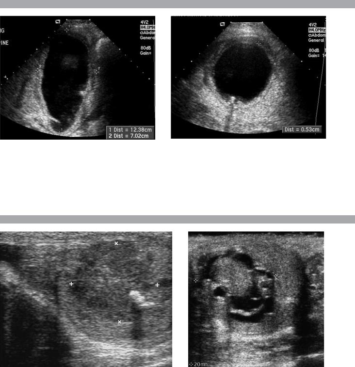
Longitudinal views of the porta hepatis in two patients. The first image is from a 21-year-old man and the second is from a 65-year-old woman.
1.Which of the patients shown in this case is more likely to have biliary obstruction?
2.Is the common duct frequently ectatic following a cholecystectomy?
3.How sensitive is ultrasound in determining the level and cause of biliary obstruction?
4.What is the theory behind giving patients a fatty meal when biliary obstruction is suspected?
C A S E 4 6
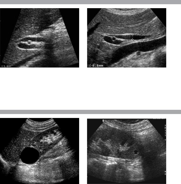
Longitudinal views of the kidney in two patients.
1.Is ultrasound a good means of characterizing lesions such as the ones shown here?
2.What are the four characteristics of these lesions?
3.What other lesions should be included in the differential diagnosis?
4.How common are these lesions?
49

A N S W E R S
C A S E 4 5
Extrahepatic Biliary Dilation
1.Both images show dilated common bile ducts. In the second image, the duct is dilated in its midportion, but it tapers to a normal caliber distally and proximally. In the first image, overlying gas obscures the distal duct, but the proximal duct is dilated where it crosses over the right hepatic artery. Because of this, it is more likely to be obstructed. Additional images showed distal duct stones in the first patient and no abnormalities in the second patient.
2.The size of the extrahepatic duct increases with age and following a cholecystectomy. Therefore, an enlarged duct is less concerning in these patients.
3.Reports indicate that sensitivities up to 80% to 90% can be obtained with good technique.
4.A fatty meal stimulates bile production by the liver and contraction of the gallbladder. In patients with obstruction, this results in enlargement of the duct.
Fatty meals also stimulate relaxation of the sphincter of Oddi so that there is no change or reduction in the size of the duct in patients who do not have obstruction. Intrinsic variability in measurements of the bile duct diameter often limits the use of this technique.
Reference
Middleton WD: The bile ducts. In: Goldberg BB (ed): Diagnostic Ultrasound. Baltimore, Williams & Wilkins, 1993, pp 146–172.
Cross-Reference
Ultrasound: THE REQUISITES, 2nd ed, pp 90–92.
Comment
Diagnosis of bile duct obstruction with sonography depends on detection of ductal dilatation. Different studies have proposed different measurements for the upper limits of normal for the extrahepatic ducts. Unfortunately, some obstructed ducts may not be dilated, and many things other than obstruction can cause dilated ducts. Therefore, rigid reliance on any single measurement value is dangerous. Important factors to realize when analyzing the common duct are that the duct normally enlarges with age (due to degeneration of the elastic fibers in the wall) and it often enlarges following a cholecystectomy (this is controversial). Thus, borderline enlarged ductal measurements are more likely to indicate obstruction in young patients and in patients with their gallbladders than in older patients or patients who have had a cholecystectomy. In addition, it is not uncommon for the common duct to be ectatic in its midportion between the liver and the head of the pancreas. Therefore, if the duct is of normal caliber proximally where it
crosses the right hepatic artery and distally within the head of the pancreas, then measurements slightly above the normal range in the midsegment are much less likely to indicate obstruction. With these caveats in mind, a reasonable value to use as the upper limit of normal for the maximum duct diameter is 6 mm. Some experts use 5 mm as the upper limit of normal and allow an additional 1 mm for each decade beyond age 50. In other words, a 5-mm duct is normal at age 50, a 6-mm duct is normal at age 60, and so forth.
C A S E 4 6
Renal Cysts
1.As in other parts of the body, ultrasound is an excellent way to evaluate the characteristics of cystic renal lesions.
2.Characteristics of a simple cyst include no perceptible wall, lack of internal echoes, sharp back wall reflection, and posterior acoustic enhancement.
3.Lesions that can simulate a simple cyst include calyx diverticulum, aneurysm, pseudoaneurysm, lymphoma, or upper pole duplication.
4.Approximately 50% of patients older than age 50 have at least one renal cyst.
Reference
Curry NS, Bissada NK: Radiologic evaluation of small and indeterminant renal masses. Urol Clin North Am 1997;24:493–505.
Cross-Reference
Ultrasound: THE REQUISITES, 2nd ed, pp 110–111.
Comment
Renal cysts are extremely common. Provided they satisfy the classic sonographic criteria for cysts, renal cysts require no further evaluation. It is important to realize that all three of the criteria for cysts may not be seen on all images of the cyst. For instance, the image that shows increased through transmission may not demonstrate an anechoic lumen. As long as the three criteria can be shown on a group of images, then a confident diagnosis of a cyst can be made.
A common dilemma is a cyst that appears to contain internal echoes. Internal echoes may be real or may be artifactual. Imaging from multiple, different approaches varies the interactions from overlying tissues and often helps to clear out artifactual internal artifacts. These types of artifacts can also be dramatically reduced or completely eliminated through the use of tissue harmonic imaging.
50
C A S E 4 7
Transverse view of the pancreas and sagittal view of the epigastrium in different patients with the same abnormality.
1.What question would you want to ask these patients?
2.How often do pancreatic adenocarcinomas have cystic components?
3.Does the differential diagnosis vary if the patient is a man or a woman?
4.Are pseudocysts more common in alcohol-induced or in gallstone-induced pancreatitis?
C A S E 4 8
Longitudinal views of the porta hepatis in two patients.
1.Where is the common duct in these patients?
2.In what percentage of patients is the duct located as shown here?
3.Which is straighter, the bile duct or the hepatic artery?
4.Which varies more in caliber, the bile duct or the hepatic artery?
51