

Colposcopymanual
.pdfChapter 7
acid stain is correlated with the colour tone or intensity, the surface shine, and the duration of the effect, and, in turn, with the degree of neoplastic change in the lesion. Higher-grade lesions are more likely to turn dense white rapidly. Abnormal vascular features such as punctation, mosaicism and atypical vessels are significant only if these are seen in acetowhite areas.
The acetic acid dehydrates cells and reversibly coagulates the nuclear proteins. Thus, areas of increased nuclear activity and DNA content exhibit the most dramatic colour change. The most pronounced effects are observed in high-grade lesions and invasive cancer. A direct correlation exists between the intensity of the dull, white colour and the severity of the lesion. Less differentiated areas are associated with an intensely opaque, dull-white appearance of lesions in the transformation zone.
Flat condyloma and low-grade CIN may uncommonly present as thin, satellite acetowhite lesions detached (far away) from the squamocolumnar junction with geographical patterns (resembling geographical regions) and with irregular, angular or digitating or feathery margins (Figures 7.9- 7.13). Many low-grade CIN lesions reveal less dense, less extensive and less complex acetowhite areas close to or abutting the squamocolumnar junction with well demarcated, but irregular, feathery or digitating margins (Figures 7.10- 7.16) compared with high-grade CIN lesions (Figures 7.17-7.27). High-grade lesions show well demarcated, regular margins, which may sometimes have raised and rolled out edges (Figures 7.25 and 7.26). High-grade lesions like CIN 2 or CIN 3 have a thick or dense, dull, chalk-white or greyish-white appearance (Figures 7.17- 7.27). They may be more extensive and complex lesions
Table 7.1: Surface extent of acetowhite areas associated with cervical neoplasia
Cervical neoplasia |
Cases |
One lip of cervix (%) |
Both lips (%) |
CIN 1 |
27 |
21 (78) |
6 (22) |
CIN 2 |
30 |
17 (57) |
13 (43) |
CIN 3 |
87 |
36 (41) |
51 (59) |
Early invasive cancer |
66 |
10 (15) |
56 (85) |
|
|
|
|
Adapted from Burghart et al., 1998 |
|
|
|
a
a
a |
a |
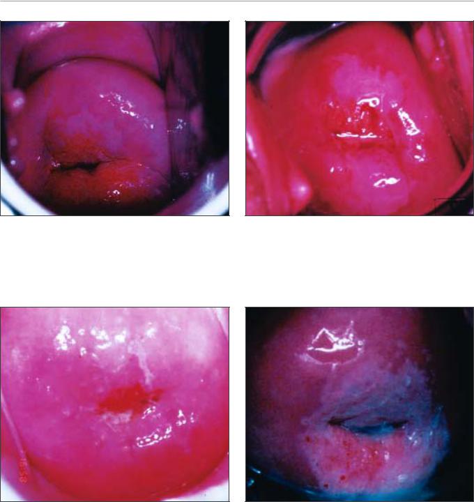
FIGURE 7.9: Geographic satellite lesion after application of 5% acetic acid (a) far away from the squamocolumnar junction, suggestive of low-grade lesion
FIGURE 7.10: Geographic satellite lesions after application of 5% acetic acid (a) far away from the squamocolumnar junction, suggestive of low-grade lesions
60
Colposcopic assessment of cervical intraepithelial neoplasia
FIGURE 7.11: Thin acetowhite lesion with geographic margins in the upper lip. Histology indicated CIN 1
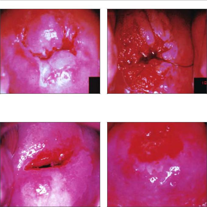
FIGURE 7.13: Mildly dense acetowhite lesions arising from the squamocolumnar junction in 12 and 6 o’clock position with irregular geographical margins, which on histology proved to be CIN 1 lesion
↑
a
FIGURE 7.12: Mildly dense, thin, elongated acetowhite lesion with regular margins abutting the squamocolumnar junction. Note the fine mosaic at the distal end of the lesion. Histology indicated CIN 1
FIGURE 7.14: Note the circumorificial acetowhite CIN 1 lesion with irregular margin and fine mosaics (a)
extending into the endocervical canal (Figures 7.22- 7.27) compared with low-grade lesions. High-grade lesions often tend to involve both the lips (Burghardt et al., 1998) (Table 7.1). Severe or early malignant lesions may obliterate the external os (Figures 7.22 and 7.25).
As lesions become more severe, their surfaces tend to be less smooth and less reflective of light, as in
normal squamous epithelium. The surfaces can become irregular, elevated and nodular relative to the surrounding epithelium (Figures 7.20 and 7.23-7.27).
The line of demarcation between normal and abnormal areas in the transformation zone is sharp and well delineated. High-grade lesions tend to have regular, sharper borders (Figures 7.17, 7.18, 7.19, 7.21,
61
Chapter 7
FIGURE 7.15: Moderately dense acetowhite lesions with irregular margins in the anterior and posterior lips (CIN 1)
FIGURE 7.17: Moderately dense acetowhite lesions with well defined margins and coarse punctations in the anterior lip and in 3 o’clock position (CIN 2 lesion)
↑
|
a |
a |
a |
|
|
|
|
FIGURE 7.16: Circumorificial, mild to dense acetowhite lesion with fine mosaic (arrow). Histology indicated CIN 1. Note the internal borders within the lesion (a)
7.23, 7.25 and 7.26) than low-grade lesions (Figures 7.13-7.16). Visualization of one or more borders within an acetowhite lesion (‘lesion within lesion’) (Figure 7.21) or a lesion with differing colour intensity (Figure 7.16) is an important observation indicating neoplastic lesions, particularly high-grade lesions. The crypt openings that are involved in high-grade precursor lesions may have thick, dense and wide acetowhite
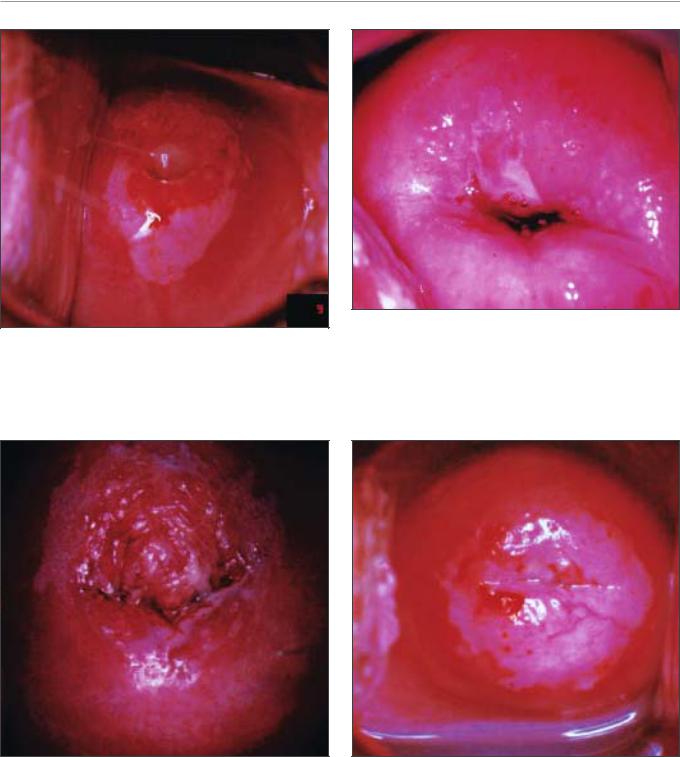
FIGURE 7.18: Dense, well defined acetowhite area with regular margins and coarse mosaic (CIN 2 lesion)
rims called cuffed crypt openings (Figure 7.26). These are whiter and wider than the mild, line-like acetowhite rings that are sometimes seen around normal crypt openings.
The cardinal features that should differentiate between the CIN lesions and immature metaplasia are the less dense and translucent nature of the acetowhitening associated with metaplasia, and the
62
Colposcopic assessment of cervical intraepithelial neoplasia
a
c c b
a
FIGURE 7.19: A dense acetowhite lesion with varying colour intensity and coarse mosaics (a) in a CIN 2 lesion
FIGURE 7.21: An acetowhite lesion arising at 12 o’clock position, abutting the squamocolumnar junction. Note the two colour intensities in the same lesion (a and b) with an internal border within the same lesion (c). This is an example of a lesion within a lesion
b
a
FIGURE 7.20: Acetowhite lesions with coarse punctation (a) and mosaics (b) in a CIN 2 lesion
lack of a distinct margin between the acetowhite areas of immature metaplasia and the normal epithelium. The line of demarcation between normal epithelium and acetowhite areas of metaplasia in the transformation zone is diffuse and invariably blends
FIGURE 7.22: A circumorificial dense opaque acetowhite area with coarse mosaics (CIN 3 lesion)
with the rest of the epithelium (Figures 6.8-6.13). The finger-like or tongue-like projections of the metaplastic epithelium often point towards the external os centripetally (Figures 6.11 and 6.12). The acetowhite lesions associated with CIN are invariably
63
Chapter 7
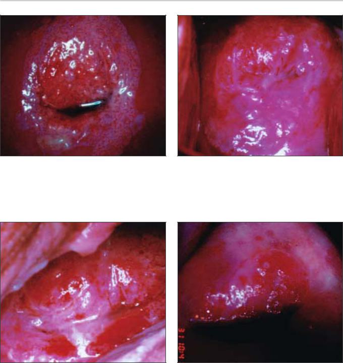
FIGURE 7.23: A dense acetowhite lesion with regular margin and coarse, irregular punctation in a CIN 3 lesion
FIGURE 7.25: Note the intensely dense, complex, acetowhite lesion (CIN 3 lesion) with raised and rolled out margins, obliterating the external os
a
↑
↑
FIGURE 7.24: Coarse mosaics (a) in a CIN 3 lesion
located in the transformation zone closer to or abutting, and appearing to arise from, the squamocolumnar junction (Figures 7.11-7.21). They spread centrifugally, pointing away from the external os. The line of demarcation between normal squamous epithelium, inflammatory lesions, and regenerating epithelium is also diffuse (Figures 9.2 and 9.5).
FIGURE 7.26: A dense acetowhite lesion with raised and rolled out margins with a cuffed crypt opening (dense arrow) and coarse mosaics with umblication, suggestive of a CIN 3 lesion
To summarize, acetowhite staining is not specific for CIN and may also occur, to some extent, in areas of immature squamous metaplasia, the congenital transformation zone, inflammation and healing and regenerative epithelium. However, acetowhite changes associated with CIN are found localized in the transformation zone, abutting the squamocolumnar
64
Colposcopic assessment of cervical intraepithelial neoplasia
FIGURE 7.27: A dense acetowhite, opaque, complex,
circumorificial CIN 3 lesion
junction and well demarcated from the surrounding epithelium. Low-grade lesions tend to be thin, less dense, less extensive, with irregular, feathery, geographic or angular margins and with fine punctation and/or mosaic; sometimes, low-grade lesions may be detached from the squamocolumnar junction; and atypical vessels are seldom observed in low-grade lesions. On the other hand, high-grade lesions are associated with dense, opaque, grey white, acetowhite areas with coarse punctation and/or mosaic and with regular and well demarcated borders; these lesions often involve both lips and may occasionally
a
a
a
harbour atypical vessels; CIN 3 lesions tend to be complex, involving the os.
After application of Lugol’s iodine solution
Lugol’s iodine solution is abundantly applied with a cotton swab to the whole of the cervix and visible parts of the vagina. The periphery of the cervix, fornices and vaginal walls must be observed until the epithelium is strongly stained dark brown or almost black by iodine. Normal vaginal and cervical squamous epithelium and mature metaplastic epithelium contain glycogen-rich cells, and thus take up the iodine stain and turn black or brown. Dysplastic epithelium contains little or no glycogen, and thus does not stain with iodine and remains mustard or saffron yellow (Figures 7.28-7.32). This colour difference is helpful in distinguishing normal from abnormal areas in the transformation zone that have shown faint acetowhitening. Columnar epithelium does not stain with iodine and immature metaplasia only partially stains, if at all. Atrophic epithelium also stains partially with iodine and this makes interpretation difficult in post menopausal women. Condylomatous lesions also do not, or only partially, stain with iodine (Figure 7.33).
Atypical epithelium of CIN may be less firmly attached to the underlying stroma, from which it may easily detach or peel off, after repeated application with different solutions, resulting in a true erosion (epithelial defect) exposing the stroma. Such true
FIGURE 7.28: Satellite lesions (a) do not stain with iodine after the application of Lugol’s iodine solution and remain as thin yellow areas (see the appearance after acetic acid application in Figure 7.10)
FIGURE 7.29: A CIN 1 lesion with a mustard yellow iodinenegative area with irregular margins (see the appearance after acetic acid application in Figure 7.15)
65
Chapter 7
FIGURE 7.30: Mustard yellow iodine-negative area in the anterior lip (CIN 2 lesion) after the application of Lugol’s iodine solution
FIGURE 7.31: Dense saffron yellow iodine-negative area of a CIN 3 lesion after the application of Lugol’s iodine solution. Note the surface irregularity.
erosions may easily be observed after iodine application, as the stroma does not stain with iodine.
Determining the nature of the lesion
The colposcopic detection of CIN essentially involves recognizing the following characteristics: the colour tone, margin and surface contour of the acetowhite
FIGURE 7.32: A dense mustard yellow iodine-negative area in the upper lip suggestive of CIN 3 lesion (see the appearance after acetic acid application in Figure 7.26)
FIGURE 7.33: A condylomatous lesion does not stain with iodine (see the appearance after acetic acid application in Figure 7.8)
epithelium in the transformation zone, as well as the arrangement of the terminal vascular bed and iodine staining. Variations in quality and quantity of the above atypical appearances help in differentiating CIN from physiological, benign, infective, inflammatory and reactive changes in the cervix. Grading schemes, based on these variations may guide the colposcopic diagnosis.
66

Colposcopic assessment of cervical intraepithelial neoplasia
Table 7.2: Modified Reid colposcopic index
Feature |
0 points |
1 point |
2 points |
Colour of |
Low-intensity acetowhitening; |
Grey-white AW with |
Dull, oyster-white; |
acetowhite |
snow-white, shiny AW; |
shiny surface |
Grey |
(AW) area |
indistinct AW; transparent AW; |
|
|
|
AW beyond the transformation |
|
|
|
zone |
|
|
AW lesion |
Feathered margins; angular, |
Regular lesions with |
Rolled, peeling edges; |
margin and |
jagged lesions; flat lesions |
smooth, straight |
internal demarcations |
surface |
with indistinct margins; |
outlines |
(a central area of high- |
configuration |
microcondylomatous or |
|
grade change and |
|
micropapillary surface |
|
peripheral area of low- |
|
|
|
grade change) |
Vessels |
Fine/uniform vessels; poorly |
Absent vessels |
Well defined coarse |
|
formed patterns of fine |
|
punctation or coarse |
|
punctuations and/or fine |
|
mosaic |
|
mosaic; vessels beyond the |
|
|
|
margin of transformation |
|
|
|
zone; fine vessels within |
|
|
|
microcondylomatous or |
|
|
|
micropapillary lesions |
|
|
Iodine |
Positive iodine uptake giving |
Partial iodine up-take |
Negative iodine uptake |
staining |
mahogany brown colour; |
by a lesion scoring 4 or |
by a lesion scoring 4 or |
|
negative uptake of lesions |
more points on above |
more points on the |
|
scoring 3 points or less on |
three categories – |
above three criteria |
|
above three categories |
variegated, speckled |
|
|
|
appearance |
|
|
|
|
|
Scoring: A score of 0 to 2 points = Likely to be CIN 1; 3-4 points = Overlapping lesion: likely to be CIN 1 - 2; 5 to 8 points = Likely to be CIN 2 - 3 lesions.
Table 7.3: Grading abnormal colposcopic findings using two categories
Grade |
Findings |
|
1. |
Insignificant |
The acetowhite epithelium is usually shiny or semitransparent. The borders |
|
|
are not sharp, with or without fine-calibre vessels (fine punctation and/or |
|
|
fine mosaic), which have ill-defined patterns and short intercapillary |
|
|
distances. There is an absence of atypical vessels. |
2. |
Significant |
Dense acetowhite or grey opaque epithelium is sharply bordered. There are |
|
|
dilated calibre, irregular shaped or coiled vessels (coarse punctation and/or |
|
|
mosaic). Atypical vessels and sometimes irregular surface contour indicate |
|
|
either imminent or invasive cancer. |
Adapted from Coppleson et al., 1993 b
67

Chapter 7
We recommend that the student should become familiar with the current colposcopic terminology given in Appendix 4 and use this to record the colposcopic findings (Stafl & Wilbanks, 1991).
The colposcopist is also encouraged to make a colposcopic prediction (or ‘diagnosis’) at the end of the colposcopic session in terms of normal (or negative), low-grade CIN, high-grade CIN, invasive cancer, other (e.g., inflammation etc.) and unsatisfactory colposcopy. Use of a scoring or grading system may guide colposcopic interpretation and diagnosis in a less subjective manner and helps
developing a systematic approach to colposcopy. The modified Reid colposcopic score (Table 7.2 and Appendix 5) based on the colposcopic index proposed by Reid & Scalzi (1985) is quite useful for this purpose. We recommend that beginners routinely use this scoring system to decide whether or not a lesion is CIN and to select biopsy sites. An alternative may be a two-class grading system developed by Coppleson et al (1993) (Table 7.3). We also recommend the student to use the above systems only when an acetowhite area is observed.
68

Chapter 8
Colposcopic diagnosis of preclinical invasive carcinoma of the cervix and glandular neoplasia
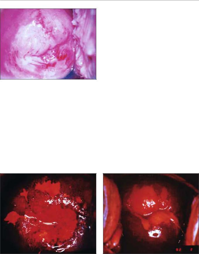
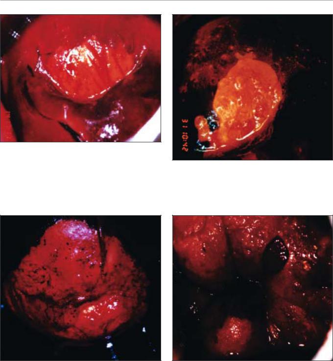
•Acetowhite lesions with atypical vessels; large, complex acetowhite lesions obliterating the os; lesions with irregular and exophytic contour; strikingly thick, chalky-white lesions with raised and rolled out margins; and lesions bleeding on touch should be thoroughly investigated to rule out the possibility of early preclinical invasive cancer.
•Appearance of atypical blood vessels may indicate the first sign of invasion; one of the earliest colposcopic signs of invasion is blood vessels breaking out from mosaic formations.
•The atypical vessel patterns are varied and may take the form of hairpins, corkscrews, waste thread, commas, tadpole and other bizarre, irregular branching patterns with irregular calibre.
•Most glandular lesions originate in the transformation zone and may be associated with concomitant CIN lesions.
•Stark acetowhiteness of individual or fused villi in discrete patches in contrast to the surrounding columnar epithelium or closely placed, multiple cuffed crypt openings in a dense acetowhite lesion may indicate glandular lesions.
•Greyish-white, dense lesions with papillary excrescences and waste thread like or character writinglike atypical vessels or lesions with strikingly atypical villous structures may be associated with glandular lesions.
Invasive carcinoma is the stage of disease that follows CIN 3 or high-grade glandular intraepithelial neoplasia. Invasion implies that the neoplastic epithelial cells have invaded the stroma underlying the epithelium by breaching the basement membrane. The term preclinical invasive cancer is applied to very early invasive cancers (e.g., stage 1) in women without symptoms and gross physical findings and clinical signs, that are diagnosed incidentally during colposcopy or by other early-detection approaches such as screening. The primary responsibility of a colposcopist is to ensure that if preclinical invasive carcinoma of the cervix is present in a woman, it will be diagnosed. Colposcopic signs of this condition are usually recognizable early on, unless the lesion is hidden at the bottom of a crypt. This chapter describes the colposcopic detection of invasive
cervical carcinomas followed by a specific consideration of cervical glandular neoplasia.
It is crucial for the colposcopist to become familiar with the signs of preclinical cervical cancer and understand the need for strict adherence to the diagnostic protocols that ensure the safety of women who are referred into their care. The use of colposcopy and directed biopsy as a diagnostic approach replaces the use of cervical cold-knife conization as the main diagnostic approach to women with cervical abnormalities. This means that the onus for diagnostic accuracy no longer rests solely on the pathologist who evaluates the cone specimen, but also on the colposcopist who provides the histological material for the pathologist’s examination. The use of ablative treatment such as cryotherapy, in which no histological specimen of the treated area is available, further
69