

Colposcopymanual
.pdfInflammatory lesions of the uterine cervix
After application of acetic acid
The liberal application of acetic acid clears the cervix and vagina of secretions, but may cause pain. Cervicovaginitis is associated with oedema, capillary dilatation, enlargement of the stromal papillae, which contain the vascular bundles, and infiltration of the
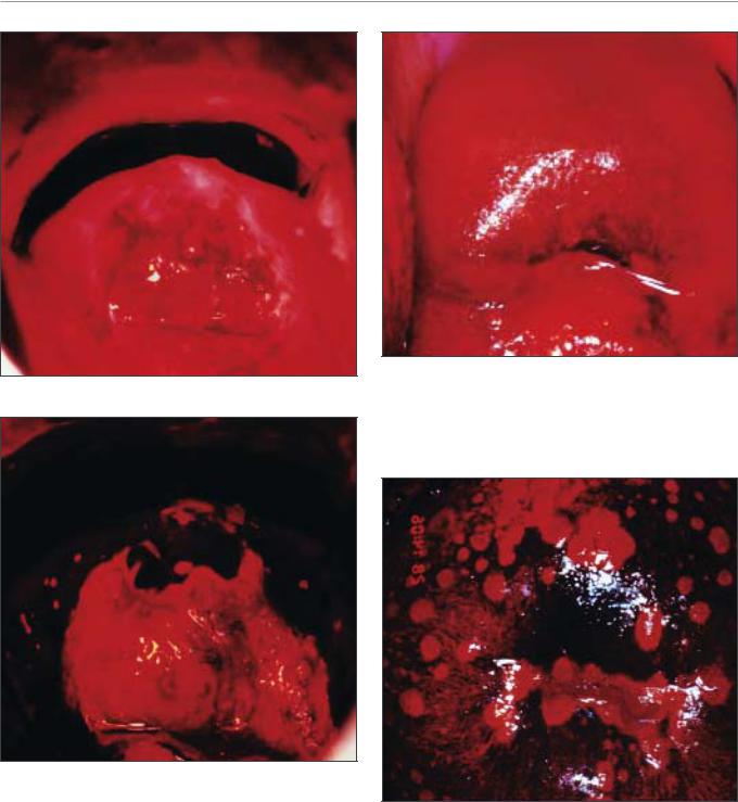
FIGURE 9.2: Chronic cervicitis: this cervix is extensively inflamed with a reddish appearance and bleeding on touch; there are ill-defined, patchy acetowhite areas scattered all over
the cervix after the application of acetic acid
stroma with inflammatory cells. Chronically inflamed cervix may appear reddish, with ill-defined, patchy acetowhite areas scattered in the cervix, not restricted to the transformation zone and may bleed on touch (Figures 9.2, 9.3). The enlarged stromal papillae appear as red spots (red punctation) in a pinkish-white background, usually in the case of T. vaginalis infection, after application of acetic acid. An inexperienced colposcopist may confuse the inflammatory punctations with those seen in cervical intraepithelial neoplasia (CIN). However, one can differentiate using the following criteria: inflammatory punctations are fine, with extremely minimal intercapillary distances, and diffusely distributed (not restricted to the transformation zone) and they involve the original squamous epithelium and vagina with intervening inflamed mucosa. As the inflammation persists and becomes chronic, it results in large, focal red punctations due to large collections of capillaries grouped together, which appear as several red spots of different sizes visible in a pinkishwhite background, producing the so-called ‘strawberry spots’ (Figure 9.4). Colposcopically, a chronically inflamed cervix may sometimes resemble invasive cervical cancer (Figure 9.5).
After application of Lugol’s iodine
The test outcome after application of Lugol’s iodine solution depends upon the desquamation and the loss
a
a
a
a |
a |
|
a
FIGURE 9.3: Chronic cervicitis: the cervix is highly inflamed and eroded with ill-defined, patchy acetowhite areas scattered all over
FIGURE 9.4: Multiple red spots (a) suggestive of Trichomonas vaginalis colpitis (strawberry appearance) (after application of 5% acetic acid)
81
Chapter 9
a a
a
After application of 5% acetic acid
After application of Lugol’s iodine
a
FIGURE 9.6: Stippled appearance (a) due to Trichomonas vaginalis colpitis after application of Lugol’s iodine
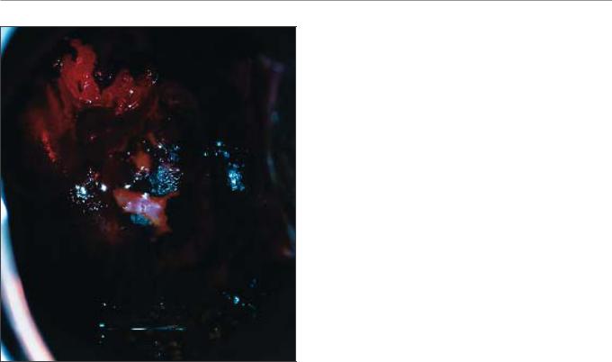
FIGURE 9.5: Colposcopic appearance of a chronically inflamed cervix showing areas of ulceration, necrosis and healing.
The regenerating areas turn somewhat white (a) after application
of cell layers containing glycogen. If desquamation is limited to the summit of the stromal papillae where the squamous epithelium is thinnest, a series of thin yellow spots are seen on a mahogany-brown background, giving a stippled appearance (Figure 9.6). When the inflammation persists and the infection becomes chronic, the small desquamated areas
FIGURE 9.7: Trichomonas vaginalis colpitis after application of Lugol’s iodine (leopard-skin appearance)
become confluent to form large desquamated areas leading to the so-called leopard-skin appearance (Figure 9.7). These features are often found with Trichomonas infection, but also may be seen with fungal and bacterial infections. If there is marked desquamation, the cervix appears yellowish-red in colour, with involvement of vagina (Figure 9.8).
82
Inflammatory lesions of the uterine cervix
In summary, inflammatory conditions of the cervix are associated with excessive, usually malodorous, mucopurulent, seropurulent or whitish discharge, red punctations, ulceration, and healing by fibrosis. The secretion is frothy with bubbles in the case of trichomoniasis and sticky cheese white in candidiasis. Inflammatory lesions of the cervix may be differentiated from CIN by their large, diffuse involvement of the cervix, extension to the vagina, red colour tone and associated symptoms such as discharge and pruritus.
FIGURE 9.8: Chronic cervicitis: there are scattered, ill-defined, patchy iodine non-uptake areas on the cervix and vagina. Also, the cervix appears yellowish red in colour
83

Chapter 10
Avoiding errors in the colposcopic assessment of the cervix and colposcopic provisional diagnosis
•A thorough knowledge of anatomy, pathophysiology and natural history of diseases of the female genital tract is essential to avoid errors in colposcopic assessment.
•Strict adherence to a diagnostic protocol and an awareness of the limitations of colposcopy are equally important.
•Regular interaction with the pathologists and clinical audits help to improve the quality of colposcopy.
•We encourage arriving at a provisional diagnosis, based on the colposcopic findings.
An adequate knowledge of pathophysiology and understanding of the natural history of diseases of the female genital tract that can be diagnosed with the colposcope and then treated are essential for satisfactory performance of colposcopy. A thorough knowledge of instrumentation, methods of examination and terminology is equally important. A high degree of accuracy in diagnosing cervical intraepithelial neoplasia (CIN) and ruling out invasive cancer may be achieved with good clinical judgement. Scrupulous adherence to a diagnostic protocol and awareness of the limitations and pitfalls of colposcopy are important.
Errors are commonly committed due to a lack of awareness and to deviation from established colposcopic protocol and practice. Good training, experience, an innate interest, and an established diagnostic algorithm will diminish the possibility of errors. These factors are particularly important in lowresource environments, where there are limited opportunities for mutual consultations and continuing education. The colposcopist should try to achieve the same degree of accuracy as a histopathologist can achieve with cervical conization specimens.
A summary of common sources of shortcomings in colposcopic practice is presented in Table 10.1. Regular interaction with the pathologist and clinical audits, to correlate colposcopic diagnoses with histological diagnoses, helps to improve the quality of colposcopy. It is important for the provider to learn the art of
taking colposcopically directed biopsies from appropriate area(s) in the transformation zone by using sharp biopsy forceps without crushing specimens. If the squamocolumnar junction is hidden in the endocervical canal, it is necessary to perform endocervical curettage (ECC) or cone biopsy in order to investigate the canal properly. It is obligatory to wait for 60 seconds after a liberal application of acetic acid for it to take full effect. Specific mention should be made of the location of the squamocolumnar junction and the acetowhite areas in relation to the junction. Careful inspection of the vagina should also be made for any extension of cervical lesions. It is best to examine the vagina when the speculum is being withdrawn at the end of each examination. Findings must be clearly and legibly documented. Using an objective scoring system such as Reid’s score (Appendix 5) is particularly helpful for beginners to arrive at a colposcopic diagnosis and to select appropriate sites for directed biopsies. Continuing education is important to enable the colposcopist to keep up with developments. Avoidance of missing or undertreating an invasive cancer and ensuring the provision of adequate treatment is largely dependent on the skills of the colposcopist.
Colposcopic provisional diagnosis
We strongly encourage the colposcopists to make a provisional diagnosis, based on the findings of
85

Chapter 10
Table 10.1: Common sources of colposcopic errors
Inadequate training and experience
Inadequate understanding of the natural history of disease
Failure to use an established diagnostic protocol or deviation from the protocol
Failure to use the largest speculum possible False squamocolumnar junction caused by
abrasion
Failure to choose appropriate biopsy sites and failure to take enough biopsies
Failure to take a biopsy when in doubt
Using a blunt, non-sharp biopsy punch to obtain tissue specimens
Failure to take a colposcopically directed biopsy Failure to perform biopsies from condylomata or
leukoplakia
Failure to wait for the full effect of acetic acid Failure to apply Lugol’s iodine solution and
examine
Failure to examine the endocervical canal adequately when the lesion limit or squamocolumnar junction is not seen
Failure to do endocervical curettage (ECC) when the lesion limit is not seen
Failure to perform excision when the lesion limit is not seen with an endocervical
speculum or when ECC is equivocal or positive Failure to perform excision when microinvasion
is suspected
Failure to inspect the vagina and vulva Failure to properly and legibly record
colposcopic findings
Failure to communicate with the pathologist Failure to correlate histological and colposcopic
findings
Failure to consult experts in difficult cases Failure to keep up with continuing education Failure to self-audit
Adapted from: Popkin (1995)
colposcopic examination. The provisional diagnosis may be in terms of normal, inflammation, leukoplakia, condyloma, low-grade CIN, high-grade CIN, early invasive cancer, overt invasive cancer, others (atrophy, cervical polyp, radiation changes, etc.) and
inconclusive. Such diagnosis is based on the evaluation of all the findings such as the characteristics of the acetowhite areas, vascular features, colour change after iodine application, surface characteristics such as ulceration, and other signs such as bleeding on touch, the nature of cervical and vaginal discharge and the findings of examination of external anogenitalia, groin and lower abdomen. These are described in detail in Chapters 6-9. Once a provisional diagnosis is made, a plan for management of the condition diagnosed should be developed. Table 10.2 provides a summary of the colposcopic findings that help in making the provisional diagnosis.
86

Table 10.2: A summary of colposcopic features guiding provisional diagnosis
Diagnosis |
|
|
Acetowhitening |
|
|
Vascular |
Iodine uptake |
Bleeding on |
Ulceration |
Discharge |
|
|
Colour tone |
Demarcation |
Margin |
Surface |
Relation to TZ |
Duration of |
features |
|
touch |
|
|
|
|
|
|
|
and SCJ |
effect |
|
|
|
|
|
Normal |
- |
- |
- |
- |
- |
- |
Normal |
Squamous |
Nil |
Nil |
Clear secretion |
|
|
|
|
|
|
|
vascular |
epithelium black |
|
|
from the |
|
|
|
|
|
|
|
pattern |
in colour; |
|
|
columnar |
|
|
|
|
|
|
|
|
columnar |
|
|
epithelium |
|
|
|
|
|
|
|
|
epithelium, no |
|
|
|
|
|
|
|
|
|
|
|
change in colour |
|
|
|
Normal, |
Pinkish white, |
Nil |
Indistinct, |
Smooth; crypt |
Restricted to TZ; |
< 1 minute |
Normal |
No or partial |
Nil |
Nil |
Clear secretion |
immature |
or snow white, |
|
blends with |
openings, |
prominent near |
|
vascular |
uptake |
|
|
from the |
metaplasia |
translucent, |
|
the rest of the |
islands of |
the SCJ |
|
pattern |
|
|
|
columnar |
|
patchy |
|
epithelium |
columnar |
|
|
|
|
|
|
epithelium |
|
acetowhite |
|
|
epithelium seen |
|
|
|
|
|
|
|
|
areas |
|
|
|
|
|
|
|
|
|
|
Normal, mature |
Light pinkish |
Nil |
Blends with |
Smooth, reveals |
Restricted to TZ |
- |
Normal |
Takes up iodine, |
Nil |
Nil |
Clear secretion |
metaplasia |
white hue. No |
|
the rest of the |
crypt openings, |
|
|
vascular |
turns black or |
|
|
from the |
|
confluent |
|
epithelium |
nabothian |
|
|
pattern |
brown |
|
|
columnar |
|
acetowhite area |
|
|
follicles |
|
|
|
|
|
|
epithelium |
Inflammation |
Pale, patchy |
Nil |
Indistinct, |
Irregular, |
Not restricted to |
< 2 minutes |
Diffusely |
Partial iodine |
May be |
May be |
Malodorous, |
|
areas, with |
|
blends with |
variegated |
TZ, may be widely |
|
distributed, |
uptake |
present |
present |
profuse, |
|
intervening red |
|
the rest of the |
appearance |
disseminated |
|
fine red |
|
|
|
mucopurulent |
|
areas and/or |
|
epithelium |
|
|
|
punctation |
|
|
|
or seropurulent |
|
necrotic areas |
|
|
|
|
|
involving cervix |
|
|
|
or non-odorous |
|
|
|
|
|
|
|
and vagina |
|
|
|
thick, sticky, |
|
|
|
|
|
|
|
|
|
|
|
white discharge |
Low-grade CIN |
Moderately |
Well demarcated |
Irregular, |
Flat, smooth or |
Mostly seen in the |
1-2 minutes |
Fine |
No uptake |
Nil |
Nil |
Nil |
|
dense, shiny, |
confluent lesions |
feathery, |
microcondyloma |
TZ, abuts the SCJ. |
|
punctation |
|
|
|
|
|
opaque, thin |
|
jagged, |
-tous or |
Very early lesions |
|
and/or mosaic |
|
|
|
|
|
lesions |
|
digitating, |
micropapillary |
may be outside TZ |
|
with in the AW |
|
|
|
|
|
|
|
angular or |
|
as satellite lesions |
|
lesion may be |
|
|
|
|
|
|
|
geographic |
|
|
|
seen |
|
|
|
|
87
diagnosis provisional colposcopic and cervix the of assessment colposcopic the in errors Avoiding

88
Table 10.2 (cont.): A summary of colposcopic features guiding provisional diagnosis
Diagnosis |
|
|
Acetowhitening |
|
|
Vascular |
Iodine uptake |
Bleeding on |
Ulceration |
Discharge |
|
|
Colour tone |
Demarcation |
Margin |
Surface |
Relation to TZ and |
Duration of |
features |
|
touch |
|
|
|
|
|
|
|
SCJ |
effect |
|
|
|
|
|
High-grade CIN |
Dull, dense, |
Well demarcated |
Regular, |
Less smooth, |
Restricted to TZ, |
2-4 minutes |
Coarse |
No iodine uptake |
May be |
Nil |
Nil |
|
greyish-white or |
confluent |
smooth |
more irregular |
abutting the SCJ |
|
punctation |
|
present in |
|
|
|
oyster-white |
lesions; internal |
outlines; |
and/or |
|
|
and/or coarse |
|
severe |
|
|
|
opaque lesion |
demarcations |
occasionally |
occasionally |
|
|
mosaic within |
|
lesions |
|
|
|
|
and borders may |
may be raised |
nodular surface |
|
|
the AW lesion |
|
|
|
|
|
|
be present |
and rolled out |
|
|
|
may be seen; |
|
|
|
|
|
|
|
|
|
|
|
atypical vessels |
|
|
|
|
|
|
|
|
|
|
|
may be seen |
|
|
|
|
|
|
|
|
|
|
|
(+) |
|
|
|
|
Preclinical |
Chalky white, |
Well demarcated |
Raised and |
Irregular, |
May involve the |
> 3 minutes |
Coarse raised |
No iodine uptake |
Surface |
May be seen |
May be present |
invasive cancer |
thick, dense, |
|
rolled out |
nodular or |
entire cervix, large |
|
mosaics and/or |
|
bleeding/- |
|
due to secondary |
|
opaque lesions |
|
margins |
mountainsand- |
complex lesions |
|
breaking |
|
oozing |
|
infection |
|
|
|
|
valley pattern |
obliterating the os |
|
mosaics |
|
common |
|
|
|
|
|
|
|
|
|
and/or, coarse |
|
|
|
|
|
|
|
|
|
|
|
punctations; |
|
|
|
|
|
|
|
|
|
|
|
atypical vessels |
|
|
|
|
|
|
|
|
|
|
|
always present |
|
|
|
|
|
|
|
|
|
|
|
(+++++) |
|
|
|
|
Overt invasive |
Dense white |
Entire cervix |
Entire cervix |
Ulceropro- |
Entire cervix |
Whiteness |
Atypical vessels |
No uptake, but |
Profuse |
Always |
Malodorous, |
cancer |
areas, may be |
replaced by |
replaced by |
liferative growth |
replaced by |
usually |
always present |
bleeding |
bleeding |
present |
blood stained, |
|
obliterated by |
growth |
growth |
|
growth extending |
obliterated by |
(++++++) |
obliterates |
|
|
purulent |
|
profuse bleeding |
|
|
|
to adjacent tissues |
bleeding |
|
iodine uptake |
|
|
discharge |
|
|
|
|
|
|
|
|
patterns |
|
|
due to secondary |
|
|
|
|
|
|
|
|
|
|
|
infection |
|
|
|
|
|
|
|
|
||||
TZ: transformation zone ; SCJ: squamocolumnar junction; AW: Acetowhitening |
|
|
|
|
|
|
|
||||
10 Chapter

Chapter 11
Management that provides continuity of care for women
•If women are diagnosed with reproductive tract infection, prompt treatment should be instituted according to the WHO guidelines.
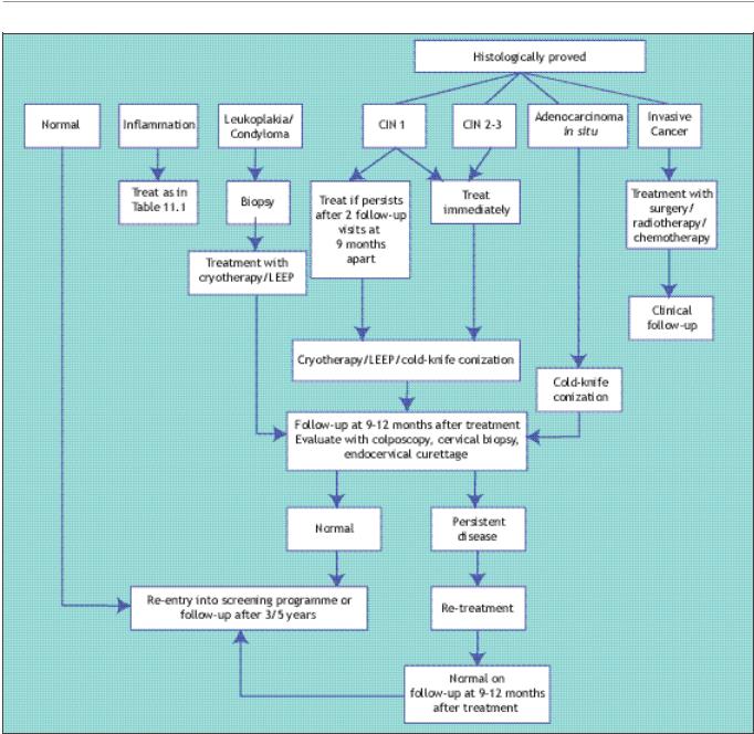
•Though it may be preferred to have the diagnosis of CIN firmly established before treatment is offered, in many low-resource settings, treatment may be offered at the first colposcopy visit, based on colposcopic findings, to maximize treatment coverage.
•The clinical management of women with CIN 1 lesions may take one of the following courses:
(i) immediate treatment or (ii) follow the woman and then treat if the lesion is persistent or progressive after 18 to 24 months.
•All women with CIN 2 and CIN 3 lesions should be treated with cryotherapy or LEEP.
•Women diagnosed with invasive cancer should be promptly referred for treatment.
•Women diagnosed with high-grade CIN during pregnancy can be reviewed at about 28 weeks gestation. If the disease is stable, the woman may be reviewed at 2-3 months post-partum for definitive diagnosis by biopsy and appropriate management of lesions.
•Women treated for CIN may be reviewed at 9-12 months after treatment.
Planning a woman’s medical management after her initial colposcopic assessment is primarily the duty of the colposcopist. It is appropriate to involve the woman, as a partner, in the decision-making process. Management usually depends on the final assessment after the colposcopic findings have been integrated with the pathology reports. Management plans also depend on whether or not the woman is pregnant. The management plan should be explicitly detailed in the medical record and communicated clearly to the patient at the earliest opportunity. Ideally, pathology reports (biopsy, endocervical curettage (ECC), loop electrosurgical excision procedure (LEEP) specimen, cytology) should be available to the colposcopist within three weeks of carrying out the colposcopy. Cryotherapy or LEEP are the two forms of therapy discussed in this manual (see Chapters 12 and 13), but it must be emphasized each has specific indications for
their use and should be used only when women fulfil all of the eligibility criteria for the specific therapy. A general plan of management that may be adapted in low-resource settings is shown in Figure 11.1.
It is generally preferable to have the diagnosis of cervical intraepithelial neoplasia (CIN) firmly established before a decision on management is taken and any treatment offered. However, there may be exceptions to this rule. For example, in many settings, particularly developing countries, women may be offered treatment at their first colposcopy visit, based on colposcopic assessment to maximize treatment coverage (otherwise patients lost to follow-up would not receive treatment for lesions). If the decision is to treat with cryotherapy, a biopsy (or biopsies) may be directed before cryotherapy, as this type of treatment does not produce a tissue specimen for histological examination. A tissue sample taken before instituting
89
Chapter 11
FIGURE 11.1: Flow chart of management decisions in cervical neoplasia and other conditions in low-resource settings. (CIN-cervical
intraepithelial neoplasia; LEEP-Loop electrosurgical excision procedure)
ablative therapy will help to confirm the histological nature of the lesion treated a posteriori. Expert colposcopists also may use this approach to maximize treatment coverage and to minimize the number of clinic visits in some settings. However, this approach may result in a significant degree of overtreatment. Even though it is assumed that treatment methods such cryotherapy and LEEP are safe, and are unlikely to be associated with long-term sequelae and complications, the long-term implications of such overtreatment remains yet to be firmly established. On the other hand, it is likely that overtreatment may, to a certain
extent, protect against future development of CIN, in view of the ablation of the transformation zone where the vast majority of CIN lesions occur.
Outcomes after colposcopic assessment
Normal colposcopy outcome
In most women with a normal cervix, assessment using the colposcope results in a satisfactory evaluation of the cervix. If the squamocolumnar junction is visible and there is no colposcopic evidence of CIN or invasive cancer, the woman should be discharged and may resume participation in the
90

Management that provides continuity of care for women
screening programme, if one is on-going in the region (which is not the case in many developing countries!). Otherwise, she may be advised to undergo a repeat screening examination after three to five years.
Reproductive tract infection
If a woman is diagnosed with reproductive tract infection, prompt treatment should be instituted following the WHO guidelines (WHO, 2001). The
Table 11.1 Treatment for reproductive tract infections
Reproductive tract |
Treatment guidelines |
|
infection |
Non-pregnant women |
Pregnant women |
T. vaginalis (Trichomoniasis) |
Metronidazole 400 mg orally, 2 times a day, for 7 days |
1st trimester: Metronidazole gel, 0.75%, 5 g, 2 times a |
|
or tinidazole 500 mg orally, 2 times a day, for 7 days |
day intravaginally, for 7 days; 2nd and 3rd trimester: |
|
or |
same as non-pregnant women. |
|
a single dose of metronidazole 2 g orally or tinidazole |
|
|
2 g orally. |
|
Candidiasis |
Clotrimazole or miconazole, 200 mg intravaginally, |
Clotrimazole or miconazole, 200 mg intravaginally, |
|
daily for 3 days |
daily for 3 days. |
|
or |
|
|
Fluconazole, 150 mg orally, as a single dose. |
|
Bacterial vaginosis |
Metronidazole 400 mg orally, 2 times a day, for |
Metronidazole gel, 0.75%, 5 g, 2 times a day |
|
7 days. |
intravaginally, for 7 days or clindamycin, 300 mg |
|
|
orally, 2 times daily, for 7 days. |
Chlamydial infection |
Doxycycline 100 mg orally, 2 times a day, for 7 days or |
Erythromycin 500 mg orally, 4 times daily, for 7 days |
|
azithromycin, 1 g orally, as a single dose. |
or amoxycillin, 500 mg orally, 3 times daily, for 7 |
|
|
days. |
Gonococcal infection |
Ciprofloxacin, 500 mg, orally, as a single dose or |
Cefixime, 200 mg orally, as a single dose or |
|
azithromycin 2 g orally as a single dose. |
ceftriaxone, 125 mg intramuscularly, as a single dose. |
Syphilis |
Benzathine penicillin, 2.4 million IU, by intramuscular |
Benzathine penicillin, 2.4 million IU, by intramuscular |
|
injection, as a single treatment; in penicillin-allergic |
injection, as a single treatment; in penicillin-allergic |
|
patients, doxycycline, 100 mg orally, 2 times a day, |
patients, erythromycin 500 mg orally, 4 times daily, |
|
for 15 days |
for 15 days. |
Lymphogranuloma venereum |
Doxycycline, 100 mg orally, 2 times daily, for 14 days |
Erythromycin 500 mg orally, 4 times a day, for |
|
or erythromycin 500 mg orally, 4 times a day, for 14 |
14 days. |
|
days. |
|
Chancroid |
Ciprofloxacin, 500 mg orally, 2 times a day, for 3 days |
Erythromycin 500 mg orally, 4 times a day, for 7 days. |
|
or erythromycin 500 mg orally, 4 times a day, for |
|
|
7 days or a single dose of azithromycin, 1 g orally. |
|
Granuloma inguinale |
Azithromycin, 1 g orally, as a single dose or |
Erythromycin 500 mg orally, 4 times a day, for 7 days. |
|
doxycycline, 100 mg orally, 2 times daily, for 14 days |
|
Genital herpes |
Acyclovir, 400 mg orally, 3 times daily, for 7 days or |
- |
|
famciclovir, 250 mg orally, 3 times daily, for 7 days. |
|
Pelvic inflammatory disease |
Ceftriaxone 250 mg as a single intramuscular injection |
Erythromycin 500 mg orally, 4 times a day, for |
(PID) |
plus doxycycline 100 mg orally, 2 times a day, for |
14 days. |
|
14 days or cefixime 800 mg orally as a single dose plus |
|
|
doxycycline as above or Ciprofloxacin 500 mg orally as |
|
|
a single dose plus doxycycline as above. |
|
|
|
|
91