

Colposcopymanual
.pdfChapter 6
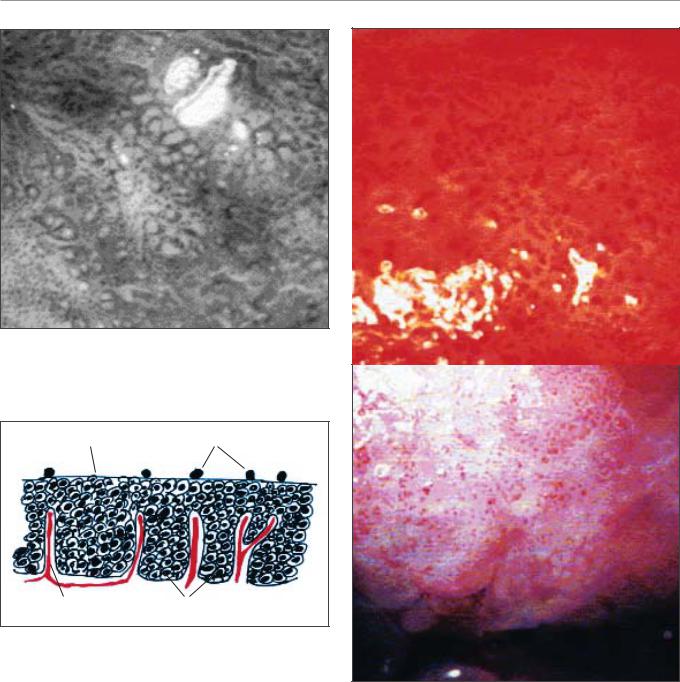
FIGURE 6.7: The colour changes in the columnar epithelium after the application of 5% acetic acid. The columnar villi turn white, obliterating the red colour of the columnar epithelium
b
a
b
b
Squamous metaplasia
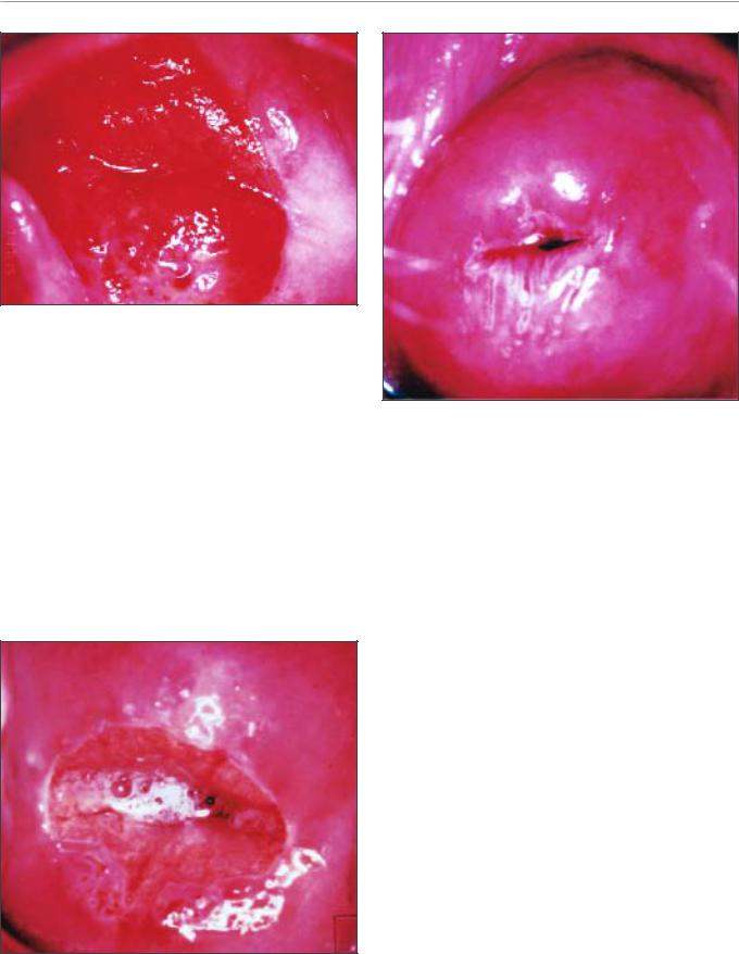
During the different stages of the development of metaplasia, a vast range of colposcopic appearances may be seen. This can present a challenge to an inexperienced colposcopist, who needs to differentiate between these normal findings and the abnormal features associated with CIN. Immature metaplastic squamous epithelium that may turn mildly white after the application of acetic acid is a common source of confusion for the beginners. It is acceptable to take a biopsy when in doubt. Colposcopically, three stages of development of squamous metaplasia may be recognized (Coppleson & Reid, 1986). In the earliest stage, the translucence of the columnar epithelial villi is lost and the villi become opaque at their tips; the villi widen and flatten and successive villi fuse in clusters and sheets with a pale pink colour (Figures 6.8, 6.9 and 6.10). Consequently the metaplastic epithelium looks like a patchily distributed pale cluster, or sheet-like areas, in the ectopic columnar epithelium.
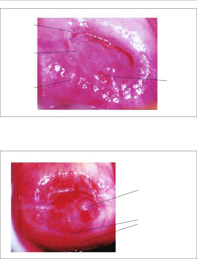
As the metaplasia progresses, the grape-like configuration of the columnar epithelium disappears and the spaces between the villi are fused with glassy, pinkish-white, fingeror tongue-like membranes pointing towards the external os (Figures 6.11 and 6.12). There may be numerous crypt openings and islands of columnar epithelium scattered throughout the metaplastic epithelium. The rims of the crypt openings may not turn white with acetic acid early in the process of metaplasia, but may turn mildly white as
b a
a
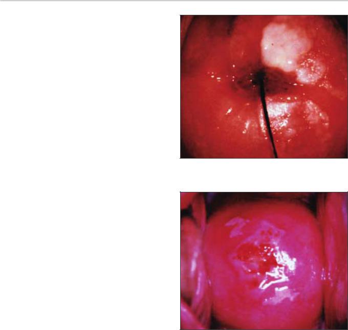
FIGURE 6.8: The earliest colposcopic changes in immature squamous metaplasia (after 5% acetic acid application) in which the tips of the columnar villi stain white (a) and adjacent villi start fusing together (b)
FIGURE 6.9: Immature squamous metaplasia: The columnar villi have fused together to form thin membrane (a). The adjacent villi are fusing together (b) (after 5% acetic acid
50
Colposcopic appearance of the normal cervix
a
↑
↑
FIGURE 6.10: The glassy, pinkishwhite immature squamous metaplastic epithelium (a) with islands of columnar epithelium (narrow arrow) and crypt opening (bold arrow) (after 5% acetic acid application)
the metaplastic process progresses. Gradually, the tongue-like metaplastic areas fuse together to form a continuously advancing glassy, shining, pinkish-white or mildly pale membrane-like area (Figure 6.13).
Finally, the immature metaplastic epithelium becomes a fully developed mature metaplastic squamous epithelium resembling the original native squamous epithelium, except for the presence of some crypt openings (Figure 6.1) and nabothian retention follicles in the metaplastic epithelium (Figures 1.11,
a
a
b
FIGURE 6.11: The prominent white line corresponds to the new squamocolumnar junction and tongues of immature squamous metaplasia (a) with crypt opening at 4-8 o’clock
positions (b) (after application of 5% acetic acid)
a
b
c
↑
FIGURE 6.12: Appearance after 5% acetic acid application: protruding tongues (a) of immature squamous metaplasia towards the external os in the lower lip and the crypt openings
(b) after application of 5% acetic acid. Some crypt openings are already covered by metaplastic epithelium (c) which may become nabothian cysts soon. Note the distal crypt opening indicated by the arrow and the pinkish white hue of the metaplastic epithelium compared to the pink colour of the original squamous epithelium
6.3 and 6.14). The retention follicles, in the beginning, may appear as white, dot-like, areas before they enlarge with progressive accumulation of mucus within the follicle, presenting as pimpleor button-like ivorywhite or mildly yellowish areas (Figures 1.11, 6.3 and 6.14). The typical vessel formations in the metaplastic epithelium include long regular branching vessels with gradually decreasing calibre and a network of regular branching vessels (Figure 6.2). These vascular patterns may be seen more prominently over the nabothian follicles (Figure 6.3).
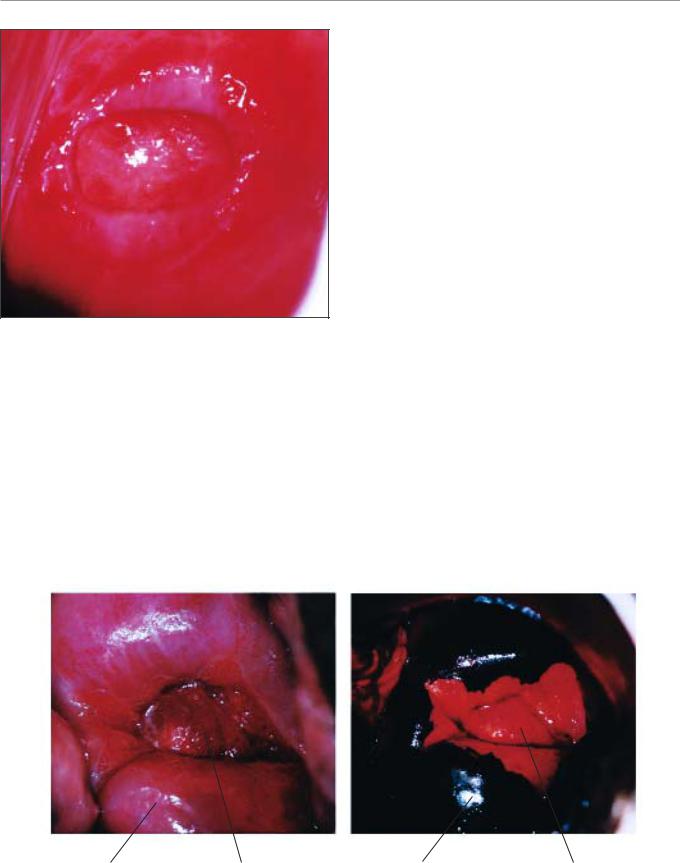
When metaplasia occurs in the epithelium covering the protruding cervical polyp, it is covered by pale white epithelium (Figure 6.15).
After application of Lugol’s iodine solution
As described in the previous chapter, glycogenated cells take iodine, so that they have a uniform dark mahogany brown colour when stained with Lugol’s iodine solution. Therefore, the normal vaginal and cervical squamous (both native and mature
51
Chapter 6
Crypt openings
Immature squamous metaplasia
Island of
columnar Mature epithelium
squamous metaplasia
FIGURE 6.13: Pale, translucent acetowhitening due to immature squamous metaplasia with several crypt openings after application of 5% acetic acid
Nabothian cyst
Dots indicating small nabothian cysts
↑ |
↑ |
FIGURE 6.14: Mature squamous metaplasia after the application of 5% acetic acid: Note the nabothian cyst at 5 o’clock position and the multiple dotlike areas indicating retention cysts. Narrow arrows indicate the distal crypt openings. The new squamocolumnar junction has receded into the cervical canal
52
Colposcopic appearance of the normal cervix
|
a |
|
a |
↑ |
a |
|
||
|
|
FIGURE 6.15: Immature squamous metaplastic epithelium (narrow arrow) on the polyp with intervening areas of columnar epithelium (a), after application of 5% acetic acid.
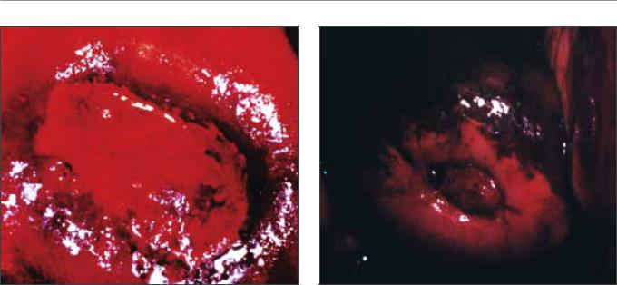
metaplastic) epithelium in women in the reproductive age group will take up the stain and become mahogany brown or black (Figure 6.16). This is helpful in distinguishing normal from abnormal areas in the transformation zone that have shown faint acetowhitening. The columnar epithelium does not stain with iodine (Figure 6.16). The immature
squamous metaplastic epithelium usually does not stain with iodine or may partially stain if it is partially glycogenated (Figure 6.17). The vascular features, so easily seen with saline, may be difficult to observe after application of Lugol’s iodine solution. Cervical polyps do not stain with iodine, as they are usually covered with columnar or immature metaplastic epithelium (Figure 6.18). If the maturation of the metaplastic epithelium varies, one may observe various fields of no uptake or partial to full iodine uptake on the polyp. In postmenopausal women, the ectocervix may not stain fully with iodine, due to atrophy of the epithelium.
Congenital transformation zone
The congenital transformation zone stains white after application of acetic acid. In this condition, the metaplastic epithelium formed during the latter portion of fetal life, lying distal to the transformation zone formed after birth, is located far out on the ectocervix, some distance from the cervical os and, in some cases, may even extend onto the vagina. It is important to recognize this as a normal condition for which no treatment is necessary.
With acetic acid, the congenital transformation zone will usually take on a mild acetowhite stain and the capillary vasculature may have a fine mosaic pattern
Squamous epithelium Columnar epithelium |
Squamous epithelium |
Columnar epithelium |
|
|
has not taken up |
|
|
iodine |
|
|
|
FIGURE 6.16: Colour changes after application of Lugol's iodine |
|
|
53
Chapter 6
a
FIGURE 6.17: An area of no or partial iodine uptake in the
immature squamous epithelium (a) (appearance after acetic acid
application is shown in figure 6.13)
(see Chapter 7). It does not take up iodine after application of Lugol’s iodine solution. If a biopsy is taken of the tissue to confirm the diagnosis, it is best
FIGURE 6.18: After application of Lugol’s iodine solution, the
endocervical polyp and the immature squamous metaplasia
surrounding the os partially take up iodine
to alert the pathologist of the colposcopic diagnosis. We emphasize that it is always necessary to provide the detailed colposcopic findings to the pathologist.
54

Chapter 7
Colposcopic assessment of cervical intraepithelial neoplasia
•The colposcopic diagnosis of cervical neoplasia depends on the recognition of four main features: intensity (colour tone) of acetowhitening, margins and surface contour of acetowhite areas, vascular features and colour changes after iodine application.
•The occurrence of abnormal features in a localised area in the transformation zone increases the probability of diagnosis of a neoplastic lesion.
•Considerable skill may be required to differentiate between low-grade CIN, immature squamous metaplasia and inflammatory lesions.
•Biopsy should be directed whenever in doubt.
•The observation of well-demarcated, dense, opaque, acetowhite area(s) in the transformation zone close to or abutting the squamocolumnar junction is the hallmark of colposcopic diagnosis of CIN.
•Low-grade CIN is often seen as thin, smooth acetowhite lesions with well-demarcated, but irregular, feathery or digitating or angular margins.
•High-grade CIN are associated with thick, dense, dull, opaque or greyish-white acetowhite areas with well-demarcated, regular margins, which sometimes may be raised and rolled out. They may be more extensive and complex lesions extending into the endocervical canal. The surface contour of the acetowhite areas associated with high-grade CIN lesions tend to be less smooth, or irregular and nodular. Visualization of one or more borders within an acetowhite lesion or an acetowhite lesion with varying colour intensity is associated with high-grade lesions.
•Abnormal vascular features such as punctation and mosaics are significant only if these are seen confined to acetowhite areas.
•Vascular features, such as fine punctation and/or fine mosaics in acetowhite areas, may be associated with low-grade CIN.
•Coarse punctation and/or coarse mosaics in acetowhite areas tend to occur in high-grade lesions.
•CIN lesions do not contain glycogen and thus do not stain with iodine and remain mustard or saffron yellow areas.
•Using a scoring system such as Reid’s colposcopic index may guide colposcopic interpretation and diagnosis.
55
Chapter 7
The colposcopic diagnosis of cervical neoplasia requires an understanding and recognition of four main features: colour tone and intensity of acetowhitening, margins and surface contour of acetowhite areas, vascular pattern and iodine staining. Colposcopy with directed biopsy is described as the reference investigation or ‘gold standard’ for the diagnosis of cervical precancer (Singer & Monaghan, 2000). Colposcopy has a reported sensitivity ranging from 87% to 99% to diagnose cervical neoplasia, but its specificity is lower, between 23% and 87% (Mitchell et al., 1998; Belinson et al., 2001).
The colposcopic features of cervical intraepithelial neoplasia (CIN) are described in this chapter to equip the student with the skills to distinguish the colposcopic findings associated with high-grade CIN (CIN 2-3) from those of low-grade lesions (CIN 1). Although the appearance of a single abnormal feature alone is not a strong indicator that a lesion is present, the occurrence of abnormal features together in a localized area in the transformation zone increases the probability of a lesion. It will become obvious during colposcopic practice that considerable skills are required to differentiate between low-grade lesions, immature squamous metaplasia and certain inflammatory conditions. The student is encouraged to obtain biopsies whenever in doubt, and to review the histopathological findings with the pathologist. Close collaboration with pathologists is obligatory and useful in improving one’s diagnostic skills. At the end of this
chapter, a system that enables the colposcopist to score abnormalities is presented. This system is useful as a basis for the choice of which area(s) to select for biopsy. It is essential to biopsy the ‘worst’ area(s) - that is, the area(s) with the most severe changes in features.
The colposcopic findings of an abnormal or atypical transformation zone can involve the whole transformation zone but more commonly affect only a portion of it and there may be multiple distinct lesions. There is usually a distinct demarcation between normal and abnormal epithelium.
The colposcopic features that differentiate an abnormal transformation zone from the normal include the following: colour tone of acetowhite areas; surface pattern of acetowhite areas; borderline between acetowhite areas and the rest of the epithelium; vascular features and colour changes after application of iodine.
After application of normal saline solution
Following application of saline, abnormal epithelium may appear much darker than the normal epithelium.
Vasculature
Using the green (or blue) filter and higher-power magnification when necessary, the best opportunity to evaluate any abnormal vasculature patterns is before the application of acetic acid, the effect of which may obscure some or all of the changes, especially in an
Coarse mosaic
Acetowhite area
Fine mosaic
Fine punctation
Coarse punctation
Umbilication
FIGURE 7.1: A schematic representation of punctation and mosaics
56
Colposcopic assessment of cervical intraepithelial neoplasia
b
a
FIGURE 7.2a: Fine punctation (a) and coarse mosaic (b) seen after application of normal saline
Mosaic |
Punctation |
Stromal capillary |
Rete pegs |
FIGURE 7.2b: Schematic diagram to show the rete pegs and the stromal capillaries which on end-on view appear as punctations
acetowhite area. The abnormalities of interest are punctation, mosaics and atypical vessels.
Capillaries: The afferent and efferent capillaries within the villi (Figure 6.4) of columnar epithelium become compressed during the normal metaplastic process and are not incorporated within the newly formed squamous epithelium. Instead, they form a fine network below the basement membrane. When CIN develops as a result of HPV infection and atypical metaplasia, the afferent and efferent capillary system may be trapped (incorporated) into the diseased dysplastic epithelium through several elongated stromal papillae (Figures 2.3 and 2.4), and a thin layer of epithelium may remain on top of these vessels. This
FIGURE 7.3: Coarse punctation before and after application of
acetic acid
forms the basis of the punctate and mosaic blood vessel patterns (Figures 7.1, 7.2 and 7.3). The terminating vessels in the stromal papillae underlying the thin epithelium appear as black points in a stippling pattern in an end-on view under the colposcope, making what are called punctate areas (Figures 7.1, 7.2 and 7.3). The inter-connecting blood vessels in the stromal papillae surrounding the rete pegs of the epithelium, running parallel to the surface, are observed colposcopically as cobbled areas of mosaic pattern (Figures 7.1 and 7.2). In mosaic areas, the epithelium
57
Chapter 7
appears as individual small, large, round, polygonal, regular or irregular blocks. Punctation and mosaic areas may be classified as either fine or coarse. Coarse changes tend to be associated with more severe degrees of abnormality. When both punctation and mosaic patterns are found to coexist, the same evaluation criteria for colposcopic prediction of disease are used as when they exist separately.
Vessels exhibiting punctation and mosaics are usually more strikingly obvious than the normal stromal vessels because these vessels penetrate into the epithelium and are thus closer to the surface. When acetic acid is applied, these abnormal vascular patterns seen to be confined to the acetowhite areas.
Fine punctation refers to looped capillaries - viewed end-on - that appear to be of fine calibre and located close to one another, producing a delicate stippling effect (Figures 7.1 and 7.2a). Fine mosaics are a network of fine-calibre blood vessels that appear in close proximity to one another, as a mosaic pattern, when viewed with the colposcope (Figure 7.1). These two vascular appearances may occur together and may be found in low-grade (CIN 1) lesions. The patterns do not necessarily appear throughout the whole lesion.
Coarse punctation (Figure 7.3) and coarse mosaics
(Figures 7.1 and 7.2) are formed by vessels having larger calibre and larger intercapillary distances, in contrast to the corresponding fine changes. Coarse punctation and mosaicism tend to occur in more severe neoplastic lesions such as CIN 2, CIN 3 lesions and early preclinical invasive cancer. Sometimes, the two patterns are superimposed in an area so that the capillary loops occur in the centre of each mosaic ‘tile’. This appearance is called umbilication (Figure 7.1).
a
FIGURE 7.4: Hyperkeratosis (leukoplakia) (a)
a
a
FIGURE 7.5: The geographic satellite lesions (a) far away from the squamocolumnar junction suggestive of condyloma
Leukoplakia (hyperkeratosis)
Leukoplakia or hyperkeratosis (Figure 7.4) is a white, well-demarcated area on the cervix that may be apparent to the unaided eye, before the application of acetic acid. The white colour is due to the presence of keratin and is an important observation. Usually leukoplakia is idiopathic, but it may also be caused by chronic foreign body irritation, HPV infection or squamous neoplasia. No matter where the area of leukoplakia is located on the cervix, it should be biopsied to rule out high-grade CIN or malignancy. It is not usually possible to colposcopically evaluate the vasculature beneath such an area.
Condylomata
An exophytic lesion on the cervix usually represents and exhibits the characteristic features of a condyloma (Figures 7.5- 7.8). Condylomata are multiple, exophytic lesions, that are infrequently found on the cervix, but more commonly in the vagina or on the vulva. Depending on their size, they may be obvious to the naked eye. They present as soft pink or white vascular growths with multiple, fine, finger-like projections on the surface, before the application of acetic acid. Under the colposcope, condylomata have a typical appearance, with a vascular papilliferous or frond-like surface, each element of which contains a central

Colposcopic assessment of cervical intraepithelial neoplasia
a
a
FIGURE 7.6: Exophytic condyloma in the posterior lip of the cervix (a) before and after 5% acetic acid application
a
FIGURE 7.7: Exophytic condyloma in the cervix (a) after application of acetic acid
capillary. Occasionally, the surface of a condyloma may have a whorled, heaped-up appearance with a brainlike texture, known as an encephaloid pattern (Figure 7.8). Often, the surface of the lesion may be densely hyperplastic. These lesions may be located within, but are more often found outside the transformation zone. After application of acetic acid, there is blanching of the surface with acetowhite change persisting for some time. A condyloma at the squamocolumnar junction can sometimes be confused with a prominent area of columnar epithelial villi. Both tend to be acetowhite, but condyloma is whiter. It is always prudent to obtain a biopsy to confirm the diagnosis of any exophytic lesion
FIGURE 7.8: Condyloma with an encephaloid (cerebriform) pattern
and to rule out malignancy. Condylomatous lesions may not take up iodine stain or may stain only partially brown.
After the application of 5% acetic acid
solution
The observation of a well demarcated, dense, opaque, acetowhite area closer to or abutting the squamocolumnar junction in the transformation zone after application of 5% acetic acid is critical. In fact, it is the most important of all colposcopic signs, and is the hallmark of colposcopic diagnosis of cervical neoplasia. The degree to which the epithelium takes up the acetic
59