

Colposcopymanual
.pdfPreface
Health care providers have observed a high incidence of cervical cancer in many developing countries in Africa, Asia, and Central and South America, and in the absence of organized early-detection programmes the mortality rates from this disease remain high. The extremely limited health-care infrastructure available in many of these countries contributes to a compelling need to build a capacity to identify cervical neoplasia in early, preventable stages, preferably even before - and not in the wake of - the introduction of early detection programmes in such settings. Colposcopy is generally regarded as a diagnostic test; it is used to assess women who have been identified to have cervical abnormalities on various screening tests.
This introductory manual for gynaecologists, pathologists, general practitioners, and nurses is intended to provide information on the principles of colposcopy and the basic skills needed to colposcopically assess cervical intraepithelial neoplasia and to provide basic treatment. Interested health professionals are expected to subsequently continue to improve their skills by undertaking a basic course of theoretical and practical training, and by referring to standard textbooks dealing with the subject more extensively. Continuing practical work is vital for acquiring, improving and sustaining necessary skills in the colposcopic diagnosis of cervical neoplasia. This manual is also intended to be a beginner’s self-learning resource and as a teaching aid for colposcopy courses for health care personnel, as well as a resource for teaching curricula for medical and nursing students in developing countries. It may also be used as a field manual in routine screening programmes.
A good understanding of the gross and microscopic anatomy of the uterine cervix, infective and inflammatory conditions of the cervix and the vagina, histology and the natural history of cervical neoplasia is absolutely essential for a correct interpretation of colposcopic findings and colposcopic diagnosis of cervical neoplasia. These aspects are dealt with in
detail in this manual, and should be well studied in conjunction with other chapters dealing with colposcopic techniques and features of cervical neoplasia and their treatment.
Generally speaking, colposcopy should not be practised unless the provider has had an opportunity to spend some time with an experienced colposcopist. Unfortunately, this is extremely difficult to arrange in most of the developing world, where the disease incidence is high (particularly in sub-Saharan Africa) and both access to such training and to a colposcope is rarely available. For instance, quite apart from colposcopy training, no colposcopy service itself is available in whole regions of Africa and Asia and Latin America. Realistically, the basic colposcopist in such situations is a self-trained health-care provider who knows how to examine the cervix, what to look for, how to make a diagnosis, and how to treat a woman with simple ablative or excisional methods. We emphasize, however, that an instructor should be available for the on-site training of new colposcopists. The limitations and far-reaching implications of incomplete understanding of cervical disease and inadequate expertise should be well appreciated by potential practitioners.
Draft versions of this manual have been used in more than 20 courses on colposcopy and management of cervical precancers conducted in Angola, Congo (Brazzaville), Guinea, Kenya, India, Mali, Mauritania, Laos and Tanzania. More than 120 doctors and nurses have been trained and initiated in colposcopy in the context of the cervical cancer prevention research initiatives in these countries as well as in other countries such as Burkina Faso, Cape Verde, Equatorial Guinea, Mozambique, Nepal, Niger, Sao Tome and Uganda. Feedback from the participants and teaching faculty of these courses has been particularly useful in revising the draft versions of the manual. The illustrations used in this manual have also largely been drawn from the above-mentioned country projects.
xi

Preface
The resource constraints for health-care systems in many developing countries are substantial, with practical challenges on how colposcopy and treatment of early cervical neoplasia can be integrated into and delivered through these health services. Awareness of these limitations will pave the way to establishing, integrating, and maintaining such services within the health-care infrastructure of developing countries. We hope that this manual will help the learner, given access to a colposcope, to start performing colposcopy
and recognizing lesions, and in effectively treating them with cryotherapy or loop electrosurgical excision procedure (LEEP). We believe that, in due course, it will catalyse and contribute to the initiation and dissemination of preventive services for cervical cancer in low-resource regions and countries.
John W. Sellors M.D.
R. Sankaranarayanan M.D.
xii

Chapter 1
An introduction to the anatomy of the uterine cervix
•The cervix, the lower fibromuscular portion of the uterus, measures 3-4 cm in length and 2.5 cm in diameter; however, it varies in size and shape depending on age, parity and menstrual status of the woman.
•Ectocervix is the most readily visible portion of the cervix; endocervix is largely invisible and lies proximal to the external os.
•Ectocervix is covered by a pink stratified squamous epithelium, consisting of multiple layers of cells and a reddish columnar epithelium consisting of a single layer of cells lines the endocervix. The intermediate and superficial cell layers of the squamous epithelium contain glycogen.
•The location of squamocolumnar junction in relation to the external os varies depending upon age, menstrual status, and other factors such as pregnancy and oral contraceptive use.
•Ectropion refers to the eversion of the columnar epithelium onto the ectocervix, when the cervix grows rapidly and enlarges under the influence of estrogen, after menarche and during pregnancy.
•Squamous metaplasia in the cervix refers to the physiological replacement of the everted columnar epithelium on the ectocervix by a newly formed squamous epithelium from the subcolumnar reserve cells.
•The region of the cervix where squamous metaplasia occurs is referred to as the transformation zone.
•Identifying the transformation zone is of great importance in colposcopy, as almost all manifestations of cervical carcinogenesis occur in this zone.
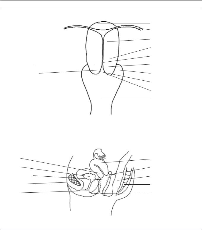
A thorough understanding of the anatomy and physiology of the cervix is absolutely essential for effective colposcopic practice. This chapter deals with the gross and microscopic anatomy of the uterine cervix and the physiology of the transformation zone. The cervix is the lower fibromuscular portion of the uterus. It is cylindrical or conical in shape, and measures 3 to 4 cm in length, and 2.5 cm in diameter. It is supported by the cardinal and uterosacral ligaments, which stretch between the lateral and posterior portions of the cervix and the walls of the bony pelvis. The lower half of the cervix, called the portio vaginalis, protrudes into the vagina through its anterior wall, and the upper half
remains above the vagina (Figure 1.1). The portio vaginalis opens into the vagina through an orifice called the external os.
The cervix varies in size and shape depending on the woman’s age, parity and hormonal status. In parous women, it is bulky and the external os appears as a wide, gaping, transverse slit. In nulliparous women, the external os resembles a small circular opening in the centre of the cervix. The supravaginal portion meets with the muscular body of the uterus at the internal cervical os. The portion of the cervix lying exterior to the external os is called the ectocervix. This is the portion of the cervix that is
1
Chapter 1
Portio vaginalis
Endocervical canal
Cervix
Bladder
Anterior fornix
Pubic bone
Urethra
Fundus Fallopian tube Body of uterus
Supravaginal cervix Internal os Endocervix
Lateral fornix
External os
Ectocervix
Vagina
Uterus Posterior fornix Rectum Sacrum
Vagina
FIGURE 1.1: Gross anatomy of the uterine cervix
readily visible on speculum examination. The portion proximal to the external os is called the endocervix and the external os needs to be stretched or dilated to view this portion of the cervix. The endocervical canal, which traverses the endocervix, connects the uterine cavity with the vagina and extends from the internal to the external os, where it opens into the
vagina. It varies in length and width depending on the woman’s age and hormonal status. It is widest in women in the reproductive age group, when it measures 6-8 mm in width.
The space surrounding the cervix in the vaginal cavity is called the vaginal fornix. The part of the fornix between the cervix and the lateral vaginal walls is called
2
An introduction to the anatomy of the uterine cervix
the lateral fornix; the portions between the anterior and posterior walls of the vagina and the cervix are termed the anterior and posterior fornix, respectively.
The stroma of the cervix is composed of dense, fibro-muscular tissue through which vascular, lymphatic and nerve supplies to the cervix pass and form a complex plexus. The arterial supply of the cervix is derived from internal iliac arteries through the cervical and vaginal branches of the uterine arteries. The cervical branches of the uterine arteries descend in the lateral aspects of the cervix at 3 and 9 o’clock positions. The veins of the cervix run parallel to the arteries and drain into the hypogastric venous plexus. The lymphatic vessels from the cervix drain into the common, external and internal iliac nodes, obturator and the parametrial nodes. The nerve supply to the cervix is derived from the hypogastric plexus. The endocervix has extensive sensory nerve endings, while there are very few in the ectocervix. Hence, procedures such as biopsy, electrocoagulation and cryotherapy are well tolerated in most women without local anaesthesia. Since sympathetic and parasympathetic fibres are also abundant in the endocervix, dilatation and curettage of the endocervix may occasionally lead to a vasovagal reaction.
The cervix is covered by both stratified nonkeratinizing squamous and columnar epithelium. These two types of epithelium meet at the squamocolumnar junction.
Stratified non-keratinizing squamous epithelium
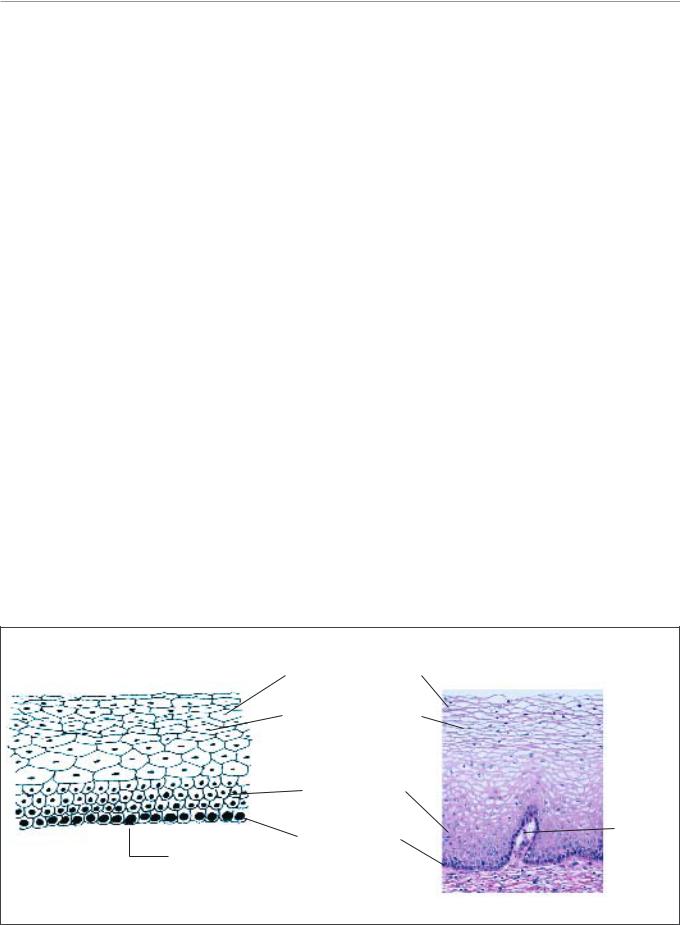
Normally, a large area of ectocervix is covered by a stratified, non-keratinizing, glycogen-containing squamous epithelium. It is opaque, has multiple (15-20) layers of cells (Figure 1.2) and is pale pink in colour. This epithelium may be native to the site formed during embryonic life, which is called the native or original squamous epithelium, or it may have been newly formed as metaplastic squamous epithelium in early adult life. In premenopausal women, the original squamous epithelium is pinkish in colour, whereas the newly formed metaplastic squamous epithelium looks somewhat pinkish-white on visual examination.
The histological architecture of the squamous epithelium of the cervix reveals, at the bottom, a single layer of round basal cells with a large darkstaining nuclei and little cytoplasm, attached to the basement membrane (Figure 1.2). The basement membrane separates the epithelium from the underlying stroma. The epithelial-stromal junction is usually straight. Sometimes it is slightly undulating with short projections of stroma at regular intervals. These stromal projections are called papillae. The parts of the epithelium between the papillae are called rete pegs.
The basal cells divide and mature to form the next few layers of cells called parabasal cells, which also have relatively large dark-staining nuclei and greenish-blue basophilic cytoplasm. Further
Superficial cell layer
Intermediate cell layer
|
Parabasal layer |
|
|
|
|
|
Stromal |
Basement |
Basal cell layer |
|
papilla |
|
|
||
|
|
|
|
membrane |
|
|
Stroma |
|
|
|
|
|
|
|
FIGURE 1.2: Stratified squamous epithelium (x 20)
3
Chapter 1
differentiation and maturation of these cells leads to the intermediate layers of polygonal cells with abundant cytoplasm and small round nuclei. These cells form a basket-weave pattern. With further maturation, the large and markedly flattened cells with small, dense, pyknotic nuclei and transparent cytoplasm of the superficial layers are formed. Overall, from the basal to the superficial layer, these cells undergo an increase in size and a reduction of nuclear size.
The intermediate and superficial layer cells contain abundant glycogen in their cytoplasm, which stains mahogany brown or black after application of Lugol’s iodine and magenta with periodic acid-Schiff stain in histological sections. Glycogenation of the intermediate and superficial layers is a sign of normal maturation and development of the squamous epithelium. Abnormal or altered maturation is characterized by a lack of glycogen production.
The maturation of the squamous epithelium of the cervix is dependent on estrogen, the female hormone. If estrogen is lacking, full maturation and glycogenation does not take place. Hence, after menopause, the cells do not mature beyond the parabasal layer and do not accumulate as multiple layers of flat cells. Consequently, the epithelium becomes thin and atrophic. On visual examination, it appears pale, with subepithelial petechial haemorrhagic spots, as it is easily prone to trauma.
Columnar epithelium
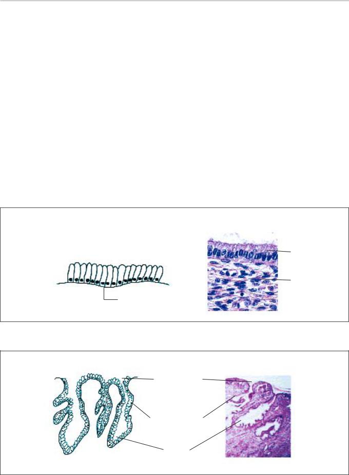
The endocervical canal is lined by the columnar epithelium (sometimes referred to as glandular epithelium). It is composed of a single layer of tall cells with dark-staining nuclei close to the basement membrane (Figure 1.3). Because of its single layer of cells, it is much shorter in height than the stratified squamous epithelium of the cervix. On visual
Columnar cells
Stroma
Basement membrane
FIGURE 1.3: Columnar epithelium (x 40)
Crypt opening
Columnar cells
Crypt
FIGURE 1.4: Crypts of columnar epithelium (x 10)
4
An introduction to the anatomy of the uterine cervix
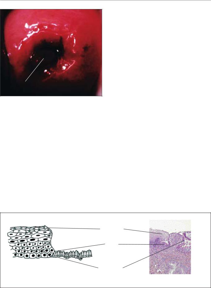
Polyp
FIGURE 1.5: Cervical polyp
examination, it appears reddish in colour because the thin single cell layer allows the coloration of the underlying vasculature in the stroma to be seen more easily. At its distal or upper limit, it merges with the endometrial epithelium in the lower part of the body of the uterus. At its proximal or lower limit, it meets with the squamous epithelium at the squamocolumnar junction. It covers a variable extent of the ectocervix, depending upon the woman’s age, reproductive, hormonal and menopausal status.
The columnar epithelium does not form a flattened surface in the cervical canal, but is thrown into multiple longitudinal folds protruding into the lumen of the canal, giving rise to papillary projections. It forms several invaginations into the substance of the cervical stroma, resulting in the formation of endocervical
crypts (sometimes referred to as endocervical glands) (Figure 1.4). The crypts may traverse as far as 5-8 mm from the surface of the cervix. This complex architecture, consisting of mucosal folds and crypts, gives the columnar epithelium a grainy appearance on visual inspection.
A localized overgrowth of the endocervical columnar epithelium may occasionally be visible as a reddish mass protruding from the external os on visual examination of the cervix. This is called a cervical polyp (Figure 1.5). It usually begins as a localized enlargement of a single columnar papilla and appears as a mass as it enlarges. It is composed of a core of endocervical stroma lined by the columnar epithelium with underlying crypts. Occasionally, multiple polyps may arise from the columnar epithelium.
Glycogenation and mitoses are absent in the columnar epithelium. Because of the lack of intracytoplasmic glycogen, the columnar epithelium does not change colour after the application of Lugol’s iodine or remains slightly discoloured with a thin film of iodine solution.
Squamocolumnar junction
The squamocolumnar junction (Figures 1.6 and 1.7) appears as a sharp line with a step, due to the difference in the height of the squamous and columnar epithelium. The location of the squamocolumnar junction in relation to the external os is variable over a woman’s lifetime and depends upon factors such as age, hormonal status, birth trauma, oral contraceptive use and certain physiological conditions such as pregnancy (Figures 1.6 and 1.7).
The squamocolumnar junction visible during childhood, perimenarche, after puberty and early
Squamous epithelium
SCJ
Columnar epithelium
FIGURE 1.6: Squamocolumnar junction (SCJ) (x 10)
5
Chapter 1
a
Original SCJ
b |
Columnar epithelium |
Original SCJ
c
Columnar epithelium
Transformation zone
External os
Original SCJ
New SCJ
d
New SCJ
 Original SCJ
Transformation zone
e |
New SCJ |
|
|
|
Transformation zone |
|
Original SCJ |
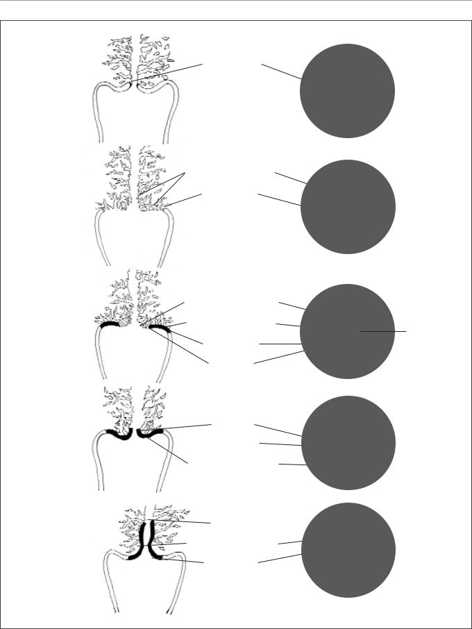
FIGURE 1.7: Location of the squamocolumnar junction (SCJ) and transformation zone; (a) before menarche; (b) after puberty and at early reproductive age; (c) in a woman in her 30s; (d) in a perimenopausal woman; (e) in a postmenopausal woman
6
An introduction to the anatomy of the uterine cervix
|
|
Columnar |
Original SCJ |
Metaplastic |
Columnar |
|
Ectropion |
External os |
Original |
squamous |
epithelium |
||
epithelium |
epithelium |
New SCJ |
||||
squamous |
||||||
|
|
|
|
External os |
||
|
|
|
epithelium |
|
||
|
|
|
|
|
a |
b |
c |
|
|
d |
|
Mature metaplastic |
External os |
New SCJ |
External os |
Mature metaplastic |
squamous epithelium |
|
|
|
squamous epithelium |
FIGURE 1.8: Location of squamocolumnar junction (SCJ)
(a)Original squamocolumnar junction (SCJ) in a young woman in the early reproductive age group. The SCJ is located far away from the external os. Note the presence of everted columnar epithelium occupying a large portion of the ectocervix producing ectropion
(b)The new SCJ has moved much closer to the external os in a woman in her 30s. The SCJ is visible as a distinct white line after the application of 5% acetic acid due to the presence of immature squamous metaplastic epithelium adjacent to the new SCJ
(c)The new SCJ is at the external os in a perimenopausal woman
(d)The new SCJ is not visible and has receded into the endocervix in a postmenopausal woman. Mature metaplastic squamous epithelium occupies most of the ectocervix
reproductive period is referred to as the original squamocolumnar junction, as this represents the junction between the columnar epithelium and the ‘original’ squamous epithelium laid down during embryogenesis and intrauterine life. During childhood and perimenarche, the original squamocolumnar junction is located at, or very close to, the external os (Figure 1.7a). After puberty and during the
reproductive period, the female genital organs grow under the influence of estrogen. Thus, the cervix swells and enlarges and the endocervical canal elongates. This leads to the eversion of the columnar epithelium of the lower part of the endocervical canal on to the ectocervix (Figure 1.7b). This condition is called ectropion or ectopy, which is visible as a strikingly reddish-looking ectocervix on visual inspection
7

Chapter 1
(Figure 1.8a). It is sometimes called ‘erosion’ or ‘ulcer’, which are misnomers and should not be used to denote this condition. Thus the original squamocolumnar junction is located on the ectocervix, far away from the external os (Figures 1.7b and 1.8a). Ectropion becomes much more pronounced during pregnancy.
The buffer action of the mucus covering the columnar cells is interfered with when the everted columnar epithelium in ectropion is exposed to the acidic vaginal environment. This leads to the destruction and eventual replacement of the columnar epithelium by the newly formed metaplastic squamous epithelium. Metaplasia refers to the change or replacement of one type of epithelium by another.
The metaplastic process mostly starts at the original squamocolumnar junction and proceeds centripetally towards the external os through the reproductive period to perimenopause. Thus, a new squamocolumnar junction is formed between the newly formed metaplastic squamous epithelium and the columnar epithelium remaining everted onto the ectocervix (Figures 1.7c, 1.8b). As the woman passes from the reproductive to the perimenopausal age group, the location of the new squamocolumnar junction progressively moves on the ectocervix towards the external os (Figures 1.7c, 1.7d, 1.7e and 1.8). Hence, it is located at variable distances from the external os, as a result of the progressive formation of the new metaplastic squamous epithelium in the exposed areas of the columnar epithelium in the ectocervix. From the perimenopausal period and after the onset of menopause, the cervix shrinks due the lack of estrogen, and consequently, the movement of the new squamocolumnar junction towards the external os and into the endocervical canal is accelerated (Figures 1.7d and 1.8c). In postmenopausal women, the new squamocolumnar junction is often invisible on visual examination (Figures 1.7e and 1.8d).
The new squamocolumnar junction is hereafter simply referred to as squamocolumnar junction in this manual. Reference to the original squamocolumnar junction will be explicitly made as the original squamocolumnar junction.
Ectropion or ectopy
Ectropion or ectopy is defined as the presence of everted endocervical columnar epithelium on the ectocervix. It appears as a large reddish area on the ectocervix surrounding the external os (Figures 1.7b and
1.8a). The eversion of the columnar epithelium is more pronounced on the anterior and posterior lips of the ectocervix and less on the lateral lips. This is a normal, physiological occurrence in a woman’s life. Occasionally the columnar epithelium extends into the vaginal fornix. The whole mucosa including the crypts and the supporting stroma is displaced in ectropion. It is the region in which physiological transformation to squamous metaplasia, as well as abnormal transformation in cervical carcinogenesis, occurs.
Squamous metaplasia
The physiological replacement of the everted columnar epithelium by a newly formed squamous epithelium is called squamous metaplasia. The vaginal environment is acidic during the reproductive years and during pregnancy. The acidity is thought to play a role in squamous metaplasia. When the cells are repeatedly destroyed by vaginal acidity in the columnar epithelium in an area of ectropion, they are eventually replaced by a newly formed metaplastic epithelium. The irritation of exposed columnar epithelium by the acidic vaginal environment results in the appearance of sub-columnar reserve cells. These cells proliferate producing a reserve cell hyperplasia and eventually form the metaplastic squamous epithelium.
As already indicated, the metaplastic process requires the appearance of undifferentiated, cuboidal, sub-columnar cells called reserve cells (Figure 1.9a), for the metaplastic squamous epithelium is produced from the multiplication and differentiation of these cells. These eventually lift off the persistent columnar epithelium (Figures 1.9b and 1.9c). The exact origin of the reserve cells is not known, though it is widely believed that it develops from the columnar epithelium, in response to irritation by the vaginal acidity.
The first sign of squamous metaplasia is the appearance and proliferation of reserve cells (Figures 1.9a and 1.9b). This is initially seen as a single layer of small, round cells with darkly staining nuclei, situated very close to the nuclei of columnar cells, which further proliferate to produce a reserve cell hyperplasia (Figure 1.9b). Morphologically, the reserve cells have a similar appearance to the basal cells of the original squamous epithelium, with round nuclei and little cytoplasm. As the metaplastic process progresses, the reserve cells proliferate and differentiate to form a thin, multicellular epithelium of immature squamous cells with no evidence of stratification (Figure 1.9c). The term immature
8