

2012 asccp updated guidelines for the management of abnormal cervical cancer screening tests and cancer precursors
.PdfASCCP Guidelines Update & S11
Figure 6.
Low-Grade Squamous Intraepithelial Lesion
The ASCUS-LSIL Triage Study showed that the natural history of LSIL approximates that of HPV-positive ASCUS (64), suggesting that women with either should be managed similarly. Analysis of the KPNC dataset confirmed that women with LSIL at ages 21Y24 years carry a lower risk of CIN 3+ than older women (33, 65). Lowgrade squamous intraepithelial lesions are highly associated with HPV infection, with a pooled estimate of HPV positivity of 77% (66). This rate appears too high to allow reflex HPV testing to select women for colposcopy efficiently. However, when co-testing is performed in women 30 years of age and older, some women have HPV-negative LSIL. In the KPNC cohort, the risk of CIN 3+ in HPV-negative women with LSIL was low, similar to that of ASC-US alone (67).
Management of Women with LSIL (Fig. 6)
For women with LSIL cytology and no HPV test or a positive HPV test, colposcopy is recommended (AI). If cotesting shows HPV-negative LSIL, repeat co-testing at 1 year is preferred, but colposcopy is acceptable. If repeat co-testing at 1 year is elected, and if the cytology is ASCUS or worse or the HPV test is positive (ie, if the co-testing result is other than HPV negative, cytology negative), colposcopy is recommended. If the co-testing result at 1 year is HPV negative and cytology negative, repeat
co-testing after an additional 3 years is recommended. If all tests are negative at that time, routine screening is recommended (BIII).
LSIL in Special Populations
Women Aged 21Y24 Years (Fig. 5). For women with LSIL who are aged 21-24 years, follow-up with cytology at 12-month intervals is recommended (BII). Colposcopy is not recommended (DII). For women with ASC-H or HSIL+ at the 12-month follow up, colposcopy is recommended. For women with ASC-US or worse at the 24-month follow up, colposcopy is recommended. For women with two consecutive negative results, return to routine screening is recommended (BII).
Pregnant Women (Fig. 7). For pregnant women with LSIL, colposcopy is preferred (BII). Endocervical curettage in pregnant women is unacceptable (EIII). For pregnant women aged 21Y24 years, follow-up according to the guidelines for management of LSIL in women aged 21Y24 years is recommended (discussed in previous paragraph). Deferring colposcopy until 6 weeks postpartum is acceptable (CIII). For pregnant women who have no cytologic, histologic, or colposcopically suspected CIN 2+ at the initial colposcopy, postpartum follow-up is recommended (BIII). Additional colposcopic and cytologic

S12 & M A S S A D E T A L .
Figure 7.
examinations during pregnancy are unacceptable for these women (DIII).
Postmenopausal Women. Acceptable options for the management of postmenopausal women with LSIL and no HPV test include obtaining HPV testing, repeat cytologic testing at 6 months and 12 months, and colposcopy (CIII). If the HPV test is negative or if CIN is not identified at colposcopy, repeat cytology in 12 months is
recommended. If either the HPV test is positive or repeat cytology is ASC-US or greater, colposcopy is recommended (AII). If two consecutive repeat cytology tests are negative, return to routine screening is recommended. (BII)
Atypical Squamous Cells, Cannot Exclude High-Grade
Squamous Intraepithelial Lesion (ASC-H)
Data from KPNC confirmed that a report of ASC-H confers higher risk for CIN 3+ over time than ASC-US or
Figure 8.
ASCCP Guidelines Update & S13
Figure 9.
LSIL (68, 69), although risk is lower than that following HSIL. This is also true for women aged 21Y24 years, although their risk of CIN 3+ is lower than that for older women with ASC-H (33). The high rate of HPV detection in women with ASC-H makes reflex HPV testing unsuitable (3). In addition, the 5-year cancer risk among women with HPV-negative ASC-H is 2%, which is too high to justify observation (68).
Management of Women With ASC-H (Fig. 8). For women with ASC-H cytology, colposcopy is recommended regardless of HPV result. Reflex HPV testing is not recommended (DII).
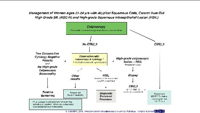
ASC-H in Special Populations Women Aged 21Y24 Years (Fig. 9)
Colposcopy is recommended (AII). Further management should follow guidelines for women aged 21Y24 years with HSIL.
High-Grade Squamous Intraepithelial Lesion (HSIL)
HSIL cytology results identify women at substantial risk. CIN 2+ is found at colposcopy in some 60% of women with HSIL (69Y71). This justifies immediate excision of the transformation zone for many women, especially those who are at risk for loss to follow-up or who have completed childbearing. Cervical cancer is found at
colposcopy in some 2% of women with HSIL, although risk rises with age and is low among women aged 21Y24 years, even with follow-up (33). Five-year cervical cancer risk is 8% among women 30 years of age and older (72). Risks are modified by HPV test results: HPVnegative HSIL co-test results, although uncommon, still carry a 5-year risk for CIN 3+ of 29%, while 7% will develop cancer (72). This precludes reflex HPV triage for HSIL. In the KPNC cohort, among women 30 years of age and older with HPV-positive HSIL, the 5-year risk of CIN 3+ was 50%, while the 5-year cancer risk was 7% (72). When HPV results are known from co-testing for women with HSIL, these risks may inform the choice between immediate diagnostic excision and colposcopy and between diagnostic excision and cytologic and colposcopic surveillance when CIN 2+ is not identified. The sensitivity of colposcopy for detecting CIN 2+ is lower than previously appreciated (73Y75), and multiple biopsies should be considered at colposcopy when large confluent or multiple discrete lesions are seen.
Management of Women With HSIL (Fig. 10). For women with HSIL cytology, immediate loop electrosurgical excision or colposcopy is acceptable, except in special populations (BII). Triage using either a program of repeat cytology alone or reflex HPV testing is unacceptable (EII). For women not managed with immediate excision, colposcopy is recommended regardless of HPV

S14 & M A S S A D E T A L .
Figure 10.
result obtained at co-testing (BII). Accordingly, reflex HPV testing is not recommended (BII).
A diagnostic excisional procedure is recommended for women with HSIL when the colposcopic examination is inadequate, except during pregnancy (BII). Women with CIN 2, CIN 3, and CIN 2,3 should be managed according to the appropriate 2012 consensus guideline (see ‘‘Management of Women With CIN 2, CIN 3, and CIN 2,3’’). Ablation is unacceptable in the following circumstances: when colposcopy has not been performed, when CIN 2,3 is not identified histologically, and when the endocervical assessment identifies CIN 2, CIN 3, CIN 2,3 or ungraded CIN (EII).
HSIL in Special Populations
Women Aged 21Y24 Years (Fig. 9). For women aged 21Y24 years with HSIL, colposcopy is recommended. Immediate treatment (ie, see-and-treat) is unacceptable. (AII) When CIN 2+ is not identified histologically, observation for up to 24 months using both colposcopy and cytology at 6-month intervals is recommended, provided the colposcopic examination is adequate and endocervical assessment is negative or CIN 1 (BIII). If CIN 2, CIN 3, or CIN 2,3 is identified histologically, management according to the 2012 consensus guideline for the management of young women with CIN 2, CIN 3, or CIN 2,3 is recommended (see ‘‘Management of Women With CIN
2, CIN 3, and CIN 2,3’’). (BIII) If during follow-up a highgrade colposcopic lesion is identified or HSIL cytology persists for 1 year, biopsy is recommended. (BIII) If HSIL persists for 24 months without identification of CIN 2+, a diagnostic excisional procedure is recommended. (BIII) A diagnostic excisional procedure is recommended for women aged 21Y24 years with HSIL when colposcopy is unsatisfactory or CIN 2, CIN 3, CIN 2,3, or ungraded CIN is identified on endocervical sampling. (BII) After two consecutive negative cytology results and no evidence of high-grade colposcopic abnormality, return to routine screening is recommended (BIII).
Atypical Glandular Cells, Cytologic Adenocarcinoma
In Situ, and Benign Glandular Changes
An AGC interpretation is poorly reproducible (76) and uncommon (77). AGC has been associated with polyps and metaplasia but also with neoplasias, including adenocarcinomas of the endometrium, cervix, ovary, fallopian tube, and other sites (78). Neoplasia risk is higher when reported as AGC favor neoplasia or frank AIS. Although the cancer risk is lower in women younger than 35 years of age with AGC, the risk of CIN 2+ is higher, and intensive assessment is warranted at all ages (78). In the KPNC cohort, CIN 3+ was found in 9% of women aged 30 years and older with AGC cytology, with cancer in 3% (72, 77). Despite its
ASCCP Guidelines Update & S15
Figure 11.
appellation, AGC cytology is most commonly associated with squamous lesions including CIN 1. However, glandular and squamous lesions often coexist, with CIN found in approximately half of women with AIS (79Y81), so identification of CIN does not preclude AIS or adenocarcinoma. Although cervical adenocarcinoma is HPV associated and can be detected with HPV testing, endometrial cancer is not, so reflex HPV testing does not identify a subgroup of women who need less invasive assessment. A negative HPV test can be useful in identifying women at greater risk for endometrial rather than cervical disease (80). Endometrial cancer risk is low in young women without endometrial cancer risk factors but is substantially greater in older women and young women with risk factors.
Benign-appearing endometrial cells and stromal cells or histiocytes are rarely associated with premalignant lesions or cancer in young women. However, in postmenopausal women, these changes can be associated with an approximately 5% risk of clinically important pathology including endometrial adenocarcinoma (81).
Management of Women With AGC or Cytologic AIS
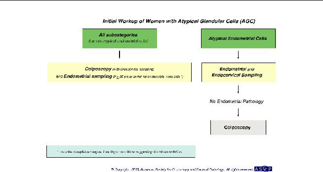
Initial Workup (Fig. 11)
For women with all subcategories of AGC and AIS except atypical endometrial cells, colposcopy with endocervical sampling is recommended regardless of HPV result (AII). Accordingly, triage by reflex HPV testing
is not recommended, and triage using repeat cervical cytology is unacceptable (DII). Endometrial sampling is recommended in conjunction with colposcopy and endocervical sampling in women 35 years of age and older with all subcategories of AGC and AIS (BII). Endometrial sampling is also recommended for women younger than 35 years with clinical indications suggesting they may be at risk for endometrial neoplasia (BII). These include unexplained vaginal bleeding or conditions suggesting chronic anovulation. For women with atypical endometrial cells, initial evaluation limited to endometrial and endocervical sampling is preferred, with colposcopy acceptable either at the initial evaluation or deferred until the results of endometrial and endocervical sampling are known; if colposcopy is deferred and no endometrial pathology is identified, colposcopy is then recommended (AII).
Subsequent Management (Fig. 12)
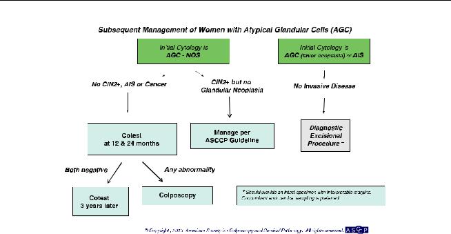
For women with AGC not otherwise specified cytology in whom CIN 2+ is not identified, co-testing at 12 months and 24 months is recommended. If both cotests are negative, return for repeat co-testing in 3 years is recommended. If any test is abnormal, colposcopy is recommended. (BII)
If CIN 2+ but no glandular neoplasia is identified histologically during the initial workup of a woman with atypical endocervical, endometrial, or glandular cells not
S16 & M A S S A D E T A L .
Figure 12.
otherwise specified, management should be according to the 2012 consensus guidelines for the lesion found (CII).
For women with AGC ‘‘favor neoplasia’’ or endocervical AIS cytology, if invasive disease is not identified during the initial colposcopic workup, a diagnostic excisional procedure is recommended (AII).
It is recommended that the type of diagnostic excisional procedure used in this setting provide an intact specimen with interpretable margins (BII). Endocervical sampling after excision is preferred (BII).
AGC or Cytologic AIS in Special Populations
Pregnant Women
The initial evaluation of AGC in pregnant women should be identical to that of nonpregnant women (BII), except that endocervical curettage and endometrial biopsy are unacceptable (EIII).
Women Aged 21Y24 Years
It is recommended that ASCCP guidelines for management of AGC be followed for all women, including those aged 21Y24 years (BII).
Management of Benign Glandular Changes. For asymptomatic premenopausal women with benign endometrial cells, endometrial stromal cells, or histiocytes, no further evaluation is recommended (BII). For postmenopausal women with benign endometrial cells, endometrial assessment is recommended (BII). For posthysterectomy pa-
tients with a cytologic report of benign glandular cells, no further evaluation is recommended (BII).
MANAGEMENT OF CIN AND HISTOLOGIC AIS
CIN 1 and No CIN Found at Colposcopy After
Abnormal Cytology
CIN 1 is the histologic manifestation of HPV infection. Although most CIN 1 lesions are associated with oncogenic HPV, HPV-16 is less common in CIN 1 than in CIN 3, and nononcogenic HPV types are also commonly found in CIN 1 lesions (82, 83). The natural history of CIN 1 is similar to that of HPV-positive ASC-US and LSIL in the absence of CIN, suggesting similar management. Regression rates are high, especially in younger women (32, 64), and progression to CIN 2+ is uncommon (64, 84).
The risk of occult CIN 3+ among women with CIN 1 at colposcopic biopsy is linked to the risk conveyed by prior cytology. KPNC data showed similar, relatively low 5-year risk of CIN 3+ when CIN 1 or no lesion was diagnosed after ASC-US or LSIL, but a substantially higher risk after HSIL, ASC-H, and AGC. For example, women with CIN 1 after LSIL or HPV-positive ASC-US had a 5-year risk of CIN 3+ of 3.8%, while those with CIN 1 after HSIL had a 5-year risk of CIN 3+ of 15% (68).
Failure to detect CIN 2+ at colposcopy in women with HSIL does not mean that a CIN 2+ lesion has been excluded, although occult carcinoma is unlikely. As a result, women with HSIL who do not have immediate diagnostic excision require close follow-up. Few studies

ASCCP Guidelines Update & S17
of the natural history of HSIL managed without treatment have been reported, and follow-up in those is limited (68); management relies on expert opinion.
Women with minor cytologic abnormalities have similar risk for CIN 3+ whether colposcopy shows CIN 1 or no lesion (64, 68). Since CIN 3+ risk is elevated for women with either HPV-16 or HPV-18 or persistent oncogenic HPV infection of any type even when cytology is negative, guidelines must provide for follow-up for women with these ‘‘lesser abnormalities’’ even when no CIN is found. These ‘‘lesser abnormalities’’ include HPV-16 or HPV-18 positivity, persistent untyped oncogenic HPV, ASC-US, and LSIL.
The management of CIN 1 in endocervical samples merits special attention. Traditional management strategies prescribed excisional therapy for women with CIN on endocervical sampling. However, these strategies preceded full understanding of the high spontaneous regression rates of CIN 1. Endocervical samples are often contaminated by ectocervical lesions. Women with CIN 1 on endocervical sampling have a low risk for CIN 2+ (85, 86) (Fukuchi E, Fetterman B, Poitras N, Kinney W, Lorey T, Little RD. Risk of cervical precancer and cancer in women with cervical intraepithelial neoplasia grade 1 on endocervical curettage. J Low Genit Tract Dis [in press]). Current guidelines on management of CIN 1 on endocervical sampling do not apply when CIN 2, CIN 3, or CIN 2,3 is specified or when the
lesion seen cannot be graded, as an associated invasive cancer cannot be excluded without a diagnostic excision procedure.
Management of Women With CIN 1 or No Lesion Preceded by ‘‘Lesser Abnormalities’’ (Fig. 13). Co-testing at 1 year is recommended (BII). If both the HPV test and cytology are negative, then age-appropriate retesting 3 years later is recommended (cytology if age is younger than 30 years, co-testing if 30 years of age or older). If all tests are negative, then return to routine screening is recommended (BII). If any test is abnormal, then colposcopy is recommended (CIII).
If CIN 1 persists for at least 2 years, either continued follow-up or treatment is acceptable (CII). If treatment is selected and the colposcopic examination is adequate, either excision or ablation is acceptable (AI). A diagnostic excisional procedure is recommended if the colposcopic examination is inadequate; the endocervical sampling contains CIN 2,CIN 3, CIN 2,3 or ungraded CIN; or the patient has been previously treated (AIII). Treatment modality should be determined by the judgment of the clinician and should be guided by experience, resources, and clinical value for the specific patient (A1II). In patients with CIN 1 and an inadequate colposcopic examination, ablative procedures are unacceptable (EI). Podophyllin or podophyllinrelated products are unacceptable for use in the vagina
Figure 13.
S18 & M A S S A D E T A L .
Figure 14.
or on the cervix (EII). Hysterectomy as the primary and principal treatment for histologically diagnosed CIN 1 is unacceptable (EII).
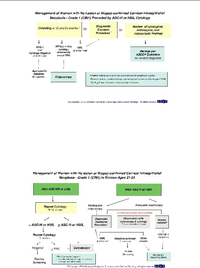
Management of Women With CIN 1 or No Lesion Preceded by ASC-H or HSIL (Fig. 14). When CIN 2+ is not identified histologically, either a diagnostic excisional procedure or observation with co-testing at 12 months and 24 months is recommended, provided in the latter case
that the colposcopic examination is adequate and the endocervical sampling is negative. (BIII). In this circumstance, it is acceptable to review the cytologic, histologic, and colposcopic findings; if the review yields a revised interpretation, management should follow guidelines for the revised interpretation (BII). If observation with cotesting is elected and both co-tests are negative, return for retesting in 3 years is recommended. If any test is abnormal, repeat colposcopy is recommended. A diagnostic
Figure 15.
ASCCP Guidelines Update & S19
excisional procedure is recommended for women with repeat HSIL cytologic results at either the 1-year or 2-year visit (CIII).
Management of Women With CIN 1 on Endocervical Sampling. When CIN 1 is detected on endocervical sampling after lesser abnormalities but no CIN 2+ is detected in colposcopic biopsies, management should follow ASCCP management guidelines for CIN 1, with the addition of repeat endocervical sampling in 12 months (BII). For women with CIN 1 on endocervical sampling and cytology reported as ASC-H, HSIL, or AGC, or with a colposcopic biopsy reported as CIN 2+, management according to the ASCCP management guidelines for the specific abnormality is recommended (BII). For women not treated, repeat endocervical sampling at the time of evaluation for the other abnormality is recommended (BII).
CIN 1 in Special Populations
Women Aged 21Y24 Years (Fig. 15). For women aged 21Y24 years with CIN 1 after ASC-US or LSIL cytology, repeat cytology at 12-month intervals is recommended.
Follow-up with HPV testing is unacceptable (EII). For women with ASC-H or HSIL+ at the 12-month follow up, colposcopy is recommended. For women with ASCUS or worse at the 24 month follow up, colposcopy is recommended. After two consecutive negative tests, routine screening is recommended. (BII)
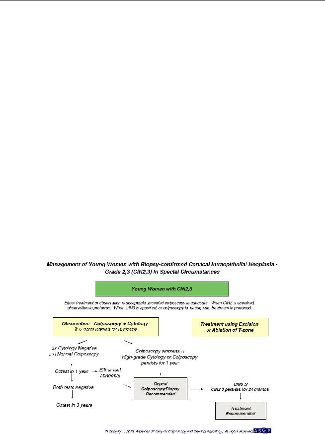
For women aged 21Y24 years with CIN 1 after ASC-H or HSIL cytology, observation for up to 24 months using both colposcopy and cytology at 6-month intervals is recommended, provided the colposcopic examination is adequate and endocervical assessment is negative (BIII). If CIN 2, CIN 3, or CIN 2,3 is identified histologically, management should follow the guideline for the management of young women with CIN 2, CIN 3, or CIN 2,3 (BIII, see ‘‘Management of Women With CIN 2, CIN 3, and CIN 2,3’’). If during follow-up a high-grade colposcopic lesion is identified or HSIL cytology persists for 1 year, biopsy is recommended (BIII). If HSIL persists for 24 months without identification of CIN 2+, a diagnostic excisional procedure is recommended (BIII). When colposcopy is inadequate or CIN 2, CIN 3, CIN 2,3 or ungraded CIN is identified on endocervical sampling, a diagnostic excision procedure is recommended (BII).
Regardless of antecedent cytology, treatment of CIN 1 in women aged 21Y24 years is not recommended (BII).
Figure 16.
S20 & M A S S A D E T A L .
Pregnant Women. For pregnant women with a histologic diagnosis of CIN 1, follow-up without treatment is recommended (BII). Treatment of pregnant women for CIN 1 is unacceptable.
CIN 2, CIN 3, and CIN 2,3
While distinction between CIN 2 and CIN 3 is difficult in individual cases, regression rates are lower and progression to cancer more common for women with CIN 3 than for those with CIN 2 (87, 88). Cervical intraepithelial neoplasia 2 remains the consensus threshold for treatment in the United States, except in special circumstances. Women with unambiguous CIN 3 have the immediate precursor to invasive cancer and should not be observed, regardless of age or concern about future fertility.
After treatment for CIN 2+, recurrence risk remains well above that of women with negative co-test results throughout observation periods that have been reported to date (89). After two negative co-tests in the first 2 years after treatment, risk is similar to that of women with a negative Pap test, suggesting a 3-year interval between surveillance examinations (89). Whether routine screening may be appropriate after three or more negative co-tests is unclear.
The objective of screening during pregnancy is to identify cervical cancer. Cervical intraepithelial neoplasia 3
does not pose a risk to the pregnancy and poses no immediate risk to the mother. Treatment during pregnancy carries substantial risk for hemorrhage and pregnancy loss.
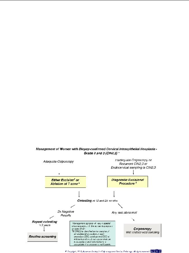
Management of Women With CIN 2, CIN 3, and CIN 2,3 (Fig. 16)
Initial Management. For women with a histologic diagnosis of CIN 2, CIN 3, or CIN 2,3 and adequate colposcopy, both excision and ablation are acceptable treatment modalities, except in pregnant women and young women (AI). A diagnostic excisional procedure is recommended for women with recurrent CIN 2, CIN 3, or CIN 2,3 (AII). Ablation is unacceptable and a diagnostic excisional procedure is recommended for women with a histologic diagnosis of CIN 2, CIN 3, or CIN 2,3 and inadequate colposcopy or endocervical sampling showing CIN 2, CIN 3, CIN 2,3, or CIN not graded (AII). Observation of CIN 2, CIN 3, or CIN 2,3 with sequential cytology and colposcopy is unacceptable, except in pregnant women and young women (EII). Hysterectomy is unacceptable as primary therapy for CIN 2, CIN 3, or CIN 2,3 (EII).
Follow-Up After Treatment. For women treated for CIN 2, CIN 3, or CIN 2,3, co-testing at 12 months and 24 months is recommended (BII). If both co-tests are
Figure 17.