

Урология - BPH. Prostatic cancer
.pdfRectal examination is by far the most important examination for the diagnosis of cancer prostate.
Cancerous hard nodules, irregular induration, obliteration of the median sulcus, a non-mobility of the rectal mucosa over the enlarged prostate suggest carcinoma of prostate.

The differentiating points between BPH and carcinoma as revealed by rectal examination are:
|
BPH |
|
Carcinoma prostate |
1. |
Size – may be quite big |
1. |
The size is usually not very big |
2. |
Consistency – firm and elastic |
2. |
Hard |
3. |
Surface - smooth |
3. |
Irregular and nodular |
4. The midline sulcus between the |
4. The sulcus is usually obliterated |
||
two lateral lobes is well felt |
|
|
|
5. |
The seminal vesicles – feel |
5. |
These may be invaded by the |
normal |
tumour and feel hard and irregular |
||
6. |
The gap between the enlarged |
6. |
This gap is obliterated by invasion |
prostate and the lateral pelvic wall is |
of the cancer |
||
clear on both sides |
|
|
|
7. The rectal mucous membrane |
7. The rectal mucous membrane is |
||
moves free over the prostate |
adherent and cannot be moved |
||

Special investigations
Blood examination. Anaemia is often detected through this examination. This may be secondary to extensive marrow invasion or
secondary to renal failure or due to haemorrhage or infection.
Blood urea, NPN, serum creatinine should be performed to detected renal function.

Urinalysis. This should be performed to exclude any infection or haemorrhage.
Liver function tests should be performed to exclude any metastasis in the liver.
Straight X-ray of the abdomen gives valuable information as bone metastasis is present or not.
Excretory urography is necessary to know: ureteric obstruction due to invasion of the tumour, ureteric obstruction due to metastasis in the lymph nodes, urinary retention.

Chest film is necessary to exclude metastasis in the lungs, involvement of hilar lymph nodes and osseous metastasis in the ribs.
Ultrasonography may give information of ureteral obstruction.

Without pathomorphological examination
the diagnosis of prostatic cancer can not be set!
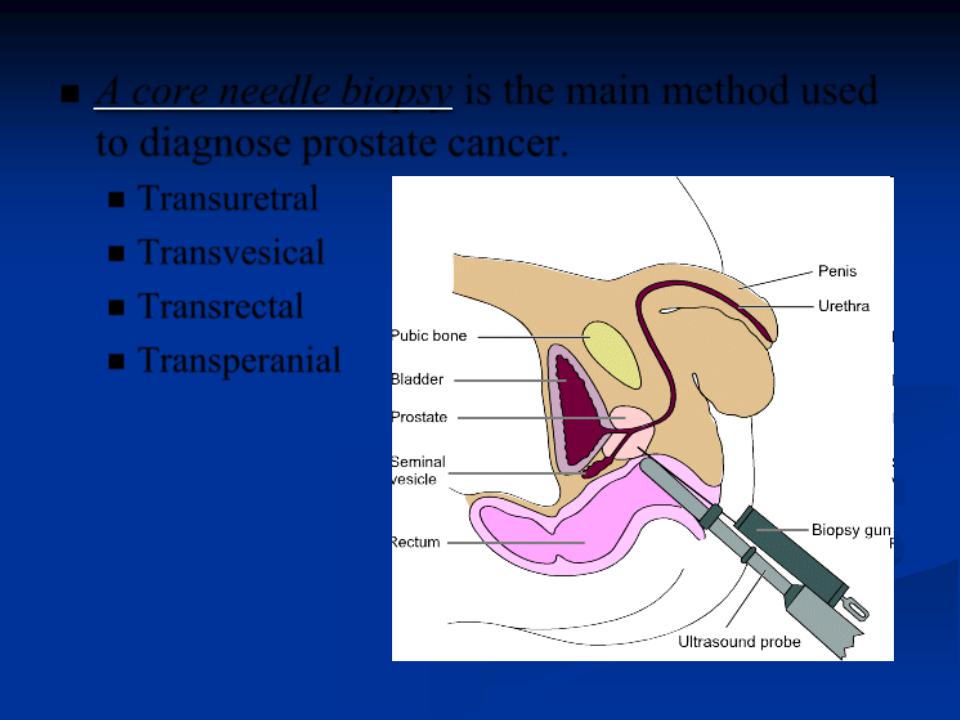
A core needle biopsy is the main method used to diagnose prostate cancer.
Transuretral
Transvesical
Transrectal
Transperanial

A PSA blood test tests the amount of prostate-specific antigen (PSA) in a sample of blood. PSA is a chemical which is made by both normal and cancerous prostate cells. If there is an abnormally high level of PSA, prostate cancer is a possibility. However, a high PSA score does not always indicate cancer and can be caused by other prostate diseases.

Additional methods – cystoscopy, oentgenologic methods: CT, MRT, nephroscintygtraphy

Conservative treatment
External Beam Radiation Therapy
Brachytherapy (Internal Radiation Therapy)
Cryosurgery
Hormone (Androgen Deprivation) Therapy:
Orchiectomy (surgical castration, Luteinizing hormone-releasing hormone (LHRH) analogs, Luteinizing hormone-releasing hormone (LHRH) antagonists
Chemotherapy - sometimes used if prostate cancer has spread outside of the prostate gland and hormone therapy isn't working.