

Chapter 1PlasticSurg
.pdfPART I ■ PRINCIPLES, TECHNIQUES, AND BASIC SCIENCE
Copyright © 2007 by Lippincott Williams & Wilkins, a Wolters Kluwer business.
Grabb and Smith's Plastic Surgery, Sixth Edition by Charles H. Thorne.
Copyright © 2007 by Lippincott Williams & Wilkins, a Wolters Kluwer business.
Grabb and Smith's Plastic Surgery, Sixth Edition by Charles H. Thorne.

CHAPTER 1 ■ TECHNIQUES AND PRINCIPLES IN PLASTIC SURGERY
CHARLES H. THORNE
Plastic surgery is a unique specialty that defies definition, has no organ system of its own, is based on principles rather than specific procedures, and, because of cosmetic surgery, is the darling of the media.
What is plastic surgery? No complete definition exists. Joe McCarthy defines it as the “problem-solving specialty.” My wife, an anesthesiologist, calls plastic surgeons the “finishers” because they come in when “the other surgeons have done all they can do and the operation has to be finished.” An even more grandiose definition is the following from a plastic surgery resident: “Plastic surgery is surgery of the skin and its contents.” There is no way to define this specialty that has acquired “turf” through a combination of tradition and innovation. What is the common denominator between craniofacial surgery and hand surgery? Between pressure sore surgery and cosmetic surgery?
Unlike other surgical specialties, plastic surgery is not organized around a specific organ system. Plastic surgery has only traditional areas of expertise and principles on which to rely for its existence and future. Because plastic surgery has loose boundaries and no specific anatomic region, it faces competition from regionally oriented specialties. Traditional areas of expertise can be lost as other specialties acquire the skills to perform the procedures developed by plastic surgeons. Consequently, plastic surgery has both freedom and vulnerability. It is this vulnerability that makes plastic surgery dependent on both the maintenance of superiority in the traditional areas of expertise and on continued innovation and acquisition of new techniques, new procedures, new problems to solve—that is, new turf.
Plastic surgery is based more on principles than on the details of specific procedures. This allows the plastic surgeon to solve unusual problems, to operate from the top of the head to the tip of the toe, to apply known procedures to other body parts, and to be innovative.
No specialty receives the attention from the lay press that plastic surgery receives. At the same time, no specialty is lesswell understood. Although the public equates plastic surgery with cosmetic surgery, the roots of plastic surgery lie in its reconstructive heritage. Cosmetic surgery, an important component of plastic surgery, is but one piece of the plastic surgical puzzle.
Plastic surgery consists of reconstructive surgery and cosmetic surgery but the boundary between the two, like the boundary of plastic surgery itself, is difficult to draw. The more one studies the specialty, the more the distinction between cosmetic surgery and reconstructive surgery disappears. Even if one asks, as an insurance company does, about the functional importance of a particular procedure, the answer often hinges on the realization that the function of the face is to look like a face (i.e., function = appearance). A cleft lip is repaired so the child will look, and therefore hopefully function, like other children. A common procedure such as a breast reduction is enormously complex when one considers the issues of appear-
ance, self-image, sexuality, and womanhood, and defies categorization as simply cosmetic or necessarily reconstructive.
This chapter outlines basic plastic surgery principles and techniques that deal with the skin. Cross-references to specific chapters providing additional information are provided. Subsequent chapters in the first section will discuss other concepts and tools that allow plastic surgeons to tackle more complex problems. Almost all wounds and all procedures involve the skin, even if it is only an incision, and therefore the cutaneous techniques described in this text are applicable to virtually every procedure performed by every specialty in surgery.
OBTAINING A FINE-LINE SCAR
“Will there be a scar?” Even the most intelligent patients ask this preposterous question. When a full-thickness injury occurs to the skin or an incision is made, there is always a scar. The question should be, “Will I have a relatively inconspicuous fineline scar?”
The final appearance of a scar is dependent on many factors, including the following: (a) Differences between individual patients that we do not yet understand and cannot predict; (b) the type of skin and location on the body; (c) the tension on the closure; (d) the direction of the wound; (e) other local and systemic conditions; and, lastly, (f) surgical technique.
The same incision or wound in two different patients will produce scars that differ in quality and aesthetics. Oily or pigmented skin produces, as a general rule, more unsightly scars (Chapter 2 discusses hypertrophic scars and keloids). Thin, wrinkled, pale, dry, “WASPy” skin of patients of English or Scotch-Irish descent usually results in more inconspicuous scars. Rules are made to be broken, however, and an occasional patient will develop a scar that is not characteristic of his or her skin type.
Certain anatomic areas routinely produce unfavorable scars that remain hypertrophic or wide. The shoulder and sternal area are such examples. Conversely, eyelid incisions almost always heal with a fine-line scar.
Skin loses elasticity with age. Stretched-out skin, combined with changes in the subcutaneous tissue, produces wrinkling, which makes scars less obvious and less prone to widening in older individuals. Children, on the other hand, may heal faster but do not heal “better,” in that their scars tend to be red and wide when compared to scars of their grandparents. In addition, as body parts containing scars grow, the scars become proportionately larger. Beware the scar on the scalp of a small child!
Just as the recoil of healthy, elastic skin in children may lead to widening of a scar, tension on a closure bodes poorly for the eventual appearance of the scar. The scar associated with a simple elliptical excision of a mole on the back will likely result
Copyright © 2007 by Lippincott Williams & Wilkins, a Wolters Kluwer business. |
3 |
|
Grabb and Smith's Plastic Surgery, Sixth Edition by Charles H. Thorne.
|
4 |
Part I: Principles, Techniques, and Basic Science |
FIGURE 1.1. Relaxed skin tension lines. (Reproduced with permission from Ruberg R. L. In: Smith DJ, ed. Plastic Surgery, A Core Curriculum. St. Louis: Mosby, 1994.)
in a much less appealing scar than an incisional wound. The body knows when it is missing tissue.
The direction of a laceration or excision also determines the eventual appearance of the scar. The lines of tension in the skin were first noted by Dupuytren. Langer also described the normal tension lines, which became known as “Langer lines.” Borges referred to skin lines as “relaxed skin tension lines” (Fig. 1.1).
Elective incisions or the excision of lesions are planned when possible so that the final scars will be parallel to the relaxed skin tension lines. Maximal contraction occurs when a scar crosses the lines of minimal tension at a right angle. Wrinkle lines are generally the same as the relaxed skin tension lines and lie perpendicular to the long axis of the underlying muscles.
Other issues, which are not related to the scar itself but to perception, determine if a scar is noticeable. Incisions and scars can be “hidden” by placing them at the junction of aesthetic units (e.g., at the junction of the lip and cheek, along the nasolabial fold), where the eye expects a change in contour (Chapter 38). In contrast, an incision in the midcheek or midchin or tip of the nose will always be more conspicuous.
The shape of the wound also affects ultimate appearance. The “trapdoor” scar results from a curvilinear incision or laceration that, after healing and contracture, appears as a depressed groove with bulging skin on the inside of the curve. Attempts at “defatting” the bulging area are never as satisfactory as either the patient or surgeon would like.
Local conditions, such as crush injury of the skin adjacent to the wound, also affect the scar. So, too, will systemic conditions such as vascular disease or congenital conditions affecting elastin and/or wound healing. Nutritional status can affect wound healing, but usually only in the extreme of malnutrition or vitamin deficiency. Nutritional status is probably overemphasized as a factor in scar formation.
Technique is also overemphasized (by self-serving plastic surgeons?) as a factor in determining whether a scar will be inconspicuous, but it is certainly of some importance. Minimizing damage to the skin edges with atraumatic technique, debridement of necrotic or foreign material, and a tension-free closure are the first steps in obtaining a fine-line scar. Ultimately,
however, scar formation is unpredictable even with meticulous technique.
Two technical factors are of definite importance in increasing the likelihood of a “good” scar. First is the placement of sutures that will not leave permanent suture marks or the prompt removal of skin sutures so disfiguring “railroad tracks” do not occur. In other words, removing the sutures may be more important than placing them! Plastic surgeons have been known to mock other specialists for using heavy-gauge suture for skin closure, but the choice of sutures is irrelevant if the sutures are removed soon enough. Sutures on the face can usually be removed in 3 to 5 days and on the body in 7 days or less. Except for wounds over joints, sutures should rarely be left in for more than 1 week. A subcutaneous layer of closure and Steri-Strips are usually sufficient to prevent dehiscence.
The second important technical factor that affects the appearance of scars is wound-edge eversion. In wounds where the skin is brought precisely together, there is a tendency for the scar to widen. In wounds where the edges are everted, or even hypereverted in an exaggerated fashion, this tendency is reduced, possibly by reducing the tension on the closure. In other words, the ideal wound closure may not be perfectly flat, but rather bulging with an obvious ridge, to allow for eventual spreading of that wound. Wound-edge eversion ALWAYS goes away. The surgeon need not ever worry that a hypereverted wound will remain that way; it will always flatten over time.
CLOSURE OF SKIN WOUNDS
While the most common method of closing a wound is with sutures, there is nothing necessarily magic or superior about sutures. Staples, skin tapes, or wound adhesives are also useful in certain situations. Regardless of the method used, precise approximation of the skin edges without tension is essential to ensure primary healing with minimal scarring.
Wounds that are deeper than skin are closed in layers. The key is to eliminate dead space, to provide a strong enough closure to prevent dehiscence while wound healing is occurring, and to precisely approximate the skin edges without tension. Not all layers necessarily require separate closure. A closure over the calf, however, is subject to motion, dependence, and stretching with walking, requiring a stronger closure than the scalp, which does not move, is less dependent, and not subject to tension in daily activities.
Except for dermal sutures, which are placed with the knot buried to prevent it from emerging from the skin during the healing process, sutures should be placed with the knot superficial to the loop of the suture (not buried), so that the tissue layers can be everted (Fig. 1.2A).
Buried dermal sutures provide strength so the external sutures can be removed early, but do not prevent the scar from spreading over time. There is no technique that reliably prevents a wound that has an inclination to widen from doing so.
Suturing Techniques
Techniques for suturing are illustrated in Figure 1.2 and are listed below.
Simple Interrupted Suture
The simple interrupted suture is the gold standard and the most commonly employed suture. The needle is introduced into the skin at an angle that allows it to pass into the deep dermis at a point further removed from the entry of the needle. This allows the width of suture at its base in the dermis to be wider
Copyright © 2007 by Lippincott Williams & Wilkins, a Wolters Kluwer business.
Grabb and Smith's Plastic Surgery, Sixth Edition by Charles H. Thorne.
|
|
Chapter 1: Techniques and Principles in Plastic Surgery |
5 |
|
A |
B |
|
C |
|
D |
E |
|
F |
|
|
G |
H |
|
|
FIGURE 1.2. Types of skin closure. A: Simple interrupted. B: Vertical mattress. C: Horizontal mattress.
D: Subcuticular continuous. E: Half-buried horizontal mattress. F: Continuous over-and-over. G: Staples.
H: Skin tapes (skin adhesive performs a similar function).
than the epidermal entrance and exit points, giving the suture a triangular appearance when viewed in cross section and everting the skin edges. Care must be taken to ensure that the suture is placed at the same depth on each side of the incision or wound, otherwise the edges will overlap. Sutures are usually placed approximately 5 to 7 mm apart and 1 to 2 mm from the skin edge, although the location and size of the needle and caliber of the suture material make this somewhat variable.
Vertical Mattress Suture
Vertical mattress sutures may be used when eversion of the skin edges is desired and cannot be accomplished with simple sutures alone. Vertical mattress sutures tend to leave the most obvious and unsightly cross-hatching if not removed early.
Horizontal Mattress Suture
Horizontal mattress sutures also provide approximation of the skin edges with eversion. They are particularly advantageous in thick glabrous skin (feet and hand). In the author’s opinion, horizontal mattress sutures are superior to their vertical counterparts.
Subcuticular Suture
Subcuticular (or intradermal) sutures can be interrupted or placed in a running fashion. In a running subcutaneous closure, the needle is passed horizontally through the superficial dermis parallel to the skin surface to provide close approximation of the skin edges. Care is taken to ensure that the sutures are placed at the same level. Such a technique obviates the need
Copyright © 2007 by Lippincott Williams & Wilkins, a Wolters Kluwer business.
Grabb and Smith's Plastic Surgery, Sixth Edition by Charles H. Thorne.
|
6 |
Part I: Principles, Techniques, and Basic Science |
for external skin sutures and circumvents the possibility of suture marks in the skin. Absorbable or nonabsorbable suture can be used, with the latter to be removed at 1 to 2 weeks after suturing.
Half-Buried Horizontal Mattress Suture
Half-buried horizontal mattress sutures are used when it is desirable to have the knots on one side of the suture line with no suture marks on the other side. For example, when insetting the areola in breast reduction, this method leaves the suture marks on the dark, pebbly areola instead of on the breast skin.
Continuous Over-and-Over Suture
Continuous over-and-over sutures, otherwise known as running simple sutures can be placed rapidly but depend on the wound edges being more-or-less approximated beforehand. A continuous suture is not nearly as precise as interrupted sutures. Continuous sutures can also be placed in a locking fashion to provide hemostasis by compression of wound edges. They are especially useful in scalp closures.
Skin Staples
Skin staples are particularly useful as a time saver for long incisions or to position a skin closure or flap temporarily before suturing. Grasping the wound edges with forceps to evert the tissue is helpful when placing the staples to prevent inverted skin edges. Staples must be removed early to prevent skin marks and are ideal for the hair-bearing scalp.
Skin Tapes
Skin tapes can effectively approximate the wound edges, although buried sutures are often required in addition to skin tape to approximate deeper layers, relieve tension, and prevent inversion of the wound edges. Skin tapes can also be used after skin sutures are removed to provide added strength to the closure.
Skin Adhesives
Skin adhesives have been developed, and may have a role in wound closure, especially in areas where there is no tension on the closure, or where strength of closure has been provided by a layer of buried dermal sutures. Adhesives, by themselves, however, do not evert the wound edges. Eversion must be provided by deeper sutures.
Methods of Excision
Lesions of the skin can be excised with elliptical, wedge, circular, or serial excision.
FIGURE 1.3. Elliptical excision. A: If the ellipse is too short, dog-ears (arrows) form at the ends of the closed wound. B: Correct method with length of ellipse at least three times the width.
Elliptical Excision
Simple elliptical excision is the most commonly used technique (Fig. 1.3). Elliptical excision of inadequate length may yield “dog-ears,” which consist of excess skin and subcutaneous fat at the end of a closure. There are several ways to correct a dogear, some of which are shown in Figure 1.4. Dog-ears are the bane of plastic surgical existence and one must be facile with their elimination. Dog-ears do not disappear on their own.
Wedge Excision
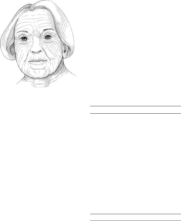
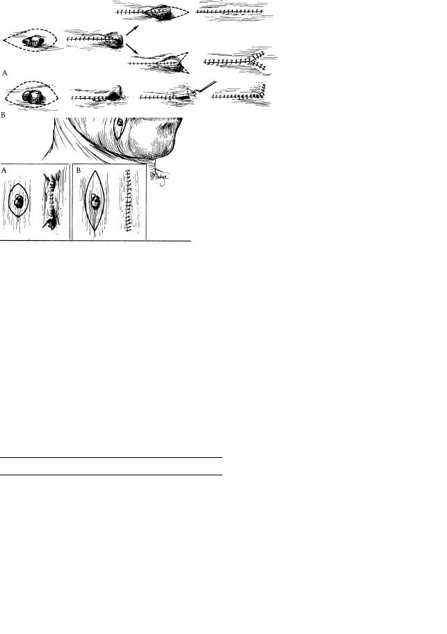
Lesions located at or adjacent to free margins can be excised by wedge excision. In some elderly patients, one third of the lower lip and one fourth of the upper lip can be excised with primary closure (Fig. 1.5)
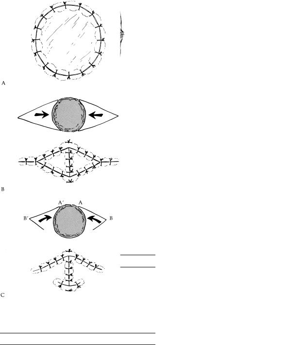
Circular Excision
When preservation of skin is desired (such as the tip of the nose) or the length of the scar must be kept to a minimum (children), circular excision might be desirable. Figure 1.6 shows some closure techniques. Figure 1.6 is included because these techniques
FIGURE 1.4. Three methods of removing a dog-ear caused by making the elliptical excision too short. A: Dog-ear excised, making the incision longer, or converted to a “Y”. B: One method of removing a dog-ear caused by designing an elliptical excision with one side longer than the other. Conversion to an “L” effectively lengthens the shorter side.
Copyright © 2007 by Lippincott Williams & Wilkins, a Wolters Kluwer business.
Grabb and Smith's Plastic Surgery, Sixth Edition by Charles H. Thorne.
Chapter 1: Techniques and Principles in Plastic Surgery |
7 |
|
FIGURE 1.5. Wedge excisions of the ear, lower eyelid and lip.
may be of value, as well as for historical purposes. Circular defects can also be closed with a purse-string suture that causes significant bunching of the skin. This is allowed to mature for many months and may result in a shorter scar on, for example, the face of a child.
Serial Excision
Serial excision is the excision of a lesion in more than one stage. Serial excision and tissue expansion (Chapter 10) are frequently employed for large lesions such as congenital nevi. The inherent viscoelastic properties of skin are used, allowing the skin to “stretch” over time. These techniques enable wound closure to be accomplished with a shorter scar than if the original lesion was elliptically excised in a single stage.
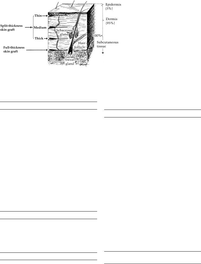
SKIN GRAFTING
Skin grafts are a standard option for closing defects that cannot be closed primarily. A skin graft consists of epidermis and some or all of the dermis. By definition, a graft is something that is removed from the body, is completely devascularized, and is replaced in another location. Grafts of any kind require vascularization from the bed into which they are placed for survival. Any tissue which is not completely removed prior to placement is not a graft.
Skin Graft Types
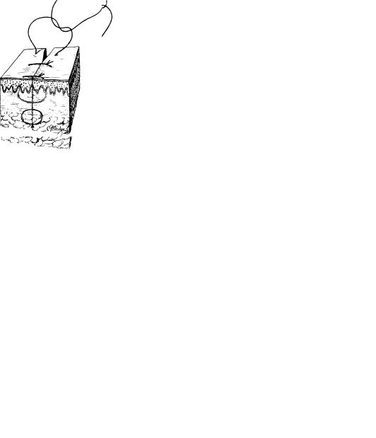
Skin grafts are classified as either split-thickness or fullthickness, depending on the amount of dermis included. Splitthickness skin grafts contain varying amounts of dermis, whereas a full-thickness skin graft contains the entire dermis
(Fig. 1.7).
All skin grafts contract immediately after removal from the donor site and again after revascularization in their final location. Primary contraction is the immediate recoil of freshly harvested grafts as a result of the elastin in the dermis. The more dermis the graft has, the more primary the contraction that will be experienced. Secondary contracture, the real nemesis, involves contraction of a healed graft and is probably a result of myofibroblast activity. A full-thickness skin graft contracts more on initial harvest (primary contraction) but less on healing (secondary contracture) than a split-thickness skin
FIGURE 1.6. Closure of wounds following circular excision. A: Skin graft. B: Sliding triangular subcutaneous pedicle flaps can be advanced to close the circular defect; the triangular defect is closed in a V-Y fashion. C: Transposition flaps based on a skin pedicle and rotated toward each other can also be used. Circular defects can also be closed by other local flaps (Figs. 1.10–1.15) or by pursestring suture.
graft. The thinner the split-thickness skin graft, the greater the secondary contracture. Granulating wounds left to heal secondarily, without any skin grafting, demonstrate the greatest degree of contracture and are most prone to hypertrophic scarring.
The number of epithelial appendages transferred with a skin graft depends on the thickness of the dermis present. The ability of grafted skin to sweat depends on the number of glands transferred and the sympathetic reinnervation of these glands from the recipient site. Skin grafts are reinnervated by ingrowth of nerve fibers from the recipient bed and from the periphery. Full-thickness skin grafts have the greatest sensory return because of a greater availability of neurilemmal sheaths. Hair follicles are also transferred with a full-thickness
Copyright © 2007 by Lippincott Williams & Wilkins, a Wolters Kluwer business.
Grabb and Smith's Plastic Surgery, Sixth Edition by Charles H. Thorne.
|
8 |
Part I: Principles, Techniques, and Basic Science |
FIGURE 1.7. Skin graft thickness.
skin graft. In general, full-thickness skin grafts demonstrate the hair growth of the donor site whereas split-thickness skin grafts, especially thin split-thickness skin grafts, are generally hairless.
Requirements for Survival of a Skin Graft
The success of skin grafting, or “take,” depends on the ability of the graft to receive nutrients and, subsequently, vascular ingrowth from the recipient bed. Skin graft revascularization or “take” occurs in three phases. The first phase involves a process of serum imbibition and lasts for 24 to 48 hours. Initially, a fibrin layer forms when the graft is placed on the recipient bed, binding the graft to the bed. Absorption of nutrients into the graft occurs by capillary action from the recipient bed. The second phase is an inosculatory phase in which recipient and donor end capillaries are aligned. In the third phase, the graft is revascularized through these “kissing” capillaries. Because the full-thickness skin graft is thicker, survival of the graft is more precarious, demanding a well-vascularized bed.
To optimize take of a skin graft, the recipient site must be prepared. Skin grafts require a vascular bed and will seldom take in exposed bone, cartilage, or tendon devoid of their periosteum, perichondrium, or paratenon. There are exceptions, however, as skin grafts are frequently successful inside the orbit or on the temporal bone, despite removal of the periosteum. Close contact between the skin graft and its recipient bed is essential. Hematomas and seromas under the skin graft will compromise its survival, and immobilization of the graft is essential.
Skin Graft Adherence
For the skin graft to take, it must adhere to the bed. There are two phases of graft adherence. The first begins with placement of the graft on the recipient bed, to which the graft adheres because of fibrin deposition. This lasts approximately 72 hours. The second phase involves ingrowth of fibrous tissue and vessels into the graft.
skin grafts result in a “pebbled” appearance that, at times, is aesthetically unacceptable. In contrast, a sheet skin graft has the advantage of a continuous, uninterrupted surface, often leading to a superior aesthetic result, but has the disadvantages of not allowing serum and blood to drain through it and the need for a larger skin graft.
Skin Graft Donor Sites
The donor site epidermis regenerates from the immigration of epidermal cells originating in the hair follicle shafts and adnexal structures left in the dermis. In contrast, the dermis never regenerates. Because split-thickness skin grafts remove only a portion of the dermis, the original donor site may be used again for a subsequent split-thickness skin graft harvest. Thus, the number of split-thickness skin grafts harvested from a donor site is directly dependent on the donor dermis thickness. Fullthickness skin graft donor sites must be closed primarily because there are no remaining epithelial structures to provide re-epithelialization.
Skin grafts can be taken from anywhere on the body, although the color, texture, thickness of the dermis, vascularity, and donor site morbidity of body locations vary considerably. Skin grafts taken from above the clavicles provide a superior color match for defects of the face. The upper eyelid skin can also be used, as it provides a small amount of very thin skin. Full-thickness skin graft harvest sites are closed primarily and are therefore of smaller size. The scalp, abdominal wall, buttocks, and thigh are common donor sites for split-thickness skin grafts. Surgeons should avoid the mistake of harvesting split-thickness skin grafts from the most accessible locations such as the anterior thigh. Although donor sites heal by reepithelialization, there is always visible evidence that an area was used as a donor site. This can vary from keloids to simple hyperor hypopigmentation. Less-conspicuous donor sites are the buttocks or scalp. Split-thickness skin grafts harvested from the scalp will have hair in them initially but no hair follicles and therefore will ultimately be hairless. The hair in the scalp donor site will return after re-epithelialization because the hair follicles were left undisturbed.
Meshed versus Sheet Skin Grafts
Multiple mechanical incisions result in a meshed skin graft, allowing immediate expansion of the graft. A meshed skin graft covers a larger area per square centimeter of graft harvest and allows drainage through the numerous holes. Meshed
Postoperative Care of Skin
Grafts and Donor Sites
Causes of graft failure include collection of blood or serum beneath the graft (raising the graft from the bed and preventing revascularization), movement of the graft on the bed
Copyright © 2007 by Lippincott Williams & Wilkins, a Wolters Kluwer business.
Grabb and Smith's Plastic Surgery, Sixth Edition by Charles H. Thorne.
Chapter 1: Techniques and Principles in Plastic Surgery |
9 |
|
|
Biologic Dressings |
|
Skin grafts can also be used as temporary coverage of wounds |
|
as biologic dressings. This protects the recipient bed from des- |
|
iccation and further trauma until definitive closure can occur. |
|
In large burns where there is insufficient skin to be harvested |
|
for coverage, skin substitutes can be used (Chapter 18). Bio- |
|
logic skin substitutes include human allografts (cadaver skin), |
|
amnion, or xenografts (such as pig skin). Allografts become |
|
vascularized (or “take”) but are rejected at approximately |
|
10 days unless the recipient is immunosuppressed (e.g., has |
|
a large burn), in which case rejection takes longer. Conversely, |
|
xenografts are rejected before becoming vascularized. Synthetic |
|
skin substitutes such as silicone polymers and composite mem- |
|
branes can also be applied, and new skin substitutes are con- |
|
stantly being developed. Human epidermis can be cultured in |
|
vitro to yield sheets of cultured epithelium that will provide |
|
coverage for large wounds. The coverage is fragile as a result |
FIGURE 1.8. Tie-over bolster dressing for skin grafts. |
of the lack of a supporting dermis. |
interrupting revascularization (immobilization techniques include the use of bolster dressings as shown in Fig. 1.8), and infection. The risk of infection can be minimized by careful preparation of the recipient site and early inspection of grafts applied to contaminated beds. Wounds that contain more than 105 organisms per gram of tissue will not support a skin graft. In addition, an infection at the graft donor site can convert a partial-thickness dermal loss into a full-thickness skin loss.
The donor site of a split-thickness skin graft heals by reepithelialization. A thin split-thickness harvest site (less than 10/1,000 of an inch) generally heals within 7 days. The donor site can be cared for in a number of ways. The site must be protected from mechanical trauma and desiccation. Xeroform, OpSite, or Adaptic can be used. Because moist, occluded wounds (donor sites) heal faster than dry wounds, the older method of placing Xeroform and drying it with a hairdryer is not optimal. An occlusive dressing, such as semipermeable polyurethane dressing (e.g., OpSite), will also significantly decrease pain at the site.
SKIN FLAPS
Unlike a skin graft, a skin flap has its own blood supply. Flaps are usually required for covering recipient beds that have poor vascularity; covering vital structures; reconstructing the full thickness of the eyelids, lips, ears, nose, and cheeks; and padding body prominences. Flaps are also preferable when it may be necessary to operate through the wound at a later date to repair underlying structures. In addition, muscle flaps may provide a functional motor unit or a means of controlling infection in the recipient area. Muscle flaps and microvascular free flaps are discussed in Chapters 5 and 8.
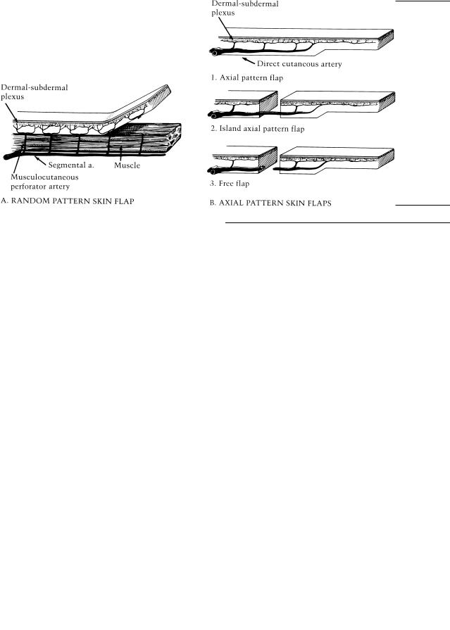
In an experimental study, Mathes et al. compared musculocutaneous flaps with “random” skin flaps to determine the bacterial clearance and oxygen tension of each (Fig. 1.9). Placement of 107 Staphylococcus aureus underneath random skin flaps in dogs resulted in 100% necrosis of the skin flaps within 48 hours; the musculocutaneous flaps, however, demonstrated long-term survival. The quantity of viable bacteria placed in wound cylinders under these flaps demonstrated an immediate reduction when placed deep to musculocutaneous flap. Oxygen
FIGURE 1.9. “Old-fashioned” classification of skin flaps. A: Random pattern. B: Axial pattern.
Copyright © 2007 by Lippincott Williams & Wilkins, a Wolters Kluwer business.
Grabb and Smith's Plastic Surgery, Sixth Edition by Charles H. Thorne.

|
10 |
Part I: Principles, Techniques, and Basic Science |
FIGURE 1.10. Rotation flap. The edge of the flap is four to five times the length of the base of the defect triangle. A back-cut or a Burow triangle can be used if the flap is under excessive tension. A: Pivot point and line of greatest tension. B: Backcut. C: Burow’s¨ triangle.
tension was measured at the distal end of the random flap and compared to that underneath the muscle of the distal portion of musculocutaneous flap as well as in its subcutaneous area. It was found that the oxygen tension in the distal random flap was significantly less than in distal muscular and cutaneous portions of the musculocutaneous flap. This study has been used to justify transfer of muscle flaps in infected wounds. It may be that well-vascularized skin flaps would be equally efficacious as muscle flaps.
Finally, a flap may be chosen because the aesthetic result will be superior. For example, a nasal defect from a skin cancer could be closed with a skin graft, leaving a visible patch. A local skin flap may require incisions in the adjacent nasal tissue, but may be aesthetically preferable in the long-term. There is no better tissue to replace nasal tissue than nasal tissue. Replace like with like.
A skin flap consists of skin and subcutaneous tissue that are transferred from one part of the body to another with a vascular pedicle or attachment to the body being maintained for nourishment. Proper planning of a flap is essential to the success of the operation. All possible sites and orientations for the flap must be considered so that the most suitable option is selected.
Planning the flap in reverse is an important principle. A pattern of the defect is transferred onto a piece of cloth toweling. The steps in the operative procedure are carried out in reverse order, using this pattern until the donor site is reached. The flap is designed slightly longer than needed, as some length will be lost in the rotation process and slight redundancy may avoid
FIGURE 1.11. Transposition flap. The secondary defect is often closed by a skin graft. A back-cut can be used if the flap is under excessive tension.
kinking of the flap blood supply. The process is repeated, being certain each time the base is held in a fixed position and not allowed to shift with the flap. Measure twice, cut once. It is easier to trim a flap that is slightly long than to add to one that is too small.
Planning a transposition or rotation flap requires attention to ensure that the line of greatest tension from the pivot point to the most distal part of the flap is of sufficient length (Figs. 1.10, 1.11 and 1.12).
Local skin flaps are of two types: flaps that rotate about a pivot point (rotation, transposition, and interpolation flaps) (Figs. 1.10 and 1.11) and advancement flaps (single-pedicle advancement, V-Y advancement, Y-V advancement, and bipedicle advancement flaps) (Figs. 1.17 and 1.18).
Flaps Rotating About a Pivot Point
Rotation, transposition, and interpolation flaps have in common a pivot point and an arc through which the flap is rotated. The radius of this arc is the line of greatest tension of the flap. The realization that these flaps can be rotated only about the pivot point is important in preoperative planning.
The rotation flap is a semicircular flap of skin and subcutaneous tissue that rotates about a pivot point into the defect to be closed (Fig. 1.10). The donor site can be closed by a skin graft or by direct suture of the wound.
Copyright © 2007 by Lippincott Williams & Wilkins, a Wolters Kluwer business.
Grabb and Smith's Plastic Surgery, Sixth Edition by Charles H. Thorne.