

3 курс / Фармакология / Essential_Psychopharmacology_2nd_edition
.pdfPrinciples of Chemical Neurotransmission |
29 |
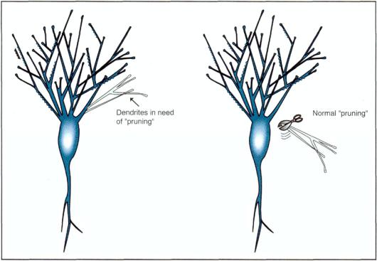
FIGURE 1—20. Synapses are formed at a furious rate between birth and age 6. However, there is competitive elimination and restructuring of synapses, a phenomenon that peaks during pubescence and adolescence, leaving about half to two-thirds of the synapses present in childhood to survive into adulthood.
and not sail past it. Other recognition molecules direct axons away by emitting repulsive axon guidance signals (RAGS) (Fig. 1 — 19).
As brain development progresses, the travel of axonal growth cones is greatly impeded but not completely lost. The fact that axonal growth is retained in the mature brain suggests that neurons continue to alter their targets of communication, perhaps by repairing, regenerating, and reconstructing synapses as demanded by the evolving duties of a neuron. A large number of recognition molecules supervise this. Some of these include not only semaphorins and collapsins but also molecules such as netrins, neuronal cellular adhesion molecules (NCAMS), integrins, cadherins, and cytokines (Table 1—4).
Interestingly, more synapses are present in the brain by age 6 than at any other time in the life cycle (Fig. 1 — 20). During the next 5 to 10 years and into adolescence, the brain then systematically removes half of all synaptic connections present at age 6. This leaves about 100 trillion synapses and up to 10,000 individual synapses for some neurons. Excitotoxicity may mediate the pruning of synaptic connections (as will be discussed in much greater detail in Chapter 4). Hopefully, neu-rodevelopmental experiences and genetic programming lead the brain to select wisely which connections to keep and which to destroy. If this is done appropriately, the individual prospers during this maturational task and advances gracefully into adulthood. Bad selections theoretically could lead to neurodevelopmental disorders such as schizophrenia or even attention deficit hyperactivity disorder.

30 Essential Psychopharmacology
FIGURE 1—21. The neuron is composed of a cell body, an axon and a dendritic tree (literally, a tree of branching dendrites). The dendritic tree is in constant flux and revises its synaptic connections throughout life.
That growth of new synapses and the pruning of old synapses then proceeds throughout a lifetime, but at a much slower pace and over shorter distances than earlier in development. Thus, the axons and dendrites of each neuron are constantly changing, establishing new connections, and removing old connections, in a manner reminiscent of the branches of a tree (Fig. 1—21). Indeed, the arborization of neuronal terminals and the dendritic tree are terms implying this constant branching (Fig. 1 — 22) and pruning (Fig. 1—23) process, which proceeds throughout the lifetime of that neuron. After the dramatic reductions in neurons before birth and in synapses during late childhood and early adolescence are complete, activity calms down considerably in the mature brain, where maintenance and remodeling of synapses continue to modest extents and over more limited distances.
Although the continuous structural remodeling of synapses in the mature brain, directed by recognition molecules, cannot approximate the pronounced long-range growth of early brain development, this restriction could be beneficial, in part because it allows structural plasticity while restricting unwanted axonal growth. This would stabilize brain function in the adult and could furthermore prevent chaotic rewiring of the brain by limiting both axonal growth away from appropriate targets and ingrowth from inappropriate neurons. On the other hand, the price of such

Principles of Chemical Neurotransmission |
31 |
FIGURE 1 — 22. The dendritic tree of a neuron can sprout branches, grow, and establish a multitude of new synaptic connections throughout its life. The process of making dendritic connections on an undeveloped neuron may be controlled by various growth factors, which act to promote the branching process and thus the formation of synapses on the dendritic tree.
growth specificity becomes apparent when a long-distance neuron in the adult brain or spinal cord dies, thus making it difficult to reestablish original synaptic connections, even if axonal growth is turned on.
As previously discussed, neurons and their supportive and neighboring glia elaborate a rich array of neurotrophic factors, which promote synaptic connections (Fig. 1—22) or eliminate them (Fig. 1—23). The potential for releasing growth factors is preserved forever, which contributes to the possibility of constant synaptic revision throughout the lifetime of that neuron. Such potential changes in synaptogenesis may provide the substrate for learning, emotional maturity, and the development of cognitive and motor skills throughout a lifetime. However, it is not clear how the brain dispenses its neurotrophic factors endogenously during normal adult physiological functioning. Presumably, demand to use neurons is met by keeping them fit and ready to function, a task accomplished by salting the brain broth with neurotrophic factors that keep the neurons healthy. Perhaps thinking and learning provoke the release of neurotrophic factors. Maybe "use it or lose it" applies to adult neurons.
32 |
Essential Psychopharmacology |
FIGURE 1—23. The dendritic tree of a neuron not only sprouts branches, grows, and establishes a multitude of new synaptic connections throughout its life, as shown in Figure 1—22, but it can also remove, alter, trim, or destroy such connections when necessary. The process of dismantling synapses and dendrites may be controlled by removal of growth factors or by a naturally occurring destructive process sometimes called excitotoxicity. Thus, there is a normal "pruning" process for removing dendrites in need of pruning.
with neurons being preserved and new connections being formed if the brain stays active. It is even possible that the brain could lose its "strength" in the absence of mental exercise. Perhaps inactivity leads to pruning of unused, "rusty" synapses, even triggering apoptotic demise of entire inactive neurons. On the other hand, mental stimulation might prevent this, and psychotherapy may even induce neurotrophic factors to preserve critical cells and innervate new therapeutic targets to alter emotions and behaviors. Only future research will clarify how to use drugs and psychotherapy to balance the seasonings in the tender stew of the brain.
Summary
The reader should now appreciate that chemical neurotransmission is the foundation of psychopharmacology. It has three dimensions, namely, space, time, and function. The spatial dimension is both that of "hard wiring" as the anatomically addressed nervous system and that of a "chemical soup" as the chemically addressed nervous system. The time dimension reveals that neurotransmission can be fast (milliseconds) or slow (up to several seconds) in onset, depending on the neurotransmitter or neuromodulator, of which there are dozens. Neurotransmission can also cause actions
Principles of Chemical Neurotransmission
33
that are short-acting (milliseconds) or very long acting (days to weeks or longer). The functional dimension of chemical neurotransmission is the process whereby an electrical impulse in one neuron is converted into a chemical message at the synaptic connection between two neurons and then into a chemical message that can alter gene expression in the second neuron.
This chapter has also emphasized a few additional points: Chemical neurotransmission sometimes occurs with more than one neurotransmitter in a single neuron. Naturally occurring neurotransmitters are often mimicked by drugs (for example, marijuana and morphine). Molecular neurobiology and its techniques demonstrate that the genetic materials of a neuron are responsible for the production of neuronal proteins in general and neurotransmitter receptors in particular. This can be modulated by physiological adaptations, by drugs, and by diseases. Finally, the neuron is dynamically modifying its synaptic connections throughout its life, in response to learning, life experiences, genetic programming, drugs, and diseases.
CHAPTER 2
RECEPTORS AND ENZYMES AS THE
TARGETS OF DRUG ACTION
I. The organization of a single receptor: Three parts of a receptor II. Synaptic teamwork
A.Ion channels
B.Transport carriers and active transport pumps
C.Neurotransmitter synaptic reuptake—an example of molecular transport us ing an active transport pump
D.Second-messenger systems
E.Ion regulation
F.Gene regulation
III. Receptors as sites of drug action
IV. Enzymes as sites of drug action
V. Summary: How drugs modify chemical neurotransmission
In Chapter 1 we discussed how modern psychopharmacology is essentially the study of chemical neurotransmission. In this chapter we will become more specific and discuss how virtually all central nervous system (CNS) drugs act in one of two very specific ways on chemical neurotransmission: first and most prominently as stimulators (agonists) or blockers (antagonists) of neurotransmitter receptors; or second, and less commonly, as inhibitors of regulatory enzymes.
Given the far-reaching importance of receptors and enzymes in our current thinking about how drugs work in the brain, this chapter will explore the properties of these very interesting targets of CNS drug action. We will first explore the organization of single receptors and how they form binding sites for neurotransmitters and drugs. We will then describe how receptors work as members of a synaptic neurotransmission team, including ions, ion channels, transport carriers, second messenger systems, transcription factors, genes, and gene products. Finally, we will
35

36 Essential Psychopharmacology
FIGURE 2 — 1. This figure is a schematic diagram of a receptor, showing that it is a protein arranged essentially as a long chain of amino acids. The chain winds in and out of the cell several times, creating three regions of the receptor: first, the extracellular portions are those parts of the chain entirely outside the neuron; second, the intracellular portions are those bits of the chain entirely inside the neuron; and third, the transmembrane portion, which comprises the regions of the receptor that reside within the membrane of the neuron.
discuss how enzymes and receptors are sites of drug actions and how such drug actions in turn modify chemical neurotransmission.
The Organization of a Single Receptor: Three Parts of a Receptor
Receptors are long chains of amino acids and therefore a type of protein (Fig. 2—1). Receptors reside partially within neuronal membranes (Figs. 2 — 1 and 2 — 2). In fact, neurotransmitter receptors can be thought of as containing three portions: an extracellular portion, a transmembrane portion and an intracellular portion (Fig. 2 — 2). The chain of amino acids constituting the receptor is not arranged in a straight line as might be implied by oversimplified representations in diagrams such as Figures

Receptors and Enzymes as the Targets of Drug Action |
37 |
FIGURE 2—2. A side view of a receptor with seven transmembrane regions is shown here. This is a common structure of many receptors for neurotransmitters and hormones. That is, the string of amino acids goes in and out of the cell several times to create three portions of the receptor: first, that part that is outside of the cell (called the extracellular portion); second, the part that is inside the receptor that is inside the cell (called the intracellular portion; and finally, the part that traverses the membrane several times (called the transmembrane portion). Throughout this text, this receptor will be represented in a simplified schematic manner with the icon shown in the small box.
2 — 1 and 2 — 2, but rather in an alpha helical manner, as a spiral around a central core (Figs. 2 — 3 and 2—4). The binding site for the neurotransmitter is inside the central core for many receptors (i.e., inside the helix of Figs. 2 — 3 and 2—4).
The extracellular binding portion of a receptor is the part of the receptor that is located outside the cell. It was originally believed that this portion of the receptor contained the selective binding site for its neurotransmitter. However, as mentioned above, it is now known that the selective binding site for a neurotransmitter is often located within the second portion of the receptor, its transmembrane regions (Figs. 2-3 and 2-4).
Some drugs may compete with the neurotransmitter for its own binding site, attempting to mimic the neurotransmitter that normally binds there or to block that neurotransmitter. As we will discuss in more detail in Chapter 3 under the topic of allosteric modulation, drugs may also act at totally separate and unique binding sites at other locations on the receptor to change the actions of the neurotransmitter on its receptor. The locations of such binding sites are still under intense investigation, but these sites may also be located in the transmembrane regions, yet separate from the neurotransmitter's binding site. This recognition site for the neurotransmitter receptor is quite unique from one receptor to the next and indeed may be one of the major distinguishing characteristics of one receptor versus another. Some receptors even have binding sites for two distinct neurotransmitters, in which case they are called co-transmitters.
The transmembrane regions (Figs. 2 — 2 and 2 — 3) probably also serve in part a structural purpose, holding the receptor in place or allowing a certain movement of the receptor relative to the membrane itself. Transmembrane regions of one neurotransmitter receptor can be quite similar to those of other neurotransmitter receptors,
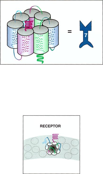
FIGURE 2 — 3. The seven transmembrane regions are not arranged in a line but rather in a circle. In the middle of this circle is a central core, where neurotransmitters find their binding sites. This figure depicts each transmembrane region as a spiral, since each is actually an alpha-helix. Also shown is how these spirals are arranged so that the seven of them form a circle. In the middle of the circle is the binding site for the neurotransmitter. Since there are seven transmembrane regions (left), the icon representing this will have the number 7 on it (right).
TOP VIEW OF THE
FIGURE 2-4. This figure shows a top view of the receptor. All that is seen are the extracellular portions of the receptors sticking out of the membrane. These extracellular regions of the receptor connect the various transmembrane regions to each other. In the center of the bits of receptor is the central core, where the neurotransmitter for that receptor binds.
38