

3 курс / Фармакология / Essential_Psychopharmacology_2nd_edition
.pdfSpecial Properties of Receptors |
79 |
FIGURE 3 — 1. Neurotransmitters have multiple receptor subtypes with which to interact. It is as though the neurotransmitter is the master key capable of unlocking each of the multiple receptor subtype locks. Drugs can be made that mimic the neurotransmitter. The most selective drugs are capable of mimicking the natural neurotransmitter's action at just one of the receptor locks. This figure shows a neurotransmitter capable of interacting with six different receptor subtypes (i.e., the master key). Also shown are six different drugs on a key chain. Each of these drugs is selective for a different single subtype of the neurotransmitter receptors.
transmitter and another member of this same family use another neurotransmitter is probably the molecular makeup of that portion of the transmembrane region that binds the neurotransmitter (see Figs. 2 — 3, 2 — 5, and 2 — 6). The molecular configuration of the neurotransmitter binding site differs from one receptor to the next in the same family. This is how different neurotransmitters can be used in the same receptor superfamily. The differences in binding sites between receptors in the same superfamily are generally based on substitution of different amino acids at a few critical places in the receptor's amino acid chain (Fig. 2 — 1). Precise substitution of amino acids in just a few key places can thus transform a receptor with binding characteristics for one neurotransmitter into a receptor with vast changes in its binding characteristics so that it now recognizes and binds an entirely different neurotransmitter. This has been previously discussed in Chapter 2 and represented in earlier figures, including Figures 2 — 1 to 2 — 3.
A second superfamily of receptors shares a common molecular makeup in which every member has four transmembrane regions, with five copies of each receptor configured around an ion channel (represented as icons in Figs. 3-3 and 3-4; see

80 Essential Psychopharmacology
FIGURE 3 — 2. Represented here is one of the two major superfamilies of neurotransmitter receptors. This superfamily is called the G protein—linked receptor superfamily. Each member of this family has a receptor containing seven transmembrane regions (shown in Figs. 2 — 1 and 2
— 2) but given here as a simple receptor icon. Each receptor in this family is linked to a G protein and also uses a second-messenger system triggered by a cooperating enzyme. A more detailed breakdown and explanation of this superfamily with a series of icons was given in Figures 2 — 25 through 2 — 22.
also Figs. 2 — 5 and 2—6). The ion channel may differ from one receptor to another in this superfamily, and the neurotransmitter may also differ from one family member to another. However, all are arranged in a similar molecular form, concentrically around the ion channel.
Another common feature of this superfamily is that not only are there multiple copies of each receptor but many different types of receptor are present. Thus, the ion channel is surrounded by multiple copies of many different receptors (Fig. 3 — 3). This allows the critical passage of ions into the cell via the ion channel to be regulated by multiple neurotransmitters and drugs rather than just a single neurotransmitter. It seems that regulating an ion channel is too important a job to be left up to a single neurotransmitter. Thus, the brain has arranged for many different gatekeepers to watch over the passage of ions into the neuron. Sometimes the various gatekeepers that have a say in the regulation of the channel compete with each other to neutralize each other. Sometimes they cooperate to boost each other's actions. There may even be two neurotransmitters that can be active at such receptors; these are known then as co-transmitters.
The ion channel itself is essentially a column of columns. By binding to the binding sites in the receptor columns, the neurotransmitter causes the opening and closing of the ion channel column in the center of all the columns (i.e., within the

Special Properties of Receptors |
81 |
FIGURE 3 — 3. The second major superfamily of neurotransmitter receptors is represented here. This superfamily is called the ligand-gated ion channel receptors. This receptor has five copies of subunits each of which have four transmembrane regions. (See also Figs. 2 — 5 and 2 — 6.) Since multiple copies of each such receptor are arranged as columns in a circle they can serve as molecular gatekeepers for an ion channel. The ion channel is located in the middle of the circle of receptors. On each receptor, there is not only the receptor binding site but also various different modulatory sites for additional neurotransmitters and drugs. In this figure, the ion channel is partially open.
column of columns) (Fig 3 — 3). This arrangement is best documented for the nicotinic acetylcholine receptor, and for the gamma-aminobutyric acid (GABA)—ben- zodiazepine receptor, but is hypothesized to be a general theme for several types of ligand-gated ion channels, including glycine receptors. However, this may not be the exact structural configuration for ion channels that use glutamate as the gatekeeping ligand.
As mentioned in Chapter 1 and represented pictorially in Figure 1—6 depicting slow and fast neurotransmission, the members of the superfamily with seven transmembrane regions linked to second-messenger systems have slower-onset modulatory signals, eventually amplified into gene activation minutes to hours later. However, the members of the superfamily with four transmembrane region ion channels have faster onset in that they immediately change the ionic condition of the neuron and thus facilitate excitatory or inhibitory neurotransmission.

82 Essential Psychopharmacology
FIGURE 3—4. Another version of the ligand-gated ion channel receptor superfamily is shown here with the ion channel opened to a greater extent than in Figure 3 — 3. The opening and closing of the ion channel is controlled by the various ligands that can bind to the different binding sites on the receptors in this family. That is why this superfamily is called ligand-gated.
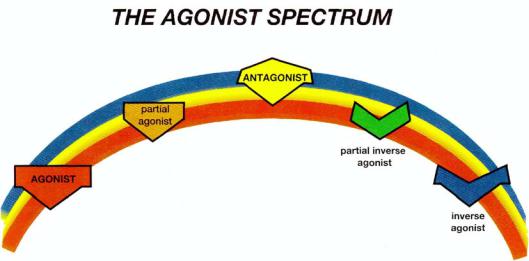
Agonists and Antagonists
Naturally occurring neurotransmitters stimulate receptors. These are called agonists. By contrast, the portfolio of options for drugs is far greater than just stimulation of receptors. In fact, a whole spectrum of possibilities exists, sometimes called the agonist spectrum (Fig. 3 — 5). Some drugs do stimulate receptors just as do the natural neurotransmitters and are therefore agonists. Other drugs actually block the actions of a natural neurotransmitter at its receptor and are called antagonists. True antagonists only exert their actions in the presence of agonist; they have no intrinsic activity of their own in the absence of agonist. Still other drugs do the opposite of what agonists do and are called inverse agonists. Thus, drugs acting at a receptor exist in a spectrum from full agonist to antagonist to inverse agonist (Fig. 3 — 5).
Examples of the actions of agonists can be taken from each of the two major molecular superfamilies. For the family of seven-transmembrane-region receptors linked to G proteins and enzymic second-messenger systems, the agonist would turn on the synthesis of second messenger to the greatest extent possible (i.e., the action
Special Properties of Receptors |
83 |
FIGURE 3-5. Shown here is the agonist spectrum. This spectrum reaches from agonists through antagonists to inverse agonists. Naturally occurring neurotransmitters are agonists. It is a common misconception that antagonists are the opposite of agonists because they block the actions of agonists. However, inverse agonists are really the opposite of agonists. Antagonists can block anything in the agonist spectrum, including inverse agonists. If an agonist is not as strong as the full agonist, it is called a partial agonist. Similarly, if an inverse agonist is partial and not as strong as a full inverse agonist, it is called a partial inverse agonist. Examples of the psychopharmacological actions of an agonist would be to reduce anxiety or to reduce pain. An inverse agonist, by analogy, would cause anxiety or pain. A partial agonist would weakly reduce anxiety or pain, and a partial inverse agonist would weakly cause anxiety or pain. An antagonist would block the full and partial agonists from reducing any anxiety or pain and would also block the full and partial inverse agonists from causing any anxiety or pain. However, an antagonist would neither reduce nor cause pain in itself.
of a full agonist). The full agonist is generally represented by the naturally occurring neurotransmitter itself, although some drugs can also act in as full a manner as the natural neurotransmitter. Often the term agonist is therefore imprecise, and the better term is full agonist.
For the family of four-transmembrane-region receptors, with multiple copies of each arranged as individual columns forming an ion channel in the center, a full agonist acts by multiple molecules of the agonist each finding the transmembrane binding site for the agonist within the receptor columns surrounding the ion channel. This in turn opens the ion channel column more completely; thus, the full agonist action (Fig. 3 — 6). At baseline the ion channel is only partially open, and a full agonist therefore opens it much more (Fig. 3 — 6).
Antagonists
Antagonists block the actions of everything in the agonist spectrum (Fig. 3 — 5). By themselves, antagonists have no intrinsic activity and therefore are sometimes referred to as "silent" (Fig. 3—7). However, in the presence of an agonist, an antagonist will block the actions of that agonist (Fig. 3-8).

84 Essential Psychopharmacology
FIGURE 3 — 6. Actions of an agonist. On the left, the ion channel is in its resting state, a balance between being opened and closed. On the right, the agonist occupies its binding site on the ligandgated ion channel receptor and as gatekeeper, opens the ion channel. This is represented as the red agonist turning the receptor red and opening the ion channel as the agonist docks into its binding site.
Inverse Agonists
Inverse agonists do the opposite of agonists. An example of their action can also be taken from receptors linked to an ion channel. By contrast to agonists and antagonists, an inverse agonist neither opens the ion channel as does an agonist (Fig. 3
— 6) nor blocks the agonist from opening the channel as does an antagonist (Fig. 3— 8); rather, it binds the neurotransmitter receptor in such a fashion as to provoke an action opposite to that of the agonist, namely, causing the receptor to close the ion channel (Fig. 3 — 9).
It might seem at first look that there is no difference between an inverse agonist and an antagonist. There is, however, a very important distinction between them. Whereas an antagonist blocks an agonist (Fig. 3 — 8), it has no particular action in the absence of the agonist, when it is thus silent (Fig. 3 — 7). An inverse agonist has an action opposite to that of an agonist (Fig. 3 — 9). Furthermore, an antagonist will actually block the action of an inverse agonist (Fig. 3 — 10), just as it will block the action of a full agonist (Fig. 3-7).

Special Properties of Receptors |
85 |
FIGURE 3 — 7. Antagonist acting alone. On the left, the ion channel is in its resting state, a balance between being open and closed. On the right, the antagonist occupies the binding site normally occupied by the agonist on the ligand-gated ion channel receptor. However, there is no consequence to this, and the ion channel neither opens further nor closes. This is represented as the yellow antagonist turning the receptor yellow and neither opening nor closing the ion channel as the antagonist docks into the binding site.
Partial Agonists
To add even more options to the actions of drugs at neurotransmitter receptors and to influence neurotransmission in even more ways, there is a class of agents known as partial agonists. A partial agonist exerts an effect similar to but weaker than that of the full agonist. Thus, in the example of the neurotransmitter system controlling an ion channel, a partial agonist would open the ion channel to a certain extent (Fig. 3 — 11) but only partially as compared with the full agonist (Fig. 3—6). Partial agonists are also blocked by antagonists (Fig. 3 — 12). It is even possible for the inverse agonists to be partial (Fig. 3 — 13). In this case, the partial inverse agonist
closes the ion channel to a lesser extent (Fig. 3 — 13) than a full inverse agonist (Fig. 3- 9).
This means that there is a spectrum of degree to which a receptor can be stimulated (Fig. 3 — 14). At one end of the spectrum, there is the full agonist, which elicits the same degree of physiologic receptor-mediated response as the natural neurotransmitter agonist itself. At the other end of the spectrum is a full inverse agonist, which in concept does the opposite of the agonist. In the middle is the

86 Essential Psychophamacology
FIGURE 3-8. Antagonist acting in the presence of an agonist. On the left, the ion channel has been opened by the agonist occupying its binding site on the ligand-gated ion channel receptor and as gatekeeper, opening the ion channel, just as in Figure 3 — 6. This is represented as the red agonist turning the receptor red and opening the ion channel as it docks into its binding site, as in Figure 3 — 6. On the right, the yellow antagonist prevails and shoves the red agonist off the binding site, reversing the agonist's actions. Since the agonist had opened the ion channel, the antagonist reverses this by partially closing the ion channel to restore the resting state. Thus, the ion channel has been caused to return to its status before the agonist acted.
antagonist, which blocks the effects of all participants in the spectrum but has no properties of its own in changing the ion channel.
The spectrum thus goes from full agonist to partial agonist to antagonist to partial inverse agonist to full inverse agonist (Fig. 3 — 14). Although this concept of agonists, antagonists, and partial agonists is well developed for several neurotransmitter systems, there are relatively few examples of inverse agonists.
Light and Dark as an Analogy for Partial Agonists
It was originally considered that a neurotransmitter could act at the receptor as does a light switch, to turn it on or off. We now know that the synapse and its receptors can function rather more like a rheostat. That is, a full agonist will turn the lights all the way on (Fig. 3 — 15), but a partial agonist will only turn the light on partially (Fig. 3 — 16). If neither full agonist nor partial agonist is present, the room is dark (Fig. 3-17).

Special Properties of Receptors |
87 |
FIGURE 3 — 9. Actions of an inverse agonist. On the left, the ion channel is in its resting state, a balance between being opened and closed. On the right, the inverse agonist occupies the binding site on the ligand-gated ion channel receptor as gatekeeper, closes the ion channel. This is the opposite of what the agonist does (cf. Fig. 3 — 6). Inverse agonist is represented as the light blue inverse agonist turning the receptor light blue and closing the ion channel as the inverse agonist docks into its binding site.
Each partial agonist has its own set point engineered into the molecule, so that it cannot make lights brighter with a higher dose. No matter how much partial agonist is given, only a certain degree of brightness will result. A series of partial agonists will differ one from the other in degree of partiality, so that theoretically all degrees of brightness can be covered within the range from "off" to "on," but each partial agonist is associated with its own unique degree of brightness.
What is so interesting about partial agonists is that they can appear as net agonists or as net antagonists, depending on the amount of naturally occurring full agonist neurotransmitter that is present. Take, for example, the case of neurotransmitters controlling an ion channel. When no full agonist neurotransmitter is present, a partial agonist will be a net agonist, that is, it will open the channel from its resting state (Fig. 3 — 18). However, when a full agonist neurotransmitter agonist is present, the same partial agonist will become a net antagonist, it will close the channel from its full agonist state (Fig. 3 — 18). Thus, a partial agonist can simultaneously boost deficient neurotransmitter activity yet block excessive neurotransmitter activity (Fig. 3-18).

88 Essential Psychopharmacology
FIGURE 3 — 10. Antagonist acting in the presence of an inverse agonist. On the left, the ion channel has been closed by the inverse agonist occupying the binding site on the ligand-gated ion channel receptor and as gatekeeper, closing the ion channel, just as shown in Figure 3—9. This is represented as the light blue inverse agonist turning the receptor light blue and closing the ion channel as it docks into its binding site, as in Figure 3-9. On the right, the yellow antagonist prevails and shoves the light blue inverse agonist off the binding site, reversing the inverse agonist's actions. Since the inverse agonist had previously closed the ion channel, the antagonist reverses this closing by opening the ion channel to restore the resting state. This causes the ion channel to return to its status before the agonist acted. In this way, the antagonist's effect on an inverse agonist's actions is similar to that on an agonist's actions, namely, it returns the ion channel to its resting state (cf. Fig. 3 — 8). However, in the case of an inverse agonist, the antagonist opens the channel, whereas in the case of an agonist, the same antagonist closes the channel (cf. Figs. 3 — 8 and 3 — 10). Thus, an antagonist can reverse either an agonist or an inverse agonist despite the fact that it does nothing on its own (Fig. 3-7).
Returning to the light switch analogy, a room will be dark when agonist is missing and the light switch is off (Fig. 3 — 17). A room will be brightly lighted when it is full of natural full agonist and the light switch is fully on (Fig. 3 — 15). Adding partial agonist to the dark room where there is no natural full agonist neurotransmitter will turn the lights up, but only as far as the partial agonist works on the rheostat (Figure 3 — 16). Relative to the dark room as a starting point, a partial agonist acts therefore as a net agonist. On the other hand, adding a partial agonist to the fully lighted room will have the effect of turning the lights down to a level of lower brightness on the rheostat (Fig. 3 — 16). This is a net antagonistic effect relative to the fully lighted room. Thus, after adding partial agonist to the dark room and to the brightly lighted room, both rooms will be equally lighted.