

3 курс / Фармакология / Essential_Psychopharmacology_2nd_edition
.pdfDepression and Bipolar Disorders |
179 |
FIGURE 5—47. Two types of norepinephrine interaction with serotonin are shown here. In the brainstem, a pathway from locus coeruleus to raphe interacts with serotonergic cell bodies there and accelerates serotonin release. A second noradrenergic pathway to target areas in the cortex also interacts with serotonin axon terminals there and brakes serotonin release.
Classical Antidepressants and the Monoamine Hypothesis
The first antidepressants to be discovered came from two classes of agents, namely, the tricyclic antidepressants, so named because their chemical structure has three rings, and the MAO inhibitors, so named because they inhibit the enzyme MAO, which destroys monoamine neurotransmitters. When tricyclic antidepressants block the NE transporter, they increase the availability of NE in the synapse, since the "vacuum cleaner" reuptake pump can no longer sweep NE out of the synapse (Figs. 5 — 16 and 5 — 18). When tricyclic antidepressants block the DA pump (Fig. 5 — 32) or the 5HT pump (Fig. 5 — 35), they similarly enhance the synaptic availability of DA or 5HT, respectively, and by the same mechanism. When MAO inhibitors block NE, DA, and 5HT breakdown, they boost the levels of these neurotransmitters (Fig. 5- 15).
Since it was recognized by the 1960s that all the classical antidepressants boost NE, DA, and 5HT in one manner or another (Figs. 5-15 and 5 — 16), the original idea was that one or another of these neurotransmitters, also chemically known as monoamines, might be deficient in the first place in depression (Fig. 5 — 14). Thus, the "monoamine hypothesis" was born. A good deal of effort was expended, especially in the 1960s and 1970s, to identify the theoretically predicted deficiencies of the monoamine neurotransmitters. This effort to date has unfortunately yielded mixed and sometimes confusing results.
Some studies suggest that NE metabolites are deficient in some patients with depression, but this has not been uniformly observed. Other studies suggest that

180 Essential Psychopharmacology
FIGURE 5—48. A schematic representation of both the excitatory and inhibitory actions of norepinephrine on serotonin release is shown here. This is the same action shown in Figure 5—47.
the 5HT metabolite 5-hydroxy-indole acetic acid (5HIAA) is reduced in the cerebrospinal fluid (CSF) of depressed patients. On closer examination, however, it has been found that only some of the depressed patients have low CSF 5HIAA, and these tend to be the patients with impulsive behaviors, such as suicide attempts of a violent nature. Subsequently, it was also reported that CSF 5HIAA is decreased in other populations who were subject to violent outbursts of poor impulse control but were not depressed, namely, patients with antisocial personality disorder who were arsonists, and patients with borderline personality disorder with self-destructive behaviors. Thus, low CSF 5HIAA may be linked more closely with impulse control problems than with depression.
Another problem with the monoamine hypothesis is the fact that the timing of antidepressant effects on neurotransmitters is far different from the timing of the antidepressant effects on mood. That is, antidepressants boost monoamines immedi-

FIGURE 5—49. A key postsynaptic regulatory receptor is the 5HT2A receptor.
FIGURE 5-50. When the postsynaptic 5HT2A receptor of Figure 5-49 is occupied by 5HT, it causes neuronal impulses in the postsynaptic neuron to be transduced via the production of second messengers.
181
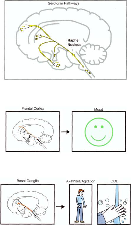
FIGURE 5 — 51. The headquarters for the cell bodies of serotonergic neurons is in the brainstem area called the raphe nucleus.
FIGURE 5 — 52. Serotonergic projections from raphe to frontal cortex may be important for regulating mood.
FIGURE 5 — 53. Serotonergic projections from raphe to basal ganglia may help control movements as well as obsessions and compulsions.
182

FIGURE 5 — 54. Serotonergic projections from raphe to limbic areas may be involved in anxiety and panic.
FIGURE 5 — 55. Serotonergic projections to the hypothalamus may regulate appetite and eating behavior.
Sleep centers |
|
Insomnia |
FIGURE 5 — 56. Serotonergic neurons in brainstem sleep centers regulate sleep, especially slow-wave sleep.
183

FIGURE 5 — 57. Serotonergic neurons descending down the spinal cord may be responsible for controlling certain spinal reflexes that are part of the sexual response, such as orgasm and ejaculation.
FIGURE 5 — 58. The chemoreceptor trigger zone in the brainstem can mediate vomiting, especially via 5HT3 receptors.
FIGURE 5-59. Peripheral 5HT3 and 5HT4 receptors in the gut may regulate appetite as well as other gastrointestinal functions, such as gastrointestinal motility.
184

Depression and Bipolar Disorders |
185 |
Table 5 — 23. Serotonin deficiency syndrome
Depressed mood
Anxiety
Panic
Phobia
Anxiety
Obsessions and compulsions
Food craving; bulimia
FIGURE 5-60. The monoamine receptor hypothesis of depression posits that something is wrong with the receptors for the key monoamine neurotransmitters. Thus, according to this theory, an abnormality in the receptors for monoamine neurotransmitters leads to depression. Such a disturbance in neurotransmitter receptors may be caused by depletion of monoamine neurotransmitters, by abnormalities in the receptors themselves, or by some problem with signal transduction of the neurotransmitter's message from the receptor to other downstream events. Depicted here is the normal monoamine neuron with the normal amount of monoamine neurotransmitter and the normal amount of correctly functioning monoamine receptors.
ately, but as mentioned earlier, there is a significant delay in the onset of their therapeutic actions, which in fact occurs many days to weeks after they have already boosted the monoamines. Because of these and other difficulties, the focus of hypotheses for the etiology of depression began to shift from the monoamine neurotransmitters themselves to their receptors. As we shall see, contemporary theories have shifted past the receptors to the molecular events that regulate gene expression.
Neurotransmitter Receptor Hypothesis
The neurotransmitter receptor theory posits that something is wrong with the receptors for the key monoamine neurotransmitters (Figs. 5—60 through 5—62). According to this theory, an abnormality in the receptors for monoamine neurotransmitters leads to depression (Fig. 5—62). Such a disturbance in neurotransmitter receptors may itself be caused by depletion of monoamine neurotransmitters (Fig. 5-61).

186 Essential Psychopharmacology
FIGURE 5 — 61. In this figure, monoamine neurotransmitter is depleted (see red circle), just as previously shown in Figure 5 — 14.
FIGURE 5 — 62. The consequences of monoamine neurotransmitter depletion, of stress, or of some inherited abnormality in neurotransmitter receptor could cause the postsynaptic receptors to abnormally up-regulate (indicated in red circle). This up-regulation or other receptor dysfunction is hypothetically linked to the cause of depression.
Depletion of monoamine neurotransmitters (cf. Fig. 5 — 60 and Fig. 5—61) has already been discussed as the central theme of the monoamine hypothesis of depression (see Figs. 5 — 13 and 5 — 14). The neurotransmitter receptor hypothesis of depression takes this theme one step further—namely, that the depletion of neurotransmitter causes compensatory up regulation of postsynaptic neurotransmitter receptors (Fig. 5 — 62).
Direct evidence of this is generally lacking, but postmortem studies do consistently show increased numbers of serotonin 2 receptors in the frontal cortex of patients who commit suicide. Indirect studies of neurotransmitter receptor functioning in patients with major depressive disorders suggest abnormalities in various neurotransmitter receptors when using neuroendocrine probes or peripheral tissues such as platelets or lymphocytes. Modern molecular techniques are exploring for abnormalities in gene expression of neurotransmitter receptors and enzymes in families with depression but have not yet been successful in identifying molecular lesions.
Depression and Bipolar Disorders |
187 |
The Monoamine Hypothesis of Gene Expression
So far, there is no clear and convincing evidence that monoamine deficiency accounts for depression; that is, there is no "real" monoamine deficit. Likewise, there is no clear and convincing evidence that excesses or deficiencies of monoamine receptors account for depression; that is, there is no pseudomonoamine deficiency due to the monoamines being there but not the monoamine receptors. On the other hand, there is growing evidence that despite apparently normal levels of monoamines and their receptors, these systems do not respond normally. For instance, probing monoaminergic receptors with drugs that stimulate them can lead to deficient output of neuroendocrine hormones. It can also lead to deficient changes in neuronal firing rates, as demonstrated on positron emission tomography (PET).
Such observations have led to the idea that depression may be a pseudomonoamine deficiency due to a deficiency in signal transduction from the monoamine neurotransmitter to its postsynaptic neuron in the presence of normal amounts of neurotransmitter and receptor. If there is a deficiency in the molecular events that cascade from receptor occupancy by neurotransmitter, it could lead to a deficient cellular response and thus be a form of pseudomonoamine deficiency (i.e., the receptor and the neurotransmitter are normal, but the transduction of the signal from neurotransmitter to its receptor is somehow flawed). Such a deficiency in molecular functioning has been described for certain endocrine diseases such as hypoparathy-roidism (parathyroid hormone deficiency), pseudohypoparathyroidism (parathyroid receptors deficient but parathyroid hormone levels normal), and pseudo- pseudohy-poparathyroidism (signal transduction deficiency leading to hypoparathyroid clinical state despite normal levels of hormone and receptor).
Perhaps a similar situation exists for depression due to a hypothesized problem within the molecular events distal to the receptor. Thus, second messenger systems leading to the formation of intracellular transcription factors that control gene regulation could be the site of deficient functioning of monoamine systems. This is the subject of much current research into the potential molecular basis of affective disorders. This hypothesis suggests some form of molecularly mediated deficiency in monoamines that is distal to the monoamines themselves and their receptors despite apparently normal levels of monoamines and numbers of monoamine receptors.
One candidate mechanism that has been proposed as the site of a possible flaw in signal transduction from monoamine receptors is the target gene for brain-derived neurotrophic factor (BDNF). Normally, BDNF sustains the viability of brain neurons, but under stress, the gene for BDNF is repressed (Fig. 5 — 63), leading to the atrophy and possible apoptosis of vulnerable neurons in the hippocampus when their BDNF is cut off (Fig. 5—64). This in turn leads to depression and to the consequences of repeated depressive episodes, namely, more and more episodes and less and less responsiveness to treatment. This possibility that hippocampal neurons are decreased in size and impaired in function during depression is supported by recent clinical imaging studies showing decreased brain volume of related structures. This provides a molecular and cellular hypothesis of depression consistent with a mechanism distal to the neurotransmitter receptor and involving an abnormality in gene expression. Thus, stress-induced vulnerability decreases the expression of genes that make neurotrophic factors such as BDNF critical to the survival and function of key neurons (Fig. 5-63). A corollary to this hypothesis is that antidepressants act to

188 Essential Psychopharmacology
FIGURE 5 — 63. The monoamine hypothesis of gene action in depression, part 1. One candidate mechanism that has been proposed as the site of a possible flaw in signal transduction from monoamine receptors is the target gene for brain-derived neurotrophic factor (BDNF). Normally, BDNF sustains the viability of brain neurons. Shown here, however, is the gene for BDNF under situations of stress. In this case, the gene for BDNF is repressed, and BDNF is not being synthesized.
reverse this by causing the genes for neurotrophic factors to be activated (see Chapter 6).
Neurokinin Hypothesis of Emotional Dysfunction
Another hypothesis for the pathophysiology of depression and other states of emotional dysfunction relates to the actions of a relatively new class of peptide neurotransmitters known as neurokinins (also sometimes called tachykinins). This hypothesis was generated by some rather serendipitous observations that an antagonist to one of the neurokinins, namely substance P, may have antidepressant actions. Classically, substance P was thought to be involved in the pain response because it is released from neurons in peripheral tissues in response to inflammation, causing "neurogenic" inflammation and pain (Fig. 5 — 65). Furthermore, substance P is present in spinal pain pathways, suggesting a role in central nervous systemmediated pain (Fig. 5 — 65). Unfortunately, however, antagonists to substance P's receptors have so far been unable to reduce neurogenic inflammation or pain of many types in human testing. On the other hand, suggestions that substance P antagonists may have improved mood, if not pain, in migraine patients led to controlled trials of such drugs in patients with depression. Although these are still early days and not all studies confirm antidepressant effects of substance P antagonists, the possibility that such