

3 курс / Фармакология / Essential_Psychopharmacology_2nd_edition
.pdfChemical Neurotransmission as the Mediator of Disease Actions |
119 |
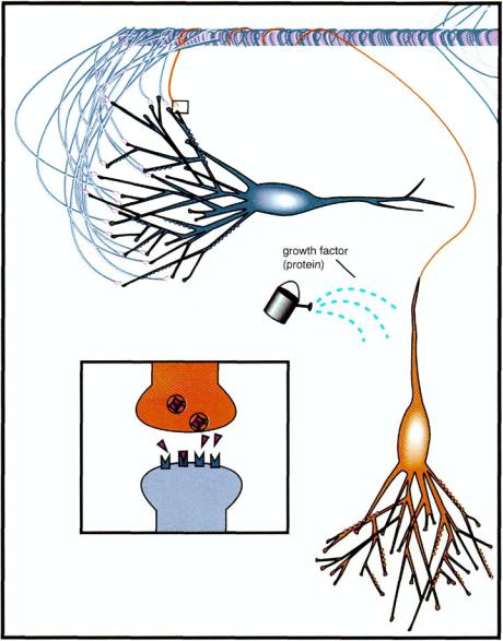
FIGURE 4—12. Shown here and in Figure 4 — 13 is a conceptually more complex mechanism of compensation for the loss of a degenerating neuron. The ailing but not yet degenerated red neuron indicated here is no longer functioning to allow normal neurotransmission with the blue neuron (see box) and is about to die. Also indicated is the application of a growth factor to the degenerating neuron. This could be conceived as either a natural reparative mechanism that the dying neuron could activate (see Fig. 1—22 and Table 1 — 3) or a drug that could mimic this.
of systemically administered neurotrophic factors may be required if treatment is going to be safe.
To complicate the potential utility of growth factors for neurodegenerative disorders is that fact that many growth factors are large protein or peptide molecules,
120 Essential Psychopharmacology
FIGURE 4—13. This figure demonstrates how a degenerating neuron might be rescued by a growth factor. In this case, the dying neuron of Figure 4—12 is salvaged by a growth factor, which restores the function of neurotransmission to reactivate normal communications between the red neuron and the blue neuron (see box).
which are unable to survive intact when administered orally and unable to cross the blood-brain barrier when administered intravenously. This has led to several different approaches to delivering neurotrophic factors to their desired targets in the CNS.
First, the protein itself can be infused directly into the cerebrospinal fluid or implanted in a biodegradable, slow-release preparation. Second, the active protein can travel across the blood-brain barrier by hiding inside a "Trojan horse" molecule

FIGURE 4—14. Transplantation of a new neuron by neurosurgical techniques is another potential mechanism for replacing the function of a degenerated neuron. In this case, the turquoise transplanted neuron makes the same neurotransmitter as the formerly red neuron made (see Fig. 4—11) prior to degenerating here. Synaptic neurotransmission is restored when the transplanted neuron takes over the lost function of the degenerated neuron (see box). This has already been performed for patients with Parkinson's disease, in which transplanted fetal substantia nigra neurons can successfully improve functional neurotransmission of degenerated substantia nigra neurons in some patients.
121
122Essential Psychopharmacology
that is normally translocated across this blood-brain barrier. Third, low molecular weight chemicals might be able to get into the brain and pharmacologically induce the formation of a trophic factor. This action, in fact, has been suggested for cholinesterase inhibitors, which not only increase acetylcholine levels but subsequently increase nerve growth factor. Finally, a high-tech idea is to transfer genes that produce the trophic factor directly into the brain by grafting cells that normally make it, by genetically engineering cells to make it, or by delivering the gene in a carrier virus. All of these possibilities are under active investigation.
A third long-term therapeutic approach to neurodegenerative disorders is transplantation of neurons. Neuronal transplantation is being investigated as a way to substitute new neurons for degenerated neurons (Fig. 4—14). This is not a Frankenstein-style transplant of an entire brain but rather a selective introduction of specific and highly specialized nerves, which produce specialized chemicals and neurotransmitters capable of compensating for and replacing the functions of the degenerated and destroyed neurons that caused disease in the first place. Transplantation of neurons into human brain is already occurring in Parkinson's disease, where dopamine-producing neurons have been successfully transplanted into the brains of patients with this condition. Experimental use of cholinergic neurons holds promise for the treatment of experimental models of Alzheimer's disease.
From Excitement to Brain Burn: Too Much Excitatory Neurotransmission Could Be Hazardous to Your Health
If Benjamin Franklin said "nothing in excess, including moderation" he may have anticipated contemporary thinking about excitatory neurotransmission. Excitatory neurotransmission with glutamate ranges from talking to neurons (Fig. 4—15), to screaming at them (Fig. 4—16), to strangling their dendrites, and even to assassinating them (Fig. 4—17).
Glutamate normally opens an ion channel so that the nerve can drink calcium (Figs. 4—15 and 4—18). Sipping calcium is exciting to a neuron and a normal reaction when glutamate is speaking pleasantly. However, when glutamate screams at a neuron, the neuron reacts by drinking more calcium (Figs. 4—16 and 4—19). Imbibing too much calcium may lead in part to excitatory symptoms such as panic, seizures, mania, or psychosis (Figs. 4—19 and 4—20). Too much calcium eventually will anger intracellular enzymes, which then generate nasty chemicals called free radicals. A small commune of free radicals can crash the chemical party in the postsynaptic dendrite and strangle it (Fig. 4 — 21). A mob of free radicals can kill the whole neuron, perhaps by triggering apoptosis (Fig. 4—22; see also Fig. 1 — 18).
Why would the neuron allow this to happen? It is possible that the brain needs this excitotoxic mechanism so that glutamate can act as a gardener in the brain, pruning worn out branches from dendritic trees so that healthy new sprouts may prosper (Fig. 1—23). However, this also equips the neuron with a powerful weapon, which can potentially be misused to cause various neurodegenerative conditions due literally to pruning neurons to death (Fig. 4—22). Such an excitotoxic mechanism could be activated if the genetic program controlling it is turned on or potentially by ingestion of toxins or toxic drugs of abuse. That is, when glutamate decides to act as an abusive bully for whatever reason, neurons may seize, panic, become manic, or become psychotic (Fig. 4—20). Furthermore, such symptoms of calcium intoxi-

FIGURE 4-15. The calcium ion is a key regulator of neuronal excitability and is constantly entering and leaving neurons through ion channels of various sorts that are conducting the normal business functions of the neuron. When this occurs at a normal rate, it modifies neuronal excitability but is not damaging to the neuron (but see Figs. 4 — 16 and 4—17).
FIGURE 4—16. Calcium may also rush into cells too quickly if its ion channels are opened too much, as is postulated to occur as a result of certain toxins, by stroke, or by neurodegenerative conditions (see Fig. 4—17).
FIGURE 4—17. If too much calcium gets into the neuron and overwhelms any sinks and buffers there, it can destroy the neuron and cause it to degenerate and die. This mechanism of excessive excitation is called excitotoxicity and is a major current hypothesis of the cause of various psychiatric and neurological disorders. This idea postulates that for such diseases, neurons are literally "excited to death."

124 Essential Psychopharmacology
FIGURE 4—18. Shown here are details of calcium entering a dendrite of the blue neuron when the red neuron excites it with glutamate during normal excitatory neurotransmission. This was shown in a more simplistic model in Figure 4—15. Glutamate released from the red neuron travels across the synapse, docks into its agonist slot on its receptor, and as ionic gatekeeper, opens the calcium channel to allow calcium to enter the postsynaptic dendrite of the blue neuron to mediate normal excitatory neurotransmission (see box).
cation may be followed by an unfortunate glutamate hangover in the form of destroyed dendrites, which can never be excited again (Fig. 4—21).
Other illnesses such as Alzheimer's disease, Parkinson's disease, amytrophic lateral sclerosis (Lou Gehrig's disease), and even schizophrenia may hire glutamate as a

FIGURE 4—19. Shown here is what may happen when excitatory neurotransmission causes too much neurotransmission. This may possibly occur during the production of various symptoms mediated by the brain, including panic attacks. It could also occur during mania, positive symptoms of psychosis, seizures, and other neuronally-mediated disease symptoms. In this case, too much glutamate is being released by the red neuron, causing too much excitation of the postsynaptic blue neuron's dendrite. Extra release of glutamate causes additional occupancy of postsynaptic glutamate receptors, opening more calcium channels and allowing more calcium to enter the blue dendrite (see box). Although this degree of excessive neurotransmission may be associated with psychiatric symptoms, it does not actually damage the neuron (but see Figs. 4 —20 and 4—21).
125

126 Essential Psychopharmacology
FIGURE 4 —20. This figure represents the concept of an electrical storm in the brain in which overexcitation and too much neurotransmission are occurring during the production of various psychiatric symptoms, including those which occur during a panic attack. This may also be a model for other disorders of excessive behavioral symptoms that imply too much neurotransmission, including mania, positive manifestation of psychosis, and seizures.
methodical undercover assassin, eliminating a whole subpopulation of predesignated neurons over a prolonged period of time. Such a systematic process would be consistent with the pace of these slow neurodegenerative disorders. In catastrophic brain diseases such as stroke and global ischemia associated with cardiac arrest, drowning, etc., a whole army of glutamate "hit men" may be hired as mass murderers. In this case, glutamate causes the massacre of an entire region of brain neurons by suddenly subjecting them to molecular mayhem.
Thus, glutamate's actions can range across a vast spectrum. It can be a friendly neuronal conversationalist or a screaming hypothetical mediator of neurological and psychiatric disorders. How might the symptoms and clinical course of various psychiatric disorders fit this model of excitotoxicity? Psychosis possibly shares some analogies with a seizure, in that excessive transmission of dopamine in the mesolimbic areas of brain may lead to symptoms of delusions, hallucinations, and thought disorder in various psychiatric disorders. Panic disorder may be analogous to a seizure in areas of the brain controlling emotions (such as the parahippocampal gyrus), leading to clinical symptoms characterized by a massive emotional discharge of panic, shortness of breath, chest pain, dizziness, feelings of impending death, or fear of losing control. Thus, disorders such as psychosis, epilepsy and panic disorder appear to involve excessive neurotransmission, which may help explain the mechanism by which they produce acute symptoms (Figs. 4—19 and 4—20).
Furthermore, these disorders seem to become more resistant to treatment the longer the disorder persists and the more poorly the symptoms are controlled, as if there were an underlying mechanism of destruction accompanying symptoms that are out of control (Figs. 4—21 through 4—23). Thus, excessive neurotransmission may itself be a cause of deficient neurotransmission. If seizures beget seizures, panic

FIGURE 4 — 21. If too much neurotransmission occurs for too long, it is hypothetically possible that this would lead to dendritic death. The mechanism for this may be tantamount to inappropriately activating the normal dendritic pruning process (indicated schematically as scissors snipping off the dendrite; see Figure 1 — 23 for a diagram of normal pruning). Thus, far too much glutamate release can cause too much opening of the gates of the calcium channel, activating an excitotoxic demise of the dendrite (see box).
127

128 Essential Psychopharmacology
FIGURE 4—22. Neurons appear to have a normal maintenance mechanism for their dendritic tree by which they are able to prune, or remove, old, unused, or useless synapses and dendrites (normal mechanism shown in Fig. 1—23). One postulated mechanism for some degenerative diseases is that this otherwise normal pruning mechanism may get out of control, eventually rendering the neuron useless or even killing it by "pruning it to death."
begets panic, psychosis begets psychosis, and mania begets mania, these symptoms are obviously not good for the brain. The psychopharmacologist must therefore act to prevent symptoms, not only because symptom control may harness the disruptive influences of excessive neurotransmission on behavior, but also because symptom control may ultimately prevent the demise of the neurons mediating these very behaviors (Figs. 4—20 to 4—23). If these disorders of excessive neurotransmission are analogous to the brain "burning" during symptomatic crises such as a seizure, psychosis, panic attack, or mania, treatments might not only "put out the fire" but also salvage the underlying neuronal substrates, which are burning as the fuel for the fire.