

3 курс / Фармакология / Essential_Psychopharmacology_2nd_edition
.pdfDepression and Bipolar Disorders |
159 |
FIGURE 5-18. Norepinephrine (NE) can also be destroyed by enzymes in the NE neuron. The principal destructive enzymes are monoamine oxidase (MAO) and catechol-O-methyl transferase (COMT). The action of NE can be terminated not only by enzymes that destroy NE, but also by a transport pump for NE, called the norepinephrine transporter, which prevents NE from acting in the synapse without destroying it. This transport pump is separate and distinct from the transport pump for tyrosine used in carrying tyrosine into the NE neuron for NE synthesis (see Fig. 5 — 17). The transport pump that terminates the synaptic action of NE is sometimes called the "NE transporter" and sometimes the "NE reuptake pump." There are molecular differences among the transporters for the NE, dopamine, and serotonin neurons. These differences can be exploited by drugs so that the transport of one monoamine can be blocked independently of another. The NE transporter is part of the presynaptic machinery, where it acts as a "vacuum cleaner," whisking NE out of the synapse, and off the synaptic receptors and stopping its synaptic actions. Once inside the presynaptic nerve terminal, NE can either be stored again for subsequent reuse when another nerve impulse arrives, or it can be destroyed by enzymes.
beta 2. More recently, adrenergic receptors have been even further subclassified on the basis of both pharmacologic and molecular differences.
For a general understanding of NE receptors, the reader should begin with an awareness of three key receptors that are postsynaptic, namely beta 1, alpha 1, and alpha 2 receptors (Fig. 5 — 19). The postsynaptic receptors for NE convert occupancy of an alpha 1, alpha 2, or beta 1 receptor into a physiological function and ultimately result in changes in gene expression in the postsynaptic neuron.
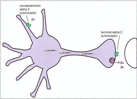
On the other hand, alpha 2 receptors are the only presynaptic noradrenergic receptors on noradrenergic neurons. They regulate NE release and so are called autoreceptors. Presynaptic alpha 2 autoreceptors are located both on the axon terminal,

160 Essential Psychopharmacology
FIGURE 5 — 19. The noradrenergic neuron is regulated by a multiplicity of receptors for NE. Pictured here are the NE transporter and several NE receptors, including the presynaptic alpha 2 autoreceptor as well as the postsynaptic alpha 1, alpha 2 and beta 1 adrenergic receptors. The presynaptic alpha 2 receptor is important because it is an autoreceptor. That is, when the presynaptic alpha 2 receptor recognizes synaptic NE, it turns off further release of NE. Thus, the presynaptic alpha 2 terminal autoreceptor acts as a brake for the NE neuron. Stimulating this receptor (i.e., stepping on the brake) stops the neuron from firing. This probably occurs physiologically to prevent too much firing of the NE neuron, since it can shut itself off once the firing rate gets too high and the autoreceptor becomes stimulated. Postsynaptic NE receptors generally act by recognizing when NE is released from the presynaptic neuron and react by setting up a molecular cascade in the postsynaptic neuron, thereby causing neurotransmission to pass from the presynaptic to the postsynaptic neuron.
(terminal alpha 2 receptors) (Fig. 5 — 19) and at the cell body (soma) and nearby dendrites (somatodendritic alpha 2 receptors) (Fig. 5—20). Presynaptic alpha 2 receptors are important because both the terminal and the somatodendritic re-ceptors are autoreceptors. That is, when presynaptic alpha 2 receptors recognize NE, they turn off further release of NE (Figs. 5—21 and 5—22). Thus, presynaptic alpha 2 autoreceptors act as a brake for the NE neuron and also cause what is known as a negative feedback regulatory signal. Stimulating this receptor (i.e., stepping on the brake) stops the neuron from firing. This probably occurs physiologically to prevent
Depression and Bipolar Disorders |
161 |
FIGURE 5 — 20. Both types of presynaptic alpha 2 autoreceptors are shown here. They are located either on the axon terminal, where they are called terminal alpha 2 receptors, or at the cell body (soma) and nearby dendrites, where they are called somatodendritic alpha 2 receptors.
overfiring of the NE neuron, since it can shut itself off once the firing rate gets too high and the autoreceptor becomes stimulated. It is worthy of note that not only can drugs mimic the natural functioning of the NE neuron by stimulating the presynaptic alpha 2 neuron, but drugs that antagonize this same receptor will have the effect of cutting the brake cable and enhancing the release of NE
Most of the cell bodies for noradrenergic neurons in the brain are located in the brainstem in an area known as the locus coeruleus (Fig. 5 — 23). The principal function of the locus coeruleus is to determine whether attention is being focused on the external environment or on monitoring the internal milieu of the body. It helps to prioritize competing incoming stimuli and fixes attention on just a few of these. Thus, one can either react to a threat from the environment or to signals such as pain coming from the body. Where one is paying attention will determine what one learns and what memories are formed as well.
Norepinephrine and the locus coeruleus are also thought to have an important input into the central nervous system's control of cognition, mood, emotions, movements, and blood pressure. Malfunction of the locus coeruleus is hypothesized to underlie disorders in which mood and cognition intersect, such as depression, anxiety, and disorders of attention and information processing. A norepinephrine deficiency syndrome (Table 5 — 21) is theoretically characterized by impaired attention, problems in concentrating, and difficulties specifically with working me-mory and the speed of information processing, as well as psychomotor retardation, fatigue, and

162 Essential Psychopharmacology
FIGURE 5 — 21. Presynaptic alpha 2 receptors are important because when they recognize NE, they turn off further release of NE. Shown here is the function of presynaptic somatodendritic autoreceptors, namely to act as a brake for the NE neuron and also to cause what is known as a negative feedback regulatory signal. Stimulating this receptor (i.e., "stepping on the brake") stops the neuron from firing. This probably occurs physiologically to prevent excessive firing of the NE neuron, since NE can shut itself off once the firing rate gets too high and the autoreceptor becomes stimulated.
apathy. Such symptoms can commonly accompany depression as well as other disorders with impaired attention and cognition, such as attention deficit disorder, schizophrenia, and Alzheimer's disease.
There are many specific noradrenergic pathways in the brain, each mediating a different physiological function. For example, one projection from the locus coeruleus to frontal cortex is thought to be responsible for the regulatory actions of NE on mood (Fig. 5 — 24); another projection to prefrontal cortex mediates the effects of NE on attention (Fig. 5—25). Different receptors may mediate these differential effects of norepinephrine in frontal cortex, postsynaptic beta 1 receptors for mood (Fig. 5—24) and postsynaptic alpha 2 for attention and cognition (Fig. 5 — 25).
The projection from the locus coeruleus to limbic cortex may regulate emotions, as well as energy, fatigue, and psychomotor agitation or psychomotor retardation (Fig. 5 — 26). A projection to the cerebellum may regulate motor movements, especially tremor (Fig. 5—27). Brainstem norepinephrine in cardiovascular centers controls blood pressure (Fig. 5—28). Norepinephrine from sympathetic neurons leaving the spinal cord to innervate peripheral tissues control heart rate (Fig. 5—29) and bladder emptying (Fig. 5 — 30).

Depression and Bipolar Disorders |
163 |
FIGURE 5 — 22. Shown here is the action of the presynaptic axon terminal alpha 2 receptors, which have the same function as the somatodendritic autoreceptors shown in Figure 5 — 21.
Dopaminergic neurons. Dopaminergic neurons utilize the neutotransmitter DA, which is synthesized in dopaminergic nerve terminals by two out of three of the same enzymes that also synthesize NE (Fig. 5 — 31). However, DA neurons lack the third enzyme, namely, dopamine beta hydroxylase, and thus cannot convert DA to NE. Therefore, it is DA that is stored and used for neurotransmitting purposes.
The DA neuron has a presynaptic transporter (reputake pump), which is unique for DA neurons (Fig. 5 — 32) but works analogously to the NE transporter (Fig. 5 — 33). On the other hand, the same enzymes that destroy NE (Fig. 5 — 18) also destroy DA (MAO and COMT) (Fig. 5-31).
Receptors for dopamine also regulate dopaminergic neurotransmission (Fig. 5 — 33). A plethora of dopamine receptors exist, including at least five pharmacological subtypes and several more molecular isoforms. Perhaps the most extensively investigated dopamine receptor is the dopamine 2 receptor, as it is stimulated by dopaminergic agonists for the treatment of Parkinson's disease and blocked by dopamine antagonist antipsychotics for the treatment of schizophrenia. Dopamine 1, 2, 3, and 4 receptors are all blocked by some atypical antipsychotic drugs, but it is not clear to what extent dopamine 1, 3, or 4 receptors contribute to the clinical properties of these drugs. Dopamine receptors can be presynaptic, where they function as autoreceptors. They provide negative feedback input, or a braking action on the release of dopamine from the presynaptic neuron. (Fig. 5 — 33).
Serotonergic neurons. Analogous enzymes, transport pumps, and receptors exist in the 5HT neuron (Figs. 5 — 34 through 5—42). For synthesis of serotonin in serotonergic

164 Essential Psychopharmacology
FIGURE 5 — 23. Most of the cell bodies for noradrenergic neurons in the brain are located in the brainstem in an area known as the locus coeruleus. This is the headquarters for most of the important noradrenergic pathways mediating behavior and other functions such as cognition, mood, emotions, and movements. Malfunction of the locus coeruleus is hypothesized to underlie disorders in which mood and cognition intersect, such as depression, anxiety, and disorders of attention and information processing.
Table 5 — 21. Norepinephrzne deficiency syndrome
Impaired attention Problems concentrating Deficiencies in working memory Slowness of information processing Depressed mood Psychomotor retardation Fatigue
neurons, however, a different amino acid, tryptophan, is transported into the brain from the plasma to serve as the 5HT precursor (Fig. 5 — 34). Two synthetic enzymes then convert tryptophan into serotonin: first tryptophan hydroxylase converts tryptophan into 5-hydroxytryptophan, which is then converted by aromatic amino acid decarboxylase into 5HT (Fig. 5-34). Like NE and DA, 5HT is destroyed by MAO and converted into an inactive metabolite (Fig. 5 — 35). Also, the 5HT neuron has a presynaptic transport pump for serotonin called the serotonin transporter (Fig. 5 — 35), which is analogous to the NE transporter in NE neurons (Fig. 5 — 18) and to the DA transporter in DA neurons (Fig. 5 — 32).
Receptor subtyping for the serotonergic neuron has proceeded at a very rapid pace, with several major categories of 5HT receptors, each further subtyped

FIGURE 5-24. Some noradrenergic projections from the locus coeruleus to frontal cortex are thought to be responsible for the regulatory actions of norepinephrine on mood. Beta 1 postsynaptic receptors may be important in transducing noradrenergic signals regulating mood in postsynaptic targets.
FIGURE 5-25. Other noradrenergic projections from the locus coeruleus to frontal cortex are thought to mediate the effects of norepinephrine on attention, concentration, and other cognitive functions, such as working memory and the speed of information processing. Alpha 2 postsynaptic receptors may be important in transducing postsynaptic signals regulating attention in postsynaptic target neurons.
FIGURE 5 — 26. The noradrenergic projection from the locus coeruleus to limbic cortex may mediate emotions, as well as energy, fatigue, and psychomotor agitation or psychomotor retardation.
165

Cerebellum |
tremor |
FIGURE 5-27. The noradrenergic projection from the locus coeruleus to the cerebellum may mediate motor movements, especially tremor.
FIGURE 5-28. Brainstem norepinephrine in cardiovascular centers controls blood pressure.
FIGURE 5 — 29. Noradrenergic innervation of the heart via sympathic neurons leaving the spinal cord regulates cardiovascular function, including heart rate, via beta 1 receptors.
166

FIGURE 5 — 30. Noradrenergic innervation of the urinary tract via sympathetic neurons leaving the spinal cord regulates bladder emptying via alpha 1 receptors.
FIGURE 5 — 31. Dopamine (DA) is produced in dopaminergic neurons from the precursor tyrosine (tyr), which is transported into the neuron by an active transport pump, called the tyrosine transporter, and then converted into DA by two of the same three enzymes that also synthesize norepinephrine (Fig. 5 — 17). The DA-synthesizing enzymes are tyrosine hydroxylase (TOH), which
produces DOPA,
and DOPA decarboxylase (DDC), which produces DA.
167
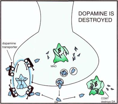
FIGURE 5 — 32. Dopamine (DA) is destroyed by the same enzymes that destroy norepinephrine (see Fig. 5 — 18), namely monoamine oxidase (MAO) and catechol-O-methyl-transferase (COMT). The DA neuron has a presynaptic transporter (reuptake pump), which is unique to the DA neuron but works analogously to the NE transporter (Fig. 5-18).
168