

Урология - Pyelonephritis
.pdfSpecial investigation
1.BLOOD EXAMINATION – reveals high neutrophil count. The ERS is increased.
2.URINE is usually scanty and highly concentrated. It is usually cloudy. Urine contains large amounts of pus and bacteria.

3. X-RAY
Straight X-ray of the abdomen may not be decisive except some obliteration of the renal shadow due to oedema of the kidney.

Excretory urography shows slight diminution of function in acute stage.
Non dilated renal pelvis is observed at 10 minutes film

IVU Acute secondary pyelonephritis
•First roengenogram: contract fluid is visualised in the left renal pelvis and vesical bladder, right kidney does not excrete contrast fluid
•Secobnd roentgenogram: dilation of right renal pelvis and upper part of the right ureter due to the roentgennegative calculus of the upper part of the ureter.

Arteriography
Chronic pyelonephritis.
A symptom of a burnt tree.

Renal carbuncle
Grey scale regimen. A round shaped zone of slightly elevated echogenity at the lover pole of kidney

Renal carbuncle
Doppler regimen: almost total absence of blood vessels at the area of carbuncle. At other parts of renal parenchyma vascularisation is normal.
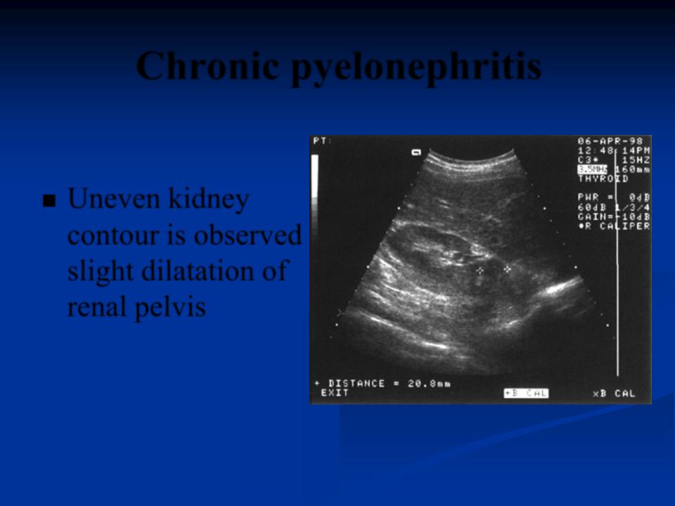
Chronic pyelonephritis
Uneven kidney contour is observed slight dilatation of renal pelvis

Complications
Perinephric absess
Sepsis
Acute renal failure
Pyonephrosis
Nephrosclerosis

Differential diagnosis
1.Acute appendicitis
2.Acute cholecystitis
3.Acute diverticulitis mimicking left sided acute pyelonephritis.
4. Pancreatitis
5.Basal pneumonia
6.Herpes Zoster