

8S Gloviczki et al
JOURNAL OF VASCULAR SURGERY
May Supplement 2011
already observed in 1948 that 58% of his patients with advanced CVD, studied with phlebography, never had a previous DVT.
Varicose veins and venous ulcers can be a great financial burden to patients and to society. Varicose veins and associated complications may lead to chronic pain, disability, decreased quality of life (QOL), loss of working days, and early retirement. In the United States, the direct medical cost of CVD has been estimated to be between $150 million and $1 billion annually.3,4 In the United Kingdom, 2% of the national health care budget per year (US $1 billion) is spent on the management of leg ulcers.1
Venous ulcer is an under-recognized and undertreated disease. A recently published supplement of the Journal of Vascular Surgery details the noble goal of the Pacific Vascular Symposium 6 (PVS6) to lead a call to action to formulate a doable and achievable plan to reduce the incidence of venous ulcers in the United States by 50% in 10 years.64
ANATOMY
During the past decade, new venous terminology has been developed and adopted by vascular societies around the world.47,49,61 The success of assigning uniform names to common veins was accompanied by new information on anatomy obtained with duplex ultrasonography, threedimensional computed tomography (CT), and magnetic resonance (MR) imaging; all these resulted in better understanding of the anatomy of veins and the pathology of
CVD.33,62
Superficial veins
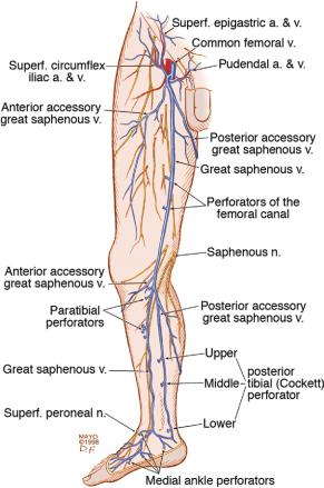
Superficial veins of the lower limbs are those located between the deep fascia, covering the muscles of the limb, and the skin. The main superficial veins are the GSV and the SSV. All previous names used to describe these vessels (greater, long, lesser) should be abandoned. The GSV originates from the medial superficial veins of the dorsum of the foot and ascends in front of the medial malleolus along the medial border of the tibia, next to the saphenous nerve (Fig 1). There are posterior and anterior accessory saphenous veins in the calf and the thigh. The saphenofemoral junction (SFJ) is the confluence of superficial inguinal veins, comprising the GSV and the superficial circumflex iliac, superficial epigastric, and external pudendal veins. The GSV in the thigh lies in the saphenous subcompartment of the superficial compartment, between the saphenous fascia and the deep fascia.
The SSV is the most important posterior superficial vein of the leg (Fig 2). It originates from the lateral side of the foot and drains blood into the popliteal vein, joining it usually just proximal to the knee crease. The intersaphenous vein (vein of Giacomini), which runs in the posterior thigh, connects the SSV with the GSV.65
Fig 1. Medial superficial and perforating veins of the lower limb. (Used with permission of Mayo Foundation for Medical Education and Research.)
Deep veins
Deep veins accompany the main arteries of the limb and pelvis. The deep veins of the calf (anterior, posterior tibial, and peroneal veins) are paired structures, and the popliteal and femoral veins may also be paired. The gastrocnemius and soleal veins are important deep tributaries. The old term superficial femoral vein has been replaced by the new term femoral vein.52 The femoral vein connects the popliteal to the common femoral vein.
The pelvic veins include the external, internal, and common iliac veins, which drain into the inferior vena cava (IVC). Large gonadal veins drain into the IVC on the right and the left renal vein on the left.
Perforating veins
Perforating veins connect the superficial to the deep venous system (Fig 1). They pass through the deep fascia that separates the superficial compartment from the deep. Communicating veins connect veins within the same system. The most important leg perforating veins are the medial calf perforators.66 The posterior tibial perforating veins (Cockett perforators in the old nomenclature) con-