

2 курс / Нормальная физиология / Учебное_пособие_по_физиологии_крови_Авдеева_Е_В_,_Репалова_Н_В_
.pdfcauses serious damage to the body cells. The platelets present in blood handle the task of limiting blood loss in the wake of an injury by helping blood to clot quickly.
2.3. Regulation (Homeostatic).
Homeostasis – is the maintenance of relatively stable conditions within the body’s internal environment despite changes occurring both inside and outside the body e.g. due to having meals, exercise, pregnancy, variations in external conditions.
Blood helps to maintain the body temperature by distributing the heat produced by the chemical activity of the cells evenly, throughout the body. When weather is very warm or during strenuous exercise there will be increased blood flow to the surface, resulting in warmer skin and faster heat loss. When environmental temperatures drop, blood flow focuses more on the important organs deep inside the body.
Blood is also responsible for controlling the concentration of hydrogen ions in the body, which are also known as pH balance. Constant pH value is very important for body functions. The administration of water and salt levels required by each cell of the body also falls under the regulatory functions of blood. Another regulatory task performed by blood is to control blood pressure and restrict it under a normal range.
3. Red blood cells.
Red blood cells (erythrocytes) also known as "RBCs". RBCs are formed in the myeloid tissue or most commonly known as red bone marrow, although when the body is under severe conditions the yellow bone marrow, which is also in the fatty places of the marrow in the body will also make RBCs. The formation of RBCs is called erythropoiesis. Red blood cells lose nuclei upon maturation, and take on a biconcave, dimpled, shape. They are about 7-8 micrometers in diameter. There are about 1000x more red blood cells than white blood cells. RBCs live about 120 days and do not self repair. RBCs contain hemoglobin which transports oxygen from the lungs to the rest of the body, such as to the muscles, where it releases oxygen load. Hemoglobin gets its red color from their respiratory pigments.
Fig. 3. Shape of erythrocytes.
11
3.1. Shape.
RBC have a shape of a disk that appears to be “caved in” or almost flattened in the middle; this is called bi-concave (fig. 3). This bi-concave shape allows RBC to carry oxygen and pass even through the smallest capillaries in the lungs. This shape also allows RBCs to stack like dinner plates and bend as they flow smoothly through the narrow blood vessels in the body. With the help of this shape the internal surface of erythrocyte is 20% higher, than the one it can obtain when it is globe-shaped.
The total surface of all erythrocytes is 3800 m2; it is 1500 times larger, than the surface of human body.
RBC lack a nucleus. The loss of nucleus causes the increase in erythrocyte capacity; the decrease of oxygen usage. Erythrocytes are used by an amount of the oxygen, which is 200 times less than the cells that form erythrocyte, that have nucleus. Erythrocytes, supporting the whole body with oxygen, use the less part of it. RBCs have a short life span of about 120 days, however, as long as our myeloid tissue works correctly, we will produce about 2-3 million RBCs per second. That is about 200 billion a day! This allows us to have more to replace those we lose.
RBC can be divided: According to size:
Normocytes - Normal sized RBC Microcytes - Small sized RBC Macrocytes - Large sized RBC
According to colour:
Normochromia - Normal coloured RBC
Hyperchromia |
- Darker, due to increased hemoglobin |
Hypochromia |
- Paler, due to decreased hemoglobin |
They are determined by measuring the:
Mean corpuscular haemoglobin (MCH)
Mean corpuscular haemoglobin concentration (MCHC)
3.2. Functions.
The main function is the
transportation of oxygen throughout the body and the ability of blood to carry out carbon dioxide which is called carbamino – hemoglobin.
maintaining the pH balance of blood. The balance can be measured by the acid and base levels in blood. Normal pH of blood ranges between 7.35-7.45; this normal blood is called Alkaline (less acidic then water). A drop in pH is called Acidic. This condition is also called Acidosis. A jump in pH higher then 7.45 is called Alkalosis.
3.3.Erythropoiesis.
Erythropoiesis is the process by which red blood cells (erythrocytes) are produced (fig. 4).
12

Fig. 4. Erythropoiesis.
It is stimulated by decreased O2 level in circulation, which is detected by the kidneys, which then secrete the hormone erythropoietin. The whole process lasts about 7 days. Through this process erythrocytes are continuously produced in the red bone marrow of large bones, at a rate of about 2 million per second in a healthy adult.
Mature red blood cells live in blood circulation for about 100 to 120 days. At the end of their lifespan, they become senescent, and are removed from circulation by the macrophages. This process is termed eryptosis, erythrocyte programmed cell death.
Regulation of erythropoiesis.
1Nervous:
sympathetic nervous system stimulates erythropoiesis;
parasympathetic – inhibits.
2 Humoral:
Somatotropic hormone, adrenocorticotropic hormone, catecholamines, hormones of thyroid gland (thyroxin) and male hormones stimulate erythropoiesis, female hormones – inhibit.
These nervous and humoral mechanisms are important for blood formation. They do not work directly but with the help of specific mediators – “hormones of blood formation”. Erythropoietin is a specific humoral stimulator of erythropoiesis (fig. 5).
13

Erythropoietin
Increased oxygencarrying
capacity
Increased number of RBS
Fig. 5. Control of red blood cell production.
Low oxygenated blood causes the kidneys and the liver to release erythropoietin which stimulates RBC production. This is a negative feedback mechanism
Within a few days many new blood cells appear in the circulating blood. (fig. 6). Vitamin B12 and folic acid are necessary. They are required for DNA synthesis,
making them necessary for the growth and division of all cells. Iron is also necessary. It is required for hemoglobin synthesis.
3.4. Destruction.
Red blood cells are broken down and hemoglobin is released. The globin part of hemoglobin is broken down into amino acid components, which in their turn are recycled by the body. Iron is recovered and returned to the bone marrow to be reused. The heme portion of the molecule experiences a chemical change and then gets excreted as bile pigment (bilirubin) by the liver. Heme portion after being broken down contributes to feces color and your skin color changing after being bruised.
14

Fig. 6. Recirculation of erythrocytes.
RBC destruction proceeds in 3 ways:
1.Mechanical destruction when RBC circulate in the vessels. Elasticity loss and erythrocyte becomes unable to pass through narrow site of the bloodstream and dwells there. One of these sites is the spleen, where the distance between trabeculations is less than 3 mkm. Erythrocytes dwell here, the part of the cells and hemoglobin are engulfed by macrophages.
2.Phagocytosis by the mononuclear phagocyte system of the liver and the spleen. Appearance of sialic acids on the erythrocyte surface. Macrophages have receptors to these groups that help in co-operation and degradation.
3.Haemolysis
The destruction of erythrocyte membrane accompanied with the release of hemoglobin into the blood plasma is called haemolysis.
15
Types of haemolysis:
1Mechanical haemolysis. It is caused by the mechanical degradation of the erythrocyte membrane.
2Osmotic haemolysis. It occurs when osmotic pressure inside the erythrocyte is higher than in blood plasma. In this case, water due to the laws of osmosis enters the erythrocyte, its volume increases and the degradation of membrane occurs.
3Chemical haemolysis. It is haemolysis, which occur under the influence of the substances that can degrade erythrocyte membrane (ether, chloroform, alcohol, bilious acids, saponine and others).
4Thermal haemolysis. It is haemolysis, which is caused by the influence of high or low temperatures. For example during deep-freeze of blood.
5Biological haemolysis. It is haemolysis, which develops after transfusion of incompatible blood and after sting of some snakes.
3.5.Variations in number of red blood cells.
3.5.1.Erythrocytosis is defined as an absolute increase in RBC mass and is also associated with an increased hematocrit (HCT) and hemoglobin concentration.
Although some people use the term polycythemia interchangeably with erythrocytosis, the two are not synonymous. Polycythemia in precise terms refers to an increased number of any hematopoietic cell in blood, be it RBCs, platelets or leukocytes. An increase in RBC number (whether relative to changes in body water or an absolute increase in RBC mass) is more precisely called erythrocythemia, but this term is not in general use and we do use the term of polycythemia (even though not inherently correct) instead of an increase in RBC (relative or absolute). Complicating matters is the term polycythemia vera, which specifically refers to a type of chronic myeloid leukemia that only affects the erythroid lineage or, in other words, a chronic erythroid leukemia.
Absolute increase in HCT or RBC mass (erythrocytosis): This is due to increased erythropoiesis and can be primary (a bone marrow disorder) or secondary, due to appropriate or inappropriate production of the erythropoietic cytokine, erythropoietin.
Relative increase in HCT or RBC count: This is due to changes in RBC numbers with respect to plasma (i.e. not a true increase in RBC mass). This can be due to loss of intravascular water, which is the most common cause of a relative increase, or proportionally increased numbers of RBC in circulation. Another reasons: much of sweating, nausea, diarrhea; scorches; shock; cholera, dysentery; hard muscle work.
3.5.2. Erythropenia – a decrease in the number of erythrocytes, associated with anemia
Absolute erythropenia is the decrease in the total amount of erythrocytes in the organism. Its reasons: increase in the haemolysis of erythrocytes (due to exposure to radiation, poisons, toxins, transfusion of the incompatible blood etc.); loss of blood; decrease in speed or stopping of erythropoiesis (because of deficit of blood
16

formation factors – iron, vitamins B6, B12, folic acid; because of erythropoietins deficiency when the kidneys are pathologic; depression of blood formation function of red bone marrow).
Relative erythropenia – decrease in erythrocytes amount in blood volume unit when the blood is diluted. Its reasons are: water retenssion in the pathologies of the kidney; injecting blood alternatives.
4. Haemoglobin.
It is composed of the protein globin (a polypeptide), and the pigment heme (fig. 7).
Structure : Hemoglobin has globular molecule, which is formed with 4 subunits. Each subunit contains heme. Heme – is Fe2+-inclusive substance, the derivative of porphyrin. Heme molecule consists of 4 pyrrol. The ion of Fe2+ is situated in the center. The hemoglobin has the ability to combine with oxygen is due to the four iron atoms associated with each heme group within the molecule.
Fig. 7. Structure of haemoglobin. |
|
|
Normal range of haemoglobin: In men |
– |
135-180 g/L |
In women |
– |
120-140 g/L |
In newborns – |
200 g/L |
|
4.1. Main physiological compositions of hemoglobin.
Physiological composition of Hb:
HbO2 – oxyhemoglobin – oxygen combines weakly with the haemoglobin molecule. Such association is called oxyhemoglobin . It is formed in lungs.
Hb – deoxyhemoglobin – when oxygen is released to the tissues of the body, the haemoglobin is called reduced haemoglobin or deoxyhemoglobin.
17
HbCO2 – carbhemoglobin – in tissues Hb combines with carbon dioxide and form carbhemoglobin.
Pathological composition of Hb:
HbCO - carboxyhemoglobin – the composition of Hb with CO.
Chemical relativity of Hb to CO is in 300 times higher than O2. That’s why carbon monoxide displaces O2 from hemoglobin, decreasing the ability of blood to bind oxygen. Even small number of CO leads to a significant increase in formation of HbCO. When concentration of CO in the air is 0.1% – 80% Hb dasn`t with O2, but with CO. When concentration of CO in the air is 1%, in few seconds it will cause death. It is dangerous because HbCO is persistent and Hb cannot transport oxygen anymore.
MetHb – methemoglobin – hemoglobin, which contains Fe3+ and has brown color. Oxidation of Fe2+ to Fe3+ in hemoglobin occurs when interacting with strong
oxidizers (KMnO4, aniline), and also with medicine of oxidative properties. In pathological conditions, when methemoglobin is formed, blood with high oxygen content circulates in the organism, but it is not entering tissues.
Fetal hemoglobin.
A variant hemoglobin, called fetal hemoglobin (HbF), is found in the developing fetus, and binds oxygen with greater affinity than adult hemoglobin. As a result, fetal blood in placenta is able to take oxygen from maternal blood.
Normal levels are:
Men: 14.0 to 16.0 g/dL (140 to 160 g/L)
Women: 12.0 to 14.0 g/dL (120 to 140 g/L)
Children: 11 to 16 g/dL (111 to 160 g/L)
Pregnant women: 11 to 14 g/dL ( usual value during pregnancy)
(110 to 140 g/L)
Dehydration or hyperhydration can greatly influence measured hemoglobin levels. If the concentration is below the normal level, this is called anemia.
Hemoglobin deficiency can be caused either by a decreased amount of hemoglobin molecules, as in anemia, or by decreased ability of each molecule to bind oxygen at the same partial pressure of oxygen. Other common causes of low hemoglobin include loss of blood, nutritional deficiency, bone marrow problems, chemotherapy, kidney failure, or abnormal hemoglobin (such as that of sickle-cell disease).
RECOMENDED LABORATORY WORKS
1.Calculation of red blood cells.
2.Calculation of hemoglobin quantity in the blood.
3.Calculation of color index of blood.
SUPPLEMENT
1. Calculation of erythrocytes.
For calculation of erythrocytes it is necessary to have: donor's blood, a microscope, count camera (Burker's camera), 3% NaCl solution.
18

The succession of the work. The calculation chamber is a thick object glass with four grooves. Narrow grounds are formed between the grooves. The middle ground of 0.1 mm is lower than the lateral ones and is divided into two parts by a perpendicular groove. At both side of the groove there are nets drifted on the glass platforms. The chamber of 0.1 mm deep is formed when cover glass is placed. Goryayev's net consists of 25 big squares, one third of the squares is divided into 16 small squares.
Fig. 8. Count camera (Burker's camera).
B – Goryayev's net, a – small square, b – big square.
In calculation of erythrocytes the hypertonic solution of NaCl (3%) is used for diluting blood (200 times). Erythrocytes shrink in this solution (for facilitation of calculation).
The chamber net is closed with cover glass. A drop of diluted blood is put on the middle ground at the edge of the cover glass. The forces of capillary tension take the fluid inside. The filled chamber is placed on the microscope table (small magnification). The number of erythrocytes in 5 big squares is counted. Egorov's rule is true in calculation, that is: "for one square the number of erythrocytes within the square is counted, on the left and upper borders. Erythrocytes which lie on the right and lower borders are not counted."
19
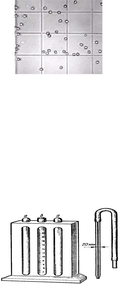
Fig. 9. Looks of red blood cells under a microscope.
The number of erythrocytes (in 1 liter of blood) is calculated by the formula:
A = E*4000*200 / 80 * 106 /L
where: A – is the total number of erythrocytes in 1 l of blood, E – is the sum of erythrocytes within 5 big squares, 200 – is dilution,
1/4000 – the volume of fluid under one small square, 80 - is number of small squares in 5 big squares.
In normal adult men it equals; 4.0*1012 to 5 .0*1012 /liter In normal adult women it equals: 3.7*10l2 to 4.7*1012 /liter
The increased quantity of RBC is called erythrocytosis. The decreased quantity of RBC is called erythropenia.
2. Determination of hemoglobin in blood by method of Saly.
For the work it is necessary to have: Saly's hemometer. It is a dark support with three test – tubes. Two extreme soldered test-tubes (a) which contain the solution of hydro-chloride hematin (16.7 g/dl or 16.7 g% or 167 g/l). The middle test – tube (b) is graduated and open; donor's blood.
Fig. 10. Saly's hemometer: a – test-tubes with standard solution, b – middle test – tube.
20