

Принципы классификации АА.
По локализации:
- каротидного бассейна (передний отдел артериального круга) - вертебробазилярного бассейна
По локализации в пределах сосудистого бассейна: ВСА
каменистый
кавернозный
параклиноидный
супраклиноидный
бифуркации ПМА-ПСоА СМА ПА ОА ЗМА
По размерам:
малые: < 4 мм обычные (средние): 5-14 мм крупные: 15-25 мм гигантские: > 25 мм
По форме:
мешотчатые (berry aneurysms) фузиформные

Интракраниальные методы лечения аневризматической болезни.
- Клипирование аневризм.
Принципы микрохирургических вмешательств:
минимальная тракция ткани мозга при подходе;
необходимость сохранения перфорантных артерий и целостности артериального круга большого мозга;
одномоментное клипирование шейки аневризмы;
тщательный гемостаз. M.G. Yasargil,


Эмболизация артериальной аневризмы микроспиралями (схема)
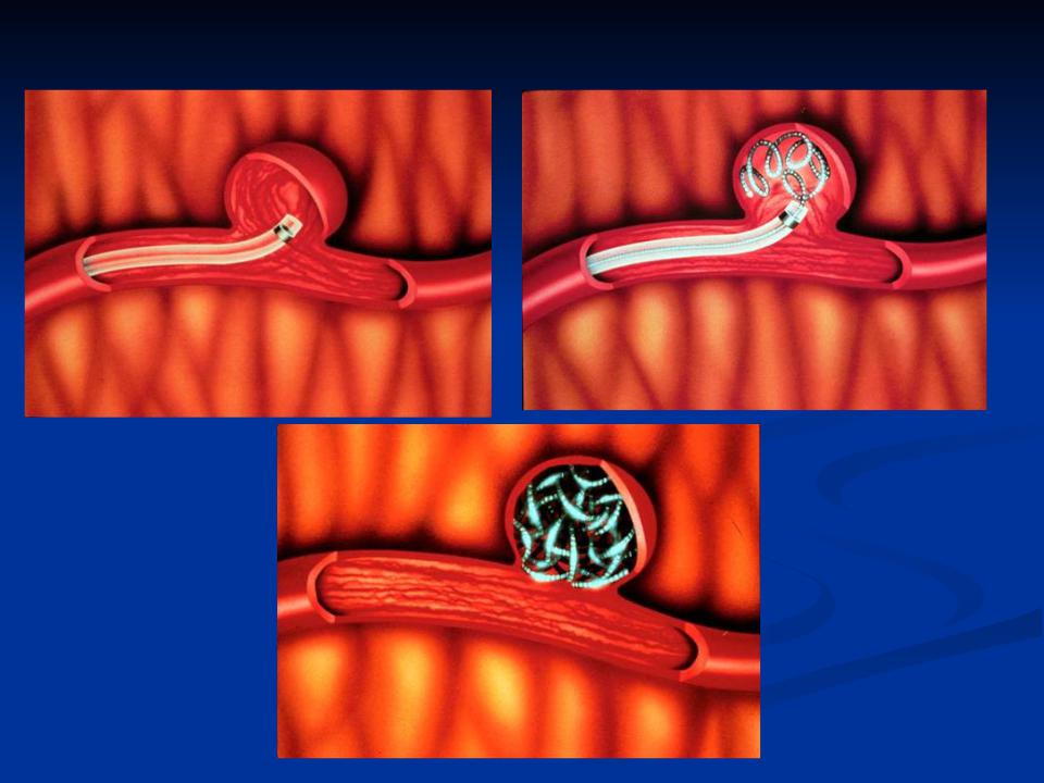
Выключение аневризм из кровотока

Виды стентов |
Embolic Protection System |
Carotid Stent System |

Артериальная
аневризма
ВСА
Велизиев круг (артериальный круг головного мозга) с гигантской аневризмой внутренней сонной артерии

CT scan demonstrating a subarachnoid hemorrhage in the right sylvian fissure and left ventricular cateter (A).Oblique right ICA angiogram revealing a large right MCA bifurcation aneurysm (B)

60-year-old male patient who presented with headache and in whom a large unruptured internal carotid aneurysm was found. Pretreatment, treatment, and follow-up images are presented.
A, Large internal carotid aneurysm before treatment.
B, Seal test before treatment of the aneurysm with Onyx. Microcatheter and balloon are in place with gentle contrast material injection into the aneurysm
C, Angiographic result immediately after Onyx treatment.
D, Follow-up angiogram 6 months after treatment, showing complete occlusion and intervening soft thickened tissue between vessel lumen and Onyx cast

45-year-old patient presenting with a partial cranial nerve III palsy and headache. No anterior cerebral artery was present on the left side and the carotid only supplied the left middle cerebral territory.
A, Lateral arterial phase carotid angiogram obtained immediately before treatment.
B, Final angiography obtained after Onyx treatment balloon catheter still present in vessel.
C, Follow-up angiography 3 months after procedure. Patient developed a complete ophthalomoplegia immediately after the procedure, which had resolved entirely by 3 months when she was asymptomatic. Note extensive orbital collaterals filling middle cerebral territory. (Images
courtesy of Peter Flynn and Steven Mckinstry, Royal Victoria Hospital,
B
Ony
x
|
50-year-old male patient with |
|
leg weakness and a possible |
|
history of subarachnoid |
|
hemorrhage. This patient was |
|
found to have a vertebral |
|
basilar junction aneurysm |
|
compressing the medulla. |
|
A, Pretreatment angiogram |
|
showing a large |
|
vertebrobasilar junction |
|
aneurysm treated on two |
|
occasions (treated with an |
|
INX stent at the second |
|
procedure after recurrence). |
|
Complete occlusion was noted |
|
at 6-month and 1-year follow- |
|
up after second procedure. |
|
B, Angiogram obtained after |
|
second treatment at 3 months |
|
after early recurrence and |
C |
after placement of an INX |
stent and re-treatment with |
|
|
Onyx. |
|
C, Unsubtracted image |
|
obtained after second |
|
procedure, showing INX stent |
|
and second cast of Onyx in |
|
inferior recurrent aneurysm |
|
pocket. |
D, Follow-up angiogram