


BENIGN NEOPLASMS OF THE LOWER GENITAL TRACT 687
Cervix
•Cervical polyps (adenoma): common; due to overgrowth of the endocervical mucosa. Sometimes arise in endometruim (pedunculated) and protrude from cervix. Very rarely malignant (1:6000), but they should be removed and hysteroscopy to rule out further polyps should be considered if symptoms warrant it.
•Nabothian cysts: mucous-retention cysts caused by blockage of endocervical mucous glands. Treatment is not required.

688 CHAPTER 23 Benign and malignant conditions
Benign neoplasms of the uterus
Uterine fibroids
These are the most common benign tumours arising from the myometrium of the uterus. Also called leiomyomata, these tumours are composed primarily of smooth muscles, but may contain fibrous tissue. Present in 20–40% of women in the reproductive age group, they have a higher incidence in Afro-Caribbean women and those with a family history of fibroids. Many women are asymptomatic, but may present with: dysmenorrhoea, menorrhagia, pressure symptoms (especially frequency), and pelvic pain. Infertility may be associated and, in <10% cases, caused solely by fibroids. In pregnancy can cause pain from degeneration, abnormal lie, and obstruction if cervical, and difficulty in CS. Association with miscarriage as yet unproven.
Types of uterine fibroids
•Submucous: >50% projection into the endometrial cavity.
•Intramural: located within the myometrium.
•Subserous: >50% of the fibroid mass extends outside the uterine contours.
•Cervical: relatively uncommon and can cause surgical difficulty due to the proximity to the bladder and the ureters.
•Pedunculated: mobile and prone to torsion.
•Parasitic: have become detached from the uterus and attached to other structures.
•IV leiomyomatosis: very rare, spread through the pelvic veins and vena cava to involve the heart.
Diagnosis
Clinical examination (hard, irregular uterine mass) may be sufficient. Transvaginal or abdominal ultrasound can differentiate the types and dimensions of the fibroids. Rarely MRI may be needed when the scan is inconclusive.
Endometrial polyps (adenoma)
These are focal overgrowth of the endometrium and are malignant in <1%. They are more common in women >40yrs, but may occur at any age. Treatment is usually resection during hysteroscopy and the polyp should be sent for histological assessment.

BENIGN NEOPLASMS OF THE UTERUS 689
Treatment options for uterine fibroids
•No treatment may be necessary if minimal symptoms.
•GnRH analogues shrink fibroids, but should only be used for this purpose prior to surgery.
•Myomectomy: open, laparoscopic, or hysteroscopic depending upon location (especially when wish to preserve fertility and when the fibroids are distinctly isolated on scan—fibroids often recur).
•Hysterectomy: women who have either completed their family or are over 45yrs—guaranteed cure of fibroids.
•Uterine artery embolization: uterine artery is catheterized generally using the unilateral approach; polyvinyl alcohol powder or gelatin sponge is used as the embolic material (minimally invasive procedure with avoidance of a general anaesthetic).
Benign neoplasms of the fallopian tube
Hydrosalpinx, pyosalpinx, and tubo-ovarian masses following pelvic inflammatory disease or endometriotic adhesions may present as a benign mass in the pelvis. The diagnosis is essentially by ultrasound and laparoscopy. Most tumours of the fallopian tubes are malignant. Although they were thought to be rare, data from series of BRCA-positive women undergoing prophylactic bilateral oophrectomy and salpingectomy (BSO) suggest that p fallopian tumours may be more common than previously thought.


BENIGN OVARIAN TUMOURS: DIAGNOSIS 691
Modified risk of malignancy index
RMI = U x M x CA125
•U = ultrasound score (0, 1, or 3).
•M = menopausal status (1 = premenopausal, 3 = post-menopausal)
•CA125 = serum cancer antigen 125 level (U/L)
Ultrasound scoring system
•1 point for each of the following features on USS:
•multilocular cyst
•evidence of solid areas
•evidence of metastases
•ascites
•bilateral lesions.
•Final U score:
•0 if no features
•1 if 1 feature
•3 if 2 or more features.
bTingulstad S, Hagen B, Skjeldestad FE, et al. (1996). Evaluation of a risk of malignancy index based on serum CA125, ultrasound findings and menopausal status in the pre-operative diagnosis of pelvic masses. Br J Obstet Gynaecol 103(8): 826–31.
Table 23.1 RMI score and ovarian cancer risk
Risk |
RMI score |
Risk of cancer |
Low |
<25 |
<3% |
Moderate |
25–250 |
20% |
High |
>250 |
75% |
Davies AP, Jacobs I, Woolas R, et al. (1993). The adnexal mass: benign or malignant? Evaluation of a risk of malignancy index. Br J Obstet Gynaecol 100(10): 927–31.
Non-gynaecological causes of pelvic masses
Other benign masses due to non-gynaecological conditions in the pelvis should always be borne in mind as a differential diagnosis:
•Bladder tumours
•Intestinal tumours
•Diverticular disease
•IBD.


694 CHAPTER 23 Benign and malignant conditions
Vulval dermatoses: lichen sclerosus
Skin conditions of the vulva can cause distressing symptoms, and be difficult to diagnose and manage. Irritation leads to scratching and excoriation, which may make the appearance clinically difficult to differentiate, especially when added to changes seen following secondary infection or use of topical creams.
Vulval dermatoses refers to a range of benign skin conditions, which generally cause white thickening of the vulval skin: lichen sclerosus; lichen planus; vulval dermatitis; vulval psoriasis.
Lichen sclerosus
•Chronic inflammatory condition (lymphocyte mediated).
•May be hereditary (association with HLA-DQ7).
•Usually confined to anogenital area.
•More common in women.
•Incidence estimated as 1:300–1:1000 women.
•Normally in peri-menopausal women, but can occur in young girls (2/3 improve at puberty—may be misdiagnosed as signs of abuse).
•Associated with other autoimmune diseases:
•e.g. thyroid disease; diabetes; vitiligo; pernicious anaemia
•20–34% have association with autoimmune disease
•up to 74% have autoantibodies.
See Colour plate 1.
Clinical presentation of lichen sclerosus
•Burning pain or itch, occasionally asymptomatic.
•Figure of 8 appearance around vulva and anus.
•White, shiny, wrinkly, atrophic appearance ‘like tissue paper’:
•may have white patches, purpura, or telangiectasia
•hyperkeratosis and lichenification if chronic scratching
•over time, can develop loss and fusion of labia minora, narrowing of introitus, resulting in problems with intercourse and micturation.
1 Long-term risk of vulval squamous cell carcinoma (~2%), so need long-term observation, follow-up, and biopsy of suspicious lesions. This can be in pcare if symptoms well controlled.

VULVAL DERMATOSES: LICHEN SCLEROSUS 695
Management of lichen sclerosus
•Biopsy for diagnosis, if not responding to treatment.
•Biopsy suspicious lesions (risk of vulval cancer) or if not responding to treatment made on clinical diagnosis.
•Check ferritin levels and treat if low.
•Screen for autoimmune conditions, if suggestive symptoms (FBC, TFTs, glucose, serum iron, autoimmune antibodies, intrinsic factor, and vitamin B12).
•Potent corticosteroids: clobetasol propionate 0.05% bd initially once a night for 4wks, alternate nights for 4wks, once or twice weekly for 4wks, then use ‘as required’ for flares; the shiny appearance will remain.
•Follow-up at 3mths to check response.
•Annual review with GP and advise urgent contact if ulcers, bleeding, or suspicious lesions.
•Referral to specialist unit for tacrolimus if symptoms not responding.
Further reading
British Association of Dermatologists. Mwww.bad.org.uk
Mwww.lichensclerosus.org
Mhttp://www.macmillan.org.uk/Cancerinformation/Cancertypes/Vulva/Pre-cancerousconditions/ Vulvallichensclerosuslichenplanus.aspx
RCOG. (2011). The management of vulval skin disorders, Green-top guideline 58. M http://www. rcog.org.uk/files/rcog-corp/GTG58Vulval22022011.pdf
696 CHAPTER 23 Benign and malignant conditions
Other vulval dermatoses
Vulval dermatitis
•Dermatitis or eczema.
•If scratched so that thickening of skin llichen simplex chronicus.
•Association with other atopic illnesses (asthma, hay fever, or eczema).
•Common irritants:
•soaps, shower gels, condoms, deodorants, creams
•if diagnosed as candidiasis, topical creams can be irritant.
Clinical presentation of vulval dermatitis
•Itch—burning and pain secondary to scratching.
•Erythema ± scaling of skin.
•No loss or fusion of labia.
Management
•Avoid irritant and apply general vulval skin care (see Box 23.1).
•Low vaginal swabs for secondary infection (e.g. Candida).
•Severe disease—treat with steroid cream—clobetasol propionate or betamethasone valerate, if less severe. Use bd initially; reduce to od, then twice weekly, as condition improves.
•Consider sedating antihistamine (e.g. chlorphenamine 4mg) at night to prevent scratching.
•Referral to dermatology for patch testing.
Lichen planus
Rare condition.
Clinical presentation of lichen planus
•Purplish papules and plaques; can have white streaks on top— ‘Wickham’s striae’. May involve mouth too.
•May cause painful, red, ulcerated areas around introitus. Occasionally can cause severe desquamative vaginitis.
•Cause itch, pain, post-coital bleeding, or discharge.
Management
•As for lichen sclerosus, including follow up, as also have an increased risk of developing vulval cancer.
•Biopsy; potent steroids; good vulval skin care; local anaesthetic gel.
Vulval psoriasis
Clinical presentation of vulval psoriasis
Classically well-defined erythematous patches, may have scaling on pubic area, but not necessarily on vulval skin.
Management
•Good vulval skin care; bland emollients; mild topical steroids.
•Other psoriatic medications often too harsh for vulval skin.

OTHER VULVAL DERMATOSES 697
Box 23.1 Vulval skin care
•Keep area clean but avoid soap:
•use soap substitutes—soap-free shower gels; bath oil; just water
•salt baths may help.
•Do not soak in hot bath.
•Allow air circulation to avoid sweating: loose underwear and bed clothes; avoid jeans, etc.
•Wear cotton next to skin: avoid synthetics (especially nylon) and wool (intrinsically itchy!).
•Avoid vaginal lubricants:
•may be irritating;
•can use saliva or oil (vegetable, olive, almond)—avoid perfumed oil.
•oils weaken condoms!
•Refer for patch testing if vulval dermatitis.
Further reading
British Association of Dermatologists. Mwww.bad.org.uk http://www.macmillan.org.uk/Cancerinformation/Cancertypes/Vulva/Pre-cancerousconditions/
Vulvallichensclerosuslichenplanus.aspx


IDIOPATHIC VULVAL ITCH AND PAIN 699
Causes of pruritis vulvae
•Infection:
•candidiasis
•threadworms
•Phthirus pubis (genital lice)
•Sarcoptes scabiei (scabies, from Latin ‘to itch’).
•Vulval dermatoses:
•lichen sclerosus
•vulval dermatitis
•lichen planus
•vulval psoriasis.
•Vulval intraepithelial neoplasia or vulval carcinoma.
•Urinary incontinence.
•Systemic conditions:
•liver failure
•uraemia.
•Medication.
•Pregnancy/menopause.
•Idiopathic.
Further reading
Mhttp://www.patient.co.uk/health/pruritus-vulvae-vulval-itch
British Association of Dermatologists. Available at: Mwww.bad.org.uk
Vulval Pain Society. Available at: Mwww.vulvalpainsociety.org/

700 CHAPTER 23 Benign and malignant conditions
Cancer screening in gynaecology: overview
The intention of screening is early identification of a disease, prompt referral for diagnostic tests, and appropriate intervention and management. Screening test does not necessarily diagnose condition or disease, but may reduce associated incidence, mortality, and morbidity.
WHO principles of screening (1968)
•The condition should be an important public health problem.
•An effective intervention should be available.
•Clear, recognizable early stage and known natural history of condition.
•There should be a suitable screening test available.
•The test should be acceptable to the population.
•Benefits of the test should outweigh the risks.
•There should be an agreed policy on who to treat.
•The total cost of finding a case should be economically balanced.
Sensitivity, specificity, and positive and negative predictive values
•Sensitivity: the ability of the screening test to detect the disease— acceptable sensitivity detects most disease.
•Specificity: the ability of the screening test not to identify those who do not have the condition—acceptable specificity excludes most without the disease.
•Positive predictive value (PPV): the proportion with a positive test result who have the disease.
•Negative predictive value (NPV): the proportion with a negative test result who do not have the disease.
This page intentionally left blank


OVARIAN AND ENDOMETRIAL CANCER SCREENING 703
Ovarian cancer screening
The efficacy of screening for ovarian cancer is not proven. The results from a large UK Collaborative Trial of Ovarian Cancer Screening (UKCTOCS) are expected in 2015. Results of the Prostate, Lung, Colorectal, Ovarian (PLCO) cancer screening trial did not demonstrate reduced mortality from ovarian cancer using a cut-off level of CA125. Currently, any screening offered to women, should be as part of a clinical trial.
•Pelvic TVS can identify ovarian cysts. In a young woman most of these will be physiological, but features suggestive of malignancy are large size of cyst, internal septa, solid areas, and increased blood flow on Doppler examination. Operator expertise is extremely important in the screening process.
•CA125 is a glycoprotein shed by 85% of epithelial tumours. Normal levels are 30–65IU/L, but false +ves are commonly seen in other malignancies (liver, pancreas), endometriosis, PID, and early pregnancy. Sensitivity is improved using serial measurements and the trends. Up to 50% of stage 1 tumours will present with a CA125 level of <30IU/L.
•Both ultrasound examination and CA125 estimation can give rise to a false +ve result in nearly 2–3% of post-menopausal women. The use of the two tests together reduces the chance of false +ves.
UK Collaborative Trial of Ovarian Cancer Screening. Mwww.ukctocs.org.uk/

704 CHAPTER 23 Benign and malignant conditions
Cervical cancer: pathology and screening
CIN is widely regarded as a necessary precursor lesion for carcinoma of the cervix. CIN is a histological diagnosis and needs persistent cervical infection with HPV to develop. There are about 15 high risk oncogenic subtypes of HPV, the most common being 16, 18, 31, and 33.
Risk factors for CIN
•Persistent high risk HPV infection.
•Multiple partners increases the risk of exposure to HPV infection.
•Smoking as a promoter.
•Immunocompromise, e.g. HIV, immunosuppressive agents.
•Use of the COCP has an association, probably due to non-barrier method and exposure to HPV.
Normal and abnormal physiology of transformation zone
The endocervix is composed of a thin secretory glandular epithelium; the ectocervix consists of a stronger stratified squamous epithelium. The two are in continuity and meet at the squamocolumar junction. Under the influence of oestrogen the glandular epithelium is pushed out onto the ectocervix and in response to low pH undergoes physiological squamous metaplasia—the transformation zone (TZ). The TZ is usually ectocervical in women of reproductive age, but tends to become endocervical in post-menopausal women.
As an area of high mitotic activity the TZ is vulnerable to HPV-driven neoplastic change, if persistent (i.e. not eradicated). Most work suggests an average of 8–10yrs from acquisition to development of cancer. Cervical cancer is therefore, in theory, a preventable disease.
Screening for cervical premalignancy
The NHS Cervical Screening Programme (NHSCSP) has been systematic since the 1980s and has since shown a 50% reduction in mortality from cervical cancer. Regular cervical screening reduces the risk of death from cervical carcinoma by 75% (but does not eliminate it).
Screening is based on the natural course of cervical cancer where CIN (dysplasia) precedes overt malignancy and is a progressive condition. However, in reality CIN may also revert back to normal. Routine screening carries a 50–70% sensitivity to detect CIN III.
HPV triage and test of cure was introduced in the UK in April 2012. Women with borderline nuclear changes or mild dyskaryosis have testing for high-risk HPV types. Women with high risk HPV are referred for colposcopy. Women without have smear follow-up at 3yrs. Similarly, following LLETZ for CIN, women without abnormal cytology or high risk HPV at the smear 6mths after treatment will return to 3-yearly smears. Women with high risk HPV or abnormal cytology will be referred back to colposcopy (see Table 23.2).

CERVICAL CANCER: PATHOLOGY AND SCREENING 705
Current English criteria for cervical screening
•Sexually active women aged 25–64.
•Three-yearly for women aged 25–50, if normal, 5-yearly till 64.
2Three-yearly screening identifies more than 95% of abnormalities tested by annual screening and is cost-effective.
2Screening in Scotland and Wales begins at 20.
Table 23.2 Management of abnormal smears
Papanicoulaou class |
Histology |
Management |
Normal |
0.1% CIN II–III |
Repeat smear in 3yrs |
Inflammatory |
6% CIN II–III |
Repeat in 6mths (colposcopy |
|
|
after 3 consecutive) |
Borderline nuclear |
20–30% CIN II–III |
High risk HPV test—refer to |
changes |
|
colposcopy if +ve; repeat smear |
|
|
3yrs if –ve |
Mild dyskaryosis |
30% CIN II–III |
High risk HPV test—refer to |
|
|
colposcopy if +ve; repeat smear |
|
|
3yrs if –ve |
Moderate dyskaryosis |
50–75% CIN II–III |
Refer to colposcopy |
Severe dyskaryosis |
80–90% CINII–III |
Refer to colposcopy |
Invasion suspected |
50% invasion |
Refer to colposcopy |
Abnormal glandular |
Adenocarcinoma of |
Refer to colposcopy |
cells |
the cervix |
|
|
|
|
Further reading
NHSCSP. (2010). Colposcopy and programme management: guidelines for the NHS cervical screening programme. M http://www.cancerscreening.nhs.uk/cervical/publications/nhscsp20. html
NHSCSP. (2011). HPV triage and test of cure protocol. M http://www.cancerscreening.nhs.uk/ cervical/hpv-triage-test-flowchart.pdf
706 CHAPTER 23 Benign and malignant conditions
Cervical cancer: cytology, colposcopy, and histology
Cervical cytology
The primary screening tool for cervical malignancy. NICE recommends liquid-based cytology for the cytological preparation of cervical cells— cleaner preparation, easier to read, inadequate cytology cut by 80%, and more cost-effective. High risk HPV testing can be performed on this sample, if indicated.
Dyskaryosis is a cytological term. False +ve and –ve rates are 10–15% and 5–15%, respectively. Due to these problems with sensitivity and specificity, abnormal cytology is further assessed by colposcopy.
Cytological markers seen with abnormal smears
•Increased nuclear/cytoplasmic ratio.
•Shape of the nucleus (poikilocytosis—abnormal shape).
•Density of the nucleus (koilocytosis—abnormal density).
•Inflammation, infection, and mitoses.
Colposcopy
Involves the magnified (6–40x) visualization of the transformation zone after application of 5% acetic acid (preferentially taken up by neoplastic cells) or Lugol’s iodine (not taken up by glycogen-deficient neoplastic cells). Upon identification of colposcopic abnormalities either:
•Directed punch biopsy to gain histological confirmation; or
•Definitive treatment (‘see and treat’).
Adequate colposcopic assessment
•Visualization of the entire transformation zone.
•Any lesion identified must be completely seen (especially upper extent).
•Problem areas: post-menopausal, post-treatment, and post-partum. See Colour plates 2–8.
Histology
CIN is a histological diagnosis and is characterized by loss of differentiation and maturation from the basal layer of the squamous epithelium upwards.
•Bottom 1/3 = CIN I.
•Bottom 2/3 = CIN II.
•Full thickness = CIN III.
•Mitotic figures are present throughout the epithelium in all grades.

CERVICAL CANCER: CYTOLOGY, COLPOSCOPY, AND HISTOLOGY 707
Referral criteria for colposcopy
•Any smear showing borderline nuclear changes or mild dyskaryosis with high risk HPV.
•Any smear showing moderate or severe dyskaryosis.
•Any smear suggestive of malignancy.
•Any smear suggestive of glandular abnormality.
•Three consecutive inadequate smears.
•Keratinizing cells (?underlying CIN).
•Post-coital bleeding.
•Abnormal-looking cervix.
Colposcopic appearances of CIN
•Aceto-white epithelium (AWE).
•Vascular abnormalities, especially mosaic and punctuation.
•Bizarre or grossly abnormal vessels are suggestive of micro-invasive carcinoma.

708 CHAPTER 23 Benign and malignant conditions
Management of cervical intraepithelial neoplasia (CIN)
CIN can be managed conservatively, by excision, by destruction, or rarely by hysterectomy. Management depends upon the grade of CIN and patient preference, but excision is the preferred treatment modality. This is usually by LLETZ.
Benefits of LLETZ
•Easy and safe.
•Usually possible with local anaesthetic.
•Tissue available for histology and assessment of excision margins.
Low grade CIN (CIN I)
Will spontaneously regress in at least 50–60% of cases within 2yrs. Malignant potential is very low but still up to 10x greater than women with normal cytology. Management options are:
•Conservative monitoring with colposcopy and/or cytology every 6mths.
•LLETZ if persistent.
High grade CIN (>CIN I)
Will progress to cancer in up to 3–5% (CIN II) and 20–30% (CIN III) within 10yrs. Spontaneous regression occurs less often.
• LLETZ recommended.
Follow-up and management after LLETZ
•Low grade: follow-up cytology and HPV testing at 6mths. If –ve, smear in 3yrs.
•High grade: follow-up cytology and high risk HPV test-of-cure at 6mths. If –ve, smear in 3yrs.
Complications of LLETZ
Short term
•Haemorrhage.
•Infection.
•Vaso-vagal reaction.
•Anxiety (disproportionately high in colposcopy clinic attenders).
Long term
•Cervical stenosis (dysmenorrhoea and/or difficulty in follow-up).
•Cervical incompetence and premature delivery (evidence suggests absolute risk of adverse effect on neonatal outcome is very low).

MANAGEMENT OF CERVICAL INTRAEPITHELIAL NEOPLASIA (CIN) 709
Further reading
NHSCSP. (2010). Colposcopy and programme management: guidelines for the NHS Cervical Screening Programme. Mwww.bsccp.org.uk/docs/public/pdf/nhscsp20.pdf
NHSCSP. (2011). HPV triage and test of cure protocol. M http://www.cancerscreening.nhs.uk/ cervical/hpv-triage-test-flowchart.pdf


MANAGEMENT OF CGIN AND HUMAN PAPILLOMAVIRUS 711
HPV vaccines—what we know
•They reliably induce excellent type-specific antibody titres vs. HPV.
•There is good trial evidence for reliable prevention of CIN (and presumably therefore ultimately cancer) and anogenital warts.
•Due to type specificity they will not prevent all cancers—there are 15 high risk HPVs (current vaccines target 2h HPVs only).
•The long-term antibody response is not yet known, although current limited data suggest it is likely to be good.
•They need to be widely used in young girls before sexual debut to be most effective—recommended at 12yrs in the UK.
•They offer no protection once HPV-infected.
2 Cost-effectiveness is still unknown and will take decades to determine, including whether they truly prevent cervical carcinoma.
CIN, VAIN, VIN + AIN
•The presence of any form of intraepithelial abnormality of the lower genital tract is a marker for a ‘field change’.
•The vagina (VAIN), vulva (VIN), and peri-anal (AIN) area are all at risk in the presence of CIN and vice versa.
•When the cervical appearances are normal with abnormal cytology the abnormal cells may be derived from elsewhere—they should be examined at colposcopy.

712 CHAPTER 23 Benign and malignant conditions
Gynaecological cancer: a multidisciplinary approach
Optimal treatment of the gynaecological cancer patient is provided by coordination of care within a multidisciplinary team (MDT). This team is made up of doctors, nurses, and allied professionals with an interest in treating gynaecological cancer patients and should, at a minimum, consist of: gynaecological oncologist (surgeon); clinical oncologist; medical oncologist; radiologist; histopathologist; colposcopist; gynaecological cancer nurse specialist; and MDT coordinator. Ideally, because of the complex nature of gynaecological cancer patients, their treatments, and the complications they may encounter, teams should also include or have ready access to a:
•Palliative care team.
•Dietician; fertility specialist.
•Lymphoedema specialist.
•Lower gastrointestinal surgeon.
•Urological surgeon.
•Stoma therapy nurse.
•Psychologist.
•Psychosexual counsellor.
The role of the team is to provide the following areas of care:
•Diagnosis, staging, primary surgical, and adjuvant treatment, and coordination of follow-up care.
•Psychological preparation for anticancer treatment and follow-up: psychosexual support is a vital aspect, since many of the treatments can have major impacts on sexual functioning, either physically
or psychologically, and may deprive women of their sense of femininity.
•Information on diagnosis, treatment plans, likely side effects, and follow-up plans.
•Access to financial, social, and psychological support: often required as patients may be young and have either young or older dependants, for whom they may be either the financial provider or primary carer.
•Advice on future fertility and treatments available.
•Aiding rehabilitation and preventing complications, e.g. provision of dilators following pelvic radiotherapy.
•Support with issues of survivorship.
•Appropriate and timely transition from active care to palliative care.
•Recruitment to clinical trials.
•Training of junior doctors and nurses.
•Audit of practice.
The cancer nurse specialist is often the central point of contact for the patient and ideally is trained to fulfill a supportive, advisory, advocacy role, in addition to being experienced in caring for women with complex medical issues and treatments.
Provision of appropriate care within an MDT has been shown to not only improve outcomes in terms of life-expectancy and cure rates, but also benefit patients’ functional, cosmetic, and psychological well-being.

GYNAECOLOGICAL CANCER: A MULTIDISCIPLINARY APPROACH 713
Sources of information for patients
Excellent patient advice and information leaflets are available from:
•Macmillan Cancer Support. Mwww.macmillan.org.uk
•Cancer Research UK. Mwww.cancerresearchuk.org
•Ovacome, an ovarian cancer support network. Mwww.ovacome. org.uk
•www.dipex.org (very useful website that uses patient experiences of disease to help patients and their carers)
•Jo’s Cervical Cancer Trust. Mwww.jostrust.org.uk/
Further reading
National Cancer Guidance Steering Group, Department of Health. (1999). Improving outcomes in gynaecological cancers—guidance for commissioners: the manual. London: NHS Executive. M www.dh.gov.uk/en/publicationsandstatistics

714 CHAPTER 23 Benign and malignant conditions
Cervical cancer: aetiology and presentation
Cervical cancer is the second most common cancer in women world-wide (83% occur in developing countries), but mortality is declining in the UK due to the success of the cervical screening programme, introduced in the 1980s (decreased from 4000 to <1000 deaths/yr)—one of the few screening tests, that meets the WHO criteria for effectiveness (see b Cancer screening in gynaecology: overview, p. 700). There are dual peaks in incidence (30–39yr age group and over 70s). The UK national screening programme is changing the spectrum of disease—i proportion of microscopic disease and adenocarcinomas.
Aetiology
The overwhelming majority of cervical cancer is associated with persistent infection with high risk HPV subtypes (mainly HPV 16 and 18). The natural history is well known; untreated high grade CIN leads to cervical cancer in 20–30% of women over 10yrs.
Risk factors for cervical cancer
•Exposure to HPV:
•early first sexual experience
•multiple partners
•non-barrier contraceptive.
•COCP and high parity may have direct hormonal effect, but difficult to show independent role from indirect effect on sexual behaviour.
•Smoking: strong dose/response effect—reduces viral clearance.
•Immunosuppression: HIV and transplant patients especially.
Presentation
•Cervical smear demonstrating ?invasion (but not reliable, so if suspect cancer, clinically need biopsy).
•Incidental at treatment for pre-invasive disease (CIN).
•PCB.
•Post-menopausal bleeding (cervical cancer present in <1% of women with PMB).
•Rarer presentations (often suggestive of advanced disease):
•heavy bleeding PV
•ureteric obstruction
•weight loss
•bowel disturbance
•fistula (vesicovaginal most common).

CERVICAL CANCER: AETIOLOGY AND PRESENTATION 715
Histology of cervical cancers
•Squamous cell carcinoma (85–90%).
•Adenocarcinoma (10–15%).
•Neuroendocrine tumour (<1%):
•originates from argyrophil cells in the cervix
•may present with carcinoid syndrome (very rare)
•median survival <2yrs.
•Clear cell carcinoma (<1%):
•<25yrs sto DES exposure in utero (not now given)
•>45yrs not associated with DES
•treat as per adenocarcinoma, but prognosis worse.
•Glassy cell carcinoma (<<1%):
•median age 35yrs
•presents with bleeding—often normal smear history
•similar prognosis to adenocarcinoma.
•Sarcoma botryoides of the cervix (<<1%):
•type of embryonal rhabdomyosarcoma;
•median age ~14yrs (range 5mths–45yrs);
•local excision (conservative surgery if possible) ± chemotherapy.
•Lymphoma of cervix (0.06%):
•no need for surgical excision
•responds well to combination chemotherapy.

|
|
|
CERVICAL CANCER: DIAGNOSIS |
717 |
|
|
Table 23.3 FIGO staging of cervical cancer |
||||
|
|
|
|
|
|
|
Stage |
Extent of disease |
5-year survival |
||
0Cervical intraepithelial neoplasia (CIN)
ILimited to cervix
Ia |
Microscopic disease |
>95% |
|
|
Ia1 |
Microscopic disease: invasion ≤3 |
|
|
|
|
mm; width ≤7 mm |
|
|
|
Ia2 |
Microscopic disease: invasion ≤5 |
|
|
|
|
mm; width ≤7 mm |
|
|
|
Ib |
Macroscopic disease or |
|
|
|
|
microscopic disease >5 mm depth |
|
|
|
|
and/or >7mm width |
|
|
|
Ib1 |
<4cm in diameter |
~90% |
|
|
Ib2 |
>4cm in diameter |
~80–85% |
|
|
II |
Extended beyond uterus |
~75–78% |
|
|
|
(parametria/vagina), but not out to |
|
|
|
|
pelvic side wall, or lower 1/3 vagina |
|
|
|
IIa |
No obvious parametrial |
|
|
|
|
involvement |
|
|
|
IIb |
Obvious parametrial involvement |
|
|
|
III |
Extension to pelvic sidewall and/or |
~47–50% |
|
|
|
lower 1/3 vagina |
|
|
|
IIIa |
Lower 1/3 vagina involved |
|
|
|
IIIb |
Extension to pelvic sidewall |
|
|
|
|
(includes all cases with |
|
|
|
|
hydronephrosis) |
|
|
|
IV |
Extension beyond true pelvis or |
~20–30% |
|
|
|
involvement of bladder/bowel |
|
|
|
|
mucosa |
|
|
|
IVa |
Extension to adjacent organs |
|
|
|
IVb |
Distant metastases |
|
|
|
|
|
|
|
|
|
|
|
|
|
|
|
|
|
|


CERVICAL CANCER: TREATMENT 719
Treatment options for cervical cancer according to stage
•Stage Ia1: local excision or total abdominal hysterectomy (risk of +ve LN <1%).
•Stage Ia2 and Ib1: lymphadenectomy + Wertheim’s hysterectomy if –ve LN (~5% +ve LN).
•Stage Ib2 and early IIa:
•chemoradiotherapy
•consider lymphadenectomy and Wertheim’s hysterectomy in very selected LN –ve cases.
•>Stage Ib2: combination chemoradiotherapy.
•Stage IVb:
•?chemotherapy and pelvic radiotherapy, if response
•?best supportive care ±palliative radiotherapy to control symptoms.
Complications of treatment
•Wertheim’s hysterectomy and lymphadenectomy:
•bleeding
•infection
•DVT/PE
•ureteric fistula
•bladder dysfunction
•lymphoedema
•lymphocysts.
•Radiotherapy:
•acute bowel and bladder dysfunction (tenesmus, mucositis, bleeding)
•5% late bowel and bladder dysfunction (ulceration, strictures, bleeding, fistula formation)
•vaginal stenosis, shortening, and dryness.

720 CHAPTER 23 Benign and malignant conditions
Ovarian cancer: aetiology
Ovarian cancer is the leading cause of death from gynaecological malignancy in the UK, with around 6500 new cases per year. The ovary is a collection of several different cell types, each of which can have neoplastic development. However, 90% are epithelial ovarian cancers (EOCs) and are commonly referred to as ovarian cancer (and will be below unless otherwise stated). Peak incidence of ovarian cancer is in women aged 75–84yrs.
Aetiology
Believed to be due to irritation of ovarian surface epithelium by damage during ovulation.
•iRisk if multiple ovulations and drisk if ovulation suppressed:
•nulliparity irisk
•early menarche and/or late menopause irisk
•COCP drisk (RR 0.5)
•pregnancy drisk.
BRCA mutations
•BRCA1 and BRCA2 gene products involved in repair of damaged DNA.
•Mutations lead to irisk of ovarian and breast cancer (see Table 23.4).
HNPCC (Lynch II syndrome)
Identified in families with strong history of colorectal, uterine, and ovarian cancer.
•Rarer than BRCA1 and BRCA2 mutations.
•Lifetime risk of ovarian cancer 712%.
•If prophylactic surgery then consider hysterectomy in addition to BSO.
Screening of genetically high-risk individuals
•Need a blood sample from a consenting affected relative (often a problem as may have died). Mutations may occur anywhere within the BRCA1 and BRCA2 genes, so the entire sequence of the BRCA genes should be screened. However, a deletion may not be found, in which case patient is still at moderately high risk.
Table 23.4 Ovarian cancer risk in BRCA1 and BRCA2 positive women
Cumulative risk by age |
BRCA1 |
BRCA2 |
30 |
0% |
0% |
40 |
3% |
2% |
50 |
21% |
2% |
60 |
40% |
6% |
70 |
46% |
12% |
Data from King MC, Marks JH, Mandell JB, et al. (2003). Breast and ovarian cancer risks due to inherited mutations in BRCA1 and BRCA2. Science 302(5645): 643–6.

OVARIAN CANCER: AETIOLOGY 721
Clinical genetics counselling
Refer for if:
•Two primary cancers (breast and/or ovary) in one 1st or 2nd degree relative.
•Three 1st and 2nd degree relatives with any of the following cancers:
•breast
•ovary
•colorectal
•stomach
•endometrial.
•Two 1st or 2nd degree relatives, one with ovarian cancer at any age, and one with breast cancer under 50.
•Two 1st or 2nd degree relatives with ovarian cancer at any age.
Management if BRCA mutation is identified
•Surveillance with repeated CA125/ TVS/USS—efficacy not proven (UKFOCCS trial results awaited).*
•Prophylactic surgery:
•BSO
•evidence that many tumours actually arise from the fallopian tubes, so remove as much tube as possible
•counsel regarding risk of finding occult tumour at time of surgery
•screen with CA125 and USS within 2mths prior to surgery (to ensure no evidence of cancer prior to prophylactic surgery, in which case a full staging laparotomy would be required rather than laparoscopic BSO)
•can still develop primary peritoneal cancer post-surgery, so risk of cancer not reduced to zero
•dbreast cancer risk following oophorectomy if premenopausal (even on combined HRT, but most will avoid).
•Theoretically drisk of breast cancer further if only oestrogen HRT required (but would need hysterectomy).
•Hysterectomy only recommended if required for other reasons (fibroids, menorrhagia, etc.).
Mhttp://www.instituteforwomenshealth.ucl.ac.uk/academic_research/gynaecologicalcancer/ gcrc/ukfocss/


OVARIAN CANCER: PRESENTATION AND INVESTIGATION 723
Management of ascites and pleural effusion
Diagnosis
•Ascitic or pleural fluid should be sampled and sent for:
•cytology
•microbiology
•biochemistry (U&E).
2Send as much fluid as possible as it may be relatively acellular.
Symptom control
•Drainage of massive tense ascites or a pleural effusion preoperatively.
•For ascitic drainage use a pig-tail drain, aseptic technique, and instill LA into skin and through abdominal wall.
2 USS guidance, especially if bowel metastases are suspected or previous abdominal surgery.
1Albumin may dprecipitously following ascitic drainage. Suggest dietitian referral and use of high-protein supplements to avoid problems with hypoalbuminaemia and severe generalized oedema.
Table 23.5 FIGO staging of ovarian cancer
Stage |
Extent of disease |
5-yr survival |
I |
Limited to ovaries |
75–90% |
Ia |
One ovary |
|
Ib |
Both ovaries |
|
Ic |
Ruptured capsule, tumour on ovarian |
|
|
surface; or positive peritoneal washings/ |
|
|
ascites |
|
II |
Limited to pelvis |
45–60% |
IIa |
Uterus or tubes |
|
IIb |
Other pelvic structures |
|
IIc |
Positive peritoneal washings/ascites |
|
III |
Limited to abdomen (including regional LN |
30–40% |
|
metastases) |
|
IIIa |
Microscopic metastases |
|
IIIb |
Macroscopic metastases <2cm |
|
IIIc |
Macroscopic metastases >2cm |
|
IV |
Distant metastases outside abdominal cavity |
<20% |
Data from Shepherd JH. (1989). Revised FIGO staging for gynaecological cancer. Br J Obstet Gynaecol 96(8): 889–92. Engel J, Eckel R, Schubert-Fritschle G, et al. (2002). Moderate progress for ovarian cancer in the last 20 years: prolongation of survival, but no improvement in the cure rate. Eur J Cancer 38(18): 2435–45.

724 CHAPTER 23 Benign and malignant conditions
Ovarian cancer: treatment
Surgery
•Current standard care for patients with a high RMI is for a staging laparotomy to be performed through a midline incision.
•Ideally this should be performed at a cancer centre by a gynaecological oncologist, as studies have demonstrated that this iprognosis.
•The laparotomy should aim to remove as much tumour as possible— ideally with no macroscopic tumour remaining.
•Achievement of optimal debulking is a positive prognostic factor.
•Full staging in stage Ia cancers is important as this affects whether adjuvant chemotherapy is required.
2Women with advanced ovarian cancer (bulky stage IIIc or IV) may benefit from neoadjuvant chemotherapy prior to interval debulking surgery after 3 cycles, followed by an additional 3 cycles postoperatively. Survival rates are similar, with reduced morbidity in the neoadjuvant group.
XThere is no evidence to support routine ‘second-look surgery’ to see if there is still tumour present after chemotherapy.
X There is no consensus on the use of interval debulking surgery (IDS) (following 3 cycles of chemotherapy), if p surgery does not achieve optimal debulking.
X The role of supraradical surgery (diaphragmatic and extensive peritoneal stripping, liver resection, splenectomy, etc.) remains contentious.
Pseudomyxoma peritonei
Mucinous cystadenocarcinomas may present with a thick, jelly-like ascites with mucinous tumour deposits throughout the abdominal cavity. Frequently these may arise from a primary tumour of the appendix and an appendicetomy is recommended as part of the debulking surgery for diagnosis.
2 Two specialist centres for pseudomyxoma peritoneii exist in the UK, at Manchester and Basingstoke. Optimal treatment requires extensive abdominal surgery (Sugarbaker technique) and intraperitoneal chemotherapy. Ideally, diagnosis should be made before surgery and patients referred for primary surgery at a specialist centre. If found intraoperatively, it is recommended that the main masses be removed (ovary and appendix) and the abdomen thoroughly washed out to remove as much jelly-like material as possible. More extensive primary surgery can limit ability of specialist centre to perform radical debulking and should be avoided, if possible.

OVARIAN CANCER: TREATMENT 725
This consists of a full surgical staging laparotomy
•Laparotomy.
•Hysterectomy.
•Bilateral salpingo-oophrectomy.
•Omentectomy.
•Lymph node sampling (pelvic and para-aortic).
•Peritoneal biopsies.
•Pelvic washings/ascitic sampling.
See Table 23.5.
726 CHAPTER 23 Benign and malignant conditions
Ovarian cancer: chemotherapy and follow-up
Adjuvant chemotherapy
Adjuvant chemotherapy (following surgery) is recommended for all patients other than those with low risk early stage disease (stage Ia–b low grade disease). For advanced disease (stage II and greater) RCTs have demonstrated that platinum agents are superior and that carboplatin = cisplatin in terms of prognosis, but has dside effects.
2Normal regimen is 6 cycles of carboplatin ± paclitaxel every 3wks.
2 Some patients with bulky stage IIIc/IV may benefit from neoadjuvant chemotherapy (3 cycles, IDS, 3 cycles).
2 Systematic review evidence suggests that paclitaxel gives small additional survival benefit, although it increases side effects (uniformly causing alopecia as well as iside effects caused by platinum agents). It has become the standard of care in women with an adequate performance status.
X RCTs have suggested that intraperitoneal (Ip) chemotherapy may improve survival (improved regional pharmacokinetics), although at the cost of increased side effects (many related to the Ip catheter or absolute dose of chemotherapy in Ip arm of trials) and should be used only as part of a clinical trial.
Investigations before starting chemotherapy
•Baseline CT scan (to assess response).
•Creatinine clearance, or
•51CrEDTA (to assess renal function).
•Histological diagnosis.
2 Assessment of renal function is needed to determine platinum agent dosing.
Follow-up
Patients are monitored using clinical examination ± tumour markers, where previously raised (every 3mths for 1st year, every 4mths 2nd year, then, if no recurrence, every 6mths for up to 5yrs). Recent RCT demonstrated no benefit of monitoring CA125 in asymptomatic women during follow-up, since there is no improvement in overall survival if treated on CA125 rise only, in the absence of symptoms.
Various novel agents are being investigated in clinical trials for primary, relapsed disease and in a maintenance setting, including antibodies against VEGF (bevacizumab) and epidermal growth factor receptor (EGFR) (cetuximab), in addition to tyrosine kinase inhibitors (gefitinib).

RARE OVARIAN TUMOURS: GERM CELL 727
Rare ovarian tumours: germ cell
Germ cell tumours account for <5% of ovarian tumours. Can arise anywhere down tract of embryological genital ridge, along which primordial germ cells migrate from yolk sac, although most occur in the ovaries. Degree of differentiation of primordial germ cell affects type of cancer produced: undifferentiated germ cells cause dysgerminomas; cells that have undergone initial differentiation can undergo embryonal or extra-embryonal differentiation, to produce choriocarcinoma/endodermal sinus tumours (yolk sac) or teratomas, respectively. Germ cell tumours most commonly occur in young women and account for 70% of ovarian tumours in the under 20s; when ~30% of these are malignant.
Dermoid cyst
Common benign ovarian tumour, often bilateral (10%), and commonly contain sebaceous material; sometimes hair and teeth.
Dysgerminoma
•Commonest malignant germ cell tumour.
•Female equivalent of a seminoma.
•80% present at stage I and so treat with conservative surgery.
•Can be bilateral (10–20%) and require close follow-up of the conserved ovary.
•Common in XY karyotypically abnormal gonads, e.g. XO/XY Turner’s syndrome mosaic, and prophylactic removal should be recommended.
2 Dysgerminomas may require chemotherapy, if more advanced. Combination chemotherapy regimens include: bleomycin, etoposide, and cisplatin (BEP); vinblastin, bleomycin, and cisplatin (VBP); and cisplatin, vincristine, methotrexate, bleomycin, dactinomycin, cyclophosphamide, and etoposide (POMB/ACE).
1Aim is to conserve fertility if appropriate in young women, but irisk of secondary malignancies following chemotherapy.
Immature teratomas
•Present most commonly in girls 10–20yrs.
•Conservative surgery/chemotherapy unless stage Ia (BEP regimen).
Endodermal sinus tumours (previously yolk sac tumours)
•Median age 18yrs at presentation.
•Raised AFP levels.
•Conservative surgery and chemotherapy (BEP or POMB/ACE).
•2yr median survival 60–70%.
Choriocarcinoma of ovary and embryonal carcinoma
•Presentation in women <20yrs.
•Raised hCG levels (choriocarcinoma) or hCG and AFP (embryonal carcinoma).
•Treat as other germ cell tumours.


|
|
RARE OVARIAN TUMOURS: OTHER |
729 |
||
|
Table 23.6 Ovarian cancer: histological subtypes |
|
|||
|
|
|
|
|
|
|
Epithelial (85–90%) |
Sex-cord stromal (5%) |
Germ cell (5%) |
|
|
|
|
|
|
|
|
Serous |
Granulosa-stromal cell |
Dysgerminoma |
|
||
cystadenocarcinoma |
tumours |
|
|
|
|
(75%) |
|
|
|
|
|
|
Mucinous |
Granulosa cell |
Embryonal carcinoma |
|
|
|
cystadenocarcinoma |
|
|
|
|
|
Endometrioid |
Thecoma |
Immature teratoma |
|
|
|
adenocarcinoma |
|
|
|
|
|
Clear cell |
Fibroma |
Mature teratoma |
|
|
|
Undifferentiated |
Androblastomas |
Stuma ovarii |
|
|
|
|
Sertoli cell |
Carcinoid |
|
|
|
|
Sertoli–Leydig cell |
Endodermal sinus tumour |
|
|
|
|
|
(yolk sac) |
|
|
|
|
Leydig cell |
Choriocarcinoma |
|
|
5% of ovarian tumours are stumours: endometrium; cervix; fallopian tube; Krukenburg tumours (breast, stomach, colon); lymphoma; melanoma; carcinoid.

This page intentionally left blank

732 CHAPTER 23 Benign and malignant conditions
Endometrial hyperplasia
Endometrial hyperplasia is a premalignant condition, that can predispose to, or be associated with, endometrial carcinoma. It is characterized by the overgrowth of endometrial cells and is caused by excess unopposed oestrogens, either endogenous or exogenous, similar to endometrial cancer, with which it shares a common aetiology (see bEndometrial cancer: aetiology and histology, p. 734).
Presentation
Endometrial hyperplasia was commonly diagnosed on endometrial biopsies of women investigated for infertility. However, these are not routinely performed, and it is now most commonly diagnosed in women over 40yrs old with irregular menstruation or in those with post-menopausal bleeding.
Histology
Endometrial sampling or formal endometrial curettage is necessary for diagnosis. Degree of hyperplasia (simple or complex) depends on the glandular:stromal ratio (much less stroma in complex hyperplasia). Atypia describes the appearance of the individual glandular cells (increased nuclear:cytoplasmic ratio—similar to CIN). Back-to-back atypical glandular cells (i.e. no stromal component) = endometrial carcinoma.
Management of endometrial hyperplasia (no atypia)
Depends on age of patient, histology, symptoms, and desire for retaining fertility.
•Exclude treatable causes of unopposed oestrogens:
•oestrogen-only HRT
•oestrogen-secreting tumour (e.g. granulosa cell tumour of ovary).
•Treat with progestagens, e.g.:
•continuous oral progestagens daily for 3–6mths: 5mg norethisterone (premenopausal); 10mg medroxyprogesterone acetate (MPA) (perimenopausal); 20mg MPA (post-menopausal)
•levonorgestrel intrauterine device if post-menopausal.
•Risk of progression to cancer:
•simple hyperplasia ~1%
•complex hyperplasia 3.5%.
•Rebiopsy only if abnormal bleeding continues.
Classification of endometrial hyperplasia
•Endometrial hyperplasia:
•simple
•complex (adenomatous).
•Atypical endometrial hyperplasia.

ENDOMETRIAL HYPERPLASIA 733
Atypical endometrial hyperplasia
X 46% of women with atypical hyperplasia will have a concurrent adenocarcinoma and, if not concurrent, there is a very high risk the woman will develop adenocarcinoma.
2Counsel about high risk of developing endometrial carcinoma.
2Unless fertility is desired or unacceptably high operative risk, recommend TAH (+ BSO if >45yrs).
3 If conservative treatment, then treat with high-dose progestagens, e.g. MPA 100mg daily. Rebiopsy every 3–6mths until progression or regression, and continue with long-term surveillance. Mirena® coil often used for maintenance treatment (if not trying to conceive). Strongly consider hysterectomy once fertility not required.

734 CHAPTER 23 Benign and malignant conditions
Endometrial cancer: aetiology and histology
Endometrial cancer predominantly affects post-menopausal women (91% of cases in >50yr olds). Worldwide differences in prevalence reflect differences in risk factors (22:100 000 in North America compared with 3.5/100 000 in Africa), and incidence is rising with increasingly ‘western’ lifestyles.
Aetiology
Presence of unopposed oestrogen (i.e. no protective effect of progesterone), whether endogenous or exogenous.
•Endogenous:
•peripheral conversion in adipose tissue of androstenedione to oestrone
•oestrogen-producing tumour (granulosa cell tumour)
•polycystic ovarian syndrome or anovulatory cycles at menarche or during climacteric period (lack of progesterone as no luteal phase).
•Exogenous:
•oestrogen-only HRT
•tamoxifen (oestrogen agonist in endometrial tissue).
Risk factors
•Obesity and conditions predisposing or associated with obesity (including type II diabetes mellitus, hypothyroidism, hypertension).
•Reduced endogenous progesterone production:
•nulliparity (pregnancy associated with high progesterone levels)
•PCOS (anovulatory cycles—no corpus luteum, no progesterone)
•early menarche/late menopause (anovulatory cycles).
•Genetic predisposition: HNPCC (Lynch II syndrome) with high risk of colorectal, endometrial, and ovarian tumours (40–60% lifetime risk of endometrial cancer; inherited as autosomal dominant condition; inherited mutation in one copy of a mismatch repair gene)
•Breast cancer (shared lifestyle risk factors and tamoxifen usage).
Protective factors
•Parity (high progesterone dose in pregnancy).
•COCP (50% dwith up to 4yrs of use up to 72% with 12 or more years ) (progesterone effect).
Histology
Endometrial cancer arises from the endometrial lining. The major prognostic indicators in endometrial cancer are their grade of differentiation and FIGO stage of disease. These factors guide use of adjuvant treatment.
1Endometrial hyperplasia with atypia (but not without) is a premalignant condition and may have coincidental cancer in at least 50% of women.

ENDOMETRIAL CANCER: AETIOLOGY AND HISTOLOGY 735
Histological types of endometrial cancer
Adenocarcinoma
•Endometrial adenocarcinoma 87%.
•Adenosquamous carcinoma* 6%.
•Clear cell or papillary serous carcinoma* 6%.
•MMMT* 1%.
* High risk of advanced disease at presentation and recurrence—all G3. (See Grading below)
Grading
•Well differentiated (G1).
•Moderately differentiated (G2).
•Poorly differentiated or high risk cell type (G3).
736 CHAPTER 23 Benign and malignant conditions
Endometrial cancer: presentation and investigation
Presentation
Most commonly presents with PMB. Younger women present with menstrual disturbance (heavy or irregular periods). 1% are picked up on routine cervical smear tests.
1 1 in 10 women with PMB will have endometrial cancer or atypical hyperplasia (Table 23.7).
3 Endometrial sampling required for women >45yrs with abnormal menstrual symptoms.
1 PV discharge and pyometra may occur instead of bleeding—have a i index of suspicion in post-menopausal women with i PV discharge (50% of post-menopausal women with pyometra have underlying carcinoma).
Investigation
History
•Presenting symptoms.
•Menstrual history.
•Parity.
•Comorbidities.
•Drug history (COCP, HRT, tamoxifen, antihypertensives, oral hypoglycaemics).
•Family history.
Examination
•Rule out other causes of bleeding (vulval, vaginal, and cervical pathology) with vulval, vaginal, and speculum examination.
•Bimanual examination: uterine size, mobility, adnexal masses.
Haematological investigations
• FBC, U&E, LFTs.
Imaging investigations
•TVUSS: <4mm endometrial thickness/echo (ET) lvery low risk of endometrial pathology in post-menopausal women (96% NPV)—no requirement for endometrial sampling.
•CT chest/abdomen/pelvis: G3 disease for preoperative staging as irisk of disease outside of uterus.
•MRI pelvis: can be used to determine local extent of tumour and presence of grossly involved pelvic lymph nodes. Not routinely recommended as staging based on histology.
•CXR (staging).
Endometrial biopsy
Perform endometrial sampling if ET ≥4mm or persistent bleeding in woman with ET <4mm (in which case consider formal hysteroscopy).
•Blind outpatient sampling (e.g. pipelle, vabra).
•Hysteroscopy: under LA as outpatient or GA as inpatient.

ENDOMETRIAL CANCER: PRESENTATION AND INVESTIGATION 737
Table 23.7 Histopathology findings in women with PMB
Histological diagnosis |
% |
Atrophy |
49.9 |
Proliferatory/secretory |
5.5 |
Benign polyps |
9.2 |
Hyperplasia |
|
No atypia |
27.8 |
Atypical hyperplasia |
5.5 |
Adenocarcinoma |
8.1 |
Not diagnostic |
14.2 |
Other disorders |
3.3 |
Data from Gredmark T, Kvint S, Havel G, et al. (1995). Histopathological findings in women with postmenopausal bleeding. Br J Obstet Gynaecol 102(2): 133–6.

738 CHAPTER 23 Benign and malignant conditions
Endometrial cancer: treatment
Treatment
Surgery
TAH and BSO and pelvic washings: this can be performed via a transverse or midline incision. Increasingly, laparoscopic hysterectomy is gaining popularity and is approved by NICE, although longer-term survival data comparisons are lacking (no difference at 3yrs).
Pelvic lyphadenectomy
Role in low-grade early disease is controversial (and is debated fiercely across the Atlantic divide!). Two RCTs (and Cochrane systematic review) suggest no survival advantage in early disease (see Table 23.8).
Adjuvant radiotherapy
Adjuvant radiotherapy limited to vault brachytherapy, if intermediate risk (see below), EBRT ±vault brachytherapy boost for high risk (G3, stage Ib) or locally advanced disease.
Role of adjuvant chemotherapy in addition to ERBT in high risk disease being examined in clinical trial (PORTEC 3).
PORTEC and ASTEC trials
•These trials compared adjuvant radiotherapy vs. no adjuvant radiotherapy in women with intermediate risk early endometrial adenocarcinoma: G1 with deep myometrial invasion (>50%); G2 with any myometrial invasion (stage Ia or Ib); and G3 with superficial invasion (stage Ia).
•Radiotherapy reduced pelvic recurrences, but gave no survival advantage to women with stage Ib endometrial cancer and intermediate risk histology.
•This was because pelvic recurrences were amenable to radiotherapy in previously non-irradiated patients.
•Vault brachytherapy reduced risk of pelvic recurrence.
Hormonal
•High dose progesterone used for advanced and recurrent disease.
•Largely aiming for palliation of symptoms (bleeding)—no survival advantage demonstrated.
Palliative radiotherapy
•EBRT given at lower dose and in few fractions to control local symptoms (e.g. bleeding).
Further reading
NICE. (2010). Treating endometrial cancer with keyhole hysterectomy. M http://www.nice.org.uk/ nicemedia/live/12355/50833/50833.pdf

|
|
ENDOMETRIAL CANCER: TREATMENT |
739 |
||
|
Table 23.8 FIGO staging of endometrial cancer |
|
|
|
|
|
|
|
|
|
|
|
Stage |
Extent of disease |
5-year survival |
|
|
|
|
|
|
|
|
|
I |
Tumour limited to uterine body |
85% |
|
|
|
Ia |
<1/2 myometrial depth invaded |
|
|
|
|
Ib |
>1/2 myometrial depth invaded |
|
|
|
|
II |
Tumour limited to uterine body and cervix* |
75% |
|
|
|
II |
Invasion into cervical stroma |
|
|
|
|
III |
Extension to uterine serosa, peritoneal cavity, |
45% |
|
|
|
|
and/or lymph nodes |
|
|
|
|
IIIa |
Extension to uterine serosa, adnexae, or |
|
|
|
|
|
positive peritoneal fluid (ascites or washings) |
|
|
|
|
IIIb |
Extension to vagina |
|
|
|
|
IIIc |
Pelvic or para-aortic lymph nodes involved |
|
|
|
|
IV |
Extension beyond true pelvis and/or |
25% |
|
|
|
|
involvement of bladder/bowel mucosa |
|
|
|
|
IVa |
Extension to adjacent organs |
|
|
|
|
IVb |
Distant metastases or positive inguinal lymph |
|
|
|
|
|
nodes |
|
|
|
*Endocervical involvement without stromal invasion now included in stage I.

740 CHAPTER 23 Benign and malignant conditions
Rare uterine malignancies
Uterine sarcomas
Uterine sarcomas are very rare, accounting for 3–5% of uterine cancers and have an incidence of 2:100 000 women. Abnormal bleeding is the most common presenting feature; other symptoms include pain and a pelvic mass. Polypoid masses may protrude through the cervical os.
1 Uterine corpus sarcomas account for 3–5% of all uterine cancers, but cause 26% of the mortality.
Types of uterine sarcomas
•Leiomyosarcoma (46%).
•Endometrial stromal sarcoma (12%).
•Carcinosarcoma (27%).
•Not specified/others (15%).
The peak incidence for leiomyosarcoma and endometrial stromal sarcoma is 50–64yrs of age. Peak incidence for carcinosarcoma is older, at 65–79yrs. Age, stage, and tumour type are important prognostic factors.
The 5-yr survival figures are:
•Leiomyosarcoma, stage I 65%, stage IV 0%.
•Carcinosarcoma, stage I 62%, stage IV 17%.
•Endometrial stromal sarcoma, stage I 85%, stage IV 37%.

VULVAL INTRAEPITHELIAL NEOPLASIA: OVERVIEW 741
Vulval intraepithelial neoplasia: overview
VIN can occur in any age group, but is more common in post-menopausal women. There has been an iin incidence of VIN over 30yrs, especially in younger women, probably reflecting changes in sexual practice, as well as irecognition. The natural history of VIN is not as well understood as CIN, but up to 9% of women with VIN may progress to vulval cancer over several years. VIN is difficult to treat and frustrating to patient and doctor.
Aetiology
VIN is a dysplastic lesion of the squamous epithelium. As with its cervical counterpart, CIN, it is associated with persistent infection with HPV in >90% of cases, especially HPV 16. HPV infection may cause multifocal disease, and patients with VIN should be carefully screened for CIN. Smoking is also associated with development of VIN.
Histology
The 2004 classification system now uses VIN to refer to previous VIN II–III, whereas VIN I is now thought to be non-specific inflammatory changes and is not premalignant.
Presentation and investigation
Symptoms are primarily those of itch, but include pain and ulceration; over 20% may be asymptomatic. Lesions may be raised and warty or flat and erythematous and are frequently found at multiple sites on the vulva (~50%). Diagnosis is made by punch or excision biopsy. Since HPV causes multifocal disease, patients require regular cervical smears.
Paget’s disease of the vulva
•Non-mammary adenocarcinoma in situ: in breast, Paget’s disease is normally associated with underlying malignancy, whereas only
10–12% with vulval Paget’s disease have an invasive adenocarcinoma component, and another ~8% have an underlying adenocarcinoma (e.g. colorectal).
•Post-menopausal women.
•Presents with itching and vulval soreness.
•Eczematous or raised and velvety appearance—may weep serous fluid.
•Extent of disease spreads well beyond clinical lesion—difficult to excise completely.
•May be associated with rectal adenocarcinoma, especially if Paget’s in perianal area.
•Treat with surgical excision and exclude underlying malignancy.
•Can recur and if does so is normally another adenocarcinoma in situ.

This page intentionally left blank

744 CHAPTER 23 Benign and malignant conditions
Vulval cancer: aetiology and investigation
Vulval carcinomas are uncommon, but approximately 90% are squamous cell carcinomas and ~5% are vulval melanomas, with basal cell, Bartholin’s gland carcinoma, and rarely sarcomas, accounting for the rest. Most occur in older women (median age at presentation 74yrs), although younger women are at risk, especially those with multifocal VIN.
Aetiology
Vulval squamous carcinomas (vulval cancer) commonly arise on a background of lichen sclerosus or VIN.
Presentation
Vulval cancers commonly present with a lump, pain, irritation, or bleeding. There may be an obvious ulcer present. Older women in particular may delay presentation due to embarrassment. Referral to secondary care may also be delayed if there is not an adequately high index of suspicion.
Investigation
History
Vulval symptoms, treatments (prescribed or self), past medical history, and performance status (see Table 23.9).
Clinical examination
Palpable groin LN, size and location of lesion, general medical condition. May be too painful for examination unless under GA, so if obvious tumour, do this at time of biopsy.
Haematological investigations
FBC, U&E, LFTs.
Imaging investigations
•CXR (staging and preoperative).
•As yet, no role of imaging groins for LN (see p. 746).
Anaesthetic review
Patients with vulval cancer are often very old and may be quite frail. Involve an anaesthetist at an early stage in the preoperative work-up. Remember that regional anaesthetic may be a preferred option in some patients.
Histology
All suspicious vulval lesions should be biopsied. Small lesions can be excised and larger lesions should have a wedge biopsy taken, including the edge of the lesion if possible (in ulcerated lesions, it may be difficult to get a diagnosis from the sloughed tissue central to the lesion).

VULVAL CANCER: AETIOLOGY AND INVESTIGATION 745
Table 23.9 ECOG performance status
Grade |
Performance criteria |
0Fully active
1Reduced physical activity, but ambulatory and able to perform light work, e.g. light housework, office work
2Ambulatory and capable of all self-care, but unable to carry out work activities. Up and about more than 50% of waking hours
3Limited self-care, confined to bed or chair more than 50% of waking hours
4Totally confined to bed or chair, cannot carry out any self-care
746 CHAPTER 23 Benign and malignant conditions
Vulval cancer: treatment
Surgery
Surgery is the mainstay of treatment in vulval cancer, both for curative intent and also for palliation.
•Patients with disease >1mm invasion should have groin lymphadenectomy performed.
•Lateral disease can have an ipsilateral LN dissection: if +ve LN, bilateral groin LN dissection is required.
•Central disease requires bilateral groin LN dissections.
The importance of lymphadenectomy was recognized in the 1940s by Way and Taussig, who developed the ‘butterfly’ incision en bloc dissection, which removed the entire vulva and inguinal LNs with all connecting tissue. These wounds frequently broke down and took many months to heal by secondary intent. The triple incision vulvectomy, with separate groin incisions, was subsequently developed to reduce morbidity. Current treatment aims for wide local excision (ideally margins >1cm) of the vulval lesion and LN dissection through separate incisions along the inguinal ligament. Occasionally, plastic surgical reconstruction is required.
2 Groin recurrence is very difficult to treat and carries a very i rate of mortality.
Complications
•Wound breakdown and infection.
•Lymphocysts.
•Lymphoedema.
•DVT/PE.
Radiotherapy +/– chemotherapy
•Can be used before surgery to shrink primary to reduced morbidity of surgery (e.g. if urethra or anus involved).
•Is used after surgery if positive groin LNs, to prevent regional recurrence. External beam radiotherapy given to treat potentially +ve pelvic LN.
•Can combine with chemotherapy.
2 Should not be used as an alternative to groin dissection (RCT halted early as 5/26 had groin recurrences, compared with none in surgical arm).
Sentinel lymph node biopsy
Morbidity i from groin LN dissection. The theory is that there is a single LN, which primarily drains the tumour. If this is identified and is negative, patient can be spared full groin dissection. Identify sentinel LN with blue dye and radiolabelled tracer. Currently under RCT investigation for safety comparing sentinel LN dissection +/– radiotherapy with groin node dissection +/– radiotherapy. GROINS II Trial changed at interim analysis, as high recurrence in +ve sentinel node + radiotherapy arm, so now full groin node dissection required if +ve sentinel node (see Table 23.10).

VULVAL CANCER: TREATMENT 747
Vulval/vaginal melanoma
•Staged, like other melanomas, rather than vulval cancer: Breslow depth and American Joint Committee on Cancer (AJCC) staging (2002).
•Most common site is the lower anterior vaginal wall (so easy to miss on speculum examination).
•Very poor prognosis—5-yr survival 13–19%.
Table 23.10 FIGO staging of vulval cancer
Stage |
Extent of disease |
5-yr survival |
0Intraepithelial neoplasia (VIN)
I |
Tumour limited to vulva or perineum <2cm |
98% |
|
diameter (–ve lymph nodes) |
|
Ia |
<1mm depth of invasion |
|
Ib |
>1mm depth of invasion or >2cm |
|
IITumour of any size with spread to adjacent perineal structures ( lower 1/3 vagina, lower 1/3 urethra, anus) with negative lymph nodes
IIITumour of any size with or without extension to adjacent perineal structures
IIIa |
1 LN metastasis >5mm; 2–3 LN metastases |
|
|
|
<5mm |
|
|
IIIb |
≥2 LN metastases (>5mm); ≥3 LN |
|
|
|
metastases (<5mm) |
|
|
IIIc |
+ve LN with extracapsular spread |
|
|
IVa |
Tumour spread to upper urethra, bladder, |
|
|
|
bowel, pelvic bones, and/or fixed/ulcerated |
|
|
|
inguinal lymph nodes |
|
|
IVb |
Distant metastases and/or +ve pelvic lymph |
|
|
|
nodes |
|
|
|
|
|
|
|
|
|
|
|
|
|
|


VAGINAL CANCER 749
Vaginal clear cell adenocarcinoma in DES-exposed women
•Probably only of historical significance.
•Appear after 14yrs of age; peak incidence of 19yrs.
•RR of developing clear cell adenocarcinoma is 40.7 (95% CI, 13.1–126.2).
•Cumulative incidence rate only 1.5:1000 DES-exposed women.
Vaginal clear cell adenocarcinoma in women not exposed to DES
• Peak incidence 50–60yrs.
1 The DES-exposed cohort is only just reaching this age, so the total effect of DES exposure is not yet known.
Table 23.11 FIGO staging of vaginal cancer
Stage |
Extent of disease |
5-yr survival* |
0 |
Intraepithelial neoplasia (VAIN) |
95% |
I |
Tumour limited to vaginal wall |
~67% |
II |
Tumour limited to vagina and subvaginal |
<39% |
|
tissue, but not extending to pelvic sidewall |
|
III |
Tumour spread to pelvic sidewall |
~33% |
IV |
Tumour spread beyond true pelvis and/or |
<19% |
|
into bladder/bowel mucosa |
|
IVa |
Tumour spread to bladder/bowel or directly |
|
|
invading beyond true pelvis |
|
IVb |
Distant metastases |
|
* Beller U, Maisonneuve P, Benedet JL, et al. (2003). Carcinoma of the vagina. Int J Gynaecol Obstet 83(Suppl 1):27–39.

750 CHAPTER 23 Benign and malignant conditions
Gestational trophoblastic disease: hydatidiform mole
Gestational trophoblastic disease (GTD) covers a spectrum of diseases caused by overgrowth of the placenta. This includes hyatidiform mole, choriocarcinoma, invasive mole, and placental site trophoblastic tumour.
•Incidence: 0.6–2.3:1000 pregnancies.
•Background: 50% of cases follow hyatidiform mole, 25% a normal pregnancy, and 25% a miscarriage or ectopic pregnancy.
Hyatidiform mole
Can be subdivided into complete and partial mole based on genetic and histological features.
Complete mole
•Consists of diffuse hydropic villi with trophoblastic hyperplasia.
•This is diploid, derived from sperm duplicating its own chromosome following fertilization of an ‘empty’ ovum. This is mostly 46XX with no evidence of fetal tissue.
Partial mole
•Consists of hydropic and normal villi.
•This is triploid (69XXX, XXY, XYY) with one maternal and two paternal haploid sets. Most cases occur following two sperms fertilizing an ovum, and a fetus may be present.
Diagnosis
Symptoms and signs (with approximate frequency)
•Irregular first-trimester vaginal bleeding (>90%).
•Uterus large for dates (25%).
•Pain from large theca lutein cysts (20%) resulting from ovarian hyperstimulation by high hCG levels.
•Vaginal passage of vesicles containing products of conception (10%).
•Exaggerated pregnancy symptoms:
•hyperemesis (10%)
•hyperthyroidism (5%)
•early pre-eclampsia (5%).
2Serum hCG is excessively high with complete moles, but levels may be within the normal range for partial moles.
Risk factors for hyatidiform mole
•Age: extremes of reproductive life (>40yrs and <15yrs of age) in complete moles, not partial moles.
•Ethnicity: x2 higher in east Asia, particularly Korea and Japan.
•Previous molar pregnancy: x10 higher risk of developing future molar
pregnancy.
GESTATIONAL TROPHOBLASTIC DISEASE: HYDATIDIFORM MOLE 751
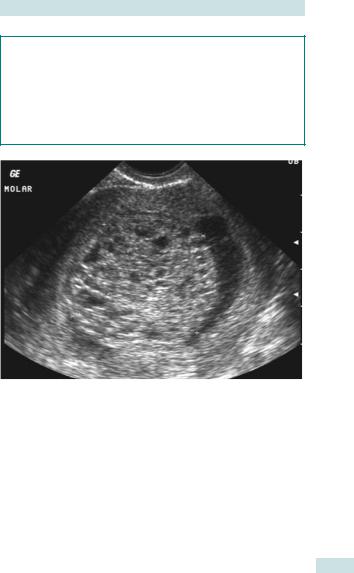
USS findings (see Fig. 23.1)
Complete mole
•‘Snowstorm’ appearance of mixed echogenecity, representing hydropic villi and intrauterine haemorrhage.
•Large theca lutein cysts.
Partial mole
Fetus may be viable, with signs of early growth restriction or structural abnormalities.
Fig. 23.1 Ultrasound of hydatidiform mole.

752 CHAPTER 23 Benign and malignant conditions
Hydatidiform mole: management
Management
•Complete mole: surgical evacuation is advisable and should be performed by an experienced surgeon as risks of uterine perforation and haemorrhage are significant. Oxytocin may be required to reduce the risk of haemorrhage, but its use is associated with a theoretical risk of tissue dissemination leading to metastatic disease to the lungs or brain and should be avoided until the uterus is evacuated if possible.
•Partial mole: surgical evacuation is preferable, unless the size of fetal parts necessitates medical evacuation.
•Histological examination of products of conception is essential to confirm diagnosis.
Treatment of persistent gestational trophoblastic disease
Risk of requiring chemotherapy is 15% after a complete mole and 0.5% after a partial mole.
Indications for chemotherapy
•Serum hCG levels >20 000IU/L at 4wks after uterine evacuation.
•Static or rising hCG after uterine evacuation in absence of new pregnancy.
•Persistent symptoms, e.g. uterine bleeding and/or abdominal pain.
•Evidence of metastases.
•Histological diagnosis of choriocarcinoma.
Prognosis
•With effective registration and treatment programme, cure rate is high (98–100%) with low chemotherapy rates (5–8%).
•Recurrence rate is low (1/55).
•Women should be advised not to conceive until hCG level has been normal for 6mths.
•hCG levels should be checked 6 and 10wks after each subsequent pregnancy.
Contraception and hormone replacement therapy
•Barrier contraception should be used until serum hCG is normal.
•The COCP and HRT are safe to use after hCG levels have returned to normal.

HYDATIDIFORM MOLE: MANAGEMENT 753
Specialist follow-up for molar pregnancy
In the UK, all women with any molar pregnancy should be registered at one of the three specialist centres (Sheffield, Dundee, London). The protocol for follow-up varies amongst the specialist centres. At Charing Cross Hospital (London), follow-up with hCG ranges from 6mths to 2yrs after uterine evacuation.
•Serum hCG should be checked fortnightly until levels are normal (<4IU/L).
•Following this, urine hCG is requested at 4-weekly intervals until 1yr post-evacuation, then every 3mths in the 2nd year of follow-up.
•If hCG normalizes within 8wks, follow-up will be limited to 6mths.
•Patients who do not have normal hCG values within 8wks of evacuation should have the 2-yr follow-up.
Further reading
Hydatidiform Mole and Choriocarcinoma UK Information and Support Service. M www. hmole-chorio.org.uk
RCOG. (2010). The management of gestational trophoblastic neoplasia, RCOG Guideline No. 38. M http://www.rcog.org.uk/files/rcog-corp/GT38ManagementGestational0210.pdf.
Sheffield Trophoblastic Tumour Screening and Treatment Centre. M http://www.chorio.group. shef.ac.uk/clin.html


GESTATIONAL TROPHOBLASTIC DISEASE: CHORIOCARCINOMA 755
Chemotherapy for GTD
•Chemotherapy continued until hCG normal for 6wks.
•More likely to have earlier menopause.
•Low risk patients (≤6):
•methotrexate and folinic acid (well tolerated with main side effects of mucositis and pleuritic chest pain)
•cure rate almost 100%.
•High risk patients (≥7):
•intensive weekly schedule of EMA (etoposide, methotrexate, and dactinomycin) alternating with CV (cyclophosphamide and vincristine)
•salvage surgery may be required (craniotomy, pleurotomy, hysterectomy)
•cure rate 95%
•increased risk of scancers
•small increased risk of miscarriage and stillbirth, but no increase in fetal abnormalities in subsequent pregnancy.
756 CHAPTER 23 Benign and malignant conditions
Principles of chemotherapy
Chemotherapy, together with radiotherapy and surgery, is used in the treatment of gynaecological cancers for control (and in some cases cure) of these diseases or palliation of their symptoms.
The cell cycle
•Chemotherapeutic agents interfere with cell division by acting on a specific phase of the cell cycle (see bPrinciples of chemotherapy, phases of the cell cycle, p. 757) (e.g. taxanes active against cells in G2/M) or non-specifically (e.g. alkylating agents exert their effects throughout the cell cycle).
•One of the characteristics of cancer cells is uncontrolled proliferation. As chemotherapy has a propensity for actively proliferating cells,
they are more vulnerable than normal cells. However, they do act on normal cells so side effects are not unusual (see bSide effects of chemotherapy: haematological and gastrointestinal, p. 758).
Practical aspects
Before embarking on chemotherapy, and indeed any other form of therapy, the intent of treatment (i.e. curative or palliative) for each patient should be discussed within the MDT and clearly explained to the patient.
It is important to weigh up the potential risks and anticipated benefits so that optimal survival and quality of life may be achieved. Factors to consider are:
•Disease-related:
•type and stage of cancer
•response to previous treatment.
•Patient-related:
•performance status (PS; the general condition of the patient); in general, chemotherapy is not indicated if PS >2
•concurrent medical problems
•nutritional status
•patient’s wishes.
•Chemotherapy is generally delivered on an outpatient basis:
•for most ovarian chemotherapy, each course of systemic treatment is composed of 6 cycles, depending on response assessed after the 3rd one
•each cycle lasts 21–28 days
•the gap between consecutive cycles enables damaged ‘normal’ cells to repair and regenerate.
•Response to treatment is assessed by:
•changes in evaluable disease on CT/MRI scans (RECIST (response evaluation criteria in solid tumours) criteria)
•trends in tumour markers (e.g. CA125 levels for ovarian cancer)
•symptoms
•signs.

PRINCIPLES OF CHEMOTHERAPY 757
Phases of the cell cycle
•G0: resting phase.
•G1: RNA and protein synthesis.
•S: DNA synthesis.
•G2: protein synthesis, mitotic spindle formation.
•M: mitosis.
ECOG performance status
•Grade 0: asymptomatic, fully active.
•Grade 1: restricted in strenuous activity by symptoms but fully ambulatory.
•Grade 2: symptomatic, able to self-care, but not work, in bed <50% of the day.
•Grade 3: symptomatic, in bed >50% of the day.
•Grade 4: confined to bed or chair, unable to self-care.
•Grade 5: dead.
RECIST criteria
•Complete: disappearance of all lesions with no evidence of new lesions on two occasions of at least 4wks apart.
•Partial: ≥30% decrease in sum of longest diameters of lesions.
•Stable disease.
•Progression: ≥20% increase in the sum of longest diameters of lesions or presence of new lesions.
Some classes of cytotoxic agents
•Antimetabolites: interfere with DNA and RNA synthesis, e.g. 5-FU, gemcitabine, methotrexate.
•Alkylating agents: form covalent bonds with DNA bases, e.g. cyclophosphamide, ifosfamide.
•Intercalating agents: bind to DNA, thus inhibiting its replication, e.g. cisplatin, carboplatin (most widely used in gynaecological cancer).
•Antitumour antibiotics: complex mechanism of action leading to inhibition of DNA synthesis, e.g. bleomycin, doxorubicin, etoposide.
•Drugs against spindle microtubules: prevent mitosis, e.g. paclitaxel, vincristine.

758 CHAPTER 23 Benign and malignant conditions
Side effects of chemotherapy: haematological and gastrointestinal
Chemotherapy is associated with a range of side effects due to its action on normal cells, as well as cancer cells. Pattern of toxicity varies between drugs, as well as between individuals. Most side effects are self-limiting. It is important to recognize and seek ways to prevent and manage them, whenever possible, to maintain good quality of life.
Haematological
Bone marrow suppression leads to a gradual fall in blood count, which eventually recovers. The nadir (period of lowest count) occurs around 7–14 days after chemotherapy.
•Neutropenic sepsis: potentially fatal, urgent action required
•neutropenia = neutrophil count ≤1.0 × 10⁹/L
•must have a high level of suspicion in any patient having chemotherapy presenting with a temperature of ≥38°C and feeling unwell
•take FBC, cultures—blood, urine, etc., start IV broad-spectrum antibiotics as per hospital protocol (e.g. pipercillin and tazobactum, and gentamicin), and inform an oncologist
•prophylaxis with granulocyte colony-stimulating factor (GCSF) and oral antibiotics, such as ciprofloxacin, may be appropriate if there have been previous episodes of neutropenic sepsis.
•Anaemia (Hb ≤11g/dL):
•may be seen after several cycles of chemotherapy
•treatment depends on severity and includes blood transfusion, iron tablets, erythropoietin.
•Thrombocytopaenia (platelet count ≤100):
•rarely problematic with chemotherapy used for gynaecological cancers
•avoid invasive procedures.
Gastrointestinal
Gastrointestinal side effects are due to loss of epithelial cells.
•Nausea and vomiting: can be prevented in most cases with effective antiemetics (e.g. steroids, domperidone, metoclopramide, and/or 5-HT3 antagonists).
•Mucositis:
•resolves spontaneously with epithelial healing
•helpful measures are good mouth care, use of local anaesthetic agents, treatment of oral candidiasis
•commonly occurs with methotrexate and 5-FU.
•Constipation: pay attention to diet and fluid intake; short-term use of laxatives.
•Diarrhoea:
•exclude infection/constipation
•control with codeine phosphate, loperamide, and rehydration.

SIDE EFFECTS OF CHEMOTHERAPY: OTHER 759
Side effects of chemotherapy: other
Alopecia
•Taxanes (e.g. paclitaxel), doxorubicin, and etoposide commonly cause temporary hair loss.
•Carboplatin and cisplatin are not usually associated with this side effect.
•Has a psychological impact on some women. Individual preference for wigs, scarves, or hats.
Neurological
These are dose-related side effects that slowly subside on dose reduction or on stopping the offending drug.
•Peripheral neuropathy:
•usually sensory changes such as numbness and tingling
•commonly seen with paclitaxel and cisplatin.
•Tinnitus: associated with cisplatin and taxanes.
Constitutional
These symptoms tend to have cumulative effects as treatment progresses, but resolve on its cessation.
•Lethargy.
•Anorexia.
Reproductive function
•Fetal abnormality:
•most chemotherapeutic agents are teratogenic and should be avoided during the 1st trimester. Some agents may be used after 12wks if necessary—data are generally reassuring but limited
•contraception is necessary until a period of time has elapsed following completion of treatment.
•Ovarian failure:
•more likely to be caused by alkylating agents than other chemotherapy
•permanent ovarian failure in premenopausal patients results in early menopause and infertility
•young women with premature menopause are predisposed to osteoporosis, cardiovascular disease, and post-menopausal
symptoms; hormone replacement therapy can be used without evidence of increase risk of cancer recurrence in most cancers.
•cryopreservation of embryos and ovarian tissue is possible for women who wish to preserve their fertility options prior to chemotherapy.


CHEMOTHERAPY FOR GYNAECOLOGICAL CANCER 761
Common chemotherapy regimens for gynaecological cancers
•Ovarian cancer:
•sensitive to platinum-based regimens, response rate ~70%
•carboplatin +/– paclitaxel (may be used neoadjuvantly, adjuvantly, or palliatively)
•>50% of patients will relapse and require further treatment; if >6mths have elapsed since initial chemotherapy, tumour more likely to be sensitive to carboplatin and therefore it is often given again
•Ip chemotherapy (instillation of agents such as cisplatin directly into the peritoneal cavity) has shown clinical benefit, but with increased toxicity; recommended to be used as part of clinical trial only).
•Endometrial cancer:
•chemotherapy has a limited role, reserved for recurrent or metastatic disease
•carboplatin +/– paclitaxel
•doxorubicin and cisplatin.
•Cervical cancer:
•cisplatin combined with radiotherapy has been shown to reduce risk of relapse for those undergoing radiotherapy after surgery
•cisplatin may be used for metastatic disease but the response rate is low.
•Vulval cancer:
•cisplatin can be used in combination with radiotherapy for patients unfit for surgery
•cisplatin may be used as sole therapy for symptom control in metastatic disease.
•Trophoblastic tumour:
•chemotherapy alone may be curative
•treatment given at specialist centres
•see bp. 750.


RADIOTHERAPY: SIDE EFFECTS 763
Radiotherapy: side effects
The side effects of radiotherapy depend on the site being irradiated. In gynaecological cancers this is the pelvis, and therefore tissues in this area are prone to damage. Problems may become apparent during and immediately after treatment (early effects) or occur months or years later (late effects).
Early side effects
•Due to damage of rapidly dividing cells such as the mucosa. Usually self-limiting.
•Skin:
•erythema
•moist desquamation
•management—aqueous cream, hydrocortisone cream.
•Mouth/bowel:
•mucositis—treat with mouthwash, analgesia, nystatin if thrush present
•nausea (less so vomiting)—antiemetics for prophylaxis or treatment
•diarrhoea—exclude infection, treat with loperamide or codeine phosphate.
•Bladder:
•cystitis causing frequency and dysuria
•management—exclude infection, ensure adequate fluid intake, oxybutynin may help.
•Tiredness:
•treat anaemia if present
•rest.
•Bone marrow suppression.

764 CHAPTER 23 Benign and malignant conditions
Radiotherapy: gynaecological cancers
Management of gynaecological cancers is multimodal in nature, combining surgery, chemotherapy, and radiotherapy. Surgery is the mainstay of treatment whenever possible. The choice of treatment depends on the type of cancer, the extent of disease, and the patient’s fitness and wishes.
Cervical cancer
•Early disease (stage Ib):
•radiotherapy is as effective as radical hysterectomy (cure rate ~80%)
•for those who have had surgery, radiotherapy may be given afterwards (adjuvant treatment) to reduce the risk of pelvic recurrence.
•Locally advanced disease (stage II–IVa): adjuvant use of external beam radiotherapy and brachytherapy ± chemotherapy.
•Recurrent disease: localized recurrence may be treated by surgical resection or radiotherapy.
•Distant metastatic disease (stage IVb): best supportive care, palliative radiotherapy; or palliative chemotherapy.
Endometrial cancer
•Early disease (stage I):
•if patient unfit for surgery, radiotherapy used as primary treatment
•if fit for surgery, brachytherapy +/– external beam therapy to the pelvis adjuvantly, especially if high risk features present (e.g. poorly differentiated tumour, deep myometrial involvement).
•Locally advanced disease (stage II–IIIc): postoperative (adjuvant) radiotherapy.
•Recurrent disease: as for cervical cancer.
Vulval cancer
•Early disease: primary radiotherapy if unfit for surgery.
•Locally advanced disease:
•radiotherapy to the pelvis after surgery if lymph nodes found to be involved
•radiotherapy in combination with chemotherapy being evaluated to shrink extensive disease before surgery.
•Recurrent disease: may be possible if the maximum dose has not been exceeded.
Ovarian cancer
•No evidence to support the use of radiotherapy in the primary or adjuvant setting.
•Radiotherapy may have a role in the palliation of localized symptoms.
Radiotherapy for symptom palliation
Radiotherapy is used to control some symptoms caused by metastatic disease. Examples are:
•Pain from bone/brain metastases.
•Bleeding from fungating tumour.
This page intentionally left blank

PAIN AND ITS MANAGEMENT 767
WHO analgesic ladder
•Step 1: non-opioid (e.g. paracetamol, NSAIDs).
•Step 2: weak opioid (e.g. codeine) + non-opioid.
•Step 3: strong opioid (e.g. morphine) + non-opioid.
Side effects of opioids
•Commonly:
•nausea
•constipation
•sedation (advise amitriptyline should be taken 3 h before bed to avoid morning ‘hangover’).
•Inform patient of potential problems.
•Give antiemetics and laxatives.
1 Do not allow concerns about potential dependence prevent you from prescribing adequate amounts of opiates.

768 CHAPTER 23 Benign and malignant conditions
Symptoms of advanced gynaecological cancer
Gastrointestinal symptoms
Nausea and vomiting |
|
|
|
• Causes: |
|
|
|
• |
ascites |
• |
bowel obstruction |
• |
constipation |
• |
brain metastases |
• |
metabolic (e.g. Ca2+) |
• |
medication. |
•Treat cause, if possible:
•radiotherapy and corticosteroids for brain metastases
•paracentesis for ascites
•hydration and bisphosphonates for hypercalcaemia.
•Antiemetics.
Constipation |
|
|
|
• Causes: |
|
|
|
• |
dfluid intake |
• |
metabolic (e.g. Ca2+) |
• |
immobility |
• |
medication (especially |
• |
lethargy |
|
opioids). |
•bowel obstruction
•Treat cause, if possible.
•Laxatives.
(Subacute) bowel obstruction
•Caused by tumour seedling within the peritoneum.
•Exclude constipation with AXR.
•Best to manage conservatively; however, if patient is reasonably fit and there is only one area of obstruction, surgery may be an option.
Other symptoms
•Vaginal discharge and bleeding:
•may be infective, therefore give metronidazole
•tranexamic acid +/– embolization for bleeding.
•Fistulae: difficult to manage, limited role for surgery.
•Fatigue: dexamethasone may give a sense of well-being.
•Anorexia and cachexia:
•caused by the secretion of pro-inflammatory cytokines
•dexamethasone or megestrol/medroxyprogesterone may improve appetite
•total parenteral nutrition is rarely indicated.
•Dyspnoea:
•causes—pleural effusion, cachexia, lymphangitis, anaemia, lung metastases, pulmonary embolism
•suspect pulmonary embolism if acute onset of shortness of breath.

PRINCIPLES OF PALLIATIVE CARE 769
Principles of palliative care
The WHO defines palliative care as the ‘active, holistic care of patients with advanced, progressive illness’.
Good communication and rapport with the patient (and her family) are essential to facilitate frank discussion of prognosis and formulate a plan of care that is most suited to her.
Goals of palliative care
•To achieve optimal symptom relief.
•To promote the best quality of life.
In order to palliate effectively, it is necessary to individualize each patient’s care and take her wishes into account. Aspects to explore include:
•Physical symptoms.
•Psychological symptoms.
•Social issues.
•Spiritual issues.
Multidisciplinary approach
•Symptoms may be controlled by pharmacological or non-pharmacological means.
•The expertise of different specialties may be needed depending on the nature of the patient’s problems, e.g. advice from dietitians about nutrition, from clinical oncologists about radiotherapy for bone pain, from surgeons about feasibility of resection for bowel obstruction.
•Palliative care is delivered at home, in a hospice, or in hospital.
Terminal care
•Institute Liverpool Care Pathway.
•Aim for a peaceful and dignified death.
•Maintain comfort and offer psychosocial-spiritual support.
•Avoid unnecessary/uncomfortable procedures.
•Review medication. Most can be stopped, but continue the following:
•analgesics
•antiemetics
•anxiolytics
•anticonvulsants.
This page intentionally left blank