

SOGC JOINT CLINICAL PRACTICE GUIDELINE Colposcopic management of abnormal cervical cytology and histology
.pdfSOGC joint clinical practice guideline
No. 284, December 2012
Colposcopic Management of Abnormal
Cervical Cytology and Histology
This clinical practice guideline has been prepared by the Executive Council of the Society of Canadian
Colposcopists and approved by the Society of Obstetricians and Gynaecologists of Canada/Society of Gynecologic Oncology of Canada/Society of Canadian Colposcopists Policy and Practice Guidelines Committee, the Executive and Council of the Society of Gynecologic Oncology of Canada and the Executive and Council of the Society of Obstetricians and Gynaecologists of Canada.
PRINCIPAL AUTHOR
James Bentley, MB ChB, Halifax NS
THE EXECUTIVE COUNCIL OF THE SOCIETY OF CANADIAN COLPOSCOPISTS
James Bentley, MB ChB, Halifax NS
Monique Bertrand, MD, London ON
Lizabeth Brydon, MD, Regina SK
Hélène Gagné, MD, Ottawa ON
Brian Hauck, MD, Calgary AB Marie-Hélène Mayrand, MD, Montreal QC Susan McFaul, MD, Ottawa ON
Patti Power, MD, St.. John’s NL Alexandra Schepansky, MD, Edmonton AB Marina Straszak-Suri, MD, Ottawa ON
SPECIAL CONTRIBUTORS
Terry Colgan, MD, Toronto ON Laurette Geldenhuys, MD, Halifax NS Mark Heywood, MD, Vancouver BC Roberta Howlett, PhD, St.. Thomas ON Linda Kapusta, MD, Mississauga ON Rachel Kupets, MD, Toronto ON Joan Murphy, MD, Toronto ON
Jill Nation, MD, Calgary AB
Vyta Senikas, MD, Ottawa ON
Michael Shier, MD, Toronto ON
Disclosure statements have been received from all authors..
Abstract
Objective: To provide a guideline for managing abnormal cytology results after screening for cervical cancer, to clarify the appropriate algorithms for follow-up after treatment, and to promote the best possible care for women while ensuring efficient use of available resources..
Outcomes: Women with abnormal cytology are at risk of developing cervical cancer; appropriate triage and treatment will reduce this risk.. This guideline will facilitate implementation of common standards across Canada, moving away from the current trend of individual guidelines in each province and territory..
Evidence: Published literature was retrieved through searches of PubMed or Medline, CINAHL, and The Cochrane Library in October 2008 using appropriate controlled vocabulary (e..g.., colposcopy, cervical dysplasia) and key words (e..g.., colposcopy management, CIN, AGC, cervical dysplasia, LEEP, LLETZ, HPV testing, cervical dysplasia triage).. Results were restricted to systematic reviews, randomized control trials/controlled clinical trials, and observational studies.. There were no date or language restrictions.. Searches were updated on a regular basis and incorporated in the guideline to July 2012.. Grey (unpublished) literature was identified through searching the websites of health technology assessment and health technology assessment-related agencies, clinical practice guideline collections, and national and international medical specialty societies..
Expert opinion from published peer-reviewed literature and evidence from clinical trials is summarized.. Consensus opinion is outlined when evidence is insufficient..
Values: The quality of the evidence is rated using the criteria described by the Canadian Task Force on Preventive Health
Care (Table 1)..
Validation: This guideline has been reviewed for accuracy from content experts in cytology, pathology, and cervical screening programs.. Guideline content was also compared with similar documents from other organizations including theAmerican Society for Colposcopy and Cervical Pathology, the British Society for Colposcopy and Cervical Pathology, and the European Cancer Network..
J Obstet Gynaecol Can 2012;34(12):1188–1202
Key Words: Cervical cytology, cervical cancer, colposcopy, treatment, follow-up, abnormalities, guidelines
This document reflects emerging clinical and scientific advances on the date issued and is subject to change. The information should not be construed as dictating an exclusive course of treatment or procedure to be followed. Local institutions can dictate amendments to these opinions. They should be well documented if modified at the local level. None of these contents may be reproduced in any form without prior written permission of the SOGC.
1188 • DECEMBER JOGC DÉCEMBRE 2012

Colposcopic Management of Abnormal Cervical Cytology and Histology
Table 1. Key to evidence statements and grading of recommendations, using the ranking of the Canadian Task Force on Preventive Health Care
Quality of evidence assessment* |
Classification of recommendations† |
|
|
|
|
I: |
Evidence obtained from at least one properly randomized |
A.. There is good evidence to recommend the clinical preventive action |
|
controlled trial |
|
II-1: Evidence from well-designed controlled trials without randomization
II-2: Evidence from well-designed cohort (prospective or retrospective) or case–control studies, preferably from more than one centre or research group
II-3: Evidence obtained from comparisons between times or places with or without the intervention.. Dramatic results in uncontrolled experiments (such as the results of treatment with penicillin in the 1940s) could also be included in this category
III:Opinions of respected authorities, based on clinical experience, descriptive studies, or reports of expert committees
B..There is fair evidence to recommend the clinical preventive action
C..The existing evidence is conflicting and does not allow to make a recommendation for or against use of the clinical preventive action; however, other factors may influence decision-making
D..Thereisfairevidencetorecommendagainsttheclinicalpreventiveaction
E..There is good evidence to recommend against the clinical preventive action
L..There is insufficient evidence (in quantity or quality) to make a recommendation; however, other factors may influence decision-making
*The quality of evidence reported in these guidelines has been adapted from The Evaluation of Evidence criteria described in the Canadian Task Force on Preventive Health Care..95
†Recommendations included in these guidelines have been adapted from the Classification of Recommendations criteria described in the Canadian Task Force on Preventive Health Care..95
Recommendations
Managing Cytological Abnormalities
The age of the patient may affect management and where pertinent will be identified in the recommendation..
The Colposcopy Examination
01.. Colposcopic findings are best described according to the terminology defined by the International Federation for Cervical Pathology and Colposcopy.. (III-C)
ABBREVIATIONS
AC |
adenocarcinoma |
AGC-N |
atypical glandular cells-favour neoplasia |
AGC-NOS |
atypical glandular cells-not otherwise specified |
AGUS |
atypical glandular cells of undetermined significance |
AIS |
Adenocarcinoma in situ |
ALTS |
ASCUS/LSIL Triage Study for Cervical Cancer |
ASC-H |
atypical squamous cells-cannot exclude |
|
high-grade squamous intraepithelial lesion |
ASCUS |
atypical squamous cells of undetermined |
|
significance |
CIN |
cervical intraepithelial neoplasia |
ECC |
endocervical curettage |
HPV |
human papillomavirus |
HR-HPV |
high-risk HPV |
HSIL |
high-grade squamous intraepithelial lesion |
LEEP/ LLETZ |
loop electrosurgical excision procedure/large loop |
|
excision of the transformation zone |
LSIL |
low-grade squamous intraepithelial lesion |
Pap smear |
Papanicolaou smear |
SCC |
squamous cell carcinoma |
TZ |
transformation zone |
02.. At colposcopy, 2 or more biopsy specimens should be taken.. (I-A)
03.. An endocervical curettage should be performed when the transformation zone is not visible in women with an AGC Pap smear and in women over 45 years old with high-grade cytology.. (II-2B)
04.. Routine HR-HPV testing for all colposcopy referrals is discouraged.. (III-C)
Managing Women With ASCUS or LSIL on Referral for Colposcopy
05.. A woman with persistent ASCUS/LSIL or ASCUS HR-HPV positive cytology should be referred for colposcopy as directed by provincial/territorial guidelines.. (III-A)
06.. A colposcopically identified lesion should be biopsied.. (III-C)
07.. If no lesion is identified, biopsies of the transformation zone should be considered.. (III-C)
Managing ASC-H
08.. A woman with an ASC-H Pap smear should have colposcopy to rule out CIN 2 or 3 and/or cancer.. (II-2A)
09.. Biopsies should be performed on any identifiable lesions at colposcopy.. (II-2A)
10..With an ASC- H Pap smear, the finding of negative colposcopy does not automatically warrant a diagnostic excisional procedure.. (III-E)
Managing HSIL
11..All women with an HSIL test result should have colposcopy.. (II-2A)
12..In the absence of an identifiable lesion at colposcopy, whether satisfactory or unsatisfactory, an endocervical curettage and directed biopsies should be performed.. (III-B)
13..In women with HSIL, when the transformation zone is not seen in its entirety and endocervical curettage and/or biopsy results
DECEMBER JOGC DÉCEMBRE 2012 • 1189

SOGC joint clinical practice guideline
are negative, a diagnostic excisional procedure should be considered.. (III-B)
Managing Atypical Glandular Cytology
(AGC-NOS, AGC-N, AIS)
14..The finding of an AGC Pap smear warrants colposcopy.. (II-2A)
15..All women with an AGC Pap smear should have an endocervical curettage.. Women over 35 years of age or with a history of abnormal bleeding should have endometrial sampling.. (II-2A)
16..Women with an AGC-N Pap smear without an identifiable lesion at colposcopy should undergo a diagnostic excisional procedure.. (II-2A)
Managing Squamous Cell Carcinoma and Adenocarcinoma on Cytology
17..Women with a cytologic diagnosis suggestive of carcinoma, with or without a visible lesion, should have colposcopy and appropriate biopsies.. (III-A)
Managing the Patient With Abnormal HPV
Test and Normal Cytology
18..Women less than 30 years old should not have HR-HPV testing done as a screen with cytology.. (II-2E)
19..Women less than 30 years old who are HR-HPV positive and have normal cytology should be followed as per provincial/ territorial guidelines; colposcopy is not required.. (III-B)
20..Women 30 years old and over who test positive for HR-HPV and have negative cytology should have HR-HPV and cytology testing repeated at 12 months.. Persistent positive HR-HPV tests warrant colposcopy.. (I-A)
Managing Abnormal Cytology in Pregnancy
21..Women with anASC-US or LSILtest result during pregnancy should have repeat cytology testing at 3 months post pregnancy.. (III-B)
22..Pregnant women with HSIL, ASC-H, or AGC should be referred for colposcopy within 4 weeks.. (III-B)
23..Endocervical curettage should not be performed during pregnancy.. (III-D)
Managing Abnormal Cytology in
Women Less Than 21 Years Old
24..Cytological screening should not be initiated in women less than 21 years old.. (II-2E)
25..If screening is done in a woman less than 21 years old, and an ASC-US or LSIL result is reported, cytology should be repeated only per provincial or territorial guidelines.. (III-B)
26..A woman less than 21 years old who has cytology results of
ASC-H, HSIL, and AGC should be referred for colposcopy.. (III-B)
Managing Histological Abnormalities
Managing CIN 1
27..The preferred option for biopsy-proven CIN 1 is observation with repeat assessment at 12 months with cytology testing.. (Colposcopy at 12 months is an acceptable option..)
Management should be according to the cytology result.. (II-1B)
28..In the case of a patient with biopsy-proven CIN 1 after HSIL or AGC, cytology and histology should be reviewed, where available.. If a discrepancy remains, then an excisional biopsy may be considered.. (III-B)
Managing CIN 2 or 3 in Women
Aged 25 Years and Over
29..CIN 2 or 3 should be treated.. Excisional procedures are preferred for CIN 3.. (II-1A)
30..Women who have positive margins should have follow-up with colposcopy and directed biopsies and/or endocervical curettage.. Treatment for recurrent or persistent CIN 2 or 3 should be by excision.. (II-1B)
Managing CIN 2 or 3 in Women Less Than 25 Years Old
31..The pathologist should be asked to clarify whether the lesion is CIN 2 or CIN 3.. (III-B)
32..CIN 2 in women less than 25 years old should be observed with colposcopy at 6-month intervals for up to 24 months before treatment is considered.. (II-2B)
33..CIN 3 in women less than 25 years old should be treated.. (III-B)
Managing Adenocarcinoma in Situ
34..If AIS is diagnosed, treatment needs to be a diagnostic excisional procedure, or type 3 transformation zone excision.. (II-2A)
35..If margins are positive after diagnostic excisional procedure, a second excisional procedure should be performed.. (II-2A)
36..If after treatment for AIS a woman has finished childbearing, a hysterectomy should be considered.. (III-B)
37..If AIS is diagnosed after LEEP is performed for CIN in a woman who has not completed her family and margins are negative, it is unnecessary to perform a further diagnostic excisional procedure.. (II-2E)
Managing Histological Abnormalities During Pregnancy
38..If CIN 2 or CIN 3 is suspected or diagnosed during pregnancy, repeat colposcopy and treatment should be delayed until 8 to 12 weeks after delivery.. (II-2A)
Follow-up Post Treatment for CIN 2 or 3
Either option is acceptable:
39..Women should be followed with cytology testing and colposcopy at 6-month intervals for 2 visits.. If both cytology and any biopsies are negative, they will then return to screening per provincial/ territorial guidelines.. (II-2B)
40..HPV testing at 6 months combined with cytology testing is acceptable.. If both are negative, women may return to screening per provincial/territorial guidelines.. (II-2B)
Managing Histological Abnormalities in Women at High Risk
41..Immunocompromised women do not require screening colposcopy.. (II-2D)
Wait Times for Colposcopy
42..Women with ASC-H or AGC should be seen in a colposcopy clinic within 6 weeks of referral.. (III-C)
43..Women with HSIL should ideally be seen in a colposcopy clinic within 4 weeks of referral.. (III-C)
44..Women with a Pap smear suggestive of carcinoma should be seen in a colposcopy clinic within 2 weeks of referral.. (III-C)
45..All other women with abnormal results should be seen in a colposcopy clinic within 12 weeks of referral.. (III-C)
1190 • DECEMBER JOGC DÉCEMBRE 2012

Colposcopic Management of Abnormal Cervical Cytology and Histology
INTRODUCTION
Over the last 30 years cervical cancer morbidity and mortality rates have dropped significantly in Canada, from approximately 30 per 100 000 to 7 per 100 000 women.1 This change has been widely attributed to the
availability of cervical screening via cytologic sampling.2
Colposcopy has evolved to become a tool for evaluating those with abnormal cytology and obtaining histological samples by biopsy. Treatment of lesions can then be performed, usually preserving fertility and making major surgery unneccesary.3 Numerous jurisdictions have developed guidelines4–8 for colposcopy, and these have been reviewed in developing this document.
Cervical cancer screening is organized within each province and territory in Canada. Screening programs issue screening and follow-up recommendations for abnormal screening results, including referral for colposcopy. The diversity and status of cervical screening in Canada has been summarized elsewhere.9
A review of the recommended age for initial screening was initiated by the American Society of Colposcopy and Cervical Pathology, which in June 2009 convened a consensus practice improvement conference that included representatives from the United States and Canada. Outcomes from this meeting included a recommendation to start screening at age 21.10 This recommendation has been incorporated into new guidelines from Quebec,11 Alberta,12 and Ontario.13
Canadian colposcopic practice is unique in several ways. Colposcopy is performed predominantly by gynaecologists in both hospital clinics and private offices. Access to HPV testing is currently limited outside teaching hospitals. The primary aim of these guidelines is to standardize the colposcopic care provided for women in Canada.
COLPOSCOPIC MANAGEMENT OF
CYTOLOGICAL ABNORMALITIES
Screening and colposcopy recommendations vary across provinces and territories and have been documented elsewhere.9 Current guidelines for colposcopic referrals can be summarized as follows: referral for colposcopy is recommended for persistent ASCUS, persistent or incident LSIL, ASC-H, HSIL, and AGC, as well as for Pap smears that suggest squamous or glandular carcinoma. HPV testing is not widely available; however, when reflex HPV testing shows the presence of oncogenic or HR-HPV with ASCUS cytology, referral for colposcopy is recommended.
THE COLPOSCOPY EXAMINATION
Colposcopy is the examination of the lower genital tract and cervix using magnification from a colposcope with a good light source. The squamocolumnar junction and transformation zone should be identified; this determines whethertheexaminationissatisfactoryornot.Aceticacidis then used to assess the size, shape, margin, and location of any neoplastic lesion. These findings can then be described according to the new nomenclature of the International Federation for Cervical Pathology and Colposcopy.14
When any lesion is identified, recent evidence supports the practice of taking at least 2 biopsy specimens to improve the accuracy of colposcopy. A biopsy should be performed and specimens taken of the most severe area found on colposcopic examination to confirm or rule out malignant lesions.15,16 Analysis of the ALTS data showed that taking 2 biopsies for a low-grade cytology referral at initial colposcopy improved the sensitivity (to detect CIN 2 or greater) to 81.8%, compared with 68.3% with 1 biopsy.15
A recent review of the utility of endocervical curettage was published using data from Calgary. Using data from over 13 000 examinations, the authors showed that 99 ECC specimens had to be taken to detect 1 additional case of CIN 2 or higher grade lesion. The largest benefit was in older women referred after high-grade cytology.17 An ECC should thus be performed in the case of unsatisfactory colposcopy, anAGCsmear,andinolderwomenwithhigh-gradecytology.
A low threshold is recommended for undertaking a biopsy. If any lesion is seen, biopsy should be completed. If only metaplasia is in question, a biopsy should be considered. Unless dictated by the appropriate algorithm, there is no role for routine HR-HPV testing in the colposcopy clinic.
Clinical Tip: HPV Vaccination
A woman’s visit to the colposcopy clinic is an opportune time to discuss HPV vaccination with her.. Even if she has not been previously vaccinated a woman may benefit from vaccination to prevent new HPV infections..
Recommendations
1. Colposcopic findings are best described according to the terminology defined by the International Federation for Cervical Pathology and Colposcopy. (III-C)
2. At colposcopy, 2 or more biopsy specimens should be taken. (I-A)
3. An endocervical curettage should be performed when the transformation zone is not visible in women with an AGC Pap smear and in women over 45 years old with high-grade cytology. (II-2B)
DECEMBER JOGC DÉCEMBRE 2012 • 1191

SOGC joint clinical practice guideline
4. Routine HR-HPV testing for all colposcopy referrals is discouraged. (III-C)
MANAGING WOMEN WITH ASCUS OR LSIL ON REFERRAL FOR COLPOSCOPY
Management of low-grade abnormalities remains controversial. A large randomized trial in the United States concluded that women with LSIL cytology results were best managed by immediate referral for colposcopy; it was noted that 83% were positive for HR-HPV and thus HPV triage would not be effective.18 The same study reported that women with ASCUS results, but negative for HRHPV, could safely be triaged away from colposcopy.18 This approach requires access to reflex HPV testing, which unfortunately is not widely available in Canada. A recent multicentre study in the United Kingdom evaluated the management of similar low-grade cytology. The study concluded that “there was no clear benefit of a policy of immediate colposcopy as although it detects more cervical intraepithelial neoplasia grade II or more severe disease, it leads to a large number of referrals with no high grade cervical intraepithelial neoplasia [and] overtreatment with associated after effects in young women.”19
With low-grade lesions, colposcopy is done to rule out potentially premalignant changes i.e., CIN 2 or 3; if these are detected, management is undertaken according to the appropriate protocol. A meta-analysis reported CIN 2+ ratesof 10%andCIN3+of 6%withanASCUSreferral.20,21 With an LSIL referral, the rates of CIN 2+ are 17% and CIN 3+ 12%.22,23 If CIN 1 is the highest grade identified at colposcopy, conservative management is recommended. If no lesion is identified at colposcopy, a random biopsy at the transformation zone should be considered. As per consensus opinion, if no dysplasia is identified at colposcopy, annual screening with the referring health care provider is recommended until 3 negative Pap smears have been reported. If all cytology is negative, women may then be followed every 2 to 3 years, consistent with provincial/ territorial guidelines.
Recommendations
5. A woman with persistent ASCUS/LSIL or ASCUS HR-HPV positive cytology should be referred for colposcopy as directed by provincial/territorial guidelines. (III-A)
6. A colposcopically identified lesion should be biopsied. (III-C)
7. If no lesion is identified, biopsies of the transformation zone should be considered. (III-C)
MANAGING ASC-H
With an ASC-H result on the Pap smear, significant pathology is typically found in the majority of cases. In a study of 517 cases from Edmonton, Alberta, CIN 2 or greater was detected in 70% of cases. Most cases were CIN 2; however, invasive carcinoma was reported in 2.9% of cases and AIS in 1.7%.24 A similar Ontario study showed CIN 2 or greater in 59.4% of cases with a stronger correlation in women under 40 years of age.25 All women with ASC-H should have colposcopy to rule out significant pathology. If colposcopy is negative, recommendations include repeat colposcopy, repeat cytology and, ideally, HR-HPV testing twice, at 6-month intervals, to ensure a significant lesion is not missed. If these repeat tests are negative, women may return to regular screening, as per provincial/territorial protocol. The finding of ASC-H with negative colposcopy does not warrant a cone biopsy or excisional procedure for diagnostic purposes.
Recommendations
8. A woman with an ASC-H Pap smear should have colposcopy to rule out CIN 2 or 3 and/or cancer. (II-2A)
9. Biopsies should be performed on any identifiable lesions at colposcopy. (II-2A)
10.With an ASC-H Pap smear, the finding of negative colposcopy does not automatically warrant a diagnostic excisional procedure. (III-E)
MANAGING HSIL
The risk of a significant lesion is high with HSIL cytology. Studies have shown CIN 2 or greater in 53% to 66% of cases in which colposcopic biopsies are undertaken, and up to 90% if an immediate LEEP is performed.26,27 Because of this high rate of significant high-grade histology, all women with an HSIL result should have colposcopy. A visual assessment and LEEP approach may be appropriate in some circumstances, but a colposcopically directed biopsy and tailored treatment is preferred.
If a lesion is not detected at colposcopy and colposcopy is not satisfactory, then a diagnostic excisional procedure should be done. This can be achieved with a cone biopsy, or LEEP using a large loop, or a second endocervical pass. However, if no lesion was detected and colposcopy was satisfactory, combined colposcopy and cytology is appropriate at 6-month intervals for 2 visits. This situation is rare. For women who have finished childbearing, a diagnostic excisional procedure should be considered.
1192 • DECEMBER JOGC DÉCEMBRE 2012

Colposcopic Management of Abnormal Cervical Cytology and Histology
Recommendations
11.All women with an HSIL test result should have colposcopy. (II-2A)
12.In the absence of an identifiable lesion at colposcopy, whether satisfactory or unsatisfactory, an endocervical curettage and directed biopsies should be performed. (III-B)
13.In women with HSIL, when the transformation zone is not seen in its entirety and endocervical curettage and/or biopsy results are negative,
a diagnostic excisional procedure should be considered. (III-B)
MANAGING ATYPICAL GLANDULAR
CYTOLOGY (AGC-NOS, AGC-N, AIS)
The finding of AGC-NOS, AGC-N, or AIS always warrants prompt referral for colposcopy in the absence of other symptomatology. Neoplastic lesions found in areas other than the cervix, including endometrium, ovary, and fallopian tube, have been identified with AGC cytology.28–30 In a Canadian report, 456 cases of AGC or AGUS were identified from a database of over 1 million Pap smears (0.043%).29 On final histologic follow-up, 7% were found to have CIN 1, 36% were found to have CIN 2 or 3, AIS was identified in 20%, carcinoma of the cervix in 9%, and endometrial pathology in 29%, including carcinoma of the endometrium in 10%. It should be noted that CIN is consistently the most frequent finding across many studies.28-31 This high rate of pathology precludes any attempt to triage using repeat cytology or HPV testing.
The diagnosis of AGC-N is associated with higher rates of abnormalities and thus, in the absence of an abnormality found by colposcopy, a diagnostic excisional procedure should be performed.32,33 These procedures includes cold knife cone biopsy and laser cone biopsy and may include LEEP if the specimen is of sufficient size. A hysterectomy is not considered a diagnostic excisional procedure. Endocervical curettage should be done in all women, and endometrial sampling should be performed in women who are over 35 years or who have a history of abnormal bleeding, including anovulation.
However, with AGC-NOS cytology and the absence of an identified lesion, women are still at risk of developing a lesion. In this situation, follow-up assessment every 6 months for 2 years includes repeat cytology testing, colposcopy, and ECC. If HR-HPV testing is available and was done at the initial colposcopy visit, women who test negative for HR-HPV may have repeat assessment
with colposcopy, cytology testing, ECC, and HR-HPV testing at 12 months. If a lesion is identified, treatment is in accordance with the guideline specific to the type of lesion. If a carcinoma is identified, referral should be made to a gynaecologic oncologist. If all follow-up is negative after 2 years, routine cytologic testing may be resumed.
Recommendations
14.The finding of an AGC Pap smear warrants colposcopy. (II-2A)
15.All women with an AGC Pap smear should have an endocervical curettage. Women over 35 years of age or with a history of abnormal bleeding should have endometrial sampling. (II-2A)
16.Women with an AGC-N Pap smear without an identifiable lesion at colposcopy should undergo a diagnostic excisional procedure. (II-2A)
MANAGING SQUAMOUS CELL CARCINOMA AND ADENOCARCINOMA ON CYTOLOGY
Women should be referred promptly for colposcopy if theirPap smearissuggestiveof carcinoma, withor without a visible lesion. Assessment should include colposcopy and directed biopsy, and ECC should be considered. If no abnormality is detected, a diagnostic excisional procedure isrecommendedtoruleoutoccultcarcinoma.Endometrial biopsy should also be contemplated in the workup of women with adenocarcinoma found by Pap smear.
Recommendation
17.Women with a cytologic diagnosis suggestive of carcinoma, with or without a visible lesion, should have colposcopy and appropriate biopsies. (III-A)
MANAGING THE PATIENT WITH ABNORMAL HPV TEST AND NORMAL CYTOLOGY
Women with ASCUS and positive reflex HR-HPV should be referred for colposcopy. However, no provincial guidelines address management of negative cytology findings combined with a positive HR-HPV result.
Women with negative cytology and positive HPV results should have both tests repeated with their primary health care provider after 12 months.34,35 If both tests are negative at 12 months, women should return to being screened according to provincial/territorial guidelines. Women with a cytological abnormality should be managed according to the cytological diagnosis. If there is persistent HR-HPV on 2tests1yearapart,referralforcolposcopyisrecommended to rule out the possibility of a high-grade lesion.
DECEMBER JOGC DÉCEMBRE 2012 • 1193

SOGC joint clinical practice guideline
Recommendations
18.Women less than 30 years old should not have HRHPV testing done as a screen with cytology. (II-2E)
19.Women less than 30 years old who are HR-HPV positive and have normal cytology should be followed as per provincial/territorial guidelines; colposcopy is not required. (III-B)
20.Women 30 years old and over who test positive for HR-HPV and have negative cytology should have HR-HPV and cytology testing repeated at 12 months. Persistent positive HR-HPV tests warrant colposcopy. (I-A)
MANAGING ABNORMAL
CYTOLOGY IN PREGNANCY
The indications for colposcopy in pregnant women are essentially the same as in non-pregnant women. If a lowgrade lesion (ASCUS or LSIL) is found during pregnancy, the Pap smear should be repeated 3 months postpartum. This practice is safe as the rate of cancer in this group is very low.36 If HSIL, ASC-H, or AGC is found, prompt evaluation with colposcopy is essential. If colposcopy is unsatisfactory in the first trimester, it should be repeated after20weeks’gestationwhen,becauseof thephysiological changes, the cervix everts itself and the squamocolumnar junction may become visible.
If CIN3orcarcinomaissuspected,biopsyisrecommended. Thereisevidencethatbiopsyinpregnancyisnotharmful.37 Women with high-grade dysplasia in pregnancy should be seen by an experienced colposcopist.
Recommendations
21.Women with an ASC-US or LSIL test result during pregnancy should have repeat cytology testing at 3 months post pregnancy. (III-B)
22.Pregnant women with HSIL, ASC-H, or AGC should be referred for colposcopy within 4 weeks. (III-B)
23.Endocervical curettage should not be performed during pregnancy. (III-D)
MANAGING ABNORMAL CYTOLOGY IN THE WOMAN LESS THAN 21 YEARS OLD
There is little evidence that screening by cytology testing in adolescents (< 21 years old) is beneficial. The incidence of cervicalcancerisverylow.TheSurveillance,Epidemiology, and End Results data from the United States showed a rate of 0.1/100 000 in women 15 to 19 years old and 1.6/100 000 in women 20 to 24 years old, compared with 15.5/100 000 in women 40 to 45 years old.38 Although
HPV infection and low-grade abnormalities found by Pap smears are common in this age group, most of these infections and related cytological changes will resolve without intervention.39,40 Screening is invasive and can have adverse psychological sequelae, especially if it leads to colposcopy referral.10,41
If this screening leads to treatment by LEEP, this can later be associated with a slightly increased risk of premature rupture of membranes and preterm delivery.42,43 HPV vaccination has recently been instituted in Canada, and the high efficacy against HPV 16 and 18 should result in fewer high-grade lesions needing treatment.44-48 The American Congress of Obstetricians and Gynecologists and several Canadian organizations have therefore recommended that screening begin at 21 years of age.11–13,49–52
In women less than 21 years, if a Pap smear has been done and abnormalities are detected at screening, management should be conservative to prevent harm. Low-grade changes, i.e., ASC-US and LSIL, regress in up to 93% of cases with conservative management. Thus women less than 21 years with ASC-US and LSIL results should have cytology testing repeated in 1 year, with referral to colposcopy only if abnormalities persist for 24 months.10 Women less than 21 years with ASC-H, HSIL, or AGC results should be referred for colposcopy.
Recommendations
24.Cytological screening should not be initiated in women less than 21 years old. (II-2E)
25.If screening is done in a woman less than 21 years old, and an ASC-US or LSIL result is reported, cytology should be repeated only per provincial or territorial guidelines. (III-B)
26.A woman less than 21 years old who has cytology results of ASC-H, HSIL, and AGC should be referred for colposcopy. (III-B)
MANAGING HISTOLOGICAL ABNORMALITIES
Once a lesion has been identified on colposcopy and biopsy has been completed, a decision must be made regarding management. The aim of treatment is to remove a potentially precancerous lesion to prevent development of carcinoma. The initial classification of cervical intraepithelial neoplasia as CIN 1, 2, or 3 was proposed by Richart in 1973 and reinforced by the World Health Organization in 1994.53 The natural history of dysplastic lesions has been more fully elucidated in recent years, and this has led pathologists to adopt a 2-tiered cytologic terminology to describe histological lesions. A recent consensus project led by the College
1194 • DECEMBER JOGC DÉCEMBRE 2012

Colposcopic Management of Abnormal Cervical Cytology and Histology
Table 2. Evolution of cervical cancer precursors53
|
|
|
|
Progression |
|
Regression, |
Persistence, |
Progression to |
towards invasive |
CIN grade |
% |
% |
CIN 3, % |
cancer, % |
CIN 1 |
57 |
32 |
11 |
1 |
CIN 2 |
43 |
35 |
22 |
5 |
CIN 3 |
32 |
< 56 |
not known |
> 12 |
|
|
|
|
|
of American Pathologists and the American Society for Colposcopy and Cervical Pathology recommended the following terminology for HPV-associated squamous lesions: “Low-grade squamous intraepithelial lesion and high-grade squamous intraepithelial lesion,” which may be further classified using the CIN terminology.51 This guideline uses the CIN terminology. The rate of progression of these dysplastic lesions has been well reviewed by Ostor52 (Table 2), and over time the therapeutic approach has been adapted to prevent harm when lesser CIN grades are unlikely to progress to invasive cancer.
Treatment modalities include excisional and ablative approaches (cryotherapy or laser ablation). The favoured method in Canada is excisional, the loop electrosurgical excisionprocedure,whichisrelativelyeasytoperformasan outpatient procedure, although there can be complications. A recent meta-analysis estimated that after a LEEP procedure the risk for preterm delivery in a subsequent pregnancy of less than 32 to 34 weeks’ gestation, was 1 in 143 treatments.42 The same research group suggested that a depth threshold of 10 mm is also a variable in reducing harm. Consequently, if the colposcopist is able to adjust the procedure to the lesion, negative sequelae in future pregnancy may be minimized.54
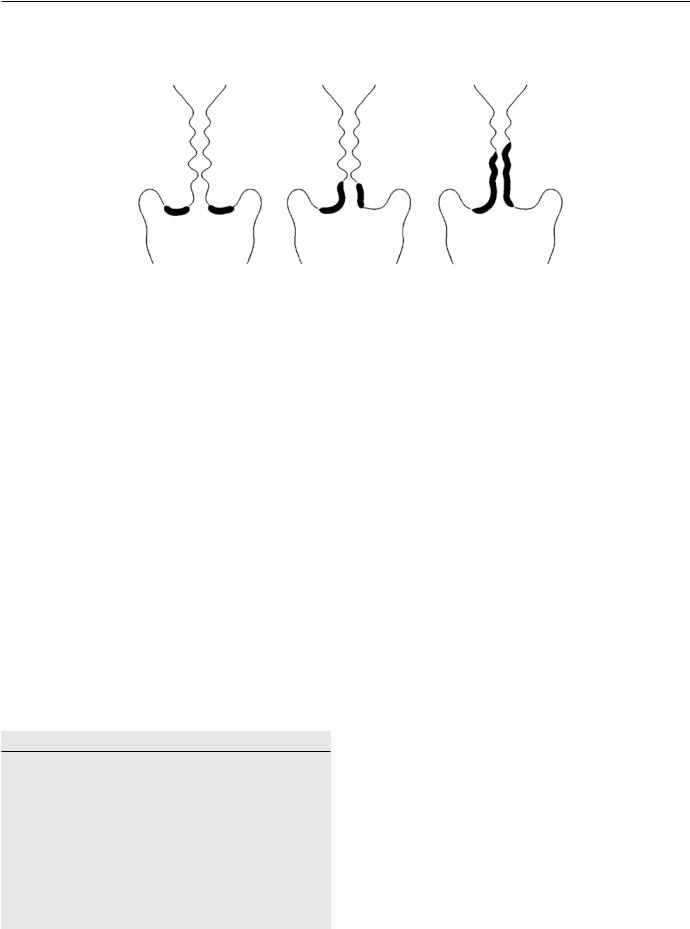
Treatment is tailored to the lesion identified on the cervix by either removal or ablation of the entire transformation zone. The International Federation of Cervical Pathology andColposcopyhasclassifiedthetransformationzoneinto three categories.17 A type 1 TZ is completely ectocervical and fully visible. A type 2 TZ is fully visible, has an endocervical component, and may have an ectocervical component. A type 3 TZ is predominantly endocervical, not fully visible, and may have an ectocervical component (Figure).
Using this classification, ablative methods can be used for a type 1 or 2 TZ if the following criteria are met: the TZ must be fully visible; a colposcopically directed diagnostic biopsy must be taken from the most dysplastic area in the TZ; there must be no suspicion of invasive disease; there
must be no suspicion of glandular disease; there is no cytological/histological disparity; and the patient has not had previous treatment.
Cryotherapy is not recommended for treatment of CIN 3.55
If excision with LEEP is used the size of loop electrode must be adjusted depending on the lesion: a type 2 TZ requires a larger loop electrode than a type 1 TZ to ensure the lesion is fully excised. If the lesion is not seen in its entirety, colposcopy is unsatisfactory and ablative therapies should not be used.17,56 Care should be taken to avoid removal of excessive cervical stroma, which would predispose women to preterm delivery, especially if very large loops are used or multiple passes taken.
A type 3 TZ with a lesion that extends into the endocervical canal or a glandular lesion requires a larger or longer excision for adequate evaluation or treatment. This document adopted the new the International Federation of Cervical Pathology and Colposcopy terminology to identify this procedure as a type 3 excision to avoid the current confusion in terminology.17 Currently, cone biopsy, diagnostic excisional procedure, laser excision and LEEP may be used but have different meanings to individual colposcopists.56
Managing CIN 1
Evidence from the recent ALTS trial has confirmed significant interobserver variability in the histological diagnosis of CIN 1, with the overlap often observed with benign HPV infection.57 Our current understanding is that CIN 1 seldom progresses to invasive disease and that it will regress without treatment within 2 to 5 years in 60% to 80% of all cases.54,57 Regression rates are even more pronounced in adolescents, with regression of low-grade squamous intra-epithelial lesions in up to 91% of cases over a 3-year period.56 This knowledge has led to a change in the treatment philosophy for CIN 1.
Conservative management with observation is preferred forCIN1.Womenshouldbefollowedwithrepeatcytology testing at 12 months, which can be done by the primary
DECEMBER JOGC DÉCEMBRE 2012 • 1195
SOGC joint clinical practice guideline
Transformation zone categories
Type I |
Type II |
Type III |
completely ectocervical |
has an endocervical |
has an endocervical |
|
component |
component |
fully visible |
fully visible |
not fully visible |
small or large |
may have ectocervical |
may have ectocervical |
ectocervical component |
component, which may |
component, which may |
|
be small or large |
be small or large |
|
|
|
care provider; any subsequent management should be according to the cytology result. If the patient requires further colposcopy and the CIN lesion persists for 24 months or longer, treatment is acceptable. If colposcopy is satisfactory, treatment may be by ablative modalities. However in an adherent patient, longer follow-up is possible, especially in women who have not completed childbearing.
The exception to a conservative approach occurs when a diagnosis of CIN 1 is preceded by HSIL or AGC cytology. Inthesesituations,histologicalfindingshavenotadequately explainedtheabnormalcytology.If specimensareavailable the cytology and histology should be reviewed and if the discrepancy is confirmed an excisional procedure may be considered.
Recommendations
27.The preferred option for biopsy-proven CIN 1 is observation with repeat assessment at 12 months with cytology testing. (Colposcopy at 12 months is an acceptable option.) Management should be according to the cytology result. (II-1B)
28.In the case of a patient with biopsy-proven CIN 1 after HSIL or AGC, cytology and histology should be reviewed, where available. If a discrepancy remains, then an excisional biopsy may be considered. (III-B)
Managing CIN 2 or 3 in Women 25 Years Old and Over
Pathologically confirmed high-grade dysplasia includes CIN 2 and CIN 3, which are treated in the same fashion in most jurisdictions.7,58-62 There are, however, differences in the rates of regression. The seminal review by Ostor showed that CIN 2 regressed in 43% of cases and progressed to CIN 3+ in 27%.52 This compares with regression of 33%, persistence of 52%, and progression to invasion in at least 12% of CIN 3 cases57 (Table 2). The true malignant potential of CIN 3 has been demonstrated in New Zealand by long-term follow-up of CIN 3 that was not treated. This showed that the invasive risk in untreated CIN 3 is 31% over 30 years. The study also noted that patients with documented persistent CIN 3 for 2 years had a 50% risk of subsequently developing invasive disease.63
For these reasons, most women with CIN 2 or 3 should be treated. Women who are younger or pregnant are managed as outlined elsewhere in this document. If colposcopy is satisfactory,i.e.,atype1or2TZ,excisionandablativetherapy are both acceptable; however, an excisional procedure is preferred for the treatment of CIN 3. If CIN 2 or 3 is identified and colposcopy is unsatisfactory, an excisional procedureshouldbeperformed.Womenareatincreasedrisk of persistent dysplasia if at treatment margins are positive for CIN, or the ECC (if done) is positive. In a meta-analysis of excisional treatment, the risk of post-treatment disease
1196 • DECEMBER JOGC DÉCEMBRE 2012

Colposcopic Management of Abnormal Cervical Cytology and Histology
was 18% for incomplete excision and 3% for complete excision.64 If the deep margins are involved, consideration should be given to repeat excision. Most women should have colposcopy repeated at 6 months.65 Hysterectomy is not recommended as initial therapy for CIN 2 or 3 but may be performed for women with persistent CIN.
Recommendations
29.CIN 2 or 3 should be treated. Excisional procedures are preferred for CIN 3. (II-1A)
30.Women who have positive margins should have follow-up with colposcopy and directed biopsies and/or endocervical curettage. Treatment for recurrent or persistent CIN 2 or 3 should be by excision. (II-1B)
Managing CIN 2 or 3 in Women Less Than 25 Years Old
As discussed earlier there is little evidence to justify routine screening in the adolescent patient. If, however, Pap screening is completed, these patients may be referred for colposcopy. Management must be modified to prevent harm. Recent evidence suggests that regression of CIN 2 in this population occurs at a rate similar to CIN 1.10,40,66,67
The evidence suggests that CIN 2 in the adolescent can be observed with repeat colposcopy and cytology every 6 months for up to 24 months. If dysplasia persists, the patient should be treated, with either ablative methods or LEEP. This is conditional on a satisfactory colposcopy. If colposcopy is unsatisfactory, treatment should consist of an excisional procedure. A recent study of regression rates of CIN 2 in women less than 25 years old (most of whom were 20 to 25 years old) found that the overall regression rate over a median of 8 months was 62%. This suggests that observation may be reasonable in women less than 25 years old.18 In some centres, high-grade histology is designated as HSIL, i.e., CIN terminology is not used. If the biopsy specimen is reported as HSIL in an adolescent woman we recommend a review of the histology using CIN terminology as suggested by the College of American Pathologists/American Society
of |
Colposcopy and Cervical Pathology consensus. |
If |
specimen is reclassified as CIN 3, treatment by an |
excisional method is preferred.
Recommendations
31.The pathologist should be asked to clarify whether the lesion is CIN 2 or CIN 3. (III-B)
32.CIN 2 in women less than 25 years old should be observed with colposcopy at 6-month intervals for up to 24 months before treatment is considered. (II-2B)
33.CIN 3 in women less than 25 years old should be treated. (III-B)
Managing Adenocarcinoma in Situ
In Canada the ratio of adenocarcinoma to squamous carcinoma of the cervix is increasing: adenocarcinoma accounts for 20% to 25% of all cervical cancer.68 This is largely because widespread availability of screening by Pap smears over several decades has led to a significant decrease in squamous cell cancers. Nevertheless, implementation of cytology quality assurance initiatives in recent years has been associated with a decrease in adenocarcinoma of the cervix.
In contrast, diagnosis of premalignant adenocarcinoma in situ occurs at a ratio of 1:50 when compared with severe squamous dysplasia.69 Consequently a colposcopist will not often see AIS, and the treatment remains controversial. Colposcopic features can be difficult to identify, and lesions often extend high into the canal.70 Bertrand and colleagues showed that in 78% of cases the highest lesion in the canal was less than 20 mm from the exocervix and none were higher than 29.9 mm.71 After a diagnosis of adenocarcinoma in situ, made by punch biopsy or endocervical curettage, a diagnosticexcisionalprocedure,ortype3TZexcisionshould be performed. Margin status is an important predictor of residual disease, and thus the method chosen for treatment must preserve the ability to assess the endocervical margin. A recent meta-analysis of 33 studies showed that the risk of residual disease was 2.6% with negative margins and 19.4% with positive margins. Invasive carcinoma was also more frequently associated with positive margins (5.2%) than with negative margins (0.1%).72 Thus, if margins are positive, a second excision is required.
If AIS is diagnosed after a LEEP procedure is completed (becauseof aCINfinding),themarginsneedtobecarefully examined. If the AIS is small and margins are clear, there is no need to perform an excisional procedure unless childbearing is complete, when hysterectomy should be considered.73 If fertility is not an issue or negative margins cannot be achieved, a hysterectomy is recommended.72
After treatment for AIS, if the woman wishes to preserve her fertility, she can be closely observed in the colposcopy clinic. She should be seen for colposcopy, ECC, and cytology testing every 6 to 12 months, for at least 5 years. HR-HPV testing can be used to reassure the patient. Thereafter the patient should have annual cytology testing.
Recommendations
34.If AIS is diagnosed, treatment needs to be a diagnostic excisional procedure, or type 3 transformation zone excision. (II-2A)
DECEMBER JOGC DÉCEMBRE 2012 • 1197