

Color.Atlas.of.Endodontics-ublog.tk
.pdf68 |
Color Atlas of Endodontics |
FIGURE 4-1 Scanning electron microscopic view of an unprepared type II canal in a mandibular incisor.
FIGURE 4-2 Scanning electron microscopic view of a prepared type II canal in a mandibular incisor. Note the areas of debris and uncleaned canal space.
A B
FIGURE 4-3 A, C-shaped morphology in this mandibular second molar complicates instrumentation. B, Resin cast of a C-shaped canal from a mandibular second molar.
irrigant should be nontoxic, capable of dissolving both
vital and necrotic pulp tissue, kill bacteria, lubricate, and remove the smear layer.3-10 At least one study has found that the smear layer seems to inhibit bacterial growth." Presently, no single irrigant possesses all of these traits.
The most common intracanal irrigant used is sodium hypochlorite. Other irrigants that have been studied alone or in combination with sodium hypochlorite in-
clude 3 % hydrogen peroxide, ethylenediaminetetraacetic acid (EDTA), citric acid, and lactic acid. 10,12-20
Sodium hypochlorite has been shown to be an effective antimicrobial agent when placed in contact with bacteria. 21-23 However, bacteria are never totally elimi nated because a sufficient volume of irrigant does not reach all aspects of the canal space or dentinal tubules.
To facilitate penetration of the canal system, the use of 27or 28-gauge irrigating needles with a Luer-Lok syringe is suggested. As the apical preparation approaches a size 45 file, the ability of the solution to reach the apical extent of the canal through a 27-gauge needle improves.24 One study comparing closed-tip needles with a

Chapter Four Canal Preparation |
69 |
side portal with other irrigation techniques found these syringe tips were the most effective in removing dye from plastic artificial canals . 25
Sonic and ultrasonically activated devices are capable of delivering larger volumes of irrigant in smaller canals, but they require special equipment and instruments. 26,27 Few of these current vibrating devices are capable of delivering sodium hypochlorite because of its corrosive action on aluminum components.
Lubricating pastes are often employed in canal preparation with sodium hypochlorite. In addition to facilitating placement of the file, these pastes also entrap debris. Dentin chips are held in suspension and are less likely to be packed in the apical portion of the canal.
Instruments
Over the second half of the twentieth century, advances in endodontic instruments and techniques allowed millions of previously condemned teeth to be saved. The early pioneers in endodontics recognized the significance of complete debridement and disinfection of the canal space in ensuring eventual endodontic success. However, their initial selection of endodontic files and reamers from various manufacturers was limited. In a cooperative effort, both the American Dental Association (ADA) Council on Dental Materials and the American Dental Manufacturers worked together to develop instrument standardization and ADA specification No. 28. 28
In the early 1970s, most accepted instrumentation techniques emphasized hand debridement with minimal use of handpiece-driven instruments except for orifice enlargement. Specification No. 28 was developed for handoperated K-type files and reamers and was first published in 1976. This specification established tip diameter, taper (increase of diameter for each millimeter of length), torsional limits, and other physical attributes for files. Speci-
fication No. 58 was developed for Hedstrom files and was published in 1982.29 Revised standards for specifications No. 28 and No. 58 were published in 1989.-10
Presently, no approved standards are available for rotary instruments, but the American National Standard/ ADA Council on Scientific Affairs has proposed Specification No. 95 for root canal enlargers. 31 This document is based largely on published reports from a Marquette University group that examined the torsional, bending, and metallurgical properties of all stainless steel engineand carbon steel engine-driven endodontic enlargers. 32-36 No recognized standards are available for nickeltitanium instruments; however, the manufacturers must meet the United States Food and Drug Administration's (FDA's) Good Manufacturing Practice Regulations before introducing a new device.
The following discussion of canal preparation techniques assumes that all files or reamers are hand operated unless otherwise stated. Instrumentation techniques that require specific instruments are identified at the time of their discussion.
FIGURE 4-4 The standardized preparation technique resulted in procedural errors when used in a curved canal. Dentin was removed from the inner wall of the canal coronal to the curve and from the outer wall of the canal apical to the curve.This resulted in an "hourglass" preparation. In the apical portion the canal was transported, or "zipped;' when larger, less flexible instruments were used at the correct working length.
Canal Preparation Techniques
From a historical perspective, clinical techniques tend to build on previously successful techniques. Therefore the various canal preparation techniques will be introduced in chronologic order, beginning with hand debridement techniques using stainless steel instruments. A few assumptions are made here to facilitate understanding of the differences among the techniques. These assumptions may not apply in actual clinical cases. The primary goal of all these techniques is to shape the canals to the apical constriction of the canal space, regardless of the radiographic appearance of the actual tooth. The guidelines for shaping outlined by Schilder are followed in this discussion . 2
Before initiating any treatment, the clinician should evaluate each case and determine the degree of difficulty. As the complexity increases, the ability to clean and shape decreases, and the potential for procedural errors increases. Procedural errors include loss of working length, transportation of the apex (or zipping), apical perforation, lateral stripping, and instrument breakage (Figure 4-4). The following criteria are suggested for evaluation of cases before treatment:
Root length-Longer roots are generally more difficult to treat.
Root width-Narrow, curved roots are at risk for apical and lateral stripping perforations.

7 0 |
Color Atlas of Endodontics |
Canal size-Small canals are more difficult to prepare and may not exhibit any natural taper.
Canal curvature-Difficulty increases as curves progress from gentle to sharp dilacerations.
Calcifications-Calcification makes location and negotiation more difficult.
Resorptions-Resorptions present potential problems for negotiation, cleaning, shaping, and obturation.
Restorations-Restorations may change the orientation of the tooth in the dental arch, block canals, and restrict vision.
Previous treatment-Teeth exhibiting previous root canal treatment are more difficult to manage for a variety of reasons. Normal anatomic landmarks may have been removed, procedural errors may be present, and debris in the root canal system may be difficult to remove.
STANDARD TECHNIQUE. John Ingle was a member of the first committee to propose standardization of endodontic instruments and introduced the classic "standardized preparation." 17 Standardized files were used sequentially to produce a canal preparation that had the same size and shape (taper) as the last standardized instrument used. The canal could then be obturated with a filling material that was also the same size and shape. Essentially, the canal was made to fit the filling material. At this same time, obturation with silver points having the same shape as the files was an accepted and popular obturation technique.
The technique was easy to perform in straight canals of mature teeth exhibiting natural taper but posed problems in small, curved canals. As the instruments got larger, the ability to finesse the stiff instruments to different lengths decreased because of the restoring force of the metal. This often resulted in ledging, apical transportation, and apical perforation, or "zipping" (see Figure 4-4). 38-40
STEP-BACK TECHNIQUE. The realization that curved canals may require less aggressive instrumentation resulted in the introduction of the step-back preparation technique.41 Several comparative debridement studies have been performed that demonstrate the superiority of the step-back preparation over the standard technique. 42 The step-back technique emphasizes keeping the apical preparation small, in its original position, and producing a gradual taper. The working length is established and then the first file to bind is set as the master apical file (MAF). Subsequent larger files are introduced at 1-mm or shorter increments (Figure 4-5). After each step back, the canal is irrigated and the MAF replaced to the established working length to remove any loosened debris. The step-back "telescoped" preparation produces a canal with greater taper compared with the standard technique and results in more dentin removal and cleaner canal walls. 43,44
FIGURE 4-5 A, To reduce procedural errors encountered with the standardized preparation technique, the step-back technique was developed. After the working length and MAF were established, successive instruments were shortened by 1-mm increments and used to develop a more tapered preparation. B, Canal bed enlargement permits the development of a tapered preparation using a more flexible process. After working length determination and establishment of the MAF, successive instruments are introduced to the initial point of binding and then rotated one half turn. No attempt is made to force the instrument to the working length or artificial predetermined length.
As an alternative to the step-back preparation, Schilder advocated canal bed enlargement .2 After determining the length determination and establishing the largest MAF, the clinician places the next larger instrument into the canal to the point of initial contact and rotates it one half turn. Force is not applied to the instrument in an apical direction and no effort is made to advance the instrument to the working length. The canal is then irrigated and the process repeated with increasingly larger instruments (see Figure 4-5). The technique allows the body of the canal to be prepared without the procedural errors inherent in the standardized preparation technique. In addition, the technique permits the natural morphology of the canal to influence
the preparation, in contrast to the more ridged incremental step-back technique. After completion of the tapering process, Schilder advocates the use of Gates Glidden drills in the canal orifice to remove coronal obstructions.
Weine emphasized pre-curving files to minimize canal alteration. However, pre-curving does not guarantee total canal symmetry and alterations occur despite the best efforts of the clinician. 45-47
Chapter Four Canal Preparation |
71 |
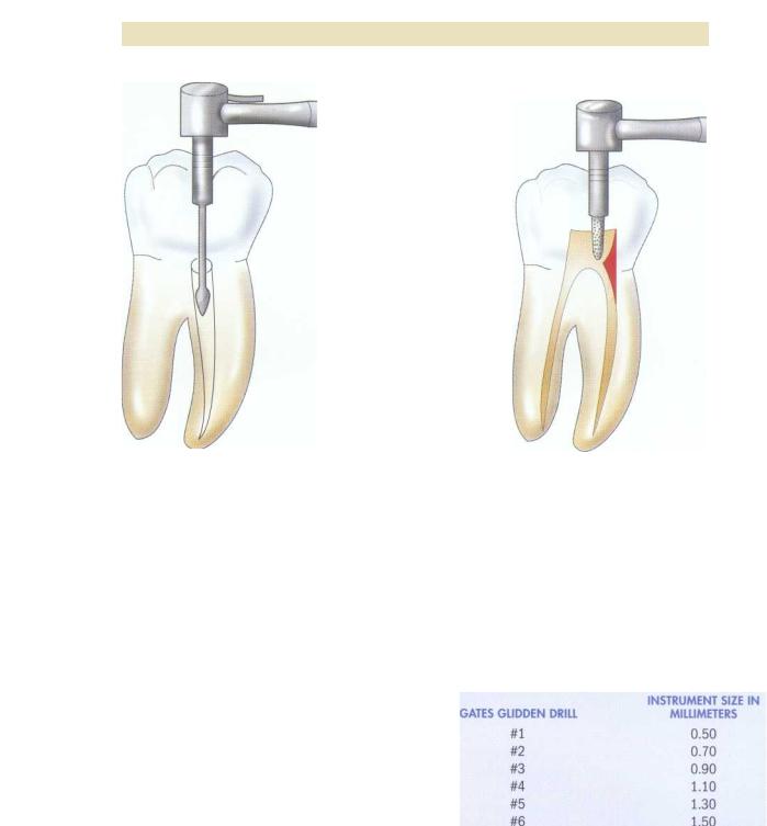
FIGURE 4-6 Gates Glidden drills are used in the coronal portion of the canal for orifice enlargement and straight line access. Before using the Gates Glidden drills, the clinician should evaluate the sizes of the canal and root as they relate to the diameter of the instrument.
FIGURE 4-7 Straight line access is facilitated by removing overhanging ledges of dentin before using the Gates Glidden drills. This can be accomplished by either slowor high-speed instrumentation.
Later variations of the original step-back technique incorporated the use of Gates Glidden #2 , #3, and #4
drills to help gain straight line access to the apical third of the canal (Figure 4-6).44,48,49 This facilitated the plac-
ing of a tapered instrument into the canal; previously, progress toward the apex was impeded when the diameter of the tapered file exceeded the diameter of the cylindrical canal. Before using Gates Glidden drills, the clinician facilitates access to the canal orifice by removing overlying dentinal structure (Figure 4-7).
Coronal enlargement before apical preparation provides straighter access to the apical region; eliminates interferences and canal irregularities in the coronal two thirds of the root; permits deeper placement of instruments that otherwise might not go to length; removes the bulk of tissue, debris, and microorganisms; and allows deeper penetration of irrigating solutions. One disadvantage of coronal enlargement and the use of Gates Glidden drills is the excessive removal of tooth structure. A comparison of the size of the Gates Glidden drills to standardized files is presented in Box 4-1. The routine use of #5 and #6 Gates Glidden instruments results in excessive removal of tooth structure and increases the incidence of stripping perforations in the furcation area.
The Gates Glidden drills are used after a size 25 MAF can be introduced to the corrected working length.
BOX 4-1
Comparison o f Gates Glidden Drills with Standardized Files
After lubricating the chamber with an irrigant, a #2 bur is placed into the canal orifice and run at medium speed with a light force. The deepest penetration of the canal occurs with the #2 Gates Glidden. Next, the #3 Gates Glidden is used at a shorter length and directed to the perimeter of the canal (Figure 4-8). Finally, a #4 Gates Glidden is used to the depth of the head of the bur to finalize the straight line access. This step helps establish the space for the hand files to reach the apical third without interference.

72 |
Color Atlas o f Endodontics |
FIGURE 4-8 Although the #1 and #2 Gates Glidden drills are used to remove dentin uniformly from the canal walls, the #3 to #6 drills have the strength to permit lateral movement and selective removal of tooth structure away from the furcation or danger zone. Directing the Gates Glidden drill toward the line angles in molars enhances straight line access and reduces the risk of stripping perforations.
STEP-DOWN TECHNIQUE. A variation on the timing of the use of the Gates Glidden drills was presented by Goerig.50 He advocated using a #4 round bur to establish coronal access, followed by Hedstrom files size 15, 20, and 25 in the coronal two thirds of the estimated canal length. Only a filing or rasping motion is used and apical patency is maintained with a size #10 K-file. Patency should be checked as necessary. A #2 Gates Glidden drill is then used 14 to 16 mm in the canal, a #3 Gates Glidden is inserted 11 to 13 mm into the canal, and a #4 Gates Glidden is used as needed by the canal size. This procedure allows straight line access to the apical one third before active instrumentation takes place, thereby eliminating many of the interferences usually encountered.
Several studies have supported this general technique with respect to reducing procedural accidents such as stripping perforations and apical transportation. Greater apical enlargement without apical transportation can be achieved if coronal obstructions are eliminated. 51,52 One group of authors demonstrated significant improvement in tactility with files following coronal pre-flaring.-53 In a direct comparison between pre-flaring before the establishment of working length and flaring after working length establishment, Morgan and Montgomery found that the "crown-down pressureless" technique resulted in a rounder canal shape
significantly more often than when the step-back flaring technique was used. 54
PASSIVE STEP-BACK TECHNIQUE. The passive step-back technique developed by Torabinejad uses a combination of hand and rotary instruments to develop a flared preparation.55 This technique provides gradual enlargement of the root in an apical to coronal direction without the application of force and reduces the risk of procedural accidents caused by transportation. It is a modification of the canal bed enlargement advocated by Schilder, in which shaping consisted of placing instruments larger than the MAF to a point of first binding and then using a reaming action to enlarge the coronal portion of the canal. This permits the morphology (shape) of the canal to influence the preparation and differs from the arbitrary step-back procedures of millimeter increments. 2
The passive step-back technique involves establishing a corrected working length using a #15 file. The #15 file is inserted to the corrected working length with light pressure and then rotated one eighth to one quarter turn. Additional K-type files between #20 and #40 are then inserted passively as far as they will go and rotated one eighth to one quarter turn with light pressure. Gates Glidden drills (#2 to #4) are then used coronally, and apical preparation is accomplished. Narrow, curved canals should not be enlarged beyond a #25 or #30 file. The apical portion of the canal is prepared by placing sequentially larger files passively in the canal and rotating them one eighth to one quarter turn. Advantages to the passive step-back technique include knowledge of canal morphology, removal of debris and minor canal obstructions, and a gradual passive enlargement of the canal in an apical to coronal direction. With this process the canal morphology influences the preparation shape.
The passive step-back technique has also been advocated for use with ultrasonic instruments after the working length is established. Copious irrigation with sodium hypochlorite is suggested. A #15 endosonic file is passively placed in the canal, driven ultrasonically for 30 seconds, and worked circumferentially. Hand instruments are then introduced into the canal. This alternating use of ultrasonic and hand instruments is performed in a stepwise fashion until a mildly flared canal is achieved. A #2 Gates Glidden drill is then inserted until it binds, withdrawn 1 to 1.5 mm, and activated with an up-and-down motion. A #3 Gates Glidden drill is used after irrigation with 2.5% sodium hypochlorite. Working length is then reestablished, and the apical preparation is completed.
The balanced force technique was developed and introduced by Roane. 56 This technique employs a unique instrument tip design and method of cutting dentin. One principle of the balanced

Chapter Four Canal Preparation |
73 |
A B
FIGURE 4-9 A, In the balanced force technique the file is placed to working length and rotated clockwise 90 degrees with light pressure to engage dentin. B, The file is then rotated counterclockwise 120 degrees while apical pressure is maintained to cut and enlarge the canal. Debris is removed with a final clockwise rotation that loads the flutes with loosened debris.
force technique is the recognition that traditional K-type files have pyramidal tips with cutting angles that can be quite aggressive with clockwise rotation. Roane modified traditional K-type files to reduce the transitional angle at the tip, producing a parabolic shape that alters the ability of the instrument to respond to distortion in a concentrated area. Forces are distributed over the file flutes and are not concentrated at the tip. The subsequent Flex-R (Union Broach, York, PA) file has a unique "safe" tip design with a guiding land area behind the tip that allows the file to follow canal curvature without binding in the outside wall of the curved canal. In addition, Roane advocated the use of a triangular crosssectional instrument because the decreased mass of the instrument and its deeper cutting flutes improve flexibility and decrease the restoring force of the instrument when placed in a curved canal.
Before instrumentation, #1 to #6 Gates Glidden drills are used for straight line access. In balanced force instrumentation, the files cut in both clockwise and counterclockwise rotation. Instruments rotated in a clockwise direction tend to move apically as the instrument engages the dentinal wall, pulling the instrument into the canal. Instruments rotated in a counterclockwise direc-
tion tend to move coronally or out of the canal. Rotation of the instrument 120 degrees ensures that each blade reaches the beginning position of the initial blade and enlarges the entire canal.
In the balanced force technique the file is placed to working length and rotated clockwise 90 degrees with light pressure to engage the dentin (Figure 4-9). The file is then rotated counterclockwise 120 degrees while apical pressure is maintained to cut and enlarge the canal. Further counterclockwise rotation ensures enlargement of the full diameter of the canal. A final clockwise rotation then loads the flutes of the file with loosened debris, and the file is withdrawn. An advantage to the technique is the ability to manipulate the files at any point in the
canal without ledging or blockage.
In small, curved canals, one study using extracted teeth demonstrated that the apical canal preparation up to a size 40 MAF remained centered in the original canal space in 80% of the cases . 57 Another study found less debris extrusion with the balanced force technique com-
pared with step-back filing and endosonic filing.58 Overall, the technique has been shown to reduce
canal transpiration compared with other techniques .59-61 However, one shortcoming of the technique is that the

74 |
Color Atlas o f Endodontics |
use of #5 and #6 Gates Glidden drills results in a higher incidence of stripping perforations. Also, no clinical studies have demonstrated superior success rates over other techniques. 62
NICKEL-TITANIUM INSTRUMENTS. In 1988, researchers at Marquette University published a study examining endodontic files that had been fabricated from nitinol orthodontic wire for use as hand instruments.63 Nickeltitanium (nitinol) alloy possesses a modulus of elasticity that is one fourth to one fifth that of stainless steel and has a wide range of elastic deformation, resulting in greater flexibility. An advantage of this increased flexibility is that the file follows the canal curvature with less deformation of the curvature during enlargement. 64 Better instrument centering in the apical preparation has been reported for both engine-driven and hand-manipulated nickel-titanium instruments. 61,66 A disadvantage of increased flexibility is the inability to pre-curve the file for introduction into canals of posterior teeth when the size of the interocclusal opening is decreased. Because of its greater elasticity, the cutting efficiency of nickel-titanium files may be reduced with clinical use compared with stainless steel. 67-69 Therefore, to take advantage of the properties of nickel-titanium files, engine-driven instruments have been developed. The present nickel-titanium
instruments incorporate a U-shaped groove with a flat land area. When the instrument is rotated, the flutes
plane the canal wall while the land area keeps the instrument centered, especially in fine, curved canals.70 -73 As might be expected, instrumentation techniques with the newer nickel-titanium files have been evolutionary as well as revolutionary. After the initial introduction of nickel-titanium files, step-back preparations using hand files and engine-driven files were common. Dentsply, Tulsa Dental (Tulsa, OK) has always advocated the use of a "crown down" technique with their Profile brand instruments. However, changes in instrument design over the past several years have resulted in at least three different sets of instruments (Profile, Quantec, and Lightspeed) and subsequent instrumentation
techniques (see Chapter 5).
EVALUATION OF CANAL PREPARATION
TECHNIQUES
Several techniques have been advocated for the evaluation of root canal debridement and enlargement. Plastic blocks with uniform, preformed curves have been used by many researchers to evaluate the appearance of the canal before and after the instrumentation technique. 26,27,42,74 This methodology provides some answers to general questions of instrument action or irrigation patterns, but does not address concerns regarding the starting point for teeth with cross-sections and curves that are far more complex than the circular artificial
canal. In addition, these studies do not provide information regarding the cleaning phase of treatment.
Some researchers have performed histologic evalua-
tions of remaining debris or pulp tissue while relying on estimates for dentinal walls that were planed.44,75,76 The
limitations of these studies lie in the fact that no method exists to "pre-test" for existing debris and therefore all results are post-test estimates. These histologic studies are extremely time-consuming and can also have artifacts from processing errors. They do not lend themselves to evaluation of the complete length of the canal.
An attempt at a "pre-test, post-test" model was developed by Bramante et a l.77 The model consisted of teeth that were mounted in dental stone or acrylic that could then be sectioned and reassembled in a jig. 77-79 This allows photographs or micrographs of the canal to be made before instrumentation. Post-instrumentation evaluation can then compare several of the aforementioned criteria with the differences found between the two photographs or micrographs. Limitations include tooth loss resulting from sectioning, a lack of precision in the instruments used to compare before and after instrumentation, and an inability to observe the entire length of the canal.
Recently a technique has been developed that eliminates many of the shortcomings of the previous studies. A three-dimensional, nondestructive technique for detailing root canal geometry by means of high-resolution tomography has been developed. 80 A micro-computed tomography scanner (microCT, cubic resolution of 34 microns) is used to record the precise canal anatomy before and after instrumentation. A three-dimensional analysis of root canal geometry by high-resolution computed tomography is then performed. The first report of this technique studied hand instrumentation with K-flex files, Lightspeed rotary instruments, Profile .04 taper rotary instruments, and GT rotary files. They found no dif-
ferences in volume removed, canal straightening, and the amount of untreated area among the techniques. Signifi-
cantly, they reported that all the techniques left more than a third of the canal surface uninstrumented. 81 This advance in the technology of canal preparation evaluation leads to only one conclusion-all instrumentation techniques fail to debride 100% of the canal space.
CLINICAL SIGNIFICANCE
Canal preparation is only one phase of endodontic treatment. Although current instruments and techniques vary, straight line access, step-back instrumentation, and apical preparation are three common concepts (Figure 4-10). Major differences in technique involve the order in which each step is accomplished and the instruments used. Although clinicians may use a specific process routinely, each case is unique and astute clinicians modify their techniques when necessary to achieve success.

Chapter Four Canal Preparation |
75 |
FIGURE 4-10 Canal preparation can be thought of in three phases: 1) straight line access, 2) step-back instrumentation, and 3) apical preparation. Although the instruments used and order of these three steps may vary, the end result should be a tapered canal in its original position with a small apical opening.
The clinical success that occurs in endodontic practice far exceeds 66%, which is the theoretical predicted success if ddbridement is the only criterion for predicting success. The importance of adequately sealing the canal after ddbridement and then sealing the coronal space are integral parts of the process and undoubtedly contribute to the reported success rates in practice.82-86
References
1.Leuck M: Root canal morphology of human mandibular incisors and canines. In Bjorndal A, Skidmore AE, editors: Anatomy and morphology of human teeth, ed 2, Iowa City, IA, 1987, Univer- sity of Iowa Press.
2.Schilder H: Cleaning and shaping the root canal, Dent Clin North Am 18(2):269, 1974.
3.Rosenfeld EE James GA, Burch BS: Vital pulp tissue response to sodium hypochlorite, J Endod 4(5):140, 1978.
4.The SD: The solvent action of sodium hypochlorite on fixed and unfixed necrotic tissue, Oral Surg Oral Med Oral Pathol Oral Radiol Endod 47:558, 1979.
5.Baumgartner JC, Cuenin PR: Efficacy of several concentrations of sodium hypochlorite for root canal irrigation, J Endod 18(12):605, 1992.
6.Svec TA, Harrison JW: Chemomechanical removal of pulpal and dentinal debris with sodium hypochlorite and hydrogen peroxide vs normal saline solution, J Endod 3(2):49, 1977.
7.Foley DB et al: Effectiveness of selected irrigants in the elimination of Bacteroides melaninogenicus from the root canal system: an in vitro study, J Endod 9(6):236, 1983.
8.McComb D, Smith DC: A preliminary electron microscopic study of root canals after endodontic procedures, J Endod 1:238, 1975.
9.McComb D, Smith DC, Beagrie GS: The results of in vivo endodontic chemomechanical instrumentation-a scanning electron
microscopic study, J Br Endod Soc 9(1):11, 1976.
10.Goldman LB et al: The efficacy of several irrigating solutions for endodontics: a scanning electron microscopic study, Oral Surg Oral Med Oral Pathol Oral Radiol Endod 52(2):197, 1981.
11.Drake DR et al: Bacterial retention in canal walls in vitro: effect of smear layer, J Endod 20(2):78, 1994.
12.Trepagnier CM, Madden RM, Lazzari EP: Quantitative study of sodium hypochlorite as an in vitro endodontic irrigant, J Endod 3(5):194, 1977.
13.Cecic PA, Peters DD, Grower MF: The comparative efficiency of final endodontic cleansing procedures in removing a radioactive albumin from root canal systems, Oral Surg Oral Med Oral Pathol Oral Radiol Endod 58(3):336, 1984.
14.Baumgartner JC, Mader CL: A scanning electron microscopic evaluation of four root canal irrigation regimens, J Endod 13(4):147, 1987.
15.Svec TA, Harrison JW The effect of effervescence on debridement of the apical regions of root canals in single-rooted teeth, J Endod 7(7):335, 1981.
16.Gambarini G: Shaping and cleaning the root canal system: a scanning electron microscopic evaluation of a new instrumentation and irrigation technique, J Endod 25(12):800, 1999.
17.Behrend GD, Cutler CW, Gutmann JL: An in-vitro study of smear layer removal and microbial leakage along root-canal fillings, Int
Endod J 29(2):99, 1996.
18.Takeda FH et al: A comparative study of the removal of smear layer by three endodontic irrigants and two types of laser, Int En-
dod J 32(1):32, 1999.
19.Yamaguchi M et al: Root canal irrigation with citric acid solution,
J Endod 22(1):27, 1996.
20.Wayman BE et al: Citric and lactic acids as root canal irrigants in vitro, J Endod 5(9):258, 1979.
21.Grahnen H, Krasse B: The effect of instrumentation and flushing of non-vital teeth in endodontic therapy, Odontol Rev 14:167,
1963.
22.Bystrom A, Sundqvist G: Bacteriologic evaluation of the efficacy of mechanical root canal instrumentation in endodontic therapy,
Scand J Dent Res 89(4):321, 1981.
23.Bystrom A, Claesson R, Sundqvist G: The antibacterial effect of camphorated paramonochlorophenol, camphorated phenol and calcium hydroxide in the treatment of infected root canals, Endod Dent Traumatol1(5):170, 1985.
24.Ram Z: Effectiveness of root canal irrigation, Oral Surg Oral Med Oral Pathol Oral Radiol Endod 44(2):306, 1977.
25.Kahn FH, Rosenberg PA, Gliksberg J: An in vitro evaluation of the irrigating characteristics of ultrasonic and subsonic handpieces and irrigating needles and probes, J Endod 21(5):277, 1995.
26.Krell KV, Johnson RJ, Madison S: Irrigation patterns during ultrasonic canal instrumentation. Part I. K-type files, J Endod 14(2):65, 1988.
27.Krell KV, Johnson RJ: Irrigation patterns of ultrasonic endodontic files. Part II. Diamond-coated files, J Endod 14(11):535, 1988.
28.American Dental Association, Council on Dental Materials: New American Dental Association specification no. 28 for endodontic files and reamers, J Am Dent Assoc 93:813, 1976.
29.American Dental Association, Council on Dental Materials: ANSI/ADA specification no. 58 for root canal files, type H (Hedstrom), J Am Dent Assoc 104:88, 1982.
30.American Dental Association, Council on Dental Materials, Instruments and Equipment: Revised ANSI/ADA specifications no.
28 for root canal files and reamers, type K, and no. 58 for root canal files, type H (Hedstrom), J Am Dent Assoc 118:239, 1989.
31.American National Standard/American Dental Association, Coun- cil on Standards: Proposed specification no. 95 for root canal en- largers, Chicago, 2000, American Dental Association.

76 |
Color Atlas o f Endodontics |
32.Luebke NH, Brantley WA: Physical dimensions and torsional properties of rotary endodontic instruments. 1. Gates Glidden drills, J Endod 16(9):438, 1990.
33.Luebke NH, Brantley WA: Torsional and metallurgical properties of rotary endodontic instruments. 2. Stainless steel Gates Glidden drills, j Endod 17(7):319, 1991.
34.Luebke NH et al: Physical dimensions, torsional performance, and metallurgical properties of rotary endodontic instruments. 3. Peeso drills, J Endod 18(1):13, 1992.
35.Luebke NH et al: Physical dimensions, torsional performance, bending properties, and metallurgical characteristics of rotary endodontic instruments. VI. Canal Master drills, J Endod 21(5):259, 1995.
36.Brantley WA et al: Performance of engine-driven rotary endodontic instruments with a superimposed bending deflection: V. Gates Glidden and Peeso drills, J Endod 20(5):241, 1994.
37.Ingle JI: A standardized endodontic technique using newly designed instruments and filling materials, Oral Surg Oral Med Oral Pathol Oral Radiol Endod 14:83, 1961.
38.Haga CS: Microscopic measurements of root canal preparations following instrumentation, J Br Endod Soc 2(3):41, 1969.
39.Jungmann CL, Uchin RA, Bucher JF: Effect of instrumentation on the shape of the root canal, J Endod 1:66, 1975.
40.Schneider SW A comparison of canal preparations in straight and curved root canals, Oral Surg Oral Med Oral Pathol Oral Radiol Endod 32(2):271, 1971.
41.Clem WH: Endodontics: the adolescent patient, Dent Clin North Am 13(2):482, 1969.
42.Weine FS, Kelly RF, Lio PJ: The effect of preparation procedures on original canal shape and on apical foramen shape, J Endod 1(8):255, 1975.
43.Coffae KP, Brilliant JD: The effect of serial preparation versus nonserial preparation on tissue removal in the root canals of extracted
mandibular human molars, J Endod 1(6):211, 1975.
44.Walton RE: Histologic evaluation of different methods of enlarging the pulp space, J Endod 2:304, 1976.
45.Weine FS et al: Pre-curved files and incremental instrumentation for root canal enlargement, J Can Dent Assoc 36(4):155, 1970.
46.Weine FS, Kelly RF, Bray KE: Effect of preparation with endodontic handpieces on original canal shape, J Endod 2(10):298, 1976.
47.Johnson WT: Instrumentation of the fine curved canals found in the mesial roots of maxillary and mandibular molars, Quintessence Int 17(5):309, 1986.
48.Weine F: Endodontic therapy, ed 5, St Louis, 1996, Mosby.
49.Mullaney TP: Instrumentation of finely curved canals, Dent Clin North Am 23(4):575, 1979.
50.Goerig AC, Michelich RJ, Schultz HH: Instrumentation of root canals in molars using the step-down technique, J Endod 8(12):550, 1982.
51.Abou-Rass M, Frank AL, Glick DH: The anticurvature filing method to prepare the curved root canal, J Am Dent Assoc 101(5):792, 1980.
52.Leeb J: Canal orifice enlargement as related to biomechanical preparation, J Endod 9(11):463, 1983.
53.Stabholz A, Rotstein 1, Torabinejad M: Effect of preflaring on tactile detection of the apical constriction, J Endod 21(2):92, 1995.
54.Morgan LF, Montgomery S: An evaluation of the crown-down pressureless technique, J Endod 10(10):491, 1984.
55.Torabinejad M: Passive step-back technique. A sequential use of ultrasonic and hand instruments, Oral Surg Oral Med Oral Pathol Oral Radiol Endod 77(4):402, 1994.
56.Roane JB, Sabala CL, Duncanson, MG, Jr: The "balanced force" concept for instrumentation of curved canals, J Endod 11(5 ):203, 1985.
57.Southard DW, Oswald RJ, Natkin E: Instrumentation of curved molar root canals with the Roane technique, J Endod 13(10):479, 1987.
58.McKendry DJ: Comparison of balanced forces, endosonic, and step-back filing instrumentation techniques: quantification of extruded apical debris, J Endod 16(1):24, 1990.
59.Sabala CL, Roane JB, Southard LZ: Instrumentation of curved canals using a modified tipped instrument: a comparison study, J Endod 14(2):59, 1988.
60.Sepic AO et al: A comparison of Flex-R files and K-type files for
enlargement of severely curved molar root canals, J Endod 15(6):240, 1989.
61.Backman CA, Oswald RJ, Pitts DL: A radiographic comparison of two root canal instrumentation techniques, J Endod 18(1):19, 1992.
62.Zuolo ML, Walton RE, Imura N: Histologic evaluation of three endodontic instrument/preparation techniques, Endod Dent Traumatol 8(3):125, 1992.
63.Walia HM, Brantley WA, Gerstein H: An initial investigation of
the bending and torsional properties of Nitinol root canal files, J Endod 14(7):346,1988.
64.Zmener O, Balbachan L: Effectiveness of nickel-titanium files for preparing curved root canals, Endod Dent Traumatol 11(3):121, 1995.
65.Glossen CR et a1: A comparison of root canal preparations using Ni-Ti hand, Ni-Ti engine-driven, and K-Flex endodontic instruments, J Endod 21(3):146, 1995.
66.Esposito PT, Cunningham CJ: A comparison of canal preparation with nickel-titanium and stainless steel instruments, J Endod 21(4):173, 1995.
67.Camps JJ, Pertot WJ: Machining efficiency of nickel-titanium K-type files in a linear motion, Int Endod J 28(6):279, 1995.
68.Schafer E, Tepel J: Cutting efficiency of Hedstrom S and U files made of various alloys in filing motion, Int Endod J 29(5):302, 1996.
69.Schafer E, Lau R: Comparison of cutting efficiency and instrumentation of curved canals with nickel-titanium and stainless-steel instruments, J Endod 25(6):427, 1999.
70.Harlan AL, Nicholls JI, Steiner JC: A comparison of curved canal instrumentation using nickel-titanium or stainless steel files with the balanced-force technique, J Endod 22(8):410, 1996.
71.Coleman CL, Svec TA: Analysis of Ni-Ti versus stainless steel instrumentation in resin simulated canals, J Endod 23(4):232, 1997.
72.Short JA, Morgan LA, Baumgartner JC: A comparison of canal centering ability of four instrumentation techniques, J Endod 23(8):503, 1997.
73.Carvalho LA, Bonetti 1, Borges MA: A comparison of molar root canal preparation using stainless-steel and nickel-titanium instruments, J Endod 25(12):807, 1999.
74.Wildey WL, Senia ES: A new root canal instrument and instrumentation technique: a preliminary report, Oral Surg Oral Med Oral Pathol Oral Radiol Endod 67(2):198, 1989.
75.Hill RL, del Rio CE: A histological comparison of the canal wall planing ability of two new endodontic files, J Endod 9(12):517, 1983.
76.Reynolds MA et al: An in vitro histological comparison of the step-back, sonic, and ultrasonic instrumentation techniques in small, curved root canals, J Endod 13(7):307, 1987.
77.Bramante CM, Berbert A, Borges RP: A methodology for evaluation of root canal instrumentation, J Endod 13(5):243, 1987.
78.Calhoun G, Montgomery S: The effects of four instrumentation techniques on root canal shape, J Endod 14(6):273, 1988.
79.Leseberg DA, Montgomery S: The effects of Canal Master, Flex-R, and K-Flex instrumentation on root canal configuration, J Endod 17(2):59, 1991.

Chapter Four
80.Peters OA et al: Three-dimensional analysis of root canal geometry by high-resolution computed tomography, J Dent Res 79(6):1405, 2000.
81.Schoenberger K, Laib A, Peters O: Four NiTi canal preparation techniques evaluated by micro computed tomography, Oral Res
Poster Res Abstr p 1, March 2000.
82.Sivers JE, Johnson WT: Restoration of endodontically treated teeth, Dent Clin North Am 36(3):631, 1992.
83.Orstavik D: Time-course and risk analyses of the development and healing of chronic apical periodontitis in man, Int Endod J 29(3):150, 1996.
Canal Preparation |
77 |
84.Sjogren U et al: Influence of infection at the time of root filling on the outcome of endodontic treatment of teeth with apical peri-
odontitis [published erratum appears in Int Endod J 31(2):148, 1998], Int Endod J 30(5):297,1997.
85.Sundqvist G et al: Microbiologic analysis of teeth with failed endodontic treatment and the outcome of conservative re-treatment,
Oral Surg Oral Med Oral Pathol Oral Radiol Endod 85(1):86, 1998.
86.Zmener O, Banegas G: Clinical experience of root canal filling by ultrasonic condensation of gutta-percha, Endod Dent Traumatol 15(2):57, 1999.